Embed Size (px)
Citation preview
IDENTIFICATION OF MARINE ANTIOXIDANTS
by
BRIGITTE E. TOWNSEND
(Under the Direction of Lyndon M. West)
ABSTRACT
Pharmaceutical science has benefited from the variety of bioactive lead compounds discovered
in marine organisms over the past several decades. The remarkable structural diversity of marine
secondary metabolites brings marine natural product chemistry to the forefront of drug
discovery. In search of novel therapeutic interventions, marine natural products research has
converged with antioxidant studies to form an exciting new research focus. Phenolic antioxidants
and indole alkaloids are among the most bioactive classes of marine metabolite, but relatively
little has been done to thoroughly investigate the biomedical potential of organisms containing
these types of metabolites. In this study, several compounds representing both of these structural
classes were identified in marine sponges collected from the Western Atlantic. Several of the
compounds were found to have high antioxidant potential when screened with the ferric-reducing
antioxidant power (FRAP) assay. Antioxidant activity of marine samples was detected primarily
in shallow and intermediate-water sponges, but no correlation was found between activity and
fraction polarity.
INDEX WORDS: Marine antioxidant, FRAP assay, chromazonorol, brominated
aplysinopsin, Smenospongia sp.

IDENTIFICATION OF MARINE ANTIOXIDANTS
by
BRIGITTE E. TOWNSEND
B.S., Liberty University, 2007
A thesis Submitted to the Graduate Faculty of The University of Georgia in Partial Fulfillment of
the Requirements for the Degree
MASTER OF SCIENCE
ATHENS, GEORGIA
2008

© 2008
Brigitte E. Townsend
All Rights Reserved

IDENTIFICATION OF MARINE ANTIOXIDANTS
by
BRIGITTE E. TOWNSEND
Major Professor: Lyndon West
Committee: Phillip Greenspan Timothy Long
Electronic Version Approved: Maureen Grasso Dean of the Graduate School The University of Georgia December 2008

iv
ACKNOWLEDGEMENTS
First of all I would like to thank my major advisor, Dr. West, for assistance throughout
this project. I am grateful for the patient instruction to a student who previously knew nothing
about natural products chemistry! Your ability to stay calm and to bring laughter to many
conversations helped remind me that research need not be stressful. All the best as you start the
new position in Florida!
Thanks to Maia, for encouraging me to stay in Athens and see this degree through. I
would not have stuck it out without your support and encouragement. Thank you for helping my
project take shape, and for your valuable input towards experiments along the way. It was great
to have someone working through calculations and new procedures along with me.
Congratulations on your Ph.D!
I would also like to thank my committee members Dr. Greenspan and Dr. Long for your
helpful suggestions, for being flexible with early graduation deadlines, and for the interesting
conversations on numerous occasions.
To Prasoon, for your incredible skill in NMR analysis! Thank you for all the assistance
you provided in the research. Also to Phillip—for the fun, and often off-topic study times we had
last year, and for all the conversations in which we battled through obstacles and doubts as new
grad students. I know you will finish strong!
I am grateful to Joy Wilson for providing suggestions, encouragement, and solutions to
problems since the very beginning when I was stuck in the airport trying to make it to the

v
interview weekend! Stopping by your office always brightened my day. Thank you as well to
Libby Rice for going out of your way in offering to help me take care of details!
It is only fitting to thank my mentor and former advisor, Dr. David DeWitt, for providing
the first opportunity for me to be involved in research. The lessons and principles you taught me
and modeled by your own work will stay with me the rest of my life.
To Dr. Dallas—your influence has been an incredible and timely blessing in my life. I am
excited about the new direction my career is taking, and look forward to joining a team that is
reaching out to prepare our hospitals and communities for whatever lies ahead. I admire your
many achievement and abilities, but most of all I am thankful for the unwavering Christian
principles that guide your life at school, at work, at home. Additionally, the love and support
your family has shown me has been one of my biggest sources of encouragement. A special
thanks to Lt. Daniel, for helping me keep in mind that there is more to life than sponges. Thank
you for your willing service to defend our country and freedoms.
To my friends: Melissa, I am truly blessed to have you for my best friend. Our
conversations have been a constant source of inspiration and laughter, and I look forward to
years to come of fun and meaningful memories! Celia—I wish the last half of my degree could
have included as many memorable excursions for coffee with you as the beginning! I missed
having you nearby. Amy—thank you so much for your patience, understanding, and helpful
suggestions. Our study breaks for story-telling will be highlights of my graduate memories.
Autumn—your sense of intrigue in my work was a great source of inspiration. Thank you for
always being ready to listen and reminding me by your encouragement to keep a sunny
disposition.

vi
Finally, without the faithful support of my family I would not be who I am today. To my
parents, brothers, and grandparents: your prayers and understanding have been a source of
motivation I should never take for granted. Thank you for the reminders that work and
responsibilities faced with perseverance and diligence brings a much deeper reward than may be
realized on the surface.

vii
TABLE OF CONTENTS
Page
ACKNOWLEDGEMENTS ........................................................................................................... iv
LIST OF SEPARATION SCHEMES ............................................................................................ ix
LIST OF FIGURES .........................................................................................................................x
CHAPTER
1 History of Natural Products Chemistry ..........................................................................1
Marine natural products in current drug research .....................................................5
Marine natural products in the Western Atlantic .....................................................9
Sponges (Phylum Porifera) .....................................................................................10
Identification and isolation of bioactive compounds ..............................................12
Emerging methods in natural products chemistry ...................................................16
Specific aims ...........................................................................................................16
2 Antioxidant Activity of Marine Organisms from the Western Atlantic ......................18
Antioxidants in the marine environment .................................................................18
Bioassays for antioxidant activity ...........................................................................19
Antioxidant activity in sponges ...............................................................................22
Specific aims ...........................................................................................................23
Results and discussion .............................................................................................24
3 Bioactive Compounds from the Bahamian Sponge Smenospongia sp. .......................28
Isolation and identification of chromazonorol ........................................................29

viii
FRAP activity of chromazonorol ............................................................................32
Isolation and identification of aplysinopsin derivatives ..........................................35
Bioactivity of brominated aplysinopsin derivatives ................................................39
Indole alkaloid antioxidants ....................................................................................40
4 Experimental Procedures .............................................................................................42
General experimental procedures ............................................................................42
Collection and preservation of marine biomaterial .................................................43
Solid-phase extraction .............................................................................................43
Extraction and isolation ...........................................................................................44
FRAP assays ............................................................................................................45
DCFH-DA assays ....................................................................................................45
REFERENCES ..............................................................................................................................47

ix
LIST OF SEPARATION SCHEMES
Page
Scheme 1: .......................................................................................................................................30
Scheme 2: .......................................................................................................................................36

x
LIST OF FIGURES
Page
Figure 1.1: Halichondria okadai and its anti-cancer metabolite Halichondrin B............................5
Figure 1.2: Marine natural products provide a potential source of new anti-cancer agents ............7
Figure 1.3: Biodiversity in the marine environment of the Bahamas’ reef system provides
resources for potential pharmaceuticals ........................................................................10
Figure 2.1: Bioactive fractions of marine sponges from the Western Atlantic. ............................25
Figure 2.2: Average FRAP equivalence values of Bahamian sponge samples from various
collection depths ............................................................................................................25
Figure 2.3: Antioxidant potential of individual marine sponge extracts varies with the polarity of
the fraction assayed. ......................................................................................................26
Figure 2.4: Antioxidant potential of individual marine sponge extracts .......................................27
Figure 3.1: Marine sponge Smenospongia sp. ...............................................................................28
Figure 3.2: 13C and 1H NMR spectra for chromazonorol (1) in CD3OD (75 and 500 MHz) ........31
Figure 3.3: 1H NMR of acetylated chromazonorol (4) in CDCl3 (500 MHz) ................................32
Figure 3.4: Average FRAP equivalence of antioxidants, chromazonorol (1), and acetylated
chromazonorol (4) .........................................................................................................33
Figure 3.5: HPLC trace of the 60% Me2CO/H2O fraction with compound 2 (top) and the 65%
Me2CO/H2O fraction with compound 3 ........................................................................37
Figure 3.6: 1H NMR of 6-bromo-2’-de-N-methylaplysinopsin (500 MHz) in CD3OD ................39
Figure 3.7: Average FRAP equivalence of antioxidants and compounds 2-3 ...............................41

1
CHAPTER 1
History of Natural Products Chemistry
Long before chemistry became the refined discipline that it is today, early cultures
experimented with chemical reactions using natural product reactants. Archeological evidence
from the Mesopotamian era reveals the use of the mortar and pestle, crucibles, and double-
rimmed earthenware vessels that could have been designed to percolate plant extracts from raw
plant material. Naturally occurring pigments provided color for cosmetics and artwork. Organic
hydrocarbons from animals and plants were used to color cloth. Dyed material was then treated
with mineral salts as a mordant to bind dye to the cloth to prevent immediate fading. Egyptians
and Assyrians combined sand with sodium carbonate, melting it to form glass that was then
fashioned into ointment bottles or artwork. Reddish-colored glass artifacts indicate addition of
gold, dissolved in a nitric- hydrochloric acid mixture, to the sand during the heating process.
These early practices could only result from ongoing chemical inquiry and primitive
experimentation.1 Throughout history, human curiosity and innovation is evident from the
creation of tools and the utilization of natural resources that increase labor effectiveness and
improve the quality of life.
Medicinal history reveals many clues about the therapeutic uses of naturally occurring
substances, whether from terrestrial or aquatic plants, microbes, or other living organisms.
Historically—and still today—many of the substances relied upon for treating diseases and
disorders are of plant origin. Ayurveda, traditional herbal medicine, can be traced to beginnings
in India as far back as 3000 B.C. with use of herbs, plant extracts, and venoms for their curative
2
properties.2 Sumerian innovations during the era from 3000-2400 B.C. constitute probably the
earliest scientific records. An unidentified Sumerian physician left detailed records of ancient
pharmaceutical practices, and many other clay tablets of information have been discovered as
well. Prescriptions often required salt, minerals, nitrates, and soaps made from alkaline salts and
fats. Other medical formulations included drugs such as henbane, poppy, and hemp. Figs, dates,
roses, thyme, and pomegranate juice constituted several medicinal plant sources. Extracts of
plants were also prescribed, with water soluble compounds easily extracted for medicines by
steeping plant material in boiling water. Additionally, hydrophobic compounds were extracted
using fermented juices, the alcohol content of which made a suitable solvent for water-insoluble
plant components.1, 3
The Egyptians formed an extensive pharmaceutical library, even importing drugs from
other empires. Prescriptions incorporated many herbs, plant compounds, and oils. Egyptians also
experimented some with toxicities, becoming the leaders of the ancient world in cures and
poisons.3 Ancient Greek culture also relied on medicinal properties of natural products in the
third and fourth centuries B.C. Comparable to the osteopathic and oriental medicine techniques
of modern society, the Greek medical schools mainly focused on diet, exercise, and mental
balance leading to health. As a culture immersed in philosophy, the Greeks were intent on
acquiring descriptions and explanations. They acquired extensive knowledge of plants and herbs
which, along with psychological treatment, comprised most of their remedies. Hippocrates'
extensive studies and writings on observed properties and uses of plants are perhaps the most
familiar historical association of natural products research. Many of these records are still
available today and form the basis for what can be inferred about the role science played in early
Greek history.

3
Preservation of scientific knowledge—from early civilizations and throughout history—
imparts insight into key uses of natural products. Although much of indigenous methodology
was based on mysticism and astrology, at least a basic grasp of healing remedies has been
present for millennia. Currently, ethnobotany provides many researchers with a valuable starting
point to investigate the medicinal properties of natural products.2, 4
Traditionally, terrestrial organisms supplied the main source of natural products.
Gradually, the field has grown to include microorganisms and marine species as well. Marine
natural products research was especially facilitated through development of SCUBA diving
techniques in the 1940’s, which extensively increased the scope of specimens available for
collection. Emergence of new separation schemes and structural elucidation methods has also
played a significant role in the expansion of natural product research.5
Compounds of interest are frequently present in such low concentrations within the
sample biomass as to be undetectable except by highly sensitive analytical techniques. Although
this is still a relevant challenge to the field, development of increasingly sensitive equipment
including 600-800 Hz NMR has steadily improved the quality of research that can be performed.
High- performance liquid chromatography (HPLC) is another method that has significantly
advanced chromatographic separation efficiency by reducing the amount of sample necessary to
obtain good product isolation.5 Since the advent of HPLC, improvements in automation, probes,
column design, and mass spectroscopy technology have impacted the versatility of column
chromatography.6
Improving methods in natural products research also addresses the need to extend
sustainability of resources. This is probably achieved most effectually with microorganisms,
which can be cultured and maintained in abundant supply. Progress proceeds more slowly in the
ability to replicate the numerous factors required to sustain marine organisms, but has already
4
met with some measure of success.5 Further research in this area is necessary, because
environmental stores of certain plants and marine organisms would be quickly depleted if
collected on a wider scale than laboratory research, such as for pharmaceutical use. Tissue
culture, genomic manipulation to optimize growth, and aquaculture are current means of
preserving the ecology of natural product sources.6-8
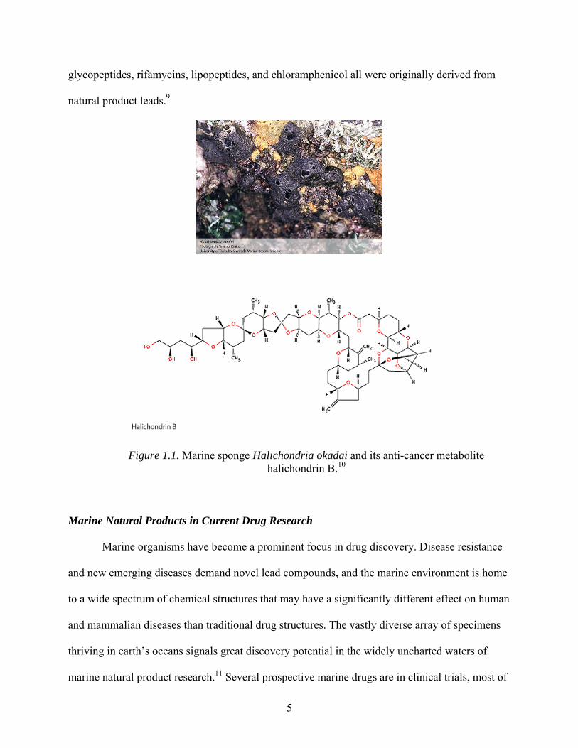
Halichondrin B provides an example of an exciting natural product discovery that was
challenged by low sample concentration and limited resources. Isolated first from the sponge
Halichondria okadai off the coast of Japan (Figure 1.1), and later from sponges in the Western
Pacific, Indian Ocean, and New Zealand coast, halichondrin B was purified and pinpointed as
having activity that affected microtubule polymerization. However, after the initial screenings,
not enough isolate was present to continue research. A collection initiative was launched by the
National Cancer Institute, the National Institute for Water and Atmospheric Research, and the
New Zealand government. The project resulted in not only an additional 300 mg of compound,
but also in successful development of aquaculture for halichondrin-producing sponges.5
Halichondrin B derivatives have since been synthesized and advanced to clinical trials as anti-
cancer agents.
In addition to halichondrin B, a significant number of drugs that have entered the market
in the past two decades are directly from natural products, semi-synthetic versions of natural
products, or synthetic variations of a natural product lead compound. Examples include
galantamine from the snow drop lily, a novel drug introduced in 2002 to treat Alzheimer’s
disease. Another potential Alzheimer’s drug, huperzine from club moss, is currently undergoing
FDA Phase II clinical trials for inhibition of acetylcholine esterase.9 Natural products have also
influenced the discovery of current anti-fungals, anti-virals, and neurological treatments. The
anti-bacterial classes including β-lactams, tetracyclins, cephalosporins, streptogramins,
5
glycopeptides, rifamycins, lipopeptides, and chloramphenicol all were originally derived from
natural product leads.9
Figure 1.1. Marine sponge Halichondria okadai and its anti-cancer metabolite halichondrin B.10
Marine Natural Products in Current Drug Research
Marine organisms have become a prominent focus in drug discovery. Disease resistance
and new emerging diseases demand novel lead compounds, and the marine environment is home
to a wide spectrum of chemical structures that may have a significantly different effect on human
and mammalian diseases than traditional drug structures. The vastly diverse array of specimens
thriving in earth’s oceans signals great discovery potential in the widely uncharted waters of
marine natural product research.11 Several prospective marine drugs are in clinical trials, most of

6
them having anti-cancer properties. The first marine natural product to be submitted to clinical
trials was didemnin B from the tunicate Trididemnum solidum. However, didemnin B was
removed during Phase II trials for toxicity reasons. More favorably, a series of bryostatin
macrolides has been isolated from a bryozoan species Bugula neritina. Some of these structures
show potential as anti-cancer, anti-depressant, anti-dementia agents. Currently in Phase II
clinical trials, the effectiveness of bryostatin-1 is indicated only in combination with another
natural product analogue, the anti-cancer drug Ara-C. Another anti-cancer drug, ecteinascidin
743, was isolated from sea squirts as early as 1969, but in insufficient quantities to bring the
product to clinical trials. Successful development of a synthetic protocol ensued, and a synthetic
version of this natural product is currently in Phase II clinical trials.5, 12, 13 Other marine natural
products are still in pre-clinical stages, such as the microtubule-binding agents peloruside A from
the New Zealand sponge Mycale hentscheli, and eleutherobin from octocoral Eleutherobia sp.,
and more recently from Erythropodium caribaeorum (Figure 1.2).10
Like terrestrial plants, marine organisms produce primary metabolites consisting of
essential proteins, lipids, and other complex molecules that form the building blocks for
structural and functional maintenance of all living organisms. Viability is dependent on
unimpaired primary metabolism. However, in most living things, additional energy is

7
OO
O
O
N
NS
H
OH
HO
O
H
H
OO
NHO
HO
Ecteinascidin 743
O
HH
O
OMe O
O
N
N
O
OAcOH
OH
Eleutherobin
O
O
HO
OCH3
HO
O
OHOCH3
HO
H3COHO
H
Peloruside
Figure 1.2. Marine natural products provide a potential source of new anti-cancer agents.10
expended on secondary metabolites. Although not vital to an organism’s survival, products of
secondary metabolism enhance resilience through protective and defensive roles. Their

8
biosynthesis is therefore a significant aspect of an organism's biochemical makeup and long-term
viability. It is these incredibly diverse molecules that constitute the focal point of natural
products research.2
Because natural products often contain small molecules having neither an extremely
polar or non-polar nature, they are ideal drug candidates. Natural products chemist Alan Harvey
reports that “the use of natural products has been the single most successful strategy for the
discovery of new medicines.”14 Soft-bodied, sessile marine invertebrates are of particular interest
pharmaceutically. Lacking external functional defense mechanisms (such as scales or shells) and
mobility to move away from danger or from a resource-depleted environment, these organisms
rely on biochemical production of—often toxic—chemical compounds. Secondary metabolites in
marine organisms serve several important ecological roles, whether to deter predators, aid food
capture, or maintain environmental boundaries against invasion of space by overgrowth of
neighboring organisms.13 These same components hold significant appeal to pharmaceutical
researchers. Therapeutic implications from the extensive variety of biologically active marine
metabolites suggest potential drugs with anti-tumor, anti-viral, and anti-inflammatory
properties.13, 15 Some species of the Hyrtios class of marine sponges produces the related
sesquiterpene compounds puuephenone and methoxypuupehenol, both of which display anti-
fungal and anti-malarial activity.16 The Caribbean sponge Cryptotethia crypta produces atypical
nucleosides which have been synthesized and used as anti-cancer and anti-viral treatments since
their discovery in the 1950’s. Halogenated metabolites are also frequently identified in sponges,
distinguishing marine metabolites from terrestrial counterparts. For centuries, Russians have
been collecting freshwater sponges with iodine-containing compounds. The sponges, collectively
called Badiaga, were ground into a powder for topical use. Polish physicians prescribed their

9
own therapeutic mixture of Badiaga powder, differing slightly from the Russian and Ukrainian
versions based on the availability of sponge species.15
Interestingly, the phylum Porifera is distinguished as the most biosynthetically active
class of marine invertebrates. Like other marine organisms, many of the metabolites emitted are
toxic, and production varies with changing environmental conditions, light exposure, and growth
rates. While the exact function of these compounds is hard to determine, it can be reasonably
proposed that they exist to keep the sponge free of barnacles, biofilms, and parasites, while also
maintaining a zone clear of ecological competitors.15 Over half of marine natural products thus
far identified are sponge-derived.17
Marine Natural Product Interest in the Western Atlantic Region
Home to the third largest reef system in the world and extensive underwater visibility, the
Caribbean waters of the Bahamas (Figure 1.3) are a wealthy resource for marine research.18 The
clarity of the water allows sufficient sunlight to support incredible underwater ecosystems of
flora and fauna. A combination of coral reefs, sessile marine invertebrates, microalgae, and
autotrophic zoozanthellae all contribute to the uniqueness of the marine environment.
Productivity is enhanced by symbiotic relationships between some marine organisms,
contributing to the diversity of nutrients and metabolites that are produced.19
While sunlight is essential to energy metabolism of phototrophic marine organisms,
excess sunlight or lack of protective mechanisms can be harmful to a reef system. Antioxidant
defenses in reef ecology are just beginning to be studied in their protective role against
deleterious effects of UV radiation. The field of marine ecology is expanding to address
oxidative stress issues through investigation of the unique relationships among corals, marine
microbes, and sponges.19-21

10
During the second half of the 19th century, explorers and fisherman began commercially
harvesting sponges from the waters around the Bahamas and the Florida Keys. By the 1930’s,
overfishing and disease had taken a visible toll on the status of the Caribbean reef system.22
Since that time, the reefs have undergone phases both of recovery and of damage from
hurricanes and coral bleaching.23
Figure 1.3. Biodiversity in the marine environment of the Bahamas’ reef system provides resources for potential pharmaceuticals.18
Sponges (Phylum Porifera)
The phylum Porifera, including thousands of varieties of sponges, is one of the first
classes of marine invertebrates to be studied by natural products chemists. Sponges are
multicellular, undifferentiated organisms equipped with an adept water-filtration system.
Although technically considered animals, physiologically sponges are among the simplest
Metazoans. Possessing no differentiated tissue to form digestive, nervous, and circulatory
systems, they are composed primarily of a porous network of channels and chambers through
which water moves. Flagellated cells (choanocytes) and phagocytotic cells (archaeocytes) lining

11
these channels propel water in the appropriate direction, and help capture nutrients that are swept
along by the water current. In the space between water channels, a gelatinous connective matrix
called the mesohyl houses skeletal materials. Following phagocytosis through apertures at the
inhalant surface (ostia), food particles are filtered through progressively smaller sieve-like
channels into the mesohyl for metabolism.7, 24 The sieves are designed because indiscrete intake
of water and particles by non-selective filter feeders results in ingestion of many particles which
are of no biochemical value to the sponge. In order to retain sufficient nutrients for metabolism
while simultaneously keeping waste levels from overwhelming the filtering system, the sponge
must constantly excrete particles from its aqueous environment. Materials that cannot be used for
energy are eliminated through apertures (oscules) in the exhalant surface. The entire sponge
biomass is involved primarily in maintaining its low-pressure water pumping system.24
Taxonomists have long been frustrated by a lack of consistent defining characteristics of
sponge morphology. Amorphous shapes, color variability within a species, and both flexibility in
size and pigment intensity depending on the degree of light exposure create classification
challenges. Unique skeletal characteristics of spicule formation provide the primary departure
point from species ambiguity, and have served as the fundamental basis of classification.
Skeletal polymorphism results from the differences in components of skeletal materials.
Depending on the Poriferan class, skeletal elements can be composed of collagenous, siliceous,
or calcareous fibers, arranged in patterns which lead to the diverse spicule arrangement. Still,
proper classification is a tedious and uncertain process. Advancements in biochemical and
histological tests aid classification of collected sponges, and other researchers have begun
exploring the interesting technique of chemotaxonomy—identification by differential metabolite
production. Chemotaxonomy has been used to distinguish sponges down to the genera, but
cannot be used to determine classification at the species level.4

12
Three classes of sponge are commonly recognized. The most prominent class,
Demospongiae, includes 95% of identified species and spans a wide range of habitats from
intertidal depths to marine trenches in fresh or brackish water. Spicules are intracellularly
produced from silica in most species, while other species are devoid of spicules. The deep-water
class, Hexactinellida, is characterized by six-membered hexactine siliceous spicules. This rather
unusual class lacks a mesohyl matrix, and is morphologically the most distinct from other
Poriferan classes. Finally, the third class Calcarea is composed of calcium carbonate-based
skeletal materials. Crystalline calciferous spicules may grow individually or as one mass.24
Despite constant engagement in the water filtration process, sponges are also proliferate
metabolite producers. Osinga et al. report that –remarkably—“the structural diversity of sponge
secondary metabolites is larger than that of any other marine phylum.”7 Although sponges were
the primary target for historical marine invertebrate research, publications presenting
biochemical information on their secondary metabolites were not available sixty years ago.24
Current research has yet to definitively link each metabolite to its role, but the functional nature
of many metabolites can be credibly theorized: toxins are most likely involved in spatial
competition, while antibacterial compounds prevent ingested bacteria from dominating sponge
metabolism. Sponge ecology become an important consideration in uncovering environmental or
biochemical factors that would suggest a need for certain types of metabolite production.7
Identification and Isolation of Bioactive Compounds
Marine organisms contain a plethora of metabolites, some of which will display
bioactivity, while many others will not. From a pharmaceutical perspective, it becomes necessary
to screen out the non-active compounds early in the research. Investigators are therefore

13
challenged to use time-effective separation schemes that will remove unwanted materials from
an extract with minimal loss of bioactive compounds.
Traditional approaches to natural products research generally focused on the chemistry of
novel compounds, without intense interest in biological activity until total isolation and
characterization was complete. Thus the process from sample collection to biological screening
often involved extensive timeframes.2, 4 Current natural product chemistry is primarily bioassay-
guided, due largely to molecular-targeted high through-put screening (HTS) which allows rapid
screening of multiple compounds at one time. The development of HTS increased the demand
for new natural products and natural product derivatives so dramatically that analytical chemists
could not supply purified compounds fast enough.25 In order to meet these new dynamics of the
field, natural products chemists had to reorganize their approach to separation schemes. Now, as
well as a continued search for novel therapeutic compounds, current research projects for natural
product scientists include assay and method development aimed at reporting quicker, more
effective methods of collection and extraction.6
Historical methods of preliminary screening involved recognizing patterns of previously
identified thin layer chromatography signatures. In contrast, modern in vitro cellular assays
screen compounds with precise molecular targets. A wide variety of assays are currently
available, aiding the search for anti-inflammatory, anti-cancer, anti-bacterial, and many other
biomedically relevant activities. This chemical-biological interface has been a significant change
in the structure of natural products approaches to chemistry. For instance, although chemical
assays have traditionally been used to characterize antioxidant activity of marine organisms,
there is a growing demand for cell-based antioxidant data. Laboratories may use a variety of
screens to speed up isolation of useful compounds.26 Organisms displaying a certain threshold of
biological activity will be selected for further research. Isolation of bioactive compounds is

14
continued in a step-wise, assay-guided manner, with each new fraction generated being subjected
to biological testing. This approach ensures that at each subsequent step in the separation
scheme, the fraction or compound pursued for purification is bioactive.
Certain disadvantages are often unavoidable when using bio-screening to identify
compounds of interest. Results processing is typically the rate-limiting step in bioassay guided
fractionation, and may impede timely investigation. Also, depending on the type of assay,
procedures can be expensive. Repeated testing at each stage of separation depletes often-
miniscule quantities of available sample material. In vivo assays in particular are expensive and
time-consuming; however, the medical field prefers in vivo results which reveal multi-target
capabilities rather than activity of an isolated point within a biochemical network.
Another advance in natural product research is the development and expansion of
databases through which characteristics of unknown compounds can be compared with
previously identified compounds to avoid overlapping research and discovery. Before the
existence of these databases, it was difficult to determine whether or not a seemingly novel
compound had already been identified in a lab across the country or in another part of the world.
Investigators needed a way to access natural product compound libraries so as to avoid spending
tedious time elucidating structures of already-known compounds. Structural dereplication—
comparison of similar structural data through the compound libraries— usually follows
preliminary screening to extracts.
Semi-purification followed by preliminary screening for activity reduces the time spent
on isolation and purification because potential hit compounds are identified in the early stages of
compound separation. Crude extracts are separated into semi-pure fractions, at which point
compounds of identical or similar fractions are grouped, profiled, and prioritized based on
interesting characteristics. Thorough natural product databases are essential to employing
15
information from existing compound profiles into a separation scheme.6 Isolation and
identification procedures have also benefited from increased automation and the availability of
simultaneous fractionation and screening methods via hyphenated techniques such as MS-NMR
and reversed-phase HPLC-MS. Renewed interest in natural products chemistry is largely due to
the combination of expanded natural product libraries and improved methodology, both of which
effectively streamlined the dereplication process.27
Drugs typically require an amphiphilic nature in order to cross both aqueous and lipid
barriers presented by a cellular environment. In a crude extract, therefore, molecules of
intermediate polarity are the compounds most likely to be compatible with physiological
considerations of adsorption, distribution, and metabolism. Unfortunately, drug-like compounds
of intermediate polarity are generally isolated in the lowest concentration from natural product
biomass, or are left undetected by traditional separation schemes. In response to this problem, a
method has been developed to achieve initial separation of fractions of intermediate polarity
from the more prominent polar and non-polar fractions.2 The low-mass, intermediately polar
fraction obtained by this method allows basic structural analysis prior to complete purification.
Compound assessment for novel or clearly recognized classes of molecules in the intermediate
polarity range is not possible by analysis of crude extract. In relation to the low amounts of
amphiphilic compounds, higher concentrations of lipids, salts, and carbohydrates will obscure
the smaller signals. In the cyclic loading method, traditional silica-based chromatography (silica
gel or C-18 stationary phase) supplies substrate to which polar compounds may become
irreversibly bound. In contrast, the macroporous polymeric resin Diaion HP20 (polystyrene-
divinylbenzene) lacks polar sites, effectively preventing irreversible binding of polar compounds
to the stationary column. Intermediately polar compounds will adhere to the HP20 substrate
while the majority of unwanted compounds are washed away. After the removal of water-

16
soluble salts and carbohydrates, using a 40% Me2CO/H2O eluant, intermediately polar
compounds are eluted with 75% Me2CO/H2O, and non-polar fats and steroids removed with
Me2CO. In order to decrease the often-tedious amount of time spent concentrating to dryness the
Me2CO/H2O–containing fractions, the fraction of interest is back-loaded onto a smaller HP20
column and metabolites eluted with Me2CO. This chromatographic loading method has been
used for metabolite extraction of numerous marine sponges, and provides an effective way of
reducing purification time of drug-like compounds.2
Emerging Methods in Natural Products Chemistry
Although traditional medicine has always recognized the significant role of natural
products, the past two decades marked a decline in pharmaceutically-funded natural products
research. Seeking to avoid the tedious—and often labor-intensive— methods characteristic of
natural product discovery, companies anticipated a surge in combinatorial chemistry libraries
that, disappointingly, has not surfaced. However, a significant change in screening methods has
emerged with the development of high throughput screening. HTS can screen more compounds
than was ever previously possible, by using robotic systems and multi-well assay plates.6 Pure
compounds are needed for screening to avoid the ambiguity of false positive and negative
results. Unfortunately, natural products chemistry does not lend itself to the HTS regime. Natural
product compounds involve more complexity than synthetic substances, and often the size and
diversity of structures makes it difficult to determine the physical and molecular properties of
those compounds. Unidentified chemical characteristics may interfere with the screening
process, challenging the accuracy of assay results.6
17
Specific Aims
In this study, a sampling of marine sponge extracts from various locations in the Western
Atlantic and collection depths will be separated into hydrophilic, hydrophobic, and amphiphilic
fractions. Each fraction will be screened for antioxidant activity using the FRAP assay. Assay-
guided isolation and purification of hit compounds will direct elucidation of potential marine
antioxidants. Additionally, in order to test the ability of the potential antioxidants to cross
membranes, a fluorescent, cell-based assay will be carried out on purified compounds using
zoozanthellae cells.

18
CHAPTER 2
Antioxidant Activity of Marine Organisms from the Western Atlantic
Antioxidants in the Marine Environment
Reactive oxygen species (ROS) are encountered by all aerobic organisms, with oxidative
proclivity being especially high at electron rich areas such as metabolic or photosynthetic sites.
Marine organisms are exposed to particularly high levels of ROS through a combination of
photosynthesis, symbiont oxygen production, and intense sunlight intensities leading to UV-
induced free radical production. The conjecture that organisms highly exposed to ROS will have
effective antioxidant mechanisms has certainly not disappointed natural product chemists. Many
species contain powerful plant-like—or completely novel— antioxidant compounds.28 Research
progress in the marine antioxidant field has led to antioxidant screening of pure compounds and
semi-pure fractions rather than crude extracts as was typical in earlier antioxidant research.
Takamatsu et al. investigated antioxidant activity of over one hundred purified marine natural
product compounds using the chemically-based DPPH (2,2-diphenyl-1-picrylhydrazyl radical)
TCL method as well as the cell-based DCFH-DA (2’,7’-dichlorodihydrofluorescein diacetate)
assay. Demonstrating that marine products are active in living cells advances the compounds of
interest one step closer to pharmaceutical applicability. Importantly, the side-by-side comparison
approach of chemical-based and cell-based assays used by Takamatsu et al. allows researchers to
distinguish chemical reductants from biological antioxidants.26
While marine antioxidants are a relatively new focus area in natural products chemistry,
emerging interest has led to several unique discoveries. A compound from brown algae exhibited

19
radical scavenging activity comparable to the common antioxidant food additive BHT. Certain
species of marine Streptomyces bacteria produce a class of superoxide scavenging molecules
called aburatubolactams.13Another research group characterized an antioxidant, anti-apoptotic
isolate from the sponge Hymeniacidon helophila. Although the protective functions of sponge
metabolites are often difficult to determine, this amino acid derivative, L-5-hydroxytryptophan,
presumably protects the intertidal Hymeniacidon sponges from oxidative stress caused by intense
sunlight exposure.29
Bioassays for Antioxidant Activity
Physiological disorders, including neurodegenerative diseases, atherosclerosis, and
cancer have been linked to oxidative stress in humans, and are a prominent target for novel
therapeutic interventions. Normally, aerobic challenges can be overcome by enzymes, chelating
agents, and non-enzymatic antioxidants which are present in aerobic organisms to protect DNA,
proteins, and lipids from the detriments of oxidation. Disease conditions may arise from
excessive oxidative stress, such as exposure to radiation.30, 31 Compounds that can be
administered pharmacologically to counteract ROS damage may be effective both in prevention
and treatment of neurodegenerative disorders.26, 32-36
At the core of oxygen utilization in biological systems are reduction-oxidation, or
“redox” reactions which involve transfer of electrons from one substrate to another. The
substrate receiving the electrons is reduced, while the substrate providing the electrons is
oxidized. Oxidation and reduction reactions must always exist in balance: one cannot occur
without the other. Redox substrates are typically classified as reductants and oxidants in
chemistry, but the terms antioxidant and pro-oxidant are more frequently encountered in
biological systems. Specifically, a pro-oxidant, (reactive oxygen species or ROS), is a

20
pathological oxidant. ROS are implicated in numerous disorders including cancer, Parkinson’s,
and Alzheimer’s disease.37 When free radicals—molecules having become charged through the
loss of an electron— are formed in consequence of aerobic metabolism, photosynthesis,
radiation, or ROS-generating conditions, they will react with the first available oxidizable
substrate. Oxidation of substrates such as DNA, proteins, or membranes can create considerable
damage inside a cell. When antioxidants are present in a cell, they replace other oxidizable
substrates as the reductant component of redox reactions. As long as the antioxidant capacity is
not overwhelmed, this mechanism protects vulnerable cellular components. Specifically, an
antioxidant is classically defined as a “substance that, when present at low concentrations
compared to those of an oxidizable substrate, significantly delays or prevents oxidation of that
substrate.”38 The products of this reaction will have little to no toxicity, as opposed to the highly
detrimental reactions of ROS with non-antioxidant substrates.37, 39
Antioxidants are classified as preventative when they prevent formation of free radicals.
Others are classified as radical scavengers, functioning to halt further propagation of chain
reactions. The final class of antioxidants consists of repair molecules which ameliorate oxidative
damage to a cell. Phenolic antioxidants often found in plant materials—and more recently in
numerous marine organisms—are thought to play a preventative role in radical formation.
Relatively little has been done to identify the mechanisms responsible for antioxidant action of
phenolic marine metabolites, however. This class of compounds, predominantly the phenolic
terpenes, forms the most bioactive category of marine structures. Utkina et al. studied a number
of marine sesquiterpenequinones to determine both antiradical and antioxidant activity.31
Antiradical activity was defined as the ability to prevent oxidation by DPPH radicals, while
antioxidant activity was tested based on the ability of the compounds to prevent, delay, or
stabilize lipid peroxidation of linseed oil. In the latter test, antioxidant capacity was quantified

21
for auto-oxidation and for lipid peroxidation induced by lipid hydroperoxides or Fe2+ molecules.
Phenolic sponge metabolites puupehenone and its analogue 15-methoxypuuepenol are potent
antiradicals in the DPPH assay, but although they slowed the process of lipid peroxidation,
neither could actually stop it. Thus both compounds qualify as preventative antioxidants, but do
not possess effective chain-breaking antioxidant potential. Both these compounds were
compared to the α-tocopherol, which is the major antioxidant component of lipid-soluble vitamin
E. Because it is known to be able to prevent as well as halt oxidative chain reactions, it is often
used as a control in antioxidant assays.31
Methods to quantify antioxidant activity have been an important component of
biomedical research. Initially created by Benzie and Strain40 to test total antioxidant power of
biological fluids, the ferric reducing antioxidant potential (FRAP) assay has since seen extended
use for plant compounds, food and nutrition chemistry, and marine natural product research.
Differing slightly in principle from other assays which quantify resistance to free radicals, the
FRAP assay evaluates the strength of an antioxidant compound as a reductant. The assay
procedure can be completed fairly quickly and lends itself to gathering preliminary data on a
large number of samples. Once the appropriate samples have been added to the FRAP reagent, a
colorimetric change to deep blue signals reduction of the ferric tripyridyltriazine complex (Fe3+-
TPTZ) to ferrous TPTZ (Fe2+-TPTZ). Molar concentration of the reductant can be plotted as a
function of color intensity as measured on a spectrophotometer.35, 40, 41
One of the disadvantages of the FRAP assay is that its scope is limited to measuring
chemically-defined reductant capacity, which may not be identical to antioxidant capacity. In this
assay, a reductant which reduces Fe3+ to Fe2+ is not necessarily an antioxidant because Fe3+ is not
a pathological oxidant. Fe2+ is actually a pro-oxidant which forms free radicals from peroxide.
Therefore the FRAP assay provides reflective antioxidant capacity, but not true antioxidant
22
power. Prior points out that antioxidants which reduce both Fe3+ and Fe2+ will be detected in the
assay, while other effective antioxidants which lack the ability to reduce Fe3+ will be
undetectable.37
Often it is necessary to obtain a variety of information about antioxidant potential other
than what can be concluded from the FRAP assay alone. Cell-based assays in particular can be
used to gain appreciable evidence of the potential in vivo antioxidant effectiveness of a
compound. One such method, the dichloroflurorescein assay, provides a straightforward
approach to measuring ROS sequestration by an antioxidant. The assay can also be used to
quantify ROS levels induced by mechanical or chemical oxidative stress factors. This cell-based
method can also be applied to measurement of antioxidant activity within an oxidative
environment. Cells readily absorb the non-polar 2’,7’-dichlorodihydrofluorescein diacetate
(DCFH-DA) probe. As the probe crosses membranous cellular boundaries, it becomes trapped
internally via hydrolyzation to the non-fluorescent polar compound 2’,7’-
dichlorodihydrofluorescein (DCF). Inside the cell, DCF fluoresces when oxidized by peroxyl
radicals are formed by treatment with a radical generator. Measuring fluorescent intensity of
oxidized DCF reveals overall oxidative stress in the cell. If antioxidants are present, fluorescent
intensity is limited to the extent that free radical scavenging counteracts the peroxyl radicals.30, 42
Antioxidant Activity in Sponges
Sponges grow at a range of depths; accordingly, not all are exposed to identical UV
intensity. Under the oxidative pressures of the marine environment, survival is influenced by the
measure of balance maintained between ROS generated within the sponge and its defensive
antioxidant mechanisms. This correlation between antioxidant activity and ecological factors
leads to the collection of sponges that appear to thriving in highly-oxidative environments. For

23
example, the metabolic products of an intertidal sponge investigated by Lysek et al. was
expected to include a significant concentration of antioxidant compounds due to its shallow
water, high-light exposure surroundings. The main constituent of the sponge Hymeniacidon
heliophila, L-5-hydroxytryptophan, exhibited anti-apoptotic activity by sequestering UV
radiation-induced ROS.29 In another investigation of sponge-derived antioxidants, Regoli et al.
examined oxidative stress levels in the sponge Petrosia ficiformis in both symbiotic and
aposymbiotic relationships. This particular sponge can be found in diverse habitats ranging from
light-exposed water to dark caves. Investigation of oxygen production in P. ficiformis revealed a
correlation between heightened antioxidant defenses and increased O2 due to symbiotic
relationships. Interestingly, exposure of symbiotic P. ficiformis to more intense radiation did not
necessarily result in increased antioxidant protection, with outer layers being especially
vulnerable to radiation stress.20 Older studies investigating the UV exposure of corals surviving
at different reef depths report that shallow water corals have heightened ability to absorb UV
radiation compared to those found in deeper water.19, 43
Specific Aims
The aims of this study were to provide evidence of antioxidant capacity in several sponge
specimens native to the Western Atlantic. Specifically, we were interested in possible
correlations between 1) antioxidant bioactivity and collection depth, and 2) fraction polarity and
concentration of antioxidant compounds. Based on previous publications involving antioxidant
assays of marine product extracts, we hypothesized that the samples collected in shallow water
or at intermediate depths would display the highest FRAP readings.

24
Results and Discussion
Lyophilized specimens of marine sponges collected from the Western Atlantic from a
range of depths were selected for comparison of antioxidant activity. Of the nine specimens
chosen for the assay, three were collected in deeper water off the coast of Panama City, Florida,
while the others had been previously collected from the Bahamas. During initial separation,
methanolic extracts of sponge material were partitioned into three discrete fractions of
decreasing polarity using the reverse-phase HP20 chromatographic method described in Chapter
1. Samples of each fraction were loaded in triplicate onto 96-well plates for preliminary
antioxidant screening based on the FRAP assay first described by Benzie and Strain.40 This
assay, used in order to determine general distribution of antioxidant compounds within the
sponges, showed ferric-reductant capability across the range of hydrophilic to hydrophobic
compounds. Marine sponge fractions were initially assayed using ~100 µg/mL of each sample
dissolved in methanol. Absorbance was measured at 600 nm and recorded 12-15 min following
addition of samples to FRAP reagent. Equivalence values for each sample were then determined
based on a linear calibration curve using known concentrations of Fe2SO4·7H2O. The fractions of
semi-purified sponge extracts exhibited variable antioxidant potential, with some of the
intermediate and shallow water specimens exhibiting activity equaling or surpassing the activity
of the control antioxidant, 100 μM α-Tocopherol, in all three fractions (Figure 2.1). While FRAP
activity was found in sponges from intermediate or shallow collection depths (Figure 2.2), the
deeper water Floridian sponges displayed little or no activity (Figure 2.3). This agrees with
previous reports of an inverse relationship between antioxidant concentration and collection
depth.44

25
4.2
17.2
6.5
4.4
30.5
29.9
44.5
6.710
.1
6.6 8.
3
2.7
12.5
19.3
7.1
0
5
10
15
20
25
30
35
40
45
50
'40% '75% '100% 'α Tocopherol
FR
AP
Eq
uiv
alen
ce (μ
M/μ
g)
BHM01-001 BHM01-069 BHM01-078 BHM01-032
Figure 2.1. Bioactive fractions of marine sponges from the Western Atlantic.
0.0
5.0
10.0
15.0
20.0
25.0
30.0
35.0
FR
AP
Eq
uiv
ale
nce
(μM
/μg
)
BHM01
-032
BHM01
-033
BHM01
-036
BHM01
-001
BHM01
-069
BHM01
-078
Contro
l
Figure 2.2. Average FRAP equivalence values of Bahamian sponge samples from
various collection depths.

26
0.02.0
4.06.08.0
10.0
12.014.016.0
18.020.0
FR
AP
Eq
uiv
ale
nc
e (μ
M/μ
g)
Hydrophilic Amphiphilic Hydrophobic
Shallow (< 3 m) Intermediate (3-15 m) Deep (> 15 m)
Figure 2.3. Average antioxidant activity distribution by polarity.
The results of this assay revealed comparable FRAP equivalence values among the three
fractions screened in the majority of sponges used in this study (Figure 2.3). Although the
intermediately polar fraction contained the majority of compounds with antioxidant activity, the
most active component was found in the non-polar fraction of Bahamian sponge, collection ID
BHM01-069 (Figure 2.4). This compound (1) was later isolated and identified as a chroman-
sesquiterpene.

27
05
10
152025
30354045
50
BHM01
-001
BHM01
-069
BHM01
-078
BHM01
-032
BHM01
-033
BHM01
-036
PC01-0
04
Contro
l
FR
AP
Eq
uiv
alen
ce (μ
M/μ
g)
40%
75%
100%
Figure 2.4. Antioxidant potential of individual marine sponge extracts varies with the
polarity of the fraction assayed.
Although the sample size of collected sponges available for screening in this study was
insufficient to determine a trend in the nature and distribution of antioxidant properties, the
majority of antioxidant compounds in these sponges were contained in the amphiphilic fraction.
Additionally, specimens collected from the 3 m to 15 m range were active, while sponges
collected at depths greater than 15 m displayed little to no FRAP activity. It would be interesting
to continue the study with a broader sample size in order to investigate statistical correlations
between distribution of antioxidant compounds and molecular polarity.

28
CHAPTER 3
Bioactive Compounds from the Bahamian Sponge Smenospongia sp.
Smenospongia sp. (Figure 3.1) was collected from the Bahamas at a depth of 10 meters.
Similar species from the genus Smenospongia have been collected from Korea and from Papua
New Guinea.45, 46
Figure 3.1. Marine sponge Smenospongia sp.
Investigation of antioxidant constituents from the marine sponge Smenospongia sp.
(collection number BHM01-069) yielded the previously described compounds chromazonorol
(1), 6-bromo-2’-de-N-methylaplysinopsin (2), and 6-bromoaplysinopsin (3).

29
Isolation and Identification of Chromazonorol
Separation of potential antioxidants from Smenospongia sp. was structured around high
levels of FRAP activity detected in crude fractions during an initial screening (Scheme 1)
Initially, semi-preparative separation of crude methanolic extract of dried Smenospongia sp.
sample was performed using the reverse-phase HP20 cyclic loading method as described in
Chapter 1. Potential antioxidant compounds were detected by FRAP screening of each of the
three fractions eluted, and all fractions showed substantial activity. A smaller HP20 column was
used to achieve further fractionation of both the 75% Me2CO/H2O and 100% Me2CO fractions
using incremental, increasing concentrations of Me2CO/H2O. The 80% Me2CO/H2O fraction
yielded a phenolic compound (1) that displayed significant FRAP activity. The molecular
formula of compound 1, C21H30O2, was determined from the HRESI of the [M+H]+ ion at m/z
315.2338, requiring seven degrees of unsaturation. Initial analysis of the 13C NMR spectrum
(Figure 3.2 and 3.3) revealed 6 carbons in the aromatic/olefinic region, 14 aliphatic carbons, and
an oxygenated carbon. These data suggested that compound 1 consisted of an aromatic ring
connected to a sesquiterpene skeleton. Analysis of the NMR data (1H, 13C, HMBC, and HSQC)
allowed identification of the compound as chromazonorol, previously isolated by Cimino et al.
in 1975 from the sponge Disidea pallescens.47 Chromazonorol shares structural similarities with
previously identified phenolic sesquiterpenes puupehenone and aureol, both bioactive marine
secondary metabolites. Puupehenone exhibits anti-fungal and anti-microbial properties against
Staphylococcus aureus ,and Clostridium tropicalis, as well as anti-malarial properties when
tested with three different strains of Plasmodium falciparum.16Additionally, puupehenone is
active as an antioxidant in both chemical DPPH and cell-based DCFH-DA assays.26

30
Scheme 1. Separation scheme of chromazonorol from the Bahamian sponge
Smenospongia sp.
O
H
H
OH
1
Smenospongia sp. (90 g)
Solids Extract
Methanol (x2)
HP20 Cyclic loading
Acetone/water
100%
70% 80% 90% Acetone
Chromazonorol (1) 32 mg
HP20 Cyclic loading
Acetone/water

31
Figure 3.2. 13C and 1H NMR spectra for chromazonorol (1) in CD3OD (75 and 500
MHz).

32
Figure 3.3. 1H NMR of acetylated chromazonorol (4) in CDCl3 (500 MHz).
FRAP Activity of Chromazonorol (1)
Several well-described, naturally occurring antioxidants were used as a comparison for
antioxidant capacity of marine compounds. Bioactivity of purified chromazonorol exhibited
more intense ferric-reductant potential than positive controls resveratrol, catechin, quercetin,
ellagic acid, and α-tocopherol. Absorbance readings of FeSO4·7H2O standards of known
concentration were used to generate a linear calibration curve from which equivalent
concentrations of antioxidant sample were obtained. Purified sponge compounds were assayed
for FRAP activity using 10 μg sample per well, while antioxidant controls were tested at a
concentration of 20 μM. The FRAP values obtained were calculated as μM FeSO4·H2O per μM
antioxidant sample, and results are shown as mean FRAP equivalence from three independent
values (Figure 3.4).

33
In order to identify the source of antioxidant activity in chromazonorol, compound 1 was
acetylated using acetic anhydride and pyridine to give the acetylated chromazonorol (4).
Compound 4 was assayed for antioxidant activity and found to have significantly reduced
activity (Figure 3.4). The antioxidant activity of chromazonorol and significantly reduced
activity of acetylated chromazonorol indicated that the phenolic hydroxyl group is essential to
the antioxidant activity of chromazonorol.
0.010.020.030.040.050.060.070.080.090.0
100.0
Resve
ratro
l
(+)C
atec
hin
Querc
etin
Ellagic
acid
Compo
und
1
Compo
und
4
FR
AP
Eq
uiv
ale
nc
e (μ
M/μ
M)
Figure 3.4. Average FRAP equivalence of antioxidants, chromazonorol (1),
and acetylated chromazonorol (4).
Puupehenone has also been found to exhibit antioxidant activity. Similar in structure to
chromazonorol, puupehenone is a phenolic sesquiterpene isolated from the sponge Hyrtios sp.
The two hydroxyl functions contribute to its strong antioxidant activity.16 As with other
phenolics, the presence of at least one hydroxyl moiety in members of this class of molecules
provides a ready substrate for free radicals. Phenolic antioxidant activity generally increases with
an increasing number of hydroxyl groups attached to the aromatic ring. This was demonstrated
by Kim et al.48-50 using the vitamin C equivalent antioxidant capacity assay to test nutritional

34
polyphenolic compounds. The increase in activity is linked to the relative ease by which the
hydroxyl proton is donated to neutralize free radicals. Electron donation is affected by
stereochemistry and bond dissociation energy, and is impeded by the presence of electron
withdrawing groups in proximity to the –OH function. Thus the position—as well as the
number— of hydroxyl groups plays a significant role in conveying antioxidant activity.48
O
H
O
OH
5
In addition to the placement and frequency of hydroxyl groups as determinants for
antioxidant capacity of phenolics, effective antioxidant compounds must also possess molecular
properties allowing efficient uptake by cells. Sometimes antioxidant activity conveyed by a
compound in a solution-based assay is lost during cell-based or in vivo screening. This may be
due to inability of the compound to cross cell membranes, or result from other properties
incompatible with the internal cellular nature. However, the reverse is also true—non-active
acetylated compounds are activated within the cell by loss of the acetyl group.26, 49 In this study,
chromazonorol (1) and acetylated chromazonorol (4) were tested for antioxidant activity in
zoozanthellae cells using the fluorescent DCFH-DA assay (data not shown). Compound 1 was
unable to prevent DCF oxidation at a concentration of 20 μg, but cells treated with compound 4
inhibited DCF oxidation. The activity of 4 in this assay may be attributed to its increased
hydrophobicity conveyed by the acetate group, facilitating cellular uptake. Similar results were
obtained by Takamatsu et al. in their investigation of marine antioxidants. Algal metabolites

35
cymopol and 7-hydroxycymopol were screened for antioxidant activity using the DPPH radical
trapping assay, along with the fluorescent DCFH-DA assay.26 The compounds were found to be
active in both assays, but the hydroxyl-containing compound displayed lower antioxidant activity
in the cell-based assay. The authors concluded that the diminished activity was a result of
increasing polarity of the molecule.
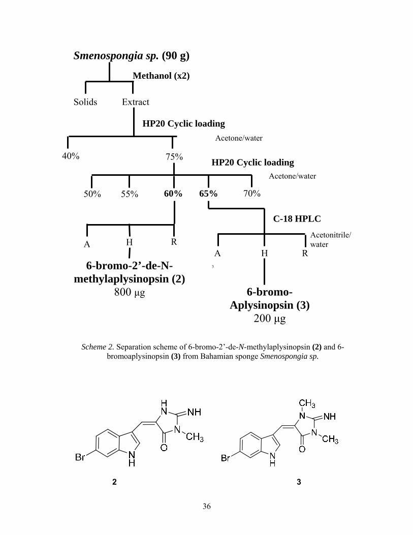
Isolation and Identification of Aplysinopsin Derivatives from Smenospongia sp.
Various indole alkaloids have been isolated from marine organisms with reported anti-
microbial or anti-inflammatory, and anti-cancer activity, representing a promising class of
compounds for pharmaceutical research. The indole alkaloid class comprises a quarter of known
marine metabolites and the diversity found within this class is accentuated by the addition of
atoms such as bromine which are not typically found in plant compounds.51 Following the
identification of chromazonorol from the 100% Me2CO fraction of Smenospongia sp. extract, the
75% Me2CO/H2O fraction from the initial HP20 column, also found to contain antioxidant
activity, was chromatographed further into five additional sub-fractions (Scheme 2). Using NMR
data, two of these sub-fractions were selected for purification by reverse-phase C-18 HPLC using
an acetonitrile/water gradient method to achieve clear separation of compounds (Figure 3.5).
Fractions were collected at 30 sec intervals and combined by peak. The fraction library was
transferred to a 96-well plate and assayed for FRAP activity to identify active compounds. The
major compound of the 60% Me2CO/H2O fraction (2) was found to be active, while the major
component of the 65% Me2CO/H2O fraction (3) displayed no activity. An additional quantity of
the 75% Me2CO/H2O fraction was then re-chromatographed by HPLC and the original active
peaks subjected to NMR analysis.
36
Scheme 2. Separation scheme of 6-bromo-2’-de-N-methylaplysinopsin (2) and 6-bromoaplysinopsin (3) from Bahamian sponge Smenospongia sp.
2 3
40%
3
C-18 HPLC
75% HP20 Cyclic loading
70% 55% 60% 65% 50%
6-bromo-2’-de-N- methylaplysinopsin (2)
800 μg
A RH
6-bromo- Aplysinopsin (3)
200 μg
A RH
Acetone/water
Acetonitrile/water
Smenospongia sp. (90 g)
Solids Extract
Methanol (x2)
HP20 Cyclic loading
Acetone/water

37
Figure 3.5. HPLC trace of the 60% Me2CO/H2O fraction with compound 2 (top) and the
65% Me2CO/H2O fraction with compound 3. The ESI of compound 2 showed [M+H]+ ion peaks at m/z 318 and 320 with nearly equal
intensity, and indicated the presence of one bromine atom. Analysis of 1H and HMBC NMR data
revealed deshielded aromatic protons of an indole moiety connected to a glycocyamidine side
chain (Figure 3.6). A search of the literature revealed a large number of indoles and bis(indole)
alkaloids have been reported from the marine environment. A comparison of the spectral data of
2 to a number of these compounds allowed it to be identified as 6-bromo-2’-de-N-
Column: Gemini C-18 (250 x 21.2 mm) Injection: 5 μg Mobile phase: Water + ACN Detectors: UV & ELSD Flow rate: 2 mL/min
Column: Gemini C-18 (250 x 21.2 mm)
Injection: 5 μg Mobile phase: Water + ACN Detectors: UV & ELSD
6-bromo-2’-de-N-methylaplysinopsin (2)
6-bromo-aplysinopsin (3)

38
methylaplysinopsin. The ESI of the compound 3 was 14 mass units different than compound 2
and showed [M+H]+ ions at m/z 332 and 334, also indicating the presence of one bromine atom.
The 1H NMR data was similar to compound 2, except that it contained an additional N-methyl
group. This suggested the proton at the R1 position of the glycoamidine side chain was replaced
with a methyl group. A comparison of the spectral of 3 allowed the compound to be identified as
6-bromoaplysinopsin. Isolation of the sesquiterpene phenols and brominated aplysinopsin
metabolites in the genus Smenospongia has been reported in the literature.45, 52 Thus, it is not
surprising to find both chromazonorol and the bromoaplysinopsin derivatives in a Bahamian
species of the sponge.
Aplysinopsin, the parent compound to the two indole molecules, is a yellow pigment that
was first identified in the sponge Aplysinopsis reticulata.53 Metabolites of marine sponges,
including species of Thorecta, Verongia, and Dercitus also yield aplysinopsin, and analogues of
this cytotoxic tryptophan derivative have been isolated from additional sponges.54 Currently
identified analogues include brominated and di-brominated versions found in Hyrtios erecta,
Smenospongia, and Dercitus, and Dentrophyllia.45, 46, 55 The analogue 6-bromoaplysinopsin was
additionally identified outside the Poriferan phylum in an anthazoan species.45, 56

39
Figure 3.6. 1H NMR of 6-bromo-2’-de-N-methylaplysinopsin (500 MHz) in CD3OD.
Bioactivity of Brominated Aplysinopsin Derivatives
Compound 2 exhibited potential antioxidant capacity comparable to resveratrol, a natural
product found in various plants that has been implicated as having anti-cancer and anti-
inflammatory properties.57 Interestingly, the methyl version exhibited high FRAP activity, while
6-bromoaplysinopsin (3), having two methyl groups connected to the indole function, was
inactive (Figure 3.8). Compounds 2-3 were also tested for activity in zoozanthellae cells using
the DCFH-DA assay. Neither compound displayed activity when tested at a concentration of 20
μg (data not shown). The limited quantity of purified compound available prevented dose-
response studies to determine if compounds 2-3 were active at higher concentrations. In a
previous study by Hu et al., 6-bromo-2’-de-N-methylaplysinopsin and 6-bromoaplysinopson
were isolated from Jamaican Smenospongia aurea and tested for in vitro anti-malarial activity
and cytotoxicity. The compound 6-bromo-2’-de-N-methylaplysinopsin showed anti-malarial

40
activity while 6-bromoaplysinopsin did not. Neither compound had cytotoxic effects at a
concentration of 5 μg/mL. Both compounds were additionally tested, along with other
aplysinopsin derivatives, for ability to displace antagonist drugs from several human serotonin
receptors. Only the two brominated derivatives exhibited binding affinity strong enough to
displace radio-labeled drug. The drug displaced from the 5-HT2C receptor was previously
studied to treat Parkinson’s disease, but had been removed from clinical trials due to toxicity.58, 59
Indole Alkaloid Antioxidants
The nitrogen of indoles plays an important role in the activity of indole alkaloids. Several
familiar physiological metabolites, including tryptophan, serotonin, and melatonin are included
in the class of indole alkaloids. The tryptamine melatonin is reported to be an effective ROS
scavenger, while methoxy-substituted tryptamines exhibit both antioxidant and pro-oxidant
activity, depending on the lipid content of their surroundings. Indole alkaloids from a variety of
natural product sources display a wide rang of bioactivity including anti-cancer, anti-
inflammatory, anti-bacterial, and other disease-countering activities. Compounds from marine
sponges in particular have shown potential anti-cancer properties, and Aoki et al. reports
selective nitric oxide synthase (NOS) inhibition by indole metabolites of the Okinawan sponge
Hyrtios erecta.32 These compounds represent a promising class from which to develop marine
pharmaceuticals.

41
0
5
10
15
20
25
30
35
40
Resve
ratro
l
(+)C
atec
hin
Que
rcet
in
Ellagic ac
id
Compo
und
2
Compo
und
3
FR
AP
Eq
uiv
ale
nce (μ
M/μ
M)
Figure 3.7. Average FRAP values of antioxidants and compounds 2-3.

42
CHAPTER 4
Experimental Procedures
General Experimental Procedures
Optical rotation was measured on a Bellington Stanlay Ltd. ADP-220 digital polarimeter.
UV spectra were obtained on a Perkin-Elmer -15 UV spectrophotometer. IR spectra were
recorded on a Thermo Electronic Corporation Nicolet IR-100 spectrophotometer. All 1D and 2D
NMR spectra were performed on a Varian Inova 500 MHz NMR spectrometer, and chemical
shifts are expressed in parts per million (δ) relative to tetramethylsilane as internal reference.
HPLC purifications were performed on Beckman-Coulter 126P solvent module system using
preparative HPLC column (Phenomenex Gemini 5µ, C18, 110A, 250 x 21.2 mm).
Solid phase Extractions (SPE) were performed on HP20 resin (3.0 g) in a 40-mL syringe-
barrel column using a 12-port manifold (Altech Associates Inc). Semi-preparative HPLC
separation were performed on a PRP-1 column (10 x 250 mm, 10 m, Hamilton) using a
Shimadzu HPLC system consisting of a Shimadzu DGU-20A online degasser, Shimadzu LC-
20AT quaternary solvent delivery system, SPD-M20A Photodiode array detector, a Shimadzu
LTII evaporative light scattering detector (ELSD) and a FRC-10A fraction collector (a
QuickSplit™ Flow Splitter (ASI, El Sobrante, CA) was used to split the flow in 1:20 to the
ELSD and fraction collector). The system was controlled with an SCL-10AVP system controller
using EZStart chromatography software. Liquid handling is performed using a Precision XS
microplate sample processor (BioTek Instruments, Inc, Winooksi, VT).

43
Collection and Preservation of Marine Biomaterial
Sponge specimens were collected using SCUBA at depths ranging from 3-90 feet in
waters of the Western Atlantic. Samples collected in the Bahamas region were given
identification numbers preceded by “BHM-01,” while sponges from the coast of Panama City
were labeled as “PC-01” samples. Biomaterial was lyophilized and stored at -20°C prior to
extraction.
Solid-Phase Extraction
The organic extracts are fractionated using a solid phase Extraction (SPE) 12-port
vacuum manifold. The organic extract (5 - 15 mL) is concentrated on to polymeric HP20 (3 g)
using a savant vacuum centrifuge system. The HP20 is transferred into a 40-mL syringe-barrel
SPE column, after washing of the column with water (20 mL) the column is eluted drop-wise
with 15 mL fractions of; 1) 40% acetone/water 2) 75% acetone/water, and 3) 100% acetone. The
eluent is collected in scintillation vials (20 mL) and is dried in a vacuum centrifuge.
Semi-preparative HPLC separation is performed on a Shimadzu HPLC system consisting
of a Shimadzu LC-20AT quaternary solvent delivery system, SPD-M20A Photodiode array
detector, evaporative light scattering detector (ELSD). We are using an evaporative light
scattering detector to estimate the quantity of compounds present in the fraction. A sample of
the 75% acetone/water fraction (5 mg) is subjected to semi-preparative HPLC separation on
reversed phase. A post-column fixed flow splitter is used to split the flow in a ratio of 1:20 to
the ELSD and the fraction collector, respectively. The elution protocol for the HPLC separation
is isocratic elution with 20% acetonitrile in water for 2.5 min, followed by a linear gradient of
acetonitrile from 20-90 % in 35 min, followed by isocratic elution with 90% for 10.0 min.

44
Extraction and Isolation
Crude extract of Smenospongia sp. (collection ID BHM01-069) was prepared by soaking
~90 g dried sponge material in 100% MeOH for 24 h. Extract was filtered, and the procedure
repeated twice. The extract was passed through an HP20 column pre-equilibrated with methanol
and separated into three discrete fractions of decreasing polarity based on the HP20
chromatographic method described previously. Polar compounds (40% Me2CO/H2O) were
eluted first as the lowest-yielding fraction, giving only 84.2 mg compound. Intermediately polar
fractions (75% Me2CO/H2O) yielded 346.1 mg compound. Non-polar fractions (100% Me2CO)
were eluted last, yielding 526.3 mg compound. Fractions were concentrated to dryness in vacuo
and stored at 4°C until further use.
Assay-guided chromatographic fractionation led to selection of a especially active
sample, collected from the Bahamas at a depth of 10 meters. This sponge, collection ID BHM01-
069, was selected for additional research. Non-polar and intermediately polar fractions (75%
Me2CO/H2O) and Me2CO) were further fractionated by semi-preparative purification using
increasing, 5% incremental concentrations of Me2CO/H2O. Isolation of the antioxidant
components was directed by results from further FRAP reactions. Preliminary 1H NMR data of
the sub-fractions was also obtained to search for typical structural features of antioxidants (such
as phenols). The 80% Me2CO/H2O was eluted as a pure compound, requiring no additional
purification, and was subjected to complete structural analysis. The hydroquinone sesquiterpene
was identified as chromazonorol (1).
Compound (2) was purified from the 60% Me2CO/H2O fraction of the intermediately
polar (75% Me2CO/H2O) extract. Structurally very different from the first antioxidant molecule,
this compound was characterized as a brominated derivative of the indole alkaloid aplysinopsin.

45
Compound 3 was isolated additionally from the 65% Me2CO/H2O fraction as the 6-bromo
analogue of methylaplysinopsin.
FRAP Assays
FRAP reagent was prepared before each assay with 300 mM acetate buffer, 10 mM
2,4,6-tripyridyl-triazine (TPTZ) solution, and 20 mM FeCl3 in a 10:1:1 ratio. Reagent was heated
to 37° prior to use. Assays were prepared in 96-well microtiter plates using 150 μL of warm
FRAP reagent with 20 μL of sample. In the initial assay, each sample was diluted in MeOH to
~100 μg/mL. In subsequent assays, 5, 10, 18, or 20 μg of sample were added to the wells.
Positive controls were tested at 10 and 20 μM concentrations. Resveratrol and catechin were
dissolved in MeOH. Quercetin and ellagic acid were first dissolved in a minimal amount of 1 M
NaOH, then further diluted to 10 and 20 μM with MeOH. A blank reading of the plate was taken
before addition of samples. Following addition of the sample compounds, absorbance readings at
593 nm were recorded at 4, 12, and 30 minutes. The 12 min reading was selected for calculation
of FRAP equivalence values.
DCFH-DA Assays
DCFH-DA (Sigma-Aldrich) was prepared as a 100 mM stock solution in MeOH and
stored away from light at -20°C.
Cultured zoozanthellae (algal) cells were centrifuged briefly to remove media, and incubated in
10 μM DCFA-DA probe for 30 min with constant agitation. Following pre-loading with the
probe, the probe was removed and cells resuspended in 36 mL seawater. A total of 190 μL cell
suspension was added to each well of black-bottom 96-well microtiter plates. Samples were
diluted to 1 μg/μL in MeOH and 5 or 20 μg added to each well. Cells were kept at 30°C and

46
subjected to radiation stress under a UV lamp for 3 h. Fluorescent intensity was recorded using a
monochromatic spectrophotometer set at excitation wavelength 485 and emission wavelength
538. Fluorescent readings remained relatively unchanged after 24 h.

47
REFERENCES
(1) Cobb, C.; Goldwhite, H. In Creations of Fire: Chemistry's Lively History from Alchemy to the Atomic Age; Plenum Press: New York, 1995.
(2) Houssen, W. E.; Jaspars, M. In Natural Products Isolation, Second Edition; Sarker, S. D., Latif, Z. and I., G. A., Eds.; Human Press Inc.: Totowa, NJ, 2006; Vol. 20, pp 353-391.
(3) Mez-Magold, L. In A History of Drugs; Barnes & Noble: Totowa, N.J, 1986.
(4) Verpoorte, R. Exploration of Nature's Chemodiversity: The Role of Secondary Metabolites as Leads in Drug Development. Drug Discovery Today, 1998, 3, 232-238.
(5) Newman, D. J.; Cragg, G. M. Marine Natural Products and Related Compounds in Clinical and Advanced Preclinical Trials. J. Nat. Prod. 2004, 67, 1216-1238.
(6) Butler, M. S. The Role of Natural Products Chemistry in Drug Discovery. J. Nat. Prod. 2004, 67, 2141-2153.
(7) Osinga, R.; Tramper, J.; Wijffels, R. H. Cultivation of Marine Sponges for Metabolite Production: Applications for Biotechnology? Trends in Biotechnology, 1998, 16, 130-134.
(8) Osinga, R.; Tramper, J.; Wijffels, R. H. Cultivation of Marine Sponges. Mar. Biotechnol. (NY) 1999, 1, 509-532.
(9) Butler, M. S. Natural Products to Drugs: Natural Product Derived Compounds in Clinical Trials. Nat. Prod. Rep. 2005, 22, 162-195.
(10) Marinebiotech.org "Drugs from the Sea" Index. http://www.marinebiotech.org/dfsindex_alpha.html (accessed October 7, 2008).
(11) Folmer, F.; Jaspars, M.; Dicato, M.; Diederich, M. Marine Natural Products as Targeted Modulators of the Transcription Factor NF-κB. Biochemical Pharmacology, 2008, 75, 603-617.
(12) Sudek, S.; Lopanik, N. B.; Waggoner, L. E.; Hildebrand, M.; Anderson, C.; Liu, H.; Patel, A.' Sherman, D.H.; Haygood, M. G. Identification of the Putative Bryostatin Polyketide Synthase Gene Cluster from "Candidatus Endobugula Sertula", the Uncultivated Microbial Symbiont of the Marine Bryozoan Bugula Neritina. J. Nat. Prod. 2007, 70, 67-74.
(13) Minh Van Chau; Kiem Van Phan; Dang Hai Nguyen Marine Natural Products and their Potential Application in the Future. ASEAN Journal on Science and Technology for Development; Vol 22, No 4 (2005) 2007.

48
(14) Harvey, A. Strategies for Discovering Drugs from Previously Unexplored Natural Products. Drug Discovery Today, 2000, 5, 294-300.
(15) Detmer Sipkema; Maurice, C. R. F.; Ronald Osinga; Johannes Tramper; René H. Wijffels Marine Sponges as Pharmacy. Marine Biotechnology 2005, 7, 142-162.
(16) Bourguet-Kondracki, M. L.; Lacombe, F.; Guyot, M. Methanol Adduct of Puupehenone, a Biologically Active Derivative from the Marine Sponge Hyrtios Species. J. Nat. Prod. 1999, 62, 1304-1305.
(17) McClintock, J. I.; Baker, B. J. In Marine chemical ecology; Boca Raton, Fla. : CRC Press, c2001.: 2001; , pp 610.
(18) Cline, W. Geography. http://www.bahamasdiving.com/6729/with_flash/html/geography.html (accessed 10/3/2008, 2008).
(19) Dunlap, W. C.; Shick, J. M. Ultraviolet Radiation-Absorbing Mycosporine-Like Amino Acids in Coral Reef Organisms: A Biochemical and Environmental Perspective. J. Phycol. 1998, 34, 418-430.
(20) Regoli, F.; Cerrano, C.; Chierici, E.; Bompadre, S.; Bavestrello, G. Susceptibility to Oxidative Stress of the Mediterranean Demosponge Petrosia Ficiformis  : Role of Endosymbionts and Solar Irradiance. Mar. Biol. 2000, 137, 453-461.
(21) Gorbi, S.; Regoli, F. Total Oxyradical Scavenging Capacity as an Index of Susceptibility to Oxidative Stress in Marine Organisms. Comments on Toxicology 2003, 9, 303.
(22) McClenachan, L.; Jackson, J. Source Survey: The Historical Ecology of the Florida Keys Coral Reef Ecosystem. September, 2007.
(23) Wilkinson, C.; Souter, D. In Status of Caribbean coral reefs after bleaching and hurricanes in 2005. Global Coral Reef Monitoring Network, and Reef and Rainforest Research Center: Townsville, 2008; pp 152.
(24) Bergquist, P. R. In Sponges; University of California Press: Berkeley, 1978; .
(25) Newman, D. J.; Cragg, G. M. Natural Products as Sources of New Drugs Over the Last 25 Years. J. Nat. Prod. 2007, 70, 461-477.
(26) Takamatsu, S.; Hodges, T. W.; Rajbhandari, I.; Gerwick, W. H.; Hamann, M. T.; Nagle, D. G. Marine Natural Products as Novel Antioxidant Prototypes. J. Nat. Prod. 2003, 66, 605-608.
(27) Lam, K. S. New Aspects of Natural Products in Drug Discovery. Trends in Microbiology 2007, 15, 279-289.
(28) Dunlap, W.; Llewellyn, L.; Doyle, J.; Yamamoto, Y. A Microtiter Plate Assay for Screening Antioxidant Activity in Extracts of Marine Organisms. Marine Biotechnology 2003, 5, 294-301.

49
(29) Lysek, N.; Kinscherf, R.; Claus, R.; Lindel, T. L-5-Hydroxytryptophan: Antioxidant and Anti-Apoptotic Principle of the Intertidal Sponge Hymeniacidon Heliophila. Z. Naturforsch. 2003, 58c, 568-572.
(30) Wan, X. S.; Zhou, Z.; Kennedy, A. R. Adaptation of the Dichlorofluorescein Assay for Detection of Radiation-Induced Oxidative Stress in Cultured Cells. Radiation Research 2003, 160, 622-630.
(31) Utkina, N. K.; Makarchenko, A. E.; Shchelokova, O. V.; Virovaya, M. V. Antioxidant Activity of Phenolic Metabolites from Marine Sponges. Chemistry of Natural Compounds 2004, 40, 373-377.
(32) Aoki, S.; Ye, Y.; Higuchi, K.; Takashima, A.; Tanaka, Y.; Kitagawa, I.; Kobayashi, M. Novel Neuronal Nitric Oxide Synthase (nNOS) Selective Inhibitor, Aplysinopsin-Type Indole Alkaloid, from Marine Sponge Hyrtios Erecta. Chem. Pharm. Bull. 2001, 49, 1372-1374.
(33) LeBel, C. P.; Bondy, S. C. Sensitive and Rapid Quantitation of Oxygen Reactive Species Formation in Rat Synaptosomes. Neurochemistry International 1990, 17, 435-440.
(34) Kanski, J.; Aksenova, M.; Stoyanova, A.; Butterfield, D. A. Ferulic Acid Antioxidant Protection Against Hydroxyl and Peroxyl Radical Oxidation in Synaptosomal and Neuronal Cell Culture Systems in Vitro: Structure-Activity Studies. The Journal of Nutritional Biochemistry 2002, 13, 273-281.
(35) Tsao, R.; Deng, Z. Separation Procedures for Naturally Occurring Antioxidant Phytochemicals. Journal of Chromatography B 2004, 812, 85-99.
(36) Elisia, I.; Kitts, D. Anthocyanins Inhibit Peroxyl Radical-Induced Apoptosis in Caco-2 Cells. Mol. Cell. Biochem. 2008, 312, 139-145.
(37) Prior, R. L.; Cao, G. In Vivo Total Antioxidant Capacity: Comparison of Different Analytical methods1. Free Radical Biology and Medicine, 1999, 27, 1173-1181.
(38) Halliwell, B.; Gutteridge, J. M. C. In Free Radicals in Biology and Medicine, 2nd ed. Clarendon Press: Oxford, 1989.
(39) Wright, J. S.; Johnson, E. R.; DiLabio, G. A. Predicting the Activity of Phenolic Antioxidants: Theoretical Method, Analysis of Substituent Effects, and Application to Major Families of Antioxidants. J. Am. Chem. Soc. 2001, 123, 1173-1183.
(40) Benzie, I. F. F.; Strain, J. J. The Ferric Reducing Ability of Plasma (FRAP) as a Measure of “Antioxidant Power”: The FRAP Assay. Analytical Biochemistry, 1996, 239, 70-76.
(41) Griffin, S. P.; Bhagooli, R. Measuring Antioxidant Potential in Corals using the FRAP Assay. Journal of Experimental Marine Biology and Ecology, 2004, 302, 201-211.
(42) Wang, H.; Joseph, J. A. Quantifying Cellular Oxidative Stress by Dichlorofluorescein Assay using Microplate Reader. Free Radical Biology and Medicine 1999, 27, 612-616.
50
(43) Dunlap, W. C.; Yamamoto, Y. Small-Molecule Antioxidants in Marine Organisms: Antioxidant Activity of Mycosporine-Glycine. Comparative Biochemistry and Physiology Part B: Biochemistry and Molecular Biology, 1995, 112, 105-114.
(44) Shick, J. M.; Lesser, M. P.; Dunlap, W. C.; Stochaj, W. R.; Chalker, B. E.; Won, J. W. Depth-Dependent Responses to Solar Ultraviolet Radiation and Oxidative Stress in the Zooxanthellate Coral Acropora Microphthalma. Mar. Biol. 1995, 122, 41.
(45) Segraves, N. L.; Crews, P. Investigation of Brominated Tryptophan Alkaloids from Two Thorectidae Sponges: Thorectandra and Smenospongia. J. Nat. Prod. 2005, 68, 1484-1488.
(46) Rho, J. -.; Lee, H. -.; Shin, H. J.; Ahn, J. -.; Kim, J. -.; Sim, C. J.; Shin, J. New Sesterterpenes from the Sponge Smenospongia Sp. J. Nat. Prod. 2004, 67, 1748-1751.
(47) Cimino, G.; De Stefano, S.; Minale, L. Ent-Chromazonarol, a Chroman-Sesquiterpenoid from the sponge Disidea Pallescens. Cellular and Molecular Life Sciences (CMLS) 1975, 31, 1117-1118.
(48) Kim, D.; Lee, C. Y. Comprehensive Study on Vitamin C Equivalent Antioxidant Capacity (VCEAC) of various Polyphenolics in Scavenging a Free Radical and its Structural Relationship. Critical Reviews in Food Science and Nutrition 2004, 44, 253.
(49) Brand-Williams, W.; Cuvelier, M. E.; Berset, C. Use of a Free Radical Method to Evaluate Antioxidant Activity. LWT - Food Science and Technology, 1995, 28, 25-30.
(50) Rice-Evans, C.; Miller, N.; Paganga, G. Antioxidant Properties of Phenolic Compounds. Trends in Plant Science, 1997, 2, 152-159.
(51) Gul, W.; Hamann, M. T. Indole Alkaloid Marine Natural Products: An Established Source of Cancer Drug Leads with Considerable Promise for the Control of Parasitic, Neurological and Other Diseases. Life Sciences 2005, 78, 442-453.
(52) Tasdemir, D.; Bugni, T. S.; Mangalindan, G. C.; Concepcion, G. P.; Harper, M. K.; Ireland, C. M. Cytotoxic Bromoindole Derivatives and Terpenes from the Philippine Marine Sponge Smenospongia Sp. Z. Naturforsch. [C]. 2002, 57, 914-922.
(53) Lovro Selič , Renata Jakscarone, Kristina Lampič , L. G.; , S. G.; -Grdadolnik, B. S., A Simple Stereoselective Synthesis of Aplysinopsin Analogs. Helv. Chim. Acta 2000, 83, 2802-2811.
(54) Stanovnik, B.; Svete, J. The Synthesis Aplysinopsins, Meridianines, and Related Compounds. Mini-Reviews in Organic Chemistry 2005, 2, 211-224.
(55) Djura, P., Faulkner, D.J. Metabolites of the Marine Sponge Dercitus. J. Org. Chem. 1980, 45, 735.
(56) Fattorusso, E.; Lanzotti, V.; Magno, S.; Novellino, E. Tryptophan Derivatives from a Mediterranean Anthozoan, Astroides Calycularis. J. Nat. Prod. 1985, 48, 924-927.

51
(57) Jang, M.; Cai, L.; Udeani, G. O.; Slowing, K. V.; Thomas, C. F.; Beecher, C. W. W.; Fong, H. H. S.; Farnsworth, N. R.; Kinghorn, A. D.; Mehta, R. G.; Moon, R. C.; Pezzuto, J. M. Cancer Chemopreventive Activity of Resveratrol, a Natural Product Derived from Grapes. Science 1997, 275, 218-220.
(58) Hu, J. -.; Schetz, J. A.; Kelly, M.; Peng, J. -.; Ang, K. K. H.; Flotow, H.; Leong, C. Y.; Ng, S. B.; Buss, A. D.; Wilkins, S. P.; Hamann, M. T. New Antiinfective and Human 5-HT2 Receptor Binding Natural and Semisynthetic Compounds from the Jamaican Sponge Smenospongia Aurea. J. Nat. Prod. 2002, 65, 476-480.
(59) Dupont, E.; Mikkelsen, B.; Jakobsen, J. Mesulergine in Early Parkinson's Disease: A Double Blind Controlled Trial. J Neurol. Neurosurg. Psychiatry 1986, 49, 390-390-395.


![14] Antioxidants](https://img.pdfslide.us/doc/110x75/577ccfa61a28ab9e78904327/14-antioxidants.jpg)