Embed Size (px)
Citation preview
Fresenius Z. Anal. Chem. 308, 229-233 (1981) [l'll~llill$ Z~it~hl'i[t flit
~) Springer-Verlag 1981
Identification of Atoms and Molecules in Human Skin After Long-Term Medical Treatment
K. D. Kupka, W. W. Schropp, Chr. Schiller, and F. Hillenkamp
Inst. f. Biophysik, Univ. Frankfurt, D-6000 Frankfurt 70, Federal Republic of Germany
Identifizierung yon Atomen und Molekiilen in mensch- licher Haut nach medizinischer Langzeit-Behandlung
Key words: Identifizierung von Atomen, Molek/.ilen in Haut ; Massenspektrometrie, Lasermikrosonde; med. Behandlung
Introduction
For a more general applicability of the L A M M A technique to biological and medical problems a better insight into the different ion yields for ions of differing origin is of considerable importance. On the one hand the detection of inorganic ions such as Na ÷ and K +, loosely bound to tissue structures or almost free in cytoplasm of cells can readily be carried out even at low ion concentration as has been demonstrated for muscle preparations [1]. Problems arise on the other hand if the a tom of interest is tightly bound to organic moieties, or for ions of comparably small yield for which high irradiances are necessary that cause strong background signals of the organic matrix. The detection and identification of specific organic molecules or mole- cular groups from the complex organic matrix is in most instances, even more difficult. A comparative study of these problems has therefore been undertaken [2].
Patients under chronic dialysis suffer from many interferences in their metabolism besides the genuine alterations of kidney function. All kinds of unphy- siological molecules and atoms can be expected to get deposited in various organ tissues. Tissue samples of such patients are therefore well suited for an assessment of the possibilities for the identification of deposits on a microscopic scale or detecting polluting or toxic sub- stances at very low tissue concentration. For this
purpose some incorporations, expected in such tissue and partially documented to be present with other methods will be used for a discussion of the general feasibility of the method: - Disturbances of the Ca 2+- and PO~--metabol ism
are of particular interest with regard to hyper- parathyroidism and vitamin D application. Cases of accumulation of CaHPO4 in the skin of dialysis patients have been reported to occur even at a macroscopic level.
- The extensive use of AI(OH)3 for compensation of disorders in the phosphate metabolism can cause dialysis dementia [3 - 5 ] . The verification of loci for aluminium deposits in tissues other than brain tissues is of interest and could open the possibility for more straight forward monitoring of the degree of patient intoxication.
- Inherent to dialysis is the transfer of plasticizers from the extracorporal blood lines into the blood serum of patients [ 6 - 8]. It is not clear, however, if and where these molecules get absorbed and wheth- er or not they are toxic in one way or another. Since skin of dialysis patients can be obtained with
relative ease and comparably little inconvenience to the patient, this tissue was chosen for the present investigation,
Materials and Methods
Skin samples have been taken from patients who had been on a chronic hemodialysis program for at least 5 years because of terminal kidney failure. Skin cylinders of 2 mm diameter and several mm in thickness were punched out of the dermis of arm or leg, frozen immediately and cut into slices of about 1 gm thickness. To keep interference from anaesthetics low chlorethyl freezing has been used and samples directly from the skin surface have been discarded.
Usual commercially available biopsy punches and a cryo- microtome were used to prepare the skin. The thin sections were recovered from the surface of a distilled water bath, suspended on electronmicroscopic copper grids and used directly as targets in the
0016-1152/81/0308/0229/S01.00
230 Fresenius Z. Anal. Chem., Band 308 (1981)
microprobe system. Some control samples were stained for light microscopical investigation.
Aluminium and plasticizer idenfification required reference measurements for the assessment of matrix effects on the ionization of these substances. In case of a luminium analysis a model matrix was designed consisting of 200 mg/ml of albumin, 130 m M Na +, 40 m M C1 , 16 m M caprylate and 16 m M N-acetyl-DL-tryptophonate, dis- solved in water. The solution was further diluted to a mass content of 10 gg of dry material per g of solution. 1 gl of this final solution was dripped onto a formvar coated electronmicroscopic mesh. Fast vacuum drying resulted in thin films of the organic matrix which subsequently were analyzed in the mass spectrometer. Different amounts of a luminium content within the matrix were obtained by using an aliquot of the solution saturated with AI(OH)3 and diluting this with an aluminium-free albumin solution.
References spectra for the plasticizer were taken in three different ways. Firstly, thin sections of regular PVC tubings used for hemo- dialysis were analyzed. Secondly, pure diethyl-2-hexylphthalate (DEHP), the most commonly used plasticizer, was dripped on formvar coated grids for analysis. Thirdly, an emulsion of DEHP in Intralipid (an emulsion of aliphatic hydrocarbons) was dripped onto the same kind of grids.
The L A M M A instrument used for these investigations has been described in detail elsewhere [9]. A Q-switched, frequency doubled ruby laser was used as the primary energy source. Pulse duration was
= 25 ns. Abou t 5 gJ energy at 2 = 347 nm wavelength were avail- able at the target surface. After at tenuation by glass filters the irradiance at the target surface was in the order of some 10 s W/cm 2 in the laser focus of about 1 gm 2 area. Since no ion reflector system was used the mass resolution of the time-of-flight mass spectrometer was about re~Am = 200, sufficient for the present investigation.
Results and Discussion
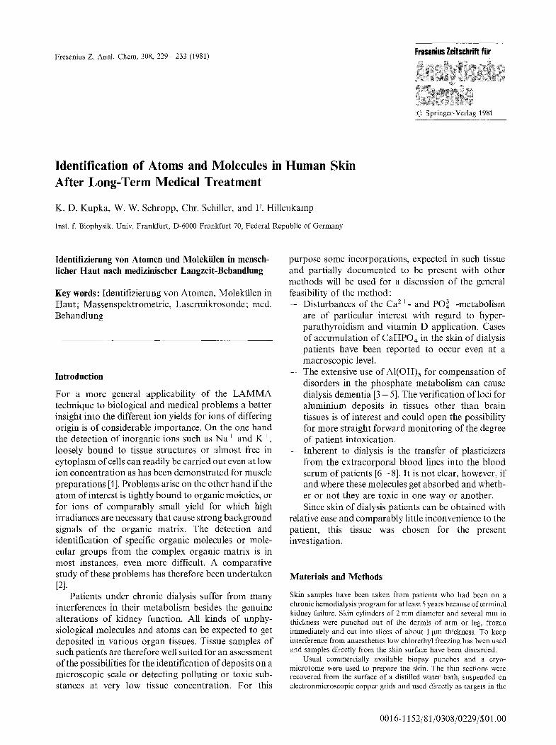
Typical spectra of skin of a dialysis patient are depicted in Fig. 1. The spectra have been taken at different irradiances thus monitoring different aspects of the sample composition. At low power no damage of the sample was seen. At this irradiance only the quasi free alkali metals either from the tissue itself or from contamination and some unspecific organic ions from the surface of the sample are detected.
Figure l a displays the typical pattern that one observes at moderate irradiance with many different organic samples. Sodium, potassium and varying amounts of calcium at relative masses mr/Z = 23, 39 and 41, and 40 are seen because they are easily ionized and are present at relatively high concentrations in most biological samples and as surface contaminants. Peaks mr/Z = 56 and 72 must be of organic origin and at lower irradiance most probably originate from the surface of the sample as well.
Figure 1 b represents the spectrum of a precipitated CaHPO4 crystal from an itching part of the skin of a dialysis patient. As expected the Ca + signal at m,/Z = 40 shows up as the base peak in the otherwise typical desorption spectrum. A strong correlation between this base peak and the signal at rn,/Z = 57, attributed to CaOH + [10] has always been observed in the spectra of such crystals.
Q
/
/
~5 50 75 100 125 150 Z 17~
Fig. l a - e . Positive ions of human skin from a patient on chronic dialysis, a and b irradiance below damage threshold E = 3 x 10⁸ W/ cm2; b probably displaying signals of an enclosed CaHPO 4 precip- itate; c irradiance above damage threshold E = 4 • 10 s W/cm 2
This example illustrates the usefulness of a "de- sorption mode" of the LAMMA method which has the advantage of spectra that display only a small number of well known peaks. It is applicable to those substances that have an ion yield comparable to the ones com- monly observed in surface desorption spectra. The simple spectra with mostly known peaks facilitate the identification of the unknown one's.
Problems arise if a substance of interest occurs at concentrations yielding signals comparable to the back- ground signal or if significant ionization of the atom or molecule under investigation occurs at higher Jr- radiances only. This applies to the two signals at mr/Z = 27 and around 139.
The peak at mr/Z = 27 in spectra b and c of Fig. 1 though small occurs regularly and could originate from small aluminium deposits in the skin. At low mass numbers like 27 the number of competing organic fragment ions is limited. Out of the biological tissue molecular ions CNH + and C2H 3 + can be formed. The limited mass resolution of the time-of-flight mass spectrometer used, does not allow for a differentiation between the aluminium ions and organic fragments due to the mass defect. Peaks at 26 and 28 have regularly been observed in spectra of industrial organic polymers at high irradiances and are attributed to C, Hm moieties.
K. D. Kupka et al. : Identification of Atoms and Molecules in Human Skin 231
AL~UN~N + A L
A L B U N I N
[ I I f I Mr 25 50 100 150 200 - -
Z
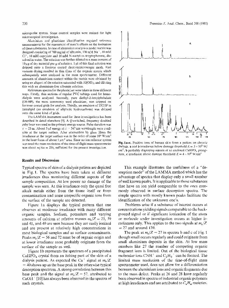
Fig. 2. Positive ions of albumin test solution (composition is given in the text) with (a) and without (b) additional AI(OH)3 content (E = 5 x 10 8 W/cm2). Original computer print
z
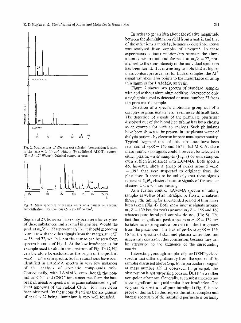
Fig. 3. Mass spectrum of plasma water of a patient on chronic hemofiltration. Positive ions (E = 2 x 10 8 W/cm 2)
Signals at 27, however, have only been seen for very few of these substances and at small intensities. Would the peak at m~/Z = 27 represent CzH~, it should moreover correlate with the other signals from the matrix at rnr/Z = 56 and 72, which is not the case as can be seen from spectra b and c of Fig. 1. At the low irradiance as for example used to obtain the spectrum of Fig. lb C2H~- can therefore be excluded as the origin of the peak a t m/Z = 27 in skin spectra. So far radical ions have been identified in LAMMA spectra in very few instances of the analysis of aromatic compounds only. Consequently, with LAMMA, even though the non- radical CN and CNO- ions sometimes form the base peak in negative spectra of organic substances, signif- icant amounts of the radical CNH § ion have never been observed. By these considerations the assignment of m/Z = 27 being aluminium is very well founded.
In order to get an idea about the relative magnitude between the aluminium ion yield from a matrix and that of the other ions a model substance as described above was analyzed from samples of 1 pg/~tm 2. In these experiments a linear relationship between the alum- inium concentration and the peak at rnr/Z = 27, nor- malized to the sum-intensity of the individual spectrum has been found. It is interesting to note that at higher mass content per area, i.e. for thicker samples, the A1 + signal vanishes. This points to the importance of using thin samples for LAMMA analysis.
Figure 2 shows two spectra of standard samples with and without aluminium additive. As expected only a negligible signal is detected at mass number 27 from the pure matrix sample.
Detection of a specific molecular group out of a complex organic matrix is an even more difficult task. The detection of signals of the phthatate plasticizer dissolved out of the blood line tubing has been chosen as an example for such an analysis. Such phthalates have been shown to be present in the plasma water of dialysis patients by electron impact mass spectrometry. Typical fragment ions of this substance have been recorded at rn/Z = 149 and 167 in E.I.M.S. At these mass numbers no signals could, however, be detected in either plasma water samples (Fig. 3) or skin samples, even at high irradiances with LAMMA. Both spectra do, however, show a group of peaks around rnr/Z = 139 + that were suspected to originate from the plasticizer. It seems to be unlikely that these signals represent C, Hm-clusters because signals of the smaller clusters 2 < n < 5 are missing.
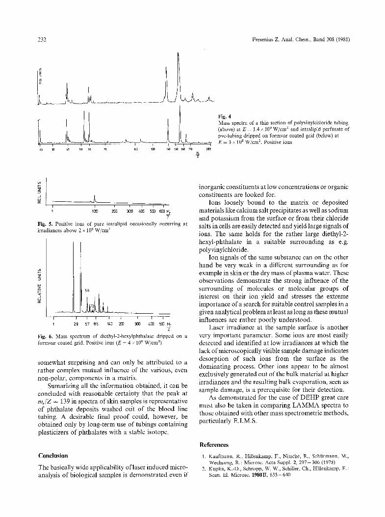
As a further control LAMMA spectra of tubing samples as well as of an intralipid perfusate, circulated through the tubing for an extended period of time, have been taken (Fig. 4). Both show intense signals around rn/Z = 139 besides peaks around m~/Z = 156 and 167 whereas pure intralipid samples do not (Fig. 5). The fact that a significant peak appears at m/Z = 139 can be taken as a strong indication that it indeed originates from the plasticizer. The lack of peaks at mr/Z = 156, 167 in the spectra of skin and plasma water does not necessarily contradict this conclusion, because they can be attributed to the influence of the surrounding matrix.
Interestingly enough samples of pure D EH P yielded spectra that differ significantly from the spectra of the samples discussed above (Fig. 6). In particular no signal at mass number /39 is observed. In principal, this observation is not surprising because D EH P is a rather non-polar substance. Generally, such substances do not show significant ion yield under laser irradiation. The very simple spectrum of pure intralipid (Fig. 5) is also proof of this fact. In this respect the rather complex and intense spectrum of the intralipid perfusate is certainly
232 Fresenius Z. Anal. Chem., Band 308 (1981)
I I I l I I
2 3 3 0 z,O 50 56 7O 100
I
1211 %0 150 150 170 200
Fig. 4 Mass spectra of a thin section of polyvinylchloride tubing (above) at E = 1.4 x 10 9 W/cm 2 and intralipid perfusate of pvc-tubing dripped on formvar coated grid (below) at E = 3 • 10 8 W/cm 2. Positive ions
. J
L I I 1 I [ [ I
1 100 200 3 0 0 /.00 500 500 N r
Fig. 5. Positive ions of pure intralipid occasionally occurring at irradiances above 2 x 10 9 W/cm 2
I I I I I
1 29 20] 300 400 500 N r
z
5 6
I I ~ I
57 @5 140
Fig. 6. Mass spectrum of diethyl-2-hexylphthalate dripped on a formvar coated grid. Positive ions (E = 4 x 10 8 W/cm 2)
somewhat surprising and can only be attributed to a rather complex mutual influence of the various, even non-polar, components in a matrix.
Sumarizing all the information obtained, it can be concluded with reasonable certainty that the peak at mr/Z = 139 in spectra of skin samples is representative of phthalate deposits washed ot/t of the blood line tubing. A desirable final proof could, however, be obtained only by long-term use of tubings containing plasticizers of phthalates with a stable isotope.
Conclusion
The basically wide applicability of laser induced micro- analysis of biological samples is demonstrated even if
inorganic constituents at low concentrations or organic constituents are looked for.
Ions loosely bound to the matrix or deposited materials like calcium salt precipitates as well as sodium and potassium from the surface or from their chloride salts in cells are easily detected and yield large signals of ions. The same holds for the rather large diethyl-2- hexyl-phthalate in a suitable surrounding as e.g. polyvinylchloride.
Ion signals of the same substance can on the other hand be very weak in a different surrounding as for example in skin or the dry mass of plasma water. These observations demonstrate the strong influence of the surrounding of molecules or molecular groups of interest on their ion yield and stresses the extreme importance of a search for suitable control samples in a given analytical problem at least as long as these mutual influences are rather poorly understood.
Laser irradiance at the sample surface is another very important parameter. Some ions are most easily detected and identified at low irradiances at which the lack of microscopically visible sample damage indicates desorption of such ions from the surface as the dominating process. Other ions appear to be almost exclusively generated out of the bulk material at higher irradiances and the resulting bulk evaporation, seen as sample damage, is a prerequisite for their detection.
As demonstrated for the case of DEHP great care must also be taken in comparing LAMMA spectra to those obtained with other mass spectrometric methods, particularly E.I.M.S.
References
1. Kaufmann, R., Hillenkamp, F., Nitsche, R., Schfirmann, M., Wechsung, R. : Microsc. Acta Suppl. 2, 297--306 (1978)
2. Kupka, K.-D., Schropp, W. W., Schiller, Ch., Hillenkamp, F. : Scan. El. Microsc. 1980II, 635-640

K. D. Kupka et al. : Identification of Atoms and Molecules in Human Skin 233
3. Dunea, G., Mahurka, S. D., Mamdani, B., Smith, E. C.: Int. Med. 88, 502-504 (1978)
4. Ward, M. K., Feest, T. G., Ellis, H. A., Parkinson, T. S., Kerr, D. N. S. : Lancet 1978, 841- 845
5. Dermott, J. R., Smith, A. I. : Lancet 1978, 901-903 6. Jaeger, R. J., Rubin, R. J.: Science 170, 460-461 (1970) 7. Omo, K., Tikeda, T., Fukumitsu, T., Tatsukana, R., Wakimoto,
T. : Eur. Dial. Transpl. Ass. XII, 5 7 l - 576 (1975)
8. Hillmann, L. S., Goodwin, S. L., Sherman, W. R. : New Engl. J. Med. 292, Nr. 8, 381-386 (1975)
9. Hillenkamp, F., Uns61d, E., Kaufmann, R., Nitsche, R. : Appl. Phys. 8, 341 (1975)
10. Heinen, H. J., Wechsung, R., Vogt, H., Hillenkamp, F., Kaufmann, R. : Acta Physica Austriaca 20, 257-272 (1979)
Received May 7, 1981