Embed Size (px)
Citation preview
DOI: 10.1002/cbic.200800219
Identification of Additional Players in the AlternativeBiosynthesis Pathway to Isovaleryl-CoA in theMyxobacterium Myxococcus xanthusHelge B. Bode,[a] Michael W. Ring,[a] Gertrud Schw�r,[a] Matthias O. Altmeyer,[a]
Carsten Kegler,[a] Ivy R. Jose,[b] Mitchell Singer,[b] and Rolf M'ller*[a]
Introduction
Leucine is not only one of the major amino acids in proteins,but it is also essential for the growth of the myxobacteriumMyxococcus xanthus.[1] M. xanthus is a predatory organism andfeeds on other bacteria and fungi; therefore leucine is not ex-pected to be in short supply during growth. Amino acid starva-tion results in the induction of the developmental life cycle in-cluding myxospore formation, which allows the species to sur-vive harsh conditions.[2] Leucine is also important for M. xan-thus because leucine-derived iso-branched fatty acids (FAs)represent the dominant FAs in M. xanthus and myxobacteria ingeneral.[3–7] The biosynthesis of these iso-FAs in myxobacteriarequires isovaleryl-CoA (IV-CoA) as starting unit. IV-CoA is usu-ally derived from the transamination of leucine to 2-ketoiso-caproic acid and subsequent oxidative decarboxylation to IV-CoA by the Bkd complex, which is also involved in the degra-dation of valine and isoleucine.[8] A reduction of the amount ofiso-FAs (e.g. , in bkd mutants) results in delayed aggregationand reduced myxospore formation under starvation conditionsin M. xanthus.[7,9, 10] This developmental phenotype can be ex-plained by the fact that iso-FA-derived etherlipids are specifi-cally produced within the myxospore and seem to representthe dominant lipids in mature myxospores.[10] In addition tothe proposed structural function(s) of leucine-derived lipids itcannot be excluded that signals derived from iso-FAs play arole in myxobacterial fruiting-body formation as previouslysuggested.[11]
One indication of the importance of leucine-derived com-pounds is the finding that M. xanthus and other myxobacteriaexhibit an alternative pathway to IV-CoA, which is also the pre-
cursor for compounds other than FAs (e.g. , myxobacterial sec-ondary metabolites like myxothiazol,[12] myxalamids[13,14] or aur-afurons[15]). Feeding experiments in M. xanthus and Stigmatellaaurantiaca led to the prediction of this alternative pathwaythat branches off the well-known mevalonate-dependent iso-prenoid biosynthesis (Scheme 1).[9, 16,17] This alternative pathwayis almost inactive during vegetative growth when leucine ispresent. However, it is highly induced in bkd mutants andduring fruiting-body formation when leucine and consequentlyIV-CoA are limited. We previously confirmed that 3-hydroxy-3-methylglutaryl-CoA (HMG-CoA) synthase (MvaS), which catalyz-es the formation of HMG-CoA from acetoacetyl-CoA andacetyl-CoA, is involved in this alternative pathway.[9]
Here, we show that mvaS is part of a five-gene operon (aibR,MXAN_4264, MXAN_4265, MXAN_4266, mvaS), the expressionof which is up-regulated in bkd mutants (Scheme 1). Moreover,the expression of several other genes is altered in bkd mutants,which might explain their complex phenotype. The leucine
Isovaleryl-CoA (IV-CoA) is usually derived from the degradation ofleucine by using the Bkd (branched-chain keto acid dehydrogen-ase) complex. We have previously identified an alternative path-way for IV-CoA formation in myxobacteria that branches fromthe well-known mevalonate-dependent isoprenoid biosynthesispathway. We identified 3-hydroxy-3-methylglutaryl-CoA (HMG-CoA) synthase (MvaS) to be involved in this pathway in Myxo-coccus xanthus, which is induced in mutants with impaired leu-cine degradation (e.g. , bkd�) or during myxobacterial fruiting-body formation. Here, we show that the proteins required for leu-cine degradation are also involved in the alternative IV-CoA bio-synthesis pathway through the efficient catalysis of the reverse
reactions. Moreover, we conducted a global gene-expression ex-periment and compared vegetative wild-type cells with bkd mu-tants, and identified a five-gene operon that is highly up-regulat-ed in bkd mutants and contains mvaS and other genes that aredirectly involved in the alternative pathway. Based on our experi-ments, we assigned roles to the genes required for the formationof IV-CoA from HMG-CoA. Additionally, several genes involved inouter-membrane biosynthesis and a plethora of genes encodingregulatory proteins were decreased in expression levels in thebkd� mutant; this explains the complex phenotype of bkd mu-tants including a lack of adhesion in developmental submerseculture.
[a] Dr. H. B. Bode, M. W. Ring, G. Schw,r, M. O. Altmeyer, Dr. C. Kegler,Prof. Dr. R. M0llerInstitut f0r Pharmazeutische BiotechnologieUniversit,t des Saarlandes, Postfach 15115066041 Saarbr0cken (Germany)Fax: (+49)681-3025473E-mail : [email protected]
[b] I. R. Jose, Prof. Dr. M. SingerDepartment of Microbiology, One Shields AvenueUniversity of California Davis, Davis CA 94616 (USA)
Supporting information for this article is available on the WWW underhttp://www.chembiochem.org or from the author.
ChemBioChem 0000, 00, 1 – 14 E 2008 Wiley-VCH Verlag GmbH&Co. KGaA, Weinheim &1&
These are not the final page numbers! ��
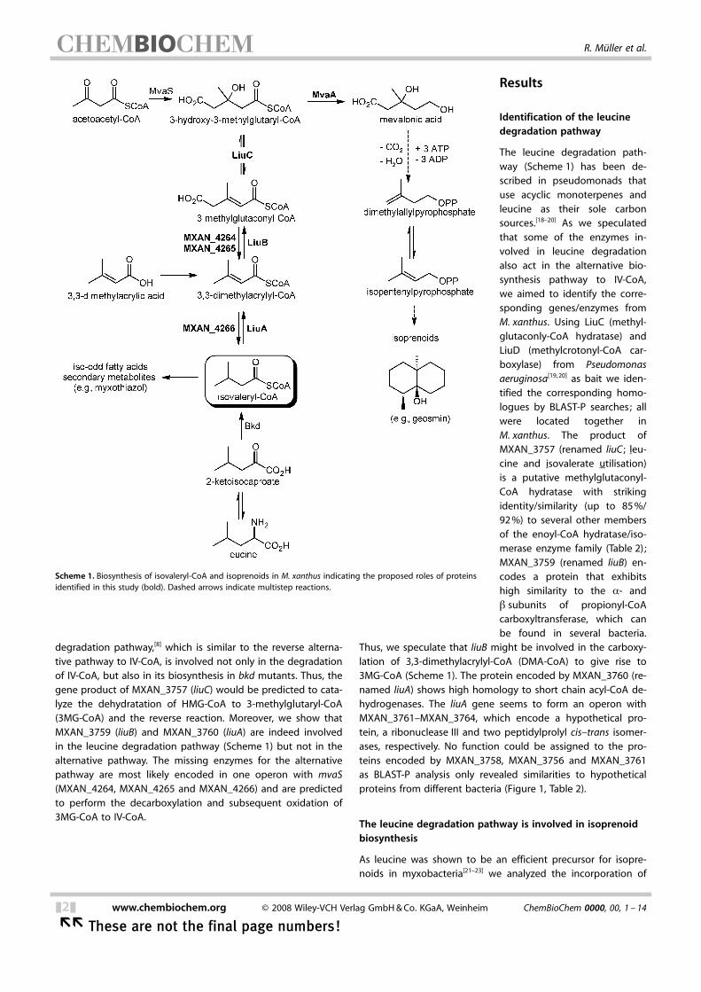
degradation pathway,[8] which is similar to the reverse alterna-tive pathway to IV-CoA, is involved not only in the degradationof IV-CoA, but also in its biosynthesis in bkd mutants. Thus, thegene product of MXAN_3757 (liuC) would be predicted to cata-lyze the dehydratation of HMG-CoA to 3-methylglutaryl-CoA(3MG-CoA) and the reverse reaction. Moreover, we show thatMXAN_3759 (liuB) and MXAN_3760 (liuA) are indeed involvedin the leucine degradation pathway (Scheme 1) but not in thealternative pathway. The missing enzymes for the alternativepathway are most likely encoded in one operon with mvaS(MXAN_4264, MXAN_4265 and MXAN_4266) and are predictedto perform the decarboxylation and subsequent oxidation of3MG-CoA to IV-CoA.
Results
Identification of the leucinedegradation pathway
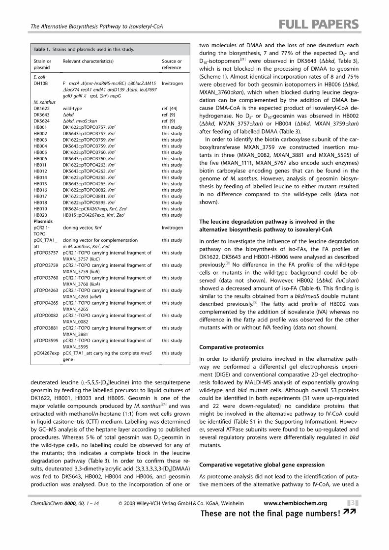
The leucine degradation path-way (Scheme 1) has been de-scribed in pseudomonads thatuse acyclic monoterpenes andleucine as their sole carbonsources.[18–20] As we speculatedthat some of the enzymes in-volved in leucine degradationalso act in the alternative bio-synthesis pathway to IV-CoA,we aimed to identify the corre-sponding genes/enzymes fromM. xanthus. Using LiuC (methyl-glutaconly-CoA hydratase) andLiuD (methylcrotonyl-CoA car-boxylase) from Pseudomonasaeruginosa[19,20] as bait we iden-tified the corresponding homo-logues by BLAST-P searches; allwere located together inM. xanthus. The product ofMXAN_3757 (renamed liuC ; leu-cine and isovalerate utilisation)is a putative methylglutaconyl-CoA hydratase with strikingidentity/similarity (up to 85%/92%) to several other membersof the enoyl-CoA hydratase/iso-merase enzyme family (Table 2);MXAN_3759 (renamed liuB) en-codes a protein that exhibitshigh similarity to the a- andb subunits of propionyl-CoAcarboxyltransferase, which canbe found in several bacteria.
Thus, we speculate that liuB might be involved in the carboxy-lation of 3,3-dimethylacrylyl-CoA (DMA-CoA) to give rise to3MG-CoA (Scheme 1). The protein encoded by MXAN_3760 (re-named liuA) shows high homology to short chain acyl-CoA de-hydrogenases. The liuA gene seems to form an operon withMXAN_3761–MXAN_3764, which encode a hypothetical pro-tein, a ribonuclease III and two peptidylprolyl cis–trans isomer-ases, respectively. No function could be assigned to the pro-teins encoded by MXAN_3758, MXAN_3756 and MXAN_3761as BLAST-P analysis only revealed similarities to hypotheticalproteins from different bacteria (Figure 1, Table 2).
The leucine degradation pathway is involved in isoprenoidbiosynthesis
As leucine was shown to be an efficient precursor for isopre-noids in myxobacteria[21–23] we analyzed the incorporation of
Scheme 1. Biosynthesis of isovaleryl-CoA and isoprenoids in M. xanthus indicating the proposed roles of proteinsidentified in this study (bold). Dashed arrows indicate multistep reactions.
&2& www.chembiochem.org E 2008 Wiley-VCH Verlag GmbH&Co. KGaA, Weinheim ChemBioChem 0000, 00, 1 – 14
�� These are not the final page numbers!
R. M0ller et al.
deuterated leucine (l-5,5,5-[D3]leucine) into the sesquiterpenegeosmin by feeding the labelled precursor to liquid cultures ofDK1622, HB001, HB003 and HB005. Geosmin is one of themajor volatile compounds produced by M. xanthus[24] and wasextracted with methanol/n-heptane (1:1) from wet cells grownin liquid casitone–tris (CTT) medium. Labelling was determinedby GC–MS analysis of the heptane layer according to publishedprocedures. Whereas 5% of total geosmin was D3-geosmin inthe wild-type cells, no labelling could be observed for any ofthe mutants; this indicates a complete block in the leucinedegradation pathway (Table 3). In order to confirm these re-sults, deuterated 3,3-dimethylacrylic acid (3,3,3,3,3,3-[D6]DMAA)was fed to DK5643, HB002, HB004 and HB006, and geosminproduction was analysed. Due to the incorporation of one or
two molecules of DMAA and the loss of one deuterium eachduring the biosynthesis, 7 and 77% of the expected D5- andD10-isotopomers
[21] were observed in DK5643 (Dbkd, Table 3),which is not blocked in the processing of DMAA to geosmin(Scheme 1). Almost identical incorporation rates of 8 and 75%were observed for both geosmin isotopomers in HB006 (Dbkd,MXAN_3760::kan), which when blocked during leucine degra-dation can be complemented by the addition of DMAA be-cause DMA-CoA is the expected product of isovaleryl-CoA de-hydrogenase. No D5- or D10-geosmin was observed in HB002(Dbkd, MXAN_3757::kan) or HB004 (Dbkd, MXAN_3759::kan)after feeding of labelled DMAA (Table 3).In order to identify the biotin carboxylase subunit of the car-
boxyltransferase MXAN_3759 we constructed insertion mu-tants in three (MXAN_0082, MXAN_3881 and MXAN_5595) ofthe five (MXAN_1111, MXAN_5767 also encode such enzymes)biotin carboxylase encoding genes that can be found in thegenome of M. xanthus. However, analysis of geosmin biosyn-thesis by feeding of labelled leucine to either mutant resultedin no difference compared to the wild-type cells (data notshown).
The leucine degradation pathway is involved in theACHTUNGTRENNUNGalternative biosynthesis pathway to isovaleryl-CoA
In order to investigate the influence of the leucine degradationpathway on the biosynthesis of iso-FAs, the FA profiles ofDK1622, DK5643 and HB001-HB006 were analysed as describedpreviously.[9] No difference in the FA profile of the wild-typecells or mutants in the wild-type background could be ob-served (data not shown). However, HB002 (Dbkd, liuC ::kan)showed a decreased amount of iso-FA (Table 4). This finding issimilar to the results obtained from a bkd/mvaS double mutantdescribed previously.[9] The fatty acid profile of HB002 wascomplemented by the addition of isovalerate (IVA) whereas nodifference in the fatty acid profile was observed for the othermutants with or without IVA feeding (data not shown).
Comparative proteomics
In order to identify proteins involved in the alternative path-way we performed a differential gel electrophoresis experi-ment (DIGE) and conventional comparative 2D-gel electropho-resis followed by MALDI-MS analysis of exponentially growingwild-type and bkd mutant cells. Although overall 53 proteinscould be identified in both experiments (31 were up-regulatedand 22 were down-regulated) no candidate proteins thatmight be involved in the alternative pathway to IV-CoA couldbe identified (Table S1 in the Supporting Information). Howev-er, several ATPase subunits were found to be up-regulated andseveral regulatory proteins were differentially regulated in bkdmutants.
Comparative vegetative global gene expression
As proteome analysis did not lead to the identification of puta-tive members of the alternative pathway to IV-CoA, we used a
Table 1. Strains and plasmids used in this study.
Strain orplasmid
Relevant characteristic(s) Source orreference
E. coliDH10B F� mcrA D(mrr-hsdRMS-mcrBC) f80lacZDM15
DlacX74 recA1 endA1 araD139 D ACHTUNGTRENNUNG(ara, leu)7697galU galK l� rpsL (Strr) nupG
Invitrogen
M. xanthusDK1622 wild-type ref. [44]DK5643 Dbkd ref. [9]DK5624 Dbkd, mvaS ::kan ref. [9]HB001 DK1622::pTOPO3757, Kmr this studyHB002 DK5643::pTOPO3757, Kmr this studyHB003 DK1622::pTOPO3759, Kmr this studyHB004 DK5643::pTOPO3759, Kmr this studyHB005 DK1622::pTOPO3760, Kmr this studyHB006 DK5643::pTOPO3760, Kmr this studyHB011 DK1622::pTOPO4263, Kmr this studyHB012 DK5643::pTOPO4263, Kmr this studyHB014 DK1622::pTOPO4265, Kmr this studyHB015 DK5643::pTOPO4265, Kmr this studyHB016 DK1622::pTOPO0082, Kmr this studyHB017 DK1622::pTOPO3881, Kmr this studyHB018 DK1622::pTOPO5595, Kmr this studyHB019 DK5624::pCK4267exp, Kmr, Zeor this studyHB020 HB015::pCK4267exp, Kmr, Zeor this studyPlasmidspCR2.1-TOPO
cloning vector, Kmr Invitrogen
pCK_T7A1_att
cloning vector for complementationin M. xanthus, Kmr, Zeor
this study
pTOPO3757 pCR2.1-TOPO carrying internal fragment ofMXAN_3757 (liuC)
this study
pTOPO3759 pCR2.1-TOPO carrying internal fragment ofMXAN_3759 (liuB)
this study
pTOPO3760 pCR2.1-TOPO carrying internal fragment ofMXAN_3760 (liuA)
this study
pTOPO4263 pCR2.1-TOPO carrying internal fragment ofMXAN_4263 (aibR)
this study
pTOPO4265 pCR2.1-TOPO carrying internal fragment ofMXAN_4265
this study
pTOPO0082 pCR2.1-TOPO carrying internal fragment ofMXAN_0082
this study
pTOPO3881 pCR2.1-TOPO carrying internal fragment ofMXAN_3881
this study
pTOPO5595 pCR2.1-TOPO carrying internal fragment ofMXAN_5595
this study
pCK4267exp pCK_T7A1_att carrying the complete mvaSgene
this study
ChemBioChem 0000, 00, 1 – 14 E 2008 Wiley-VCH Verlag GmbH&Co. KGaA, Weinheim www.chembiochem.org &3&
These are not the final page numbers! ��
The Alternative Biosynthesis Pathway to Isovaleryl-CoA
global DNA microarray approach to examine vegetative geneexpression patterns in bkd mutant cells and wild-type cells. Asthe alternative pathway is highly active in bkd cells we expect-ed putative genes involved in this pathway to exhibit signifi-cantly higher expression levels in bkd� cells. As described inthe Experimental Section, wild-type and bkd mutant cells were
grown to a density of 5N108 cellsmL�1 (midexponentialphase), total cellular RNA was harvested, and the RNA wasused for comparative DNA microarray studies. Six independentbiological experiments were performed, and based on signifi-cance analysis of microarrays (SAM)[25] approximately 509genes were statistically altered in their expression patterns(> or <2.5-fold) in bkd mutant cells compared to wild-typecells. Of these, the largest effect was seen on genes the expres-sion of which was suppressed in the bkd mutant strain(471 genes), while 38 genes showed an increase in expression.A partial list of these genes is presented in Table 5 and a com-plete list of all significantly down- and up-regulated genes (>or <2.5-fold) is provided in Table S2. The corresponding geneproducts primarily fall into three groups: 1) hypothetical orconserved hypothetical proteins (~37%); 2) membrane pro-teins, membraneassociated proteins or proteins involved inmembrane/lipid ACHTUNGTRENNUNGassociated processes (25%), and 3) regulatorygenes (11%).The highest up-regulation was observed for MXAN_4263
(6.1-fold). It shows similarity to TetR-like regulators and wastherefore renamed aibR (alternative isovaleryl-CoA biosynthesis
Figure 1. Organization of the genomic regions that encode proteins involved in A) the leucine degradation pathway and B) other genes postulated to be in-volved in the alternative IV-CoA biosynthesis pathway in Myxococcus xanthus DK1622. Numbers refer to the M. xanthus gene nomenclature (MXAN_). Genesshown in white are not thought to be involved in IV-CoA formation. For a detailed description of the respective proteins see Table 2 and the text.
Table 2. Proteins located in the gene cluster involved in leucine degradation and in the alternative IV-CoA biosynthesis pathway, their deduced function,protein size and closest homologues.
Protein Size Deduced function Closest homologue[aa] Protein Origin Identities/ Accession number
positives [%]
MXAN_3756 654 hypothetical protein STIAU_6492 Stigmatella aurantiaca 62/74 EAU65620LiuC 258 3-methylglutaconyl-CoA hydratase STIAU_6496 S. aurantiaca 85/92 EAU65644MXAN_3758 119 hypothetical protein STIAU_6515 S. aurantiaca 66/80 EAU64347LiuB 513 carboxyl transferase (a and b subunit) STIAU_6516 S. aurantiaca 86/93 EAU64343LiuA 381 short chain acyl-CoA dehydrogenase STIAU_6517 S. aurantiaca 85/95 EAU64345MXAN_3761 288 hypothetical protein STIAU_6518 S. aurantiaca 61/63 EAU64351MXAN_3762 260 ribonuclease III STIAU_6520 S. aurantiaca 63/80 EAU64340MXAN_3763 268 peptidylprolyl cis–trans isomerase STIAU_6521 S. aurantiaca 71/80 EAU64336MXAN_3764 198 peptidylprolyl cis–trans isomerase MXAN_1176 M. xanthus 69/82 ABF87515AibR 228 TetR-like transcriptional regulator STIAU_3247 S. aurantiaca 76/90 EAU65108MXAN_4264 265 glutaconate CoA-transferase, subunit A STIAU_3246 S. aurantiaca 76/84 EAU65098MXAN_4265 246 glutaconate CoA-transferase, subunit B STIAU_3245 S. aurantiaca 80/88 EAU65091MXAN_4266 345 dehydrogenase, Zn binding STIAU_3244 S. aurantiaca 82/92 EAU65096MvaS 418 3-hydroxy-3-methylglutaryl-CoA synthase STIAU_3242 S. aurantiaca 78/88 EAU65112MXAN_5157 102 hypothetical protein SACE_2015 Saccharopolyspora erythraea 57/70 CAM01325Bcap 249 branched-chain amino acid permease RHA1_ro07263 Rhodococcus sp. RHA1 45/61 ABG99027
Table 3. Incorporation [%] of labelled precursors in geosmin in differentM. xanthus strains.
Strain Condition [D3]Geosmin [D5]Geosmin ACHTUNGTRENNUNG[D10]Geosmin
DK1622 [D3]leucine 5.3 –[a] –[a]
[D6]DMAA –[a] 0.0[b] 0.0[b]
DK5643 (Dbkd) [D3]leucine 0.0[b] –[a] –[a]
[D6]DMAA –[a] 7.2 78HB001 (liuC) [D3]leucine 0.0[b] –[a] –[a]
HB002 (Dbkd, liuC) [D6]DMAA –[a] 0.0[b] 0.0[b]
HB003 (liuB) [D3]leucine 0.0[b] –[a] –[a]
HB004 (Dbkd, liuB) [D6]DMAA –[a] 0.0[b] 0.0[b]
HB005 (liuA) [D3]leucine 0.0[b] –[a] –[a]
HB006 (Dbkd, liuA) [D6]DMAA –[a] 7.6 75
[a] Not expected and not detected; [b] expected but not detected.
&4& www.chembiochem.org E 2008 Wiley-VCH Verlag GmbH&Co. KGaA, Weinheim ChemBioChem 0000, 00, 1 – 14
�� These are not the final page numbers!
R. M0ller et al.
regulator), which is the first gene within a five-gene operon(Figure 1) that includes mvaS (MXAN_4267; 2.62-fold up-regu-lated). The mvaS gene was already shown to be up-regulatedin a bkd mutant.[9] However, in an early draft version of theM. xanthus genome[26] mvaS was located on a different stretchof contiguous DNA and thus no operon structure could be de-duced for mvaS and its neighbouring genes in previous work.Other genes in this operon are MXAN_4264 and MXAN_4265which show similarity to the two subunits of glutaconyl-CoAtransferases. Additionally, MXAN_4266, which encodes a pro-tein that is similar to Zn-dependent dehydrogenases, was iden-tified in the operon and was found to be up-regulated by2.73-fold.The only other gene that might be involved in the alterna-
tive pathway due to its predicted enzymatic function wasMXAN_5158 renamed bcap as it encodes a putative branched-chain amino acid permease that forms an operon with a genethat encodes a hypothetical protein (Figure 1, Tables 2 and 5).Additionally, several sequences encoding regulatory genes andtransporters as well as hypothetical proteins were up-regulated(Table 5). The majority of genes that were down-regulatedencode hypothetical or regulatory proteins (Tables 5 and S2).
Additionally, several genes encoding proteins involved in lipo-polysaccharide (MXAN_6398, MXAN_4726, MXAN_1639) orsugar biosynthesis (MXAN_6497) were found to be down-regu-lated.In order to confirm the microarray data we performed quan-
titative reverse transcript PCR (qRT-PCR) analysis of selectedgenes as described previously.[9] As expected, aibR, MXAN_4264, MXAN_4265 and MXAN_4266 showed a similar transcrip-tion profile as described for mvaS.[9] Overall, the high level oftranscripts in the bkd mutant was decreased to wild-typelevels by the addition of IVA (Figure 2A–D). No such resultscould be obtained for bcap, liuC and liuA, which were alsoACHTUNGTRENNUNGanalyzed (Figure 2E–G). We also analyzed the expression ofMXAN_5020 (mvaA) and MXAN_5021, which encode the HMG-CoA reductase and isopentenyldiphosphate isomerase, respec-tively. Both enzymes are involved in the transformation ofHMG-CoA to isopentenyldiphosphate and dimethylallydiphos-phate, respectively, which are the universal building blocks ofthe isoprenoids. However, no difference between the expres-sion of the latter two genes was observed between the wild-type and bkd mutant cells with or without the addition of IVA(Figure 2H, mvaA, and data not shown, MXAN_5021).
Table 4. Fatty acid composition [% of total fatty acids] of vegetative cells of M. xanthus DK1622 (wild type), DK5643 (Dbkd), HB002 (Dbkd, liuC ::kan), HB012(Dbkd, aibR ::kan), HB015 (Dbkd, MXAN_4265::kan) ; both with and without the addition of isovalerate to the growth medium (1 mm) ; HB19 (Dbkd, mvaS ::-kan, mvaS+) and HB020 (Dbkd, MXAN_4265::kan, mvaS+). The two key fatty acids iso-15:0 and 16:1w5c are shown in bold.
DK1622 DK5643 HB002 HB012 HB015 HB019 HB020� +IVA � +IVA � +IVA � +IVA
12:0 0.07 0.04 0.07 0.02 0.11 0.12iso-13:0 0.31 0.07 0.41 0.20 0.14 0.19iso-14:0 0.22 0.08 0.11 0.02 0.14 0.12 0.1614:1 isomer 1 1.41 0.39 1.19 0.14 0.63 0.50 1.05 1.50 0.3814:1 isomer 2 0.05 0.17 0.12 0.17 0.08 1.05 0.35 0.2514:0 5.79 5.36 3.77 4.44 2.22 11.64 5.71 12.13 4.54 9.08 8.13iso-15:1w9c 0.32 1.01 0.61 4.54 0.26iso-15:1 isomer 2 0.04iso-15:0 40.10 19.17 52.16 4.01 47.76 3.81 58.85 2.70 56.87 41.37 3.4715:0 1.75 8.46 1.93 18.06 1.54 56.87 12.15 0.82 2.15 14.0715:1 isomer 1 2.43 3.06 1.17 3.91 1.23 0.82 3.24 2.05 1.37 5.2215:1 isomer 2 3.35 0.21 2.06 0.21 2.09 2.05 4.43 1.22 1.47 4.33iso-16:0 0.01 7.17 1.42 7.11 0.63 1.22 2.67 0.19 1.93 2.5116:2w5c,11c 5.46 3.77 5.10 2.74 4.24 0.19 5.20 4.00 6.07 5.49 4.2816:1w11c 0.90 1.48 0.51 1.58 0.49 6.07 2.44 1.10 2.9716:1w5c 17.04 29.14 10.92 37.93 10.87 41.01 17.10 38.23 13.24 15.83 36.1816:0 1.58 5.88 1.85 7.04 1.44 13.24 3.27 11.32 1.41 3.64 12.31iso-17:2w5c,11c 1.93 0.28 1.97 0.04 2.68 1.41 1.28 1.37iso-17:1w11c 0.69 0.27 0.55 0.76 1.28 0.49 0.66iso-17:1w5c 1.73 1.04 1.98 0.18 3.38 0.49 1.14 1.34iso-17:0 2.89 4.66 2.82 1.46 4.88 1.14 4.80 0.31 3.48 4.71 0.7114:0 3-OH 0.45 1.43 0.35 5.96 0.35 3.48 0.32 2.11 0.26 0.59 1.53iso-15:0 3-OH 2.15 1.32 1.85 0.34 2.78 0.26 1.42 0.14 1.82 1.94 0.2216:0 2-OH 0.35 1.02 0.28 1.74 0.29 1.82 0.14 0.94 0.11 0.08 1.5816:0 3-OH 0.38 0.37 0.33 0.90 0.14 0.11 1.88 0.11 0.39 1.53iso-17:0 2-OH 4.10 3.90 3.28 1.51 6.57 0.11 2.96 0.21 2.95 1.42 0.46iso-17:0 3-OH 1.82 0.38 0.25 0.33 0.53 2.95 0.24 0.03 0.23 0.76 0.10iso-15:0 DMA[a] 2.19 0.51 2.06 2.60 0.23 0.49 0.59iso-15:0 OAG[b] 0.74 0.22 0.49 0.08 0.94 0.49 0.25
[a] Dimethylacetal ; [b] 1-O-alkylglycerol.
ChemBioChem 0000, 00, 1 – 14 E 2008 Wiley-VCH Verlag GmbH&Co. KGaA, Weinheim www.chembiochem.org &5&
These are not the final page numbers! ��
The Alternative Biosynthesis Pathway to Isovaleryl-CoA
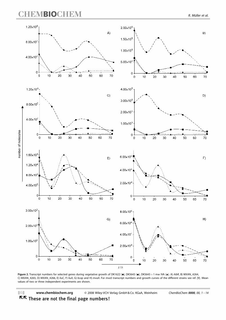
Figure 2. Transcript numbers for selected genes during vegetative growth of DK1622 (&), DK5643 (*), DK5643+1 mm IVA (~). A) AibR, B) MXAN_4264,C) MXAN_4265, D) MXAN_4266, E) liuC, F) liuA, G) bcap and H) mvaA. For mvaS transcript numbers and growth curves of the different strains see ref. [9] . Meanvalues of two or three independent experiments are shown.
&6& www.chembiochem.org E 2008 Wiley-VCH Verlag GmbH&Co. KGaA, Weinheim ChemBioChem 0000, 00, 1 – 14
�� These are not the final page numbers!
R. M0ller et al.
Functional analysis of genes found up-regulated in the bkdmutant
Plasmid insertions into aibR and MXAN_4265 in a wild-typebackground resulted in no change of phenotype with respectto fatty acid and/or geosmin biosynthesis (data not shown) asalso described for a mvaS mutant. However, in a Dbkd back-ground both double mutants showed the phenotype of a bkd/mvaS strain, which is characterized by a very low amount ofiso-FAs (Table 4) and the production of only trace amounts ofgeosmin (Table 3). This phenotype could be complemented towild-type levels by the addition of IVA or DMAA.However, as an insertion into any gene of the aibR--mvaS
operon would most likely exhibit polar effects and thus directlyinfluence expression of mvaS, we complemented the Dbkd/mvaS (DK5624) and Dbkd/MXAN_4265 (HB015) double mu-tants genetically by addition of a copy of mvaS under controlof the constitutive T7A1-promotor;[27] this resulted in strainsHB019 (Dbkd, mvaS ::kan, mvaS+) and HB020 (Dbkd, MXAN_4265::kan, mvaS+). Whereas the fatty acid profile of HB019 wassimilar to the wild-type cells as expected, no complementationwas observed for HB020 (Table 4).Despite several attempts, we were not able to generate a
bcap mutant by plasmid insertion.
Discussion
The leucine degradation pathway is involved in theACHTUNGTRENNUNGalternative pathway to IV-CoA
Plasmid insertion into genes of M. xanthus that have beenidentified by homology searches by using genes from differentPseudomonas strains[19,20] confirmed the involvement of severalgenes in the degradation of leucine also in the alternativepathway to IV-CoA. Mutants of liuA–C still produce isoprenoids,like geosmin, but do not perform leucine-dependent isopre-noid biosynthesis (Table 3). Leucine is a major amino acid con-stituent of bacteria, which are a major food source for M. xan-thus. Therefore, leucine also seems to be a major source forisoprenoids via the leucine degradation pathway that entersthe mevalonate dependent isoprenoid biosynthesis via HMG-CoA.[8] A bkd/liuC double mutant shows a similar phenotype toa bkd/mvaS mutant strain, which produces only residualamounts of iso-FAs. The remaining trace amounts of iso-FAsmight be derived from a second bkd activity.[7] This indicatesthat the liuC-encoded methylglutaconyl-CoA hydratase cataly-ses the hydration of 3-methylglutaconyl-CoA to HMG-CoA andits reverse dehydration. No reduction in the amount of iso-FAswas observed for bkd/liuB or bkd/liuA double mutants. TheLiuB protein shows similarity to the a- and b subunits (the car-boxyltransferase subunits) of a biotin dependent carboxylase,and therefore might well be involved in the carboxylation ofdimethylacryloyl-CoA, whereas the decarboxylation might becatalyzed by a different enzyme (see below). LiuA shows simi-larities to acyl-CoA dehydrogenases, and might be involved inthe oxidation of IV-CoA to DMA-CoA and the reverse reduc-tion. However, at least eleven similar dehydrogenase-encoding
genes are present in the genome[28] which might complementa liuA mutant. Interestingly, this does not seem to occur in theoxidative reaction as a liuA mutant shows no geosmin produc-tion from leucine. However, a bkd/liuA mutant shows the pro-duction of geosmin from labelled DMAA as this complementsthe liuA block of the pathway (Table 3). Alternatively, MXAN_4266 might catalyze the reduction of DMA-CoA to IV-CoA (seebelow).The fact that the expression of genes involved in leucine
degradation/IV-CoA biosynthesis is not increased in bkd mu-tants might indicate that the expression is already maximal(due to the effective use of leucine as carbon source) or thattranslational regulation occurs.We also tried to identify the biotin carboxylase partner of
the LiuB carboxyltransferase. However, disruption of three pos-sible candidates, MXAN_0082, MXAN_3881 and MXAN_5595,did not result in a difference compared to the wild-type cellswith respect to fatty acid or geosmin biosynthesis (data notshown). This might indicate that either one of two remaininggenes that encode such enzymes, MXAN_1111 or MXAN_5767,might be the missing subunit or that these enzymes can func-tionally complement each other, which seems to be morelikely. Interestingly, LiuB is the only “orphan” carboxylase subu-nit as all other carboxylase subunit encoding genes are foundassociated with other carboxylase subunits (data not shown).
Comparative global expression confirmed the complexACHTUNGTRENNUNGphenotype of the bkd mutant
Comparative proteome analysis between wild-type and bkdmutant cells led to the identification of several changes in theproteome of the bkd mutant (Table S1) but did not lead toidentification of enzymes putatively involved in the alternativepathway to IV-CoA. Luckily, comparison of global expressionpattern between wild-type and bkd cells under vegetative con-ditions was much more successful and led to the identificationof a five-gene operon, which was highly expressed in bkd cells(Table 5, Figure 2). Interestingly, only little direct overlap(MXAN_0433, MXAN_0709—both hypothetical proteins—andMXAN_1450—OmpA-related protein) could be observed be-tween the proteome and transcriptome data. This can be dueto a much smaller number of protein spots that have been an-alyzed compared to an almost complete list of genes. The factthat less abundant proteins are much more difficult to detectcompared to less abundant transcripts might be anotherreason for this discrepancy. Additionally, from the way the pro-teome comparisons were made, a difference between no pro-tein at all (sample 1) and small amounts of protein (sample 2)would only be detected in a DIGE experiment but not in thestandard Coomassie experiment with the software used (seethe Experimental Section).
The missing genes of the alternative pathway to IV-CoA areencoded in one operon with mvaS
From the transcriptome analysis the aibR–mvaS operon wasidentified, in which mvaS, which encodes the HMG-CoA syn-
ChemBioChem 0000, 00, 1 – 14 E 2008 Wiley-VCH Verlag GmbH&Co. KGaA, Weinheim www.chembiochem.org &7&
These are not the final page numbers! ��
The Alternative Biosynthesis Pathway to Isovaleryl-CoA
thase, was already identified as being part of the alternativepathway.[9] From the other proteins encoded in this operonAibR shows similarity to TetR-like regulators, MXAN_4264 andMXAN_4265 encode two subunits of a glutaconyl-CoA transfer-ase and MXAN_4266 encodes a dehydrogenase. Glutaconyl-CoA transferases are involved in fermentative glutamate degra-dation and catalyze the transfer of a CoA moiety from acetyl-CoA to glutaconate, a glutamate degradation product.[29] Theresulting glutaconyl-CoA is then decarboxylated to crotonyl-CoA, which is subsequently converted to two acetyl-CoA unitsas shown in Acidaminococcus fermentans.[30,31] The glutaconyl-CoA decarboxylase involved in this process was shown to be amembrane bound sodium pump that consists of four subunitsof which no homologues can be found in M. xanthus, exceptfor the carboxylase a subunit, which shows highest identity/similarity to LiuB (27%/49%) but also to AccB (acetyl-CoA car-boxylase, 24%/43%) and PccB (propionyl-CoA carboxylase,24%/41%). Although glutaconyl-CoA and 3-methylglutaconly-CoA—believed to be an intermediate in the alternative path-way to IV-CoA—differ only in one methyl group, all intermedi-ates in the alternative pathway seem to be CoA-bound andthus no activation of a free acid seems to be required. More-over, MXAN_4264, MXAN_4265 and MXAN_4266 are notACHTUNGTRENNUNGinvolved in the leucine-dependent isoprenoid biosynthesis asa bkd/aibR mutant, which produces only trace amounts ofACHTUNGTRENNUNGgeosmin, can be complemented by the addition of DMAA(Table 3). Moreover, an involvement of glutaconyl-CoA in theconversion of mevalonate to 3MG-CoA in a mevalonate shuntpathway, which was proposed more than 30 years ago,[32] canbe excluded as we performed feeding experiments with [1,2-13C2]mevalonolactone in DK1622 and DK5643, but could notsee any incorporation of label into iso-FAs (data not shown).Similar results were observed earlier from feeding experimentsin bkd mutants of S. aurantiaca Sg a15 which clearly incorpo-rates labelled mevalonolactone (the lactone form of mevalo-nate) into isoprenoids like aurachin but not into isovaleryl-CoAderived compounds like myxothiazol.[17]
In order to elucidate the importance of MXAN_4264, MXAN_4265 and MXAN_4266 with respect to the alternative pathway,we genetically complemented strain HB015 (Dbkd/MXAN_4265) and as a control DK5624 (Dbkd/mvaS) with an intactcopy of mvaS. No complementation of the fatty acid profilewas observed for HB020 (Dbkd, MXAN_4265::kan, mvaS+)whereas wild-type levels of iso-FAs were detected in HB019(Dbkd, mvaS ::kan, mvaS+). This clearly demonstrates a functionof at least MXAN_4265 or MXAN_4266 in the transformation of3-methylglutaconyl-CoA to isovaleryl-CoA. Our current hypoth-esis is, that MXAN_4264 and MXAN_4265 are involved in thedecarboxylation of 3-methylglutaconyl-CoA to 3,3-dimethyl-acrylyl-CoA, and MXAN_4266 is involved in the final reductionto IV-CoA. While the latter reaction sounds reasonable for theencoded enzyme, no decarboxylase function has to our knowl-edge been assigned to enzymes that show homology to CoAtransferases in the literature. Currently MXAN_4264, MXAN_4265 and MXAN_4266 are expressed in order to evaluate thishypothesis in in vitro experiments that employ recombinantenzymes.
Database analysis of the aibR–mvaS operon revealed that itis identical to an operon studied by the Kroos group severalyears ago.[33] However, at the time of their work, no informa-tion about the fifth gene in the operon (mvaS) was availabledue to the small contig size in the draft genome sequence,[26]
as already mentioned. Their aim was to characterize theW4514 regulatory region. The W4514 region is the site of aTn5 lac insertion in the M. xanthus genome that does notdepend on C signalling for expression and yet is expressedwith a timing during development similar to that of promotersthat depend on C signalling. The C signal is the best character-ized signal required for fruiting-body formation in M. xan-thus.[34] It is a protein encoded by the csgA gene, which is in-volved in aggregation and finally also sporulation during de-velopment of M. xanthus. The current hypothesis suggests thatthe CsgA protein is transferred between cells in a cell–cell con-tact dependent manner and it is involved in the expression ofseveral other developmental genes.[35] The W4514 Tn5 lac is in-serted in the third codon of MXAN_4265 and Kroos and collea-gues could show that expression is strongly induced duringdevelopment. This fits well to our previous experiments thatshow that the activity of the alternative pathway to IV-CoA canalso be detected during development in the wild-type.[9] More-over, they could show that MXAN_4263 (aibR) negatively regu-lates the whole operon, but did not detect direct binding ofMXAN_4263 to the promoter region. They speculated that thismight be due to additional low-molecular weight factors thatare missing in the in vitro binding assay similar to the case oftryptophan and the trp repressor.[36] In fact, given the currentknowledge one can speculate even more, that IV-CoA or oneof the pathway intermediates that lead to IV-CoA might besuch a factor. In order to learn more about the function of thisoperon, in vitro studies of the purified proteins are in progressto identify their substrates and regulation.The gene MXAN_5158 (bcap) encoding a branched chain
amino acid transporter was also highly expressed in the bkdmutant. Surprisingly, no mutants in bcap could be obtained inthe wild-type or bkd mutant cells by plasmid insertions despiteseveral attempts. This is surprising as we have usually had noproblems in the generation of mutants using this approach.This might suggest that bcap is essential as it might be an im-portant leucine transporter in M. xanthus, although severalother transporters (most with unknown substrate specificity)can be found in the genome (data not shown).Mutants in the bkd genes have a complex phenotype; they
show not only a reduction in the amount of iso-FAs to half ofthe amount of the wild-type cells,[5, 7] but they are also delayedin aggregation, produce less myxospores[7,10] and can hardlydevelop in submerse culture. For the latter it is crucial that thecells settle at the bottom of a microtiter plate or Petri dish.Whereas the wild-type forms a tightly bound layer of cells atthe bottom of the well, the cell layer of a bkd mutant is veryloose and cells brake away from the bottom very easily (un-published observations). The reason for this might be achange in the exopolysaccharide/fibril (or slime) compositionor in the composition of the outer membrane. Therefore, it isnot surprising that the expression of genes involved in lipid or
&8& www.chembiochem.org E 2008 Wiley-VCH Verlag GmbH&Co. KGaA, Weinheim ChemBioChem 0000, 00, 1 – 14
�� These are not the final page numbers!
R. M0ller et al.
membrane biosynthesis like MXAN_6398, MXAN_4726 andMXAN_1639 (Table 5) is down regulated in a bkd mutant. Theobserved down regulation of lipoproteins like MXAN_2391,MXAN_5414 or MXAN_4653 might also be involved in the non-adhesive phenotype of the bkd mutant.In order to correlate these data to the bkd phenotype, we
compared wild-type and bkd mutant cells in a Trypan blueassay that measures fibril polysaccharide content.[37] In accord-ance with the nonadhesive phenotype and the down-regulat-ed genes in the bkd mutant, only half (4.4%) the level ofTrypan blue binding could be identified compared to the wild-type cells (8.7%); this indicates that only half the amount offibril material is present in the mutants.Interestingly, many more genes could be identified the ex-
pression levels of which were down- rather than up-regulated.Moreover, many of the down-regulated genes encode variousforms of regulatory proteins that might be involved in thecomplex phenotype of the bkd mutant.As M. xanthus contains 18 gene clusters for the biosynthesis
of secondary metabolites[28,38] it is not too surprising that sev-eral of them are influenced in the bkd mutant: expression ofMXAN_4601, MXAN_3619 and taP (MXAN_3932) is strongly re-duced in a bkd mutant. In almost all secondary-metabolite pro-ducing organisms many more biosynthesis gene clusters thansecondary metabolites can be identified,[39] and the biosynthe-sis gene clusters that cannot be correlated to actual com-pounds are usually called “silent”. However, the identificationof MXAN_4601 and MXAN_3619 proves that the correspond-ing biosynthesis gene clusters are not silent but are expressedin wild-type cells as is also the case for taP, which is involvedin myxovirescin biosynthesis.[40] The reason why no secondarymetabolite has been identified for these two biosynthesisgene clusters might simply be a very low production of thecorresponding compound just to name one possible reason.With respect to secondary metabolism, one can conclude fromthese data that actually 12 of the 18 biosynthesis gene clustersare expressed in the wild-type cells during vegetative growth;this is in fact a very high number. Five secondary metaboliteshave already been isolated and correlated to the respectivebiosynthesis gene clusters[14,40–42] and the expression of five ad-ditional biosynthesis gene clusters have been shown by analy-sis of the proteome.[43]
In summary, we could identify the leucine degradation path-way in M. xanthus, which also serves as anabolic pathway toisoprenoids and is involved in the alternative pathway to IV-CoA. Moreover, global expression analysis of a bkd mutant andwild-type cells resulted in the identification of an operon thatharbours a set of genes (aibR, MXAN_4264, MXAN_4265,MXAN_4266, mvaS) that are most likely all involved in the al-ternative IV-CoA biosynthesis pathway. Furthermore, severaladditional genes have been identified that are either up- ordown-regulated, and reveal a complex regulatory network thatis changed in the bkd mutant. In vitro work is currently inprogress to biochemically characterise the proteins encodedby the genes in the operon(s) identified in this study.Moreover, our data offer a link between the myxobacterial
“lifestyle”, leucine biosynthesis and the alternative pathway to
IV-CoA. It has been a puzzle for a long time why M. xanthusfails to synthesize leucine as one of the most common aminoacids although its large genome would easily have the capaci-ty for leucine biosynthesis. A possible explanation for the pred-atory lifestyle of M. xanthus would be that prey are a conven-ient source of leucine. However, it would be very inefficient todegrade all the available prey leucine to IV-CoA, which is re-quired for iso-FA synthesis. The more efficient solution mightbe to use leucine directly for protein biosynthesis and to sup-plement the pathway to IV-CoA and subsequently iso-FAs andsecondary metabolites with endogenous pathways. Similarly,leucine degradation to IV-CoA during starvation conditions(leucine depletion) seems to be very risky as large amounts ofleucine is required for protein synthesis to complete myxobac-terial development. This theory would help to explain why thealternative pathway to IV-CoA is up-regulated during myxobac-terial development.
Experimental Section
Strains, culture conditions, mutant construction and comple-mentation strategy : Myxococcus xanthus DK1622 (wild type)[44]
and all of its descendants were routinely grown in CTT medium[45]
with kanamycin (40 mgmL�1) where appropriate. Feeding experi-ments with [D3]leucine and [D6]DMAA have been described previ-ously.[9,17, 21] For the construction of mutants, internal fragments(~600 bp) of MXAN_3757, MXAN_3759, MXAN_3760, MXAN_4263,MXAN_4265, MXAN_5158, MXAN_0082, MXAN_3881 and MXAN_5595, were amplified by PCR from genomic DNA of DK1622 byusing the oligonucleotides 3757-1 (5’-CCGGAATTCAAGGTCGACGC-3’), 3757-2 (5’-AAGGCCTCTGCGGCGTTGAT-3’), 3759-1 (5’-TTCGTG-ACHTUNGTRENNUNGGAGGACGCGAAGCT-3’), 3759-2 (5’-TCTTCCACCTTCTTGCCGCC-3’),3760-1 (5’-AAGCCGTATGCCCGTGAGTG-3’), 3760-2 (5’-TCCACGTTC-ACHTUNGTRENNUNGTCCAGGACGAG-3’), 4263-1 (5’-ACGAACACCGGAGGACGGAA-3’),4263-2 (5’-CGTGCTCGTGGAGGATGATG-3’), 4265-1 (5’-TGGACATCA-ACHTUNGTRENNUNGCCCCAGCGGAGA-3’), 4265-2b (5’-AACTTCGTCCGCGGCTTGTCC-3’),5158-1 (5’-ATGGGGCATGTGGATCGAAG-3’), 5158-2 (5’-GAACGCCAC-ACHTUNGTRENNUNGGGACTCGTCCA-3’), 06613-1 (5’-TGGCCATTGGTCCGTCTC-3’), 06613-2 (5’-GGTAGCCAATCGCCCGAG-3’), 02236-1 (5’-ATCACCCTGGAG-ACHTUNGTRENNUNGGGCGAC-3’), 02236-2 (5’-TCCTCCTGCCGGGAGATG-3’), 5595-1 (5’-ACGGTCGCCGTCTATTCGGA-3’) and 5595-2 (5’-GGAACATTCCCGCTC-ACHTUNGTRENNUNGCAGAC-3’), respectively. The resulting fragments were cloned intopCR2.1-TOPO (Invitrogen) to give plasmids pTOPO3757,pTOPO3759, pTOPO3760, pTOPO4263, pTOPO4265, pTOPO5158,pTOPO0082, pTOPO3881 and pTOPO5595, which were introducedinto M. xanthus DK1622 or DK5643 (Dbkd)[9] by electroporation asdescribed previously.[14, 46] This resulted in strains HB001–HB006,HB011, HB012, HB014–HB018 (Table 1). The correct integration ofeach plasmid was confirmed for all strains by using a PCR protocolbased on a plasmid- and gene-specific primer pair as describedpreviously.[46]
For construction of an mvaS expression plasmid a 1.3 kbp frag-ment containing the MXAN_4267 (mvaS) gene was amplified fromgenomic DNA of M. xanthus DK1622 by using oligonucleotides4267exp_HindIIINdeI-1 (5’-ATATAAGCTTGCATATGAAGAAGCGCGTG-ACHTUNGTRENNUNGGGAATC-3’) and 4267exp_XhoI-2 (5’-ATATCTCGAGGTCAGTTC-ACHTUNGTRENNUNGCCTTCGGCGTAC-3’). This product, which contained the gene withan NdeI restriction site (underlined) at its start codon (in bold), wasdigested with HindIII and XhoI (restriction sites underlined) andcloned into pBluescript SK(+) (Stratagene). The gene was isolatedfrom the resulting plasmid with NdeI and XhoI and cloned into
ChemBioChem 0000, 00, 1 – 14 E 2008 Wiley-VCH Verlag GmbH&Co. KGaA, Weinheim www.chembiochem.org &9&
These are not the final page numbers! ��
The Alternative Biosynthesis Pathway to Isovaleryl-CoA
pCK_T7A1_att. For the construction of pCK_T7A1_att a pCR2.1-TOPO vector containing an insert was digested with EcoRI and reli-gated to create pCR2.1_EcoRI, which had a single EcoRI restrictionsite. For insertion of a terminator the annealed oligonucleotidester_dw (5’-GGCCCAAAAAGGATCTTCACCTAGATCCTTTTTCTAGAT-
ACHTUNGTRENNUNGGCA-3’) and ter_up (5’-TCTAGAAAAAGGATCTAGGTGAAGATCCTTT-ACHTUNGTRENNUNGTTG-3’) were ligated into the Mph1103I/Bsp120I digested pCR2.1_EcoRI vector to give pTOPO_ter. The zeocin resistance gene and itspromoter was PCR amplified from the vector pPICZ B (Invitrogen)by using the primers ET_zeo_box_1 (5’-CTGGCGGCCGTTACTAGT-
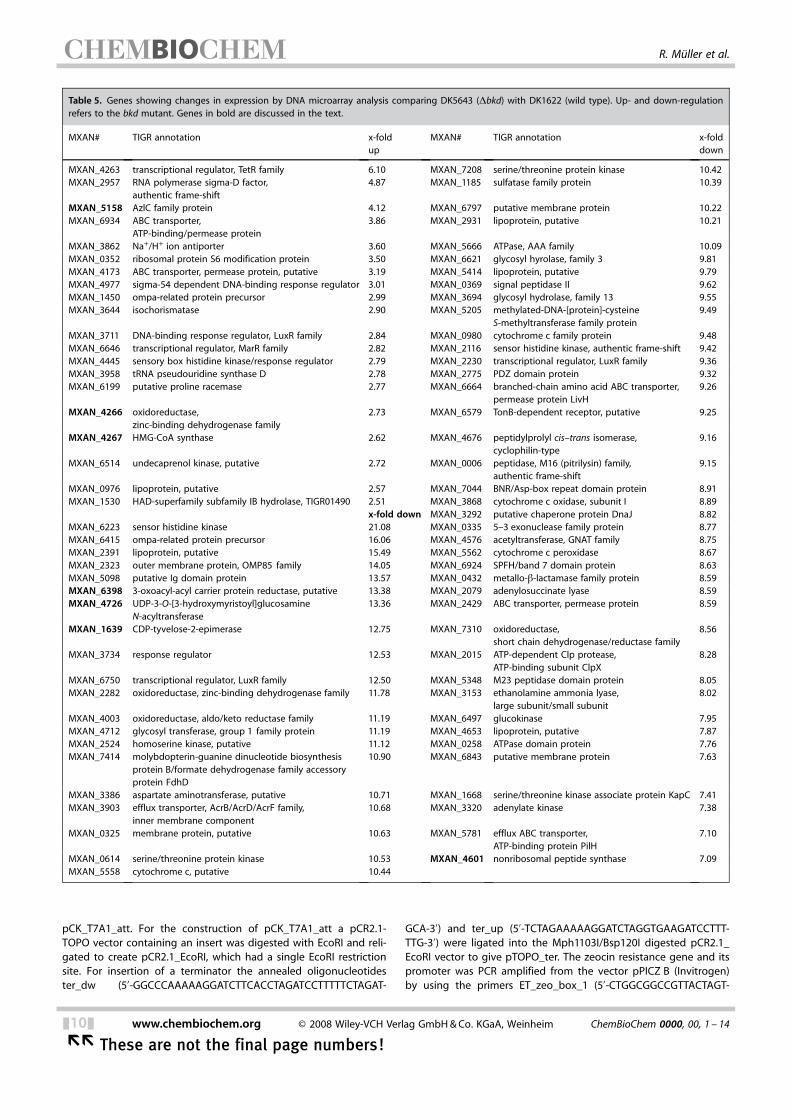
Table 5. Genes showing changes in expression by DNA microarray analysis comparing DK5643 (Dbkd) with DK1622 (wild type). Up- and down-regulationrefers to the bkd mutant. Genes in bold are discussed in the text.
MXAN# TIGR annotation x-foldup
MXAN# TIGR annotation x-folddown
MXAN_4263 transcriptional regulator, TetR family 6.10 MXAN_7208 serine/threonine protein kinase 10.42MXAN_2957 RNA polymerase sigma-D factor,
authentic frame-shift4.87 MXAN_1185 sulfatase family protein 10.39
MXAN_5158 AzlC family protein 4.12 MXAN_6797 putative membrane protein 10.22MXAN_6934 ABC transporter,
ATP-binding/permease protein3.86 MXAN_2931 lipoprotein, putative 10.21
MXAN_3862 Na+/H+ ion antiporter 3.60 MXAN_5666 ATPase, AAA family 10.09MXAN_0352 ribosomal protein S6 modification protein 3.50 MXAN_6621 glycosyl hyrolase, family 3 9.81MXAN_4173 ABC transporter, permease protein, putative 3.19 MXAN_5414 lipoprotein, putative 9.79MXAN_4977 sigma-54 dependent DNA-binding response regulator 3.01 MXAN_0369 signal peptidase II 9.62MXAN_1450 ompa-related protein precursor 2.99 MXAN_3694 glycosyl hydrolase, family 13 9.55MXAN_3644 isochorismatase 2.90 MXAN_5205 methylated-DNA-[protein]-cysteine
S-methyltransferase family protein9.49
MXAN_3711 DNA-binding response regulator, LuxR family 2.84 MXAN_0980 cytochrome c family protein 9.48MXAN_6646 transcriptional regulator, MarR family 2.82 MXAN_2116 sensor histidine kinase, authentic frame-shift 9.42MXAN_4445 sensory box histidine kinase/response regulator 2.79 MXAN_2230 transcriptional regulator, LuxR family 9.36MXAN_3958 tRNA pseudouridine synthase D 2.78 MXAN_2775 PDZ domain protein 9.32MXAN_6199 putative proline racemase 2.77 MXAN_6664 branched-chain amino acid ABC transporter,
permease protein LivH9.26
MXAN_4266 oxidoreductase,zinc-binding dehydrogenase family
2.73 MXAN_6579 TonB-dependent receptor, putative 9.25
MXAN_4267 HMG-CoA synthase 2.62 MXAN_4676 peptidylprolyl cis–trans isomerase,cyclophilin-type
9.16
MXAN_6514 undecaprenol kinase, putative 2.72 MXAN_0006 peptidase, M16 (pitrilysin) family,authentic frame-shift
9.15
MXAN_0976 lipoprotein, putative 2.57 MXAN_7044 BNR/Asp-box repeat domain protein 8.91MXAN_1530 HAD-superfamily subfamily IB hydrolase, TIGR01490 2.51 MXAN_3868 cytochrome c oxidase, subunit I 8.89
x-fold down MXAN_3292 putative chaperone protein DnaJ 8.82MXAN_6223 sensor histidine kinase 21.08 MXAN_0335 5–3 exonuclease family protein 8.77MXAN_6415 ompa-related protein precursor 16.06 MXAN_4576 acetyltransferase, GNAT family 8.75MXAN_2391 lipoprotein, putative 15.49 MXAN_5562 cytochrome c peroxidase 8.67MXAN_2323 outer membrane protein, OMP85 family 14.05 MXAN_6924 SPFH/band 7 domain protein 8.63MXAN_5098 putative Ig domain protein 13.57 MXAN_0432 metallo-b-lactamase family protein 8.59MXAN_6398 3-oxoacyl-acyl carrier protein reductase, putative 13.38 MXAN_2079 adenylosuccinate lyase 8.59MXAN_4726 UDP-3-O-[3-hydroxymyristoyl]glucosamine
N-acyltransferase13.36 MXAN_2429 ABC transporter, permease protein 8.59
MXAN_1639 CDP-tyvelose-2-epimerase 12.75 MXAN_7310 oxidoreductase,short chain dehydrogenase/reductase family
8.56
MXAN_3734 response regulator 12.53 MXAN_2015 ATP-dependent Clp protease,ATP-binding subunit ClpX
8.28
MXAN_6750 transcriptional regulator, LuxR family 12.50 MXAN_5348 M23 peptidase domain protein 8.05MXAN_2282 oxidoreductase, zinc-binding dehydrogenase family 11.78 MXAN_3153 ethanolamine ammonia lyase,
large subunit/small subunit8.02
MXAN_4003 oxidoreductase, aldo/keto reductase family 11.19 MXAN_6497 glucokinase 7.95MXAN_4712 glycosyl transferase, group 1 family protein 11.19 MXAN_4653 lipoprotein, putative 7.87MXAN_2524 homoserine kinase, putative 11.12 MXAN_0258 ATPase domain protein 7.76MXAN_7414 molybdopterin-guanine dinucleotide biosynthesis
protein B/formate dehydrogenase family accessoryprotein FdhD
10.90 MXAN_6843 putative membrane protein 7.63
MXAN_3386 aspartate aminotransferase, putative 10.71 MXAN_1668 serine/threonine kinase associate protein KapC 7.41MXAN_3903 efflux transporter, AcrB/AcrD/AcrF family,
inner membrane component10.68 MXAN_3320 adenylate kinase 7.38
MXAN_0325 membrane protein, putative 10.63 MXAN_5781 efflux ABC transporter,ATP-binding protein PilH
7.10
MXAN_0614 serine/threonine protein kinase 10.53 MXAN_4601 nonribosomal peptide synthase 7.09MXAN_5558 cytochrome c, putative 10.44
&10& www.chembiochem.org E 2008 Wiley-VCH Verlag GmbH&Co. KGaA, Weinheim ChemBioChem 0000, 00, 1 – 14
�� These are not the final page numbers!
R. M0ller et al.
ACHTUNGTRENNUNGGGATCCGAGCTCGGTACCAAGCTTGGCGTAATGGATCTGATCAGCACG-ACHTUNGTRENNUNGTGTTGACA-3’) and ET_zeo_box_2 (5’-TCGCCGCAGCCGAACGACC-ACHTUNGTRENNUNGGAGCGCAGCGAGTCAGTGAGCGAGGAAGCGGTGATCACACGTGTCA-ACHTUNGTRENNUNGGTCCTGCTCCTCGGCCACG-3’) to give a 570 bp fragment, whichwas subsequently cloned into pTOPO_ter by standard ET-cloningprocedures.[47–49] The lacZ promoter region was deleted at thesame time to give pTOPO_zeo_core. This plasmid was digestedwith BamHI/HindIII, and the T7AI promoter sequence was insertedthrough the annealed oligonucleotides T7A1_up (5’-AGCTTAT-ACHTUNGTRENNUNGCAAAAAGAGTATTGACTTAAAGTCTAACCTATAGGATACTTACAGCCAT-ACHTUNGTRENNUNGCGAGAGGTGTACATATGG-3’) and T7A1_dw (5’-GATCCCATATGTACA-ACHTUNGTRENNUNGCCTCTCGATGGCTGTAAGTATCCTATAGGTTAGACTTTAAGTCAATACTC-ACHTUNGTRENNUNGTTTTTGATA-3’) into the respective restriction sites to give thevector pCK_T7. In order to clone genes for over-expression down-stream of the T7AI promoter the restriction sites Bsp1407I andNdeI (sequences italicised) were included in the T7A1-up/dw pri-mers so that the ribosome binding site was placed six nucleotidesupstream of the ATG start codon (bold). In the final step the Mx8-phage derived attP site and intP were amplified from the vectorpSWU105 (Dale Kaiser, unpublished) by employing the primers in-tegrase_for_1 (5’-GGATGGATCTAGACAGACGGCCGCGCTTGT-3’) andintegrase_rev_1 (5’-CGGCTTTCGCGACATGGAGGACT-3’). The result-ing PCR product was digested with XbaI and ligated into the PsiI/XbaI digested vector to create the pCK_T7A1_att (for a map andnucleotide sequence of pCK_T7A1_att see Figures S1 and S2 in theSupporting Information).
The resulting mvaS complementation plasmid pCK4267exp was in-troduced into M. xanthus mutants DK5624 (Dbkd, mvaS ::kan)[9] andHB015 (Dbkd, MXAN_4265::kan) by electroporation as describedpreviously;[14,46] this resulted in strains HB19 (Dbkd, mvaS ::kan,mvaS+) and HB020 (Dbkd, MXAN_4265::kan, mvaS+).
Analytical procedures : Fatty acids were analyzed as their methylesters as described.[9] For geosmin analysis, a cell pellet from about20 mL of culture was resuspended in methanol (500 mL) and ex-tracted with heptane (500 mL) by being shaken at room tempera-ture for 20 min. Samples were centrifuged to achieve phase sepa-ration, and an aliquot of the heptane phase (2 mL) was injectedinto an Agilent 6890N gas chromatograph equipped with a 5973NEI-MS (Agilent, Waldbronn, Germany) by using a pulsed split-lessinjection technique. The column was an Agilent DB-5ht (30 mN0.25 mmN0.1 mm), and the mobile phase was helium at1 mLmin�1. The GC inlet and GC–MS transfer-line temperatureswere 250 8C and 280 8C, respectively. The column temperature washeld at 90 8C for 5 min, then increased to 140 8C at 5 8Cmin�1, to300 8C at 30 8Cmin�1 at which it was held for 10 min, then de-creased to 90 8C at 30 8Cmin�1. To improve sensitivity, the scanrange of the MS was narrowed to m/z 50–200 with the samplingrate adjusted to achieve a scan rate of about 2.5 Hz. Identificationand quantitation of geosmin and its isotopomers were done withthe AMDIS software.
Analysis of Trypan blue binding to fibrils was performed as de-scribed previously.[37]
Proteome analysis : For proteome analysis, wild-type and bkdmutant cells (25 mL each) were grown in Erlenmeyer flasks(250 mL), and cells were harvested after 24 h at OD600 1.59(DK1622) and OD600 1.52 for the bkd� culture (after 24 h). There-fore, the cultures were centrifuged (Eppendorf 5810R) at 3250g at4 8C for 10 min. The pellets were washed two times in 4 8C in coldPBS buffer and centrifuged under the same conditions. The cellpellets were stored at �20 8C.
For cell lysis, the pellets were resuspended in 2x lysis buffer (2 mL;7m urea, 2m thiourea, 4% CHAPS (3-([3-cholamidopropyl]dimethy-lammonio)-1-propanesulfonate), 2% (v/v) pharmalyte 3–10, 2%DTT). Cell lysis was performed by using a French press (3N ). Thesuspension was centrifuged and the supernatant was transferredto a new vial and proteins were precipitated with acetone/metha-nol.[50] The tube was incubated at �20 8C for 24 h and centrifugedas described above. The supernatant was removed quantitativelyand the cell pellets were resuspended at 4 8C in 500–600 mL DIGElabel buffer (7m urea, 2m thiourea, 4% CHAPS, 30 mm Tris, pH 8.5).For the estimation of protein concentrations the Bradford proteinassay was performed[51] with 1:10 and 1:20 dilutions of the samplesand by using “Dye concentrate” (Biorad) and a microtiterplatereader (Bio-Tek EL808); samples were measured at 595 nm.
DIGE labelling[52] was performed at 4 8C in the dark. Each sample(50 mg) was labelled with Cy3 or Cy5, and a 1:1 mixture (50 mg)was labelled with Cy2 to generate the internal standard. After30 min the labelling reaction was quenched by adding l-lysine(final concentration 1 mm). All samples were then pooled. We usedthe “under-labelling” technique, that is, both protein samples(150 mg) without fluorescent labels were added to the mixture toensure spots in Coomassie staining. Samples were loaded on drystrips (pH 3–11NL, Amersham) by following the manufacturer’s in-structions for in-gel-rehydration. We also applied each sample(500 mg) to individual dry strips for conventional Coomassie gels.First dimension (isoelectric focusing), equilibration, and second di-mension (SDS-PAGE) was performed as described previously.[53]
DIGE gels were scanned by using a THYPHOON 9410 (Amersham).For DIGE image analysis the DeCyder software package was used.DIGE spots that showed a different intensity higher than threefoldin the comparison of the two samples were set to pick list. All gelswere stained with colloidal Coomassie.[54] All Coomassie stainedgels were scanned on a densitometer (ImageScanner, Amersham)and the images analyzed by IMAGE Master 5.0 (Swiss institute ofbioinformatics). Coomassie spots that showed a different intensityhigher than twofold in the comparison of the two samples wereset to pick list, and all selected spots were cut out and subjectedto in-gel-digestion as described previously.[53]
As matrix compound, a-cyanocinnamic acid was dissolved to satu-ration in water/acetonitrile (1:1; 0.1% trifluoracetic acid) and tryp-sin digests (2 mL) were mixed with the matrix solution (2 mL). An ali-quot from this mixture (1–1.5 mL) was spotted on a 384 MALDIAnchor chipV target plate (Bruker Daltonics) and dried at roomtemperature. Mass profiles were generated by using a MALDI ToFULTRAflex mass spectrometer (Bruker Daltonics), and covered arange from 800–3500 Da. Peak detection included SNAP algorithm,SN ratio higher than 25 and peptide-typical isotopic distribution.Mass spectra were automatically filtered for trypsin or keratinpeaks. Optional C18 ZipTips (Millipore) purification was used by fol-lowing the manufacturer’s instructions to purify and concentratedpeptides. The elution was carried out with matrix solution (1.5 mL)and spotted directly on the target plate.
The monoisotopic masses were transferred to MASCOT (Matrix Sci-ence) and the masses were analyzed by using the in-house data-base of the translated genome of Myxococcus xanthus DK1622. Thesearch parameters were specified as follows: mass tolerance:“100 ppm”, fixed modifications: “carbamidomethyl”, variable modi-fications: “oxidized methionines”, protease: “trypsin”, missed clea-vages: “0”. The MOWSE scoring algorithm[55] identified proteinsthat had a score higher than 51, which corresponds to an identifi-cation probability of 95%.
ChemBioChem 0000, 00, 1 – 14 E 2008 Wiley-VCH Verlag GmbH&Co. KGaA, Weinheim www.chembiochem.org &11&
These are not the final page numbers! ��
The Alternative Biosynthesis Pathway to Isovaleryl-CoA
DNA microarrays : The construction of the PCR-generated DNA mi-croarrays that contained probes against the 7235 open readingframes of M. xanthus, has been previously described.[26,56,57] Proc-essing of the DNA arrays, cDNA synthesis, microarray hybridizationand posthybridization processing were performed as described.[58]
Six independent biological replica pairs of DK1622 (wild type) andDK5643 (Dbkd) were used for the analysis, and each independentwild type vs. bkd mutant pair was handled and processed identical-ly. Briefly, each pair of strains was grown at 28 8C to a density of5N108 cellsmL�1, the cells were centrifuged, the supernatants wereremoved, and the cell pellets were quick-frozen in liquid nitrogen.Total cellular RNA was isolated from quick-frozen cells by using thehot-phenol method.[59] Total RNA (30 mg) from matched cultureswas used to synthesize cDNA with pdN6 primers (10 mg; Amer-sham Pharmacia) in the presence of RNase inhibitor (40 mgmL�1;Promega). Reverse transcriptase reaction times were modified asfollows: 10 min at 37 8C, then 42 8C for 100 min, followed by a10 min incubation at 50 8C. RNA was hydrolyzed and neutralized asdescribed by Jakobsen et al.[26] and purified with Micron 30 filters(Amicon); the cDNA was eluted and dried with a SpeedVac concen-trator (Savant). The dried cDNA was resuspended in sodium bicar-bonate (0.1m, pH 9.0; 9 mL), and incubated for 5 min at 37 8C. ThecDNA was labelled with Cy3 (DK1622) or Cy5 (DK5643) from Amer-sham Pharmacia by addition of dye (2 mL) dissolved in dimethylsulfoxide (10 mL) and incubated for 1 h in the dark. The labelledcDNA was purified with the QIA-quick PCR kit (Qiagen) as de-scribed by the manufacturer, and concentrated on a Micron 30spin filter (Amicon). Labelled cDNA was then dried with a SpeedVacconcentrator (Savant) and resuspended in hybridization buffer(45 mL). Hybridization and posthybridization processing of theslides were performed as described previously.[26, 57,60]
Posthybridized DNA microarrays were scanned with a GenePix4000A microarray scanner and read by using GenePix Pro 3.0(Axon, Inc.). The GenePix array list (gal) file, MyxoGALv2.gal, thatcorresponded to the M. xanthus DNA microarrays, was constructedby using GalFileMaker v1.2 (DeRisi laboratory website; http://derisilab.ucsf.edu). Spots were flagged and removed from analysesbased on stringent criteria for shape, signal intensity and back-ground by using GenePix Pro 3.0 (Axon, Inc.). Analyses were per-formed with all unflagged spots. All array analyses, including hier-archical clustering and statistical analysis, were performed by usingCluster (Eisen Software; http://rana.lbl.gov/EisenSoftware.htm),Java TreeView software[61] (http://sourceforge.net/projects/jtreeview), and significance analysis of microarrays (SAM)[62] (http://www-stat.stanford.edu/~ tibs/SAM). All DNA microarray resultsused for this study have been submitted to Gene Expression Omni-bus (GEO) at NCBI (http://www.ncbi.nlm.nih.gov/projects/geo/).Series accession number: GSE10818; sample accession numbers:GSM273060, GSM273080, GSM273081, GSM273082, GSM273083,GSM273084.
qRT-PCR analysis : qRT-PCR of MXAN_3757, MXAN_3760, MXAN_4263, MXAN_4266, MXAN_5020, MXAN_5021 and MXAN_5158 wasperformed as described previously with vegetative cultures ofDK1622 and DK5643, with and without the addition of isovalerate(IVA; 1 mm).[9]
Acknowledgements
R.M. is grateful to the Deutsche Forschungsgemeinschaft for fi-nancial support. The authors would like to thank Taifo Mahmudfor various helpful discussions during the course of this project
and W. Lorenzen for help with the GC/MS analyses. This workwas in part supported by the National Institutes of Health PublicHealth Service grant GM354592 to M.S.
Keywords: biosynthesis · isovaleryl-CoA · leucinedegradation · Myxococcus xanthus · natural products
[1] A. P. Bretscher, D. Kaiser, J. Bacteriol. 1978, 133, 763–768.[2] M. E. Diodati, R. E. Gill, L. Plamann, M. Singer in Myxobacteria: Multicellu-
larity and Differentiation (Ed. : D. E. Whitworth), ASM, Washington, 2008,pp. 43–76.
[3] J. C. Ware, M. Dworkin, J. Bacteriol. 1973, 115, 253–261.[4] D. B. Kearns, A. Venot, P. J. Bonner, B. Stevens, G. J. Boons, L. J. Shimkets,
Proc. Natl. Acad. Sci. USA 2001, 98, 13990–13994.[5] H. B. Bode, J. S. Dickschat, R. M. Kroppenstedt, S. Schulz, R. M'ller, J.
Am. Chem. Soc. 2005, 127, 532–533.[6] J. S. Dickschat, H. B. Bode, R. M. Kroppenstedt, R. M'ller, S. Schulz, Org.
Biomol. Chem. 2005, 3, 2824–2831.[7] D. R. Toal, S. W. Clifton, B. A. Roe, J. Downard, Mol. Microbiol. 1995, 16,
177–189.[8] G. Michal, Biochemical Pathways, Spektrum, Heidelberg, 1999.[9] H. B. Bode, M. W. Ring, G. Schw�r, R. M. Kroppenstedt, D. Kaiser, R.
M'ller, J. Bacteriol. 2006, 188, 6524–6528.[10] M. W. Ring, G. Schwar, V. Thiel, J. S. Dickschat, R. M. Kroppenstedt, S.
Schulz, H. B. Bode, J. Biol. Chem. 2006, 281, 36691–36700.[11] J. Downward, D. Toal, Mol. Microbiol. 1995, 16, 171–175.[12] B. Silakowski, H. U. Schairer, H. Ehret, B. Kunze, S. Weinig, G. Nordsiek, P.
Brandt, H. Blçcker, G. Hçfle, S. Beyer, R. M'ller, J. Biol. Chem. 1999, 274,37391–37399.
[13] B. Silakowski, G. Nordsiek, B. Kunze, H. Blçcker, R. M'ller, Chem. Biol.2001, 8, 59–69.
[14] H. B. Bode, P. Meiser, T. Klefisch, N. Socorro D. J. Cortina, D. Krug, A.Gçhring, G. Schw�r, T. Mahmud, Y. A. Elnakady, R. M'ller, ChemBioChem2007, 8, 2139–2144.
[15] B. Kunze, H. Reichenbach, R. M'ller, G. Hçfle, J. Antibiot. 2005, 58, 244–251.
[16] T. Mahmud, H. B. Bode, B. Silakowski, R. M. Kroppenstedt, M. Xu, S.Nordhoff, G. Hçfle, R. M'ller, J. Biol. Chem. 2002, 277, 32768–32774.
[17] T. Mahmud, S. C. Wenzel, E. Wan, K. W. Wen, H. B. Bode, N. Gaitatzis, R.M'ller, ChemBioChem 2005, 6, 322–330.
[18] J. A. Aguilar, A. N. Zavala, C. DYaz-PZrez, C. Cervantes, A. L. DYaz-PZrez, J.Campos-GarcYa, Appl. Environ. Microbiol. 2006, 72, 2070–2079.
[19] K. Fçrster-Fromme, B. Hçschle, C. Mack, M. Bott, W. Armbruster, D. Jen-drossek, Appl. Environ. Microbiol. 2006, 72, 4819–4828.
[20] B. Hçschle, V. Gnau, D. Jendrossek, Microbiology 2005, 151, 3649–3656.[21] J. S. Dickschat, H. B. Bode, T. Mahmud, R. M'ller, S. Schulz, J. Org. Chem.
2005, 70, 5174–5182.[22] H. B. Bode, S. C. Wenzel, H. Irschik, G. Hçfle, R. M'ller, Angew. Chem.
2004, 116, 4257–4262; Angew. Chem. Int. Ed. 2004, 43, 4163–4167.[23] H. B. Bode, B. Zeggel, B. Silakowski, S. C. Wenzel, H. Reichenbach, R.
M'ller, Mol. Microbiol. 2003, 47, 471–481.[24] J. S. Dickschat, S. C. Wenzel, H. B. Bode, R. M'ller, S. Schulz, ChemBio-
Chem 2004, 5, 778–787.[25] V. G. Tusher, R. Tibshirani, G. Chu, Proc. Natl. Acad. Sci. USA 2001, 98,
5116–5121.[26] J. S. Jakobsen, L. Jelsbak, R. D. Welch, C. Cummings, B. Goldman, E.
Stark, S. Slater, D. Kaiser, J. Bacteriol. 2004, 186, 4361–4368.[27] S. Rachid, K. Gerth, I. Kochems, R. M'ller, Mol. Microbiol. 2007, 63, 1783–
1796.[28] B. S. Goldman, W. C. Nierman, D. Kaiser, S. C. Slater, A. S. Durkin, J. Eisen,
C. M. Ronning, W. B. Barbazuk, M. Blanchard, C. Field, C. Halling, G.Hinkle, O. Iartchuk, H. S. Kim, C. Mackenzie, R. Madupu, N. Miller, A.Shvartsbeyn, S. A. Sullivan, M. Vaudin, R. Wiegand, H. B. Kaplan, Proc.Natl. Acad. Sci. USA 2006, 103, 15200–15205.
[29] W. Buckel, U. Dorn, R. Semmler, Eur. J. Biochem. 1981, 118, 315–321.[30] W. Buckel, Eur. J. Biochem. 1986, 156, 259–263.[31] W. Buckel, H. Liedtke, Eur. J. Biochem. 1986, 156, 251–257.[32] J. Edmond, G. Popj[k, J. Biol. Chem. 1974, 249, 66–71.[33] T. Hao, D. Biran, G. J. Velicer, L. Kroos, J. Bacteriol. 2002, 184, 3348–3359.
&12& www.chembiochem.org E 2008 Wiley-VCH Verlag GmbH&Co. KGaA, Weinheim ChemBioChem 0000, 00, 1 – 14
�� These are not the final page numbers!
R. M0ller et al.
[34] D. Kaiser, Annu. Rev. Microbiol. 2004, 58, 75–98.[35] “Contact-Dependent Signaling in Myxococcus xanthus : The Function of
the C-Signal in Fruiting Body Morphogenesis”, L. Søgaard-Andersen inMyxobacteria: Multicellularity and Differentiation, (Ed. : D. E. Whitworth),ASM Press, Washington, 2008, pp. 77–91.
[36] A. Joachimiak, R. L. Kelley, R. P. Gunsalus, C. Yanofsky, P. B. Sigler, Proc.Natl. Acad. Sci. USA 1983, 80, 668–672.
[37] W. P. Black, Z. Yang, J. Bacteriol. 2004, 186, 1001–1008.[38] “Secondary Metabolism in Myxobacteria”, H. B. Bode, R. M'ller in Myxo-
bacteria: Multicellularity and Differentiation, (Ed. : D. Whitworth), ASMPress, Chicago, 2007, pp. 259–282.
[39] H. B. Bode, R. M'ller, Angew. Chem. 2005, 117, 6988–7007; Angew.Chem. Int. Ed. 2005, 44, 6828–6846.
[40] V. Simunovic, J. Zapp, S. Rachid, D. Krug, P. Meiser, R. M'ller, ChemBio-Chem 2006, 7, 1206–1220.
[41] P. Meiser, H. B. Bode, R. M'ller, Proc. Natl. Acad. Sci. USA 2006, 103,19128–19133.
[42] S. C. Wenzel, P. Meiser, T. Binz, T. Mahmud, R. M'ller, Angew. Chem.2006, 118, 2354–2360; Angew. Chem. Int. Ed. 2006, 45, 2296–2301.
[43] C. Schley, M. O. Altmeyer, R. Swart, R. M'ller, H. CG, J. Proteome Res.2006, 5, 2760–2768.
[44] D. Kaiser, Proc. Natl. Acad. Sci. USA 1979, 76, 5952–5956.[45] J. Hodgkin, D. Kaiser, Proc. Natl. Acad. Sci. USA 1977, 74, 2938–2942.[46] H. B. Bode, M. W. Ring, D. Kaiser, A. C. David, R. M. Kroppenstedt, G.
Schw�r, J. Bacteriol. 2006, 188, 5632–5634.[47] T. M. Binz, S. C. Wenzel, H. J. Schnell, A. Bechthold, R. M'ller, ChemBio-
Chem 2008, 9, 447–454.[48] S. C. Wenzel, F. Gross, Y. Zhang, J. Fu, F. A. Stewart, R. M'ller, Chem. Biol.
2005, 12, 349–356.[49] J. P. Muyrers, Y. Zhang, A. F. Stewart, Genet. Eng. (NY) 2000, 22, 77–98.[50] R. Mastro, M. Hall, Anal. Biochem. 1999, 273, 313–315.[51] M. M. Bradford, Anal. Biochem. 1976, 72, 248–254.
[52] R. Tonge, J. Shaw, B. Middleton, R. Rowlinson, S. Rayner, J. Young, F.Pognan, E. Hawkins, I. Currie, M. Davison, Proteomics 2001, 1, 377–396.
[53] S. Schneiker, O. Perlova, O. Kaiser, K. Gerth, A. Alici, M. O. Altmeyer, D.Bartels, T. Bekel, S. Beyer, E. Bode, H. B. Bode, C. J. Bolten, J. V. Choud-huri, S. Doss, Y. A. Elnakady, B. Frank, L. Gaigalat, A. Goesmann, C.Groeger, F. Gross, L. Jelsbak, L. Jelsbak, J. Kalinowski, C. Kegler, T. Knaub-er, S. Konietzny, M. Kopp, L. Krause, D. Krug, B. Linke, T. Mahmud, R.Martinez-Arias, A. C. McHardy, M. Merai, F. Meyer, S. Mormann, J.Munoz-Dorado, J. Perez, S. Pradella, S. Rachid, G. Raddatz, F. Rosenau, C.Ruckert, F. Sasse, M. Scharfe, S. C. Schuster, G. Suen, A. Treuner-Lange,G. J. Velicer, F. J. Vorholter, K. J. Weissman, R. D. Welch, S. C. Wenzel, D. E.Whitworth, S. Wilhelm, C. Wittmann, H. Blçcker, A. P'hler, R. M'ller, Nat.Biotechnol. 2007, 25, 1281–1289.
[54] R. Westermeier, Proteomics 2006, 6 Suppl 2, 61–64.[55] D. J. C. Pappin, P. Hojrup, A. J. Bleasby, Curr. Biol. 1993, 3, 327–332.[56] M. E. Diodati, F. Ossa, N. B. Caberoy, I. R. Jose, W. Hiraiwa, M. M. Igo, M.
Singer, A. G. Garza, J. Bacteriol. 2006, 188, 1733–1743.[57] V. D. Pham, C. W. Shebelut, M. E. Diodati, C. T. Bull, M. Singer, Microbiolo-
gy 2005, 151, 1865–1874.[58] M. E. Diodati, F. Ossa, N. B. Caberoy, I. R. Jose, W. Hiraiwa, M. M. Igo, M.
Singer, A. G. Garza, J. Bacteriol. 2006, 188, 1733–1743.[59] J. Sambrook, E. F. Fritsch, T. Maniatis, Molecular Cloning: A Laboratory
Manual, Cold Spring Harbor Laboratory Press, New York, 1989.[60] M. E. Diodati, F. Ossa, N. B. Caberoy, I. R. Jose, W. Hiraiwa, M. M. Igo, M.
Singer, A. G. Garza, J. Bacteriol. 2006, 188, 1733–1743.[61] A. J. Saldanha, Bioinformatics 2004, 20, 3246–3248.[62] V. G. Tusher, R. Tibshirani, G. Chu, Proc. Natl. Acad. Sci. USA 2001, 98,
5116–5121.
Received: April 3, 2008Published online on && &&, 2008
ChemBioChem 0000, 00, 1 – 14 E 2008 Wiley-VCH Verlag GmbH&Co. KGaA, Weinheim www.chembiochem.org &13&
These are not the final page numbers! ��
The Alternative Biosynthesis Pathway to Isovaleryl-CoA
FULL PAPERS
H. B. Bode, M. W. Ring, G. Schw,r,M. O. Altmeyer, C. Kegler, I. R. Jose,M. Singer, R. M0ller*
&& –&&
Identification of Additional Players inthe Alternative Biosynthesis Pathwayto Isovaleryl-CoA in theMyxobacterium Myxococcus xanthus
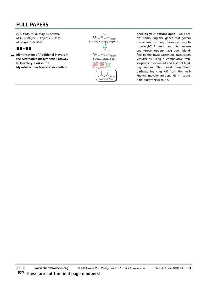
Keeping your options open : Two oper-ons harbouring the genes that governthe alternative biosynthesis pathway toisovaleryl-CoA (red) and its reversecounterpart (green) have been identi-fied in the myxobacterium Myxococcusxanthus by using a comparative tran-scriptome experiment and a set of feed-ing studies. This novel biosyntheticpathway branches off from the well-known mevalonate-dependent isopre-noid biosynthesis route.
&14& www.chembiochem.org E 2008 Wiley-VCH Verlag GmbH&Co. KGaA, Weinheim ChemBioChem 0000, 00, 1 – 14
�� These are not the final page numbers!