Embed Size (px)
Citation preview

Identification and transcriptional
regulation of eukaryotic
small nuclear RNA genes.
PhD in Biochemistry and Molecular Biology XXI cycle: 2006-2008
Gloria Fiorino
Department of Biochemistry and Molecular Biology University of Parma, Italy
PhD Coordinator: Prof. Gian Luigi Rossi PhD Tutor: Prof. Giorgio Dieci

Contents
Small nuclear RNAs: a brief introduction…………………….……………….....pag. 1
Promoter regulatory elements
in S. cerevisiae Pol II-transcribed snRNA genes………………………………pag. 17
Characterization of novel snRNA gene-like
transcriptional units in the human genome……………………………………pag. 50
References……………………………………………………………………………pag. 83

1
Introduction
Small nuclear RNAs: a brief
introduction

2
Introduction
An abundance of RNA regulators: non coding RNAs
RNA molecules are active participants in the regulation, catalysis and control of many
fundamental cellular processes, role that was thought to be restricted to proteins
until a few years ago. These regulatory RNAs, that do not encode a protein, are
referred to as non-protein-coding RNAs (ncRNAs). They are characterized by
different size, tissue specific expression pattern and biological functions. Despite
their differences, they elicit their biological responses through one of the three basic
mechanism: catalysing biological reactions, binding to and modulating the activity of
a protein, or base-pairing with a target nucleic acid.
The structure of a ncRNA is crucial for its functionality; as happens for proteins,
ncRNAs fold into specific higher order structures that impart a function to the
molecule. Often these RNAs require several partner proteins; that is, the functional
unit is the non-coding ribonucleoprotein (ncRNP) (fig. 1). The steps of ncRNP
assembly are often not well defined but can be regulated to control the activity of the
complex.
ncRNA activities are important at many levels of gene expression such as splicing,
mRNA turnover, gene silencing and translation (see table 1). Regulatory RNAs
include small interfering and micro RNA, mainly involved in postranscriptional
regulation, and other “structural molecules” including ribosomal, transfer, small
nucleolar, small cytoplasmic (sc), and small nuclear (sn) RNAs that function in the
processing, translation and degradation of other RNA molecules. Recent studies had
drawn the attention on the regulatory roles of such “structural RNA class”. Pol III-
transcribed Y RNAs are required for chromosomal DNA replication (Christov et al.,
2006), while 7SK RNA and U1 snRNA influence messenger RNA transcription; the
first represses transcript elongation by Pol II by binding the elongation factor P-TEFb
(Nguyen et al., 2001) while the second co-purifies with the multisubunit transcription
factor TFIIH and is involved both in transcription and initiation by pol II (Kwek et al.,
2002). Human RNAse P RNA is required for transcription of tRNA genes, thus linking
transcription with processing in regulation of tRNA gene expression (Jarrous et al.,
2007). The line between regulatory and structural classes has became less evident
since the recent discovery of snRNA-like transcription units that encode anti-sense
transcripts capable of post-transcriptional gene regulation (Pagano et al., 2007).

3
Introduction
Fig. 1 The structure of a non -coding RNA is crucial for its function . ncRNA genes
can be transcribed by either RNA pol I, II, III. ncRNAs fold into specific structures that
impart a function to the molecule. Often these RNAs are incorporated into large
complexes that contain proteins and sometimes other nucleic acids (Goodrich and Kugel,
2006)
Interestingly many of these ncRNA are expressed in a tissue-specific manner,
suggesting specific and regulated functions of the RNAs, rather than fundamental
housekeeping roles played in all tissues. The small cytoplasmic BC200 RNA (and its
rodent functional counterpart BC1) are specifically detected in the central nervous
system, where they could be involved in translation of dendritic mRNAs (Lin, Y. et al.
(2001). Dittmar et al. (2006) demonstrated tissue-specific differences in the
expression of individual tRNA species.
Finally the widespread functions of ncRNA makes it likely that alterations in their
levels and activity will compromise diverse cellular processes; a putative role of
BC1/BC200 in memory processes is in agreement with a recent study showing that
neocortical expression of BC200 RNA is up-regulated in Alzheimer disease (AD)
brains (Mus et al., 2007).

4
Introduction
ncRNA Species Functions
7SKsnRNA Human Inhibition of P-TEFb and RNAPII elongation
H1 RNA
(RNase P RNA) Human tRNA maturation, RNAPIII transcription
U1 snRNA Human/yeast mRNAsplicing, stimulation of RNAPII transcription/
mRNA splicing
U2 snRNA Human/yeast mRNAsplicing, stimulation of RNAPII elongation/
mRNA splicing
U4 snRNA Human/yeast mRNAsplicing
U5 snRNA Human/yeast mRNAsplicing
U6 snRNA Human/yeast mRNAsplicing
MRP RNA Human/yeast rRNA processing, mithocondrial DNA replication/
cell cycle progression
U7 snRNA Human/yeast Histone pre-mRNA 3’end formation
C/D snoRNAs Human/yeast 2’ O-methylation of rRNA, snRNAs and tRNAs;
rRNA processing
H/ACA snoRNAs Human/yeast Pseudouridylation of rRNAs, snRNAs and tRNAs;
rRNA processing
tRNA Human/yeast mRNA translation
Y scRNA Human Nc RNA degradation;chromosomal replication
Vault RNA Human Nucleocytoplasmic trafficking, multidrug resistance
SRP RNA Human Protein translocation to the endoplasmic reticulum
miRNA Human Gene silencing; translational repression or
cleavage of target mRNas
SINE-encoded
RNAs Human
RNa editing; alternative splicing; chromosomal
recombination; gene-expression regulation; cell
stress response; miRNA target
siRNA Human Gene silencing; cleavage of RNAs derived from
viruses, retroelements and repeat sequences
BC200 Human Possible role in translation of dendritic mRNA
Table 1. Functions of ncRNA in human and yeast spec ies.

5
Introduction
Fig. 2 Feautures of Sm - and Lsm -class small nuclear RNAs . Sm-class snRNA
contain three important recognition elements: a 5’-trimethylguanosine (TMG) cap, an Sm-
protein-binding site (Sm site) and a 3’ stem-loop structure. Lsm-class snRNA contain a
5’-monomethylphosphate (MPG) cap, a 3’ stem and terminate in a stretch of uridine
residues (Lsm site).
The snRNAs
snRNAs comprise a small group of highly abundant, non-polyadenylated, non-
protein-coding transcripts that function in the nucleoplasm. They assemble with
numerous protein factors to form small nuclear ribonucleoproteins (snRNPs). The
snRNAs can be divided into two classes: Sm-class RNAs (U1, U2, U4, U5, U7) are
characterized by a 5’-trimethylguanosine cap, a 3’ stem loop and by their ability to
associate to a group of seven Sm proteins, through the so-called Sm site. Lsm-class
RNAs (U6) contain a monomethylphosphate cap and a 3’ stem-loop, terminating in a
stretch of uridines (fig 2). With the exception of the U7 snRNP, which function in
histone pre-mRNA 3’ processing, the other snRNPs form the core of the
spliceosome and catalyse the removal of introns from pre-mRNA (Matera et al.,
2007).

6
Introduction
Fig. 3 Structure of the H. sapiens (Hs), A. thaliana (At) and D. melanogaster (Dm)
snRNA promoters. (Hernandez, 2001)
snRNA gene promoter structure
Promoters of snRNA genes are characterized by typical features. First, snRNA genes
usually contain transcriptional elements that are unique to this gene group. Secondly,
the promoters of both RNA pol II and RNA pol III snRNA genes within a species are
structurally related. Thirdly, although snRNA promoters are highly conserved within
the same species, they vary greatly between different genera indicating that they
have evolved rapidly. Fig. 3 shows the structure of snRNA promoters from Homo
sapiens (Hs) Arabidopsis thaliana (At) and Drosophila melanogaster (Dm) and
illustrates how snRNA promoters have diverged during evolution, maintaining close
similarity between those recognized by pol II and those recognized by pol III
(Hernandez, 2001).

7
Introduction
Members of the human snRNA and some related scRNA gene families are
characterized by a diagnostic arrangement of promoter elements, minimally including
a distal sequence element (DSE) that serves as enhancer and a proximal sequence
element (PSE) that is located in the core promoter region upstream from the start sit
of transcription. Some genes contain a TATA box located adjacently to the PSE. The
TATA element acts as a determinant for polymerase specificity: the combination of
the extragenic PSE and TATA elements directs recruitment of the RNA polymerase
III-specific transcriptional machinery whereas the absence of a TATA box specifies
recruitment of the RNA pol II-specific transcriptional apparatus. U1 and U2 snRNA
promoters and the U6 snRNA promoters serve respectively as prototypic pol II and
pol III snRNA promoters. Both DSE and PSE can be interchanged between pol II and
pol III promoters with no effect on polymerase specificity, while mutation in the TATA
box induces RNA polymerase II transcription from the U6 promoter and insertion of
the TATA box conversely causes RNA polymerase III transcription from the U2
promoter (Lobo and Hernandez ,1989; Mattaj et al.,1988). Other categories of RNA
polymerase III-transcribed genes, such as vault RNA, contain a combination of
extragenic PSE-like and TATA promoter elements along with canonical intragenic
elements typically utilized for RNA polymerase III transcription of transfer RNA genes
(Vilalta et al., 1994). The A. thaliana snRNA promoters consist of an upstream
sequence element (USE) and a TATA box. The USE is a plant snRNA gene-specific
enhancer and the spacing between the USE and the TATA box is the major
determinant of RNA polymerase specificity (Waibel and Filipowicz, 1990).
In D. melanogaster both Pol II and III promoters contain a quite conserved 21 bp
element called the PSEA located at about 42 bp upstream of the start site. A PSEB
or a canonical TATA box are located downstream the PSEA in Pol II and Pol III
snRNAs respectively (Zamrod et al., 1993). Polymerase specificity is determined by
position 19 and 20 of the PSEA, g/aG in Pol II and TC in Pol III promoters ( Jensen,
1998). Transcription of Schizosaccharomyces pombe U2 gene is directed by two
essential promoter elements: spUSE centered at -55, which functions as an activator
and a TATA box at -26 (Zhou and Lobo-Ruppert, 2001). In S. cerevisiae only the Pol
III U6 snRNA transcripton has been studied. The promoter contains a TATA box and
other two elements are required for correct efficient transcription: an A box located
within the coding region, as in tRNA genes, and a B box located at an anomalous
position in 3’ flanking region (Eschenlauer , 1993).

8
Introduction
Fig. 4 (A) sequence comparison of snRNA genes core promoters. Consensus
sequence is indicated below (Jawdekar and Henry, 2008) (B) Schematic
representation of SNAPc subunit organization. (Hernandez, 2001)
The PSE binding factor
The PSE sequence (fig 4A) is recognized by the snRNA activating protein complex
(SNAPc), also known as the PTF transcription factor (PTF) or PSE-binding protein
(PBP). SNAPc is composed of at least five proteins SNAP190, SNAP 50, SNAP45,
SNAP43 and SNAP19 (fig.4B). DNA binding by SNAPc requires both SNAP190 and
SNAP50 which directly bind to DNA via their Myb and zinc-fingers domains
respectively ( Wong et al, 1998; Jawdekar et al, 2006). SNAP43 is required for
assembly of SNAP190 and SNAP50 into a DNA binding competent complex (Hinkley
et al., 2003). These subunits are widely conserved through evolution, with omologues
in vertebrate species, Drosophila, Trypanosomes and plants. In contrast human
SNAP19 and SNAP45 subunits are dispensable for transcription in vitro and are not
so widely conserved, suggesting that these vertebrate-specific SNAPc subunits may
have acquired specialized regulatory roles for snRNA transcription (Mittal et al.,
1999). SNAPc plays a central role in snRNA transcription, being involved in pre-
initiation complex assembly, including direct promoter recognition, and serving as
target for numerous activators and repressor of transcription. For example SNAP19
can either interact with the Oct-1 activator (Ford et al., 1998) and p53 tumor
suppressor ( Gridasova and Henry, 2000).

9
Introduction
Pre-initiation complex assembly at snRNA gene promo ter
Pol II and Pol III transcribed genes share the initial promoter recognition by SNAPc
complex.
For TATA-less snRNA genes, SNAPC binding is a prelude to the recruitment of
traditional components of the general transcription machinery, including TBP,
TFIIA,TFIIB,TFIIE, and TFIIF (Kuhulman et al., 1999), used also for mRNA
transcription by RNA polymerase II. The role, if any of TFIIH in snRNA gene
transcription is not clear. Interestingly U1 snRNA was found associated to TFIIH to
stimulate RNA polymerase II transcription of certain mRNA genes (Kwek et al., 2002)
suggesting that U1 snRNA may contribute to RNA polymerase II regulation. An
intriguing possibility is that TFIIH complexes differing by the presence or absence of
U1 snRNA participate in active transcription of mRNA and snRNA genes.
SNAPc associates with TBP (Sadowski et al., 1996); although no homologues for
SNAPC have been described in yeast, it appears that a role for SNAPC in
coordinating TBP activity at snRNA genes is evolutionarily conserved.
As with TATA-less RNA polymerase II-transcribed snRNA genes, SNAPC recruits
TBP to RNA polymerase III-transcribed genes; the SNAPC/TBP juxtaposition results
in the recruitment of a TFIIB-related factor called Brf2 (Schramm et al, 2000). Brf2
differs substantially from Brf1 used for transcription of other RNA polymerase III-
transcribed genes in humans, and from the Brf used for U6 snRNA gene transcription
in yeast. Promoter recruitment of TFIIB for RNA polymerase II transcription or the
TFIIB-related Brf2 factor for RNA polymerase III transcription, as dictated by the
absence or presence of the TATA box, serves as a critical determinant for utilization
of different RNA polymerases in humans. The assembly of SNAPC, TBP, and Brf2
facilitates recruitment of the SANT- 187 domain containing protein Bdp1 (Jawdekar
et al., 2006), a factor globally utilized for all RNA polymerase III transcription
(Schramm and Hernandez,2002).

10
Introduction
Fig. 5 Factors involved in human snRNA gene transcription. The transcription of
human snRNA genes by RNA polymerases II (A) and III (B) involves a combination of
shared factors including the Staf and Oct-1 activators and the general transcription
factors SNAPC and TBP along with additional factors specialized for transcription by only
one polymerase. RNA polymerase II and III termination is directed by the 3′ box or by
the TTTT terminator, respectively. The numbers within SNAPC represent the apparent
molecular weights of each subunit (Jawdekar and Henry, 2008).

11
Introduction
Fig. 6 Assembly of the human U6 snRNA initiation co mplex (Hernandez, 2001)
Activation of snRNA gene transcription The high-level expression of snRNA in cells depends upon the DSE, typically located
at about 200 bp upstream the transcription start site. The DSE is composed by
various protein binding site, but one of them is almost invariably the octamer
sequence ATGCAAAT, recognized by the POU domain activator protein Oct-1
(Carbon et al., 1987). In addiction the DSE contains a Staf-responsive element
recognized by the snRNA transcriptional activator Staf, also known as SPH binding
factor (Myslinski et al., 1998). Some snRNA genes harbor Sp1 binding sites adjacent
to the DSE. These proteins function as activators synergizing to recruit SNAPc. The
mechanism is better understood for Oct-1, wherein direct contacts between the Oct-
POU domain and the SNAP190 subunit of SNAPc contributes to increased SNAPc
recruitment (Mittal, 1996). The strict spacing between the DSE and PSE is
conserved in most, but not all, snRNA genes, and at least for U6 and 7SK snRNA
gene expression, a nucleosome positioned between these promoter elements
contributes to activated transcription (Boyd et al, 2000; Zhao et al, 2001) spatially
juxtaposing the DSE and PSE to facilitate direct interactions between Oct-1 and
SNAP190 (fig 6). This observation suggest that factors modulating chromatin
structure are important for regulated transcription. Consistently, a higher proportion of
histone H3 at U6 promoters is acetylated in cells that maintain higher levels of RNA
polymerase III transcription (GWJ, unpublished observations). The opposing activity
of histone deacetylase (HDAC) factors is also important for transcriptional repression
by the Retinoblastoma (RB) and p53 tumor suppressor proteins.
12
Introduction
Transcription and 3’-end formation of snRNA genes.
In common with pre-mRNAs, newly pol II-transcribed pre-snRNAs are co-
transcriptionally capped at the 5’-end by several enzymatic activities, with the
attachment of a 7-methyl-guanosine (m7G) residue to the γ-phosphate through a 5’-
phosphoester linkage (Mattaj, 1986). Transcription and RNA processing reactions
have been found to be closely linked in vivo; at the heart of this lies the repetitive
CTD (C-terminal domain) of Pol II largest subunit, which serves to recruit and interact
with many processing factors. Recruitment of processing factors by Pol II CTD is
closely linked to its position along the gene and the phosphorylation state of specific
serine residues. Recently phosphorylation of the serine in position 5 (Ser5) and 2
(Ser2) of the CTD has been related to activation of 5’-end capping and 3’-end
formation of snRNA transcripts (Sylvain and Murphy, 2008). Pol II transcribed genes
transcription in fact is linked with downstream RNA processing events. Indeed,
appropriate snRNA termination depends upon the 3′ box, which is recognized by
RNA polymerase II when recruited to PSE-containing genes (Hernandez and Lucito,
1988); if snRNA promoter is replaced by a mRNA promoter, 3’-end is not formed and
the transcripts are polyadenilated. One candidate target in coupling U1 and U2
snRNA transcription to subsequent 3′ end processing is the Integrator complex,
which is composed of at least twelve polypeptides and associates with the carboxy
terminal domain (CTD) of the RNA polymerase II largest subunit. Intriguingly, serine
7 of the CTD is also specifically required for both snRNA gene transcription and
downstream 3′ end formation but has no effects on mRNA transcription and is also
critical for recruitment of the Integrator complex to U1 and U2 snRNA genes (Egloff et
al., 2007).
In constrast to the vertebrate snRNAs, the 3’-ends of two snRNAs, U2 and U5L, in
S.cerevisiae are formed by processing events unlinked to transcription (Chanfreau et
al., 1997; Abuo Elela and Ares, 1998). Rnt1p, a homolog of bacterial RNase III, is
strongly implicated in 3’-end formation; in fact U2 and U5L snRNA accumulation is
impaired in cells that have a temperature-sensitive mutation in the RNT1 gene and
purified, recombinant Rnt1p can cleave in vitro putative RNA stem-loop structures
formed by U2 and U5 3’-flanking regions. However in the rnt1 mutant cells the
steady-state levels of U1, U4, and U5S are increased at the restrictive temperature
(Chanfreau et al., 1997), suggesting that their 3’ ends may be generated by an

13
Introduction
alternative mechanism. In the fission yeast Schizosaccharomyces pombe Pac1p, a
RNAse III ortholog, is required for correct end-formation (Dewang et al., 1999). As in
S. pombe and in S. cerevisiae, studies in plants (Connelly and Filipowicz, 1993) and
in sea urchins (Wendelburg and Marzluff, 1992) indicate that the formation of the
3’ends of snRNAs can be dissociated from transcription.
Lsm-class snRNA genes are transcribed by Pol III; the run of uridines that forms the
Lsm-binding site at the 3’-end also doubles as a Pol III transcription terminator. U6
snRNA is O-methylated on the 5’ terminal (γ) phosphate in humans. Synthesis of the
γ-methyl phosphate cap of U6 RNA in human cells is dependent upon structural
determinants at the base of a conserved 5’-terminal stem (Singh et al,1990).
However is not known whether yeast U6 RNA receives a γ-methyl phosphate cap.
snRNA nuclear export system.
In higher eukaryotes, Lsm class snRNAs never leave the nucleus, whereas the
biogenesis of the Sm-class snRNPs requires export in the cytoplasmic for
maturations events; the 5’ cap structure and the length of the RNA are the key
determinant in nuclear export. Following transcription and 3’ processing in the
nucleus, newly transcribed Sm-class snRNAs are transported to the cytoplasm by an
export complex that contains the snRNA-specific export adaptor protein PHAX, the
export receptor chromosome region maintainance CRM1, the cap-binding complex
CBC and a Ran GTPase. These factors dissociate from the pre snRNA in the
cytoplasm after binding with the survival of motor neuron (SMN) complex and
desphorilation of PHAX (Segref et al., 2001). The SMN recognizes specific sequence
elements (the Sm protein-binding site and the 3’ stem-loop) and recruits a set of
seven Sm proteins to form the core RNP. Following assembly on the Sm core, the 7-
methylguanosine (m7G) cap is hypermethylated to form 2,2,7-trimethylguanosine
(TMG) cap structure and the 3’ end is trimmed by an unidentified exonuclease (Exo).
Methylguanosine caps of yeast snRNAs are also hypermethylated but the subcellular
compartment in which hypermethylation occurs is not known. The formation of the
TMG cap triggers the assembly of the import complex, which includes the import
adaptor snurportin-1 (SPN) and the import receptor importin-β (Imp-β ). After reimport
into the nucleus, snRNPs together with numerous other splicing factors assemble
into the functional spliceosome.

14
Introduction
Fig. 7 SnRNA export system. Following transcription by Pol II, pre-snRNA are exported
to the cytoplasm.. The snRNA-export complex consist of export adaptor protein (PHAX),
the export receptor chromosome region maintainance (CRM1), the cap-binding complex
(CBC) and the GTP-bound form of Ran GTPase. These factor dissociates in the
cytoplasm. The survival of motor neuron (SMN) recruits the Sm-proteins to form the Sm-
core RNP. Following assembly on the Sm core, the 7-methylguanosine (m7G) cap is
hypermethylated by trimethylguanosine syntase-1 (TGS1) to form 2,2,7-
trimethylguanosine (TMG) cap structure and the 3’ end is trimmed by an unidentified
exonuclease (Exo). TMG and associated Sm-proteins provide a targeting signal for re-
import into the nucleus by the import adaptor snurportin-1 (SPN) and the import receptor
importin-β (Imp-β ).

15
Introduction
Cell cycle regulation of snRNA gene transcription.
The demand for snRNA and other non coding RNAs depends upon the metabolic
state of the cell and therefore their transcription is regulated during changes in cells
growth and cells cycle progression. Recent evidence suggests that transcription of
snRNA genes is restricted at various points during the cell cycle by the protein kinase
CK2 and the RB 300 tumor suppressor protein. Currently, the cell cycle regulation of
RNA polymerase III transcription is better understood; RNA polymerase III
transcription is most active during the late G1, S and G2 phases of the cell cycle and
is repressed during the M and G0/early G1 phases. The protein kinase CK2 is
suggested to play a major role in M-phase repression of snRNA transcription. CK2
associates with multiple components of the polymerase III transcriptional apparatus,
including the polymerase itself, and SNAPC, and phosphorylates both the Bdp1
component of the Brf2–TFIIIB complex (Hu et al., 2004) and the SNAP190
component of SNAPC (Gu et al., 2005) reducing their promoter association. RB
tumor suppressor protein silences RNA polymerase III transcription at the G0/early
G1 stage the cell cycle. Interestingly, a role for RB family members for the regulation
of snRNA gene transcription by RNA polymerase II has not been observed (Hirsch et
al., 2004), suggesting that the distinct usage of SNAPC and Brf2–TFIIIB specifies RB
targeting of RNA polymerase III-transcribed snRNA genes. As the products of these
non-coding RNA genes contribute substantially to the biosynthetic capacity of the
cell, the repression of snRNA gene transcription and other RNA polymerase III
transcripts by RB likely plays an important role in growth control limiting tumor
progression. During U6 repression, RB stably associates with the promoter via
protein–protein interactions with components of SNAPC and Brf2–TFIIIB. Thus,
during snRNA gene repression, RB likely inhibits steps subsequent to RNA
polymerase III recruitment, potentially including promoter escape, open complex
formation, elongation, and termination.

16
Introduction
snRNAs function: pre-mRNA splicing. Pre-messenger RNA splicing refers to a process catalyzed by the spliceosome, in
which non coding intron sequences are excised and coding exons are ligated
together through a two-step reaction to form mature mRNA. In the first of the two
nucleophilic reactions, the 2’-OH of an intronic “branch point” adenosine attacks the
phosphodiester backbone at the 5’-splice site, creating a branched lariat intermediate
and a free 3’-OH on the 5’ exon. This hydroxyl then attacks the 3’-splice site, ligating
the exons and releasing the intron in the lariat form. The chemistry of these two
trans-esterifications reactions is relatively simple; however pre-mRNA splicing
requires accuracy and precise regulation and is therefore catalyzed by the
spliceosome, one of the largest and most complex molecular machines in the cell.
The spliceosome is composed of over 200 different proteins and five RNA
components (U1, U2, U4, U5, U6 snRNAs) that form a dynamic and elaborate
network of RNA-RNA, RNA-protein and protein-protein interactions (Nilsen, 2003).
The snRNPs have at least three important functions: recognition of splicing sites and
branchpoint sequences, folding of pre-mRNA into a reactive structure and alignment
of splice sites and finally possibly direct contribution to catalysis.
In the early stages of spliceosome assembly the 5’ splice site base pairs with U1
snRNA; U1 base pairing is well established in both yeast and mammalian splicing
system. U2 snRNP recognizes the branch point adenosine, and a short
intermolecular helix forms between U2 snRNA and the consensus sequence within
the intron . This U2-intron duplex serves to position the branch point adenosine for its
role as the nucleophile during the first catalytic step. The U4/U6.U5 tri-snRNP next
joins the spliceosome and U1 at the 5’ splice site is exchanged for U5 snRNA on the
exon side of the splice site.
When tri-snRNP joins the spliceosome U6 snRNA, after U4 snRNA dissociation, is
also exchanged for U1 but on the intro side of the 5’ splice site and forms a base
pairing interaction with the U2 snRNA that juxtaposes the 5’ splice site and branch
site, the reactant of the first of the two trans-esterification reactions. The highly
conserved 5’ end of the U2 snRNA is made up of several stem-loop and single
stranded regions that interact with the U6 snRNA and the snRNP proteins. Finally the
two exons are bound and kept in alignment partially via interactions with a conserved
loop in U5 snRNA.

17
Promoter regulatory elements
in S. cerevisiae Pol II-transcribed
snRNA genes

18
Abstract
Small nuclear RNAs (U1 to U6) are abundant capped RNAs synthesized by RNA
Polymerase (Pol) II, with the exception of the U6 snRNA, whose gene is transcribed
by Pol III.
Even if the function of these RNAs has been studied in detail, little is known about
their transcription. In particular, in Saccharomyces cerevisiae, only the Pol III U6
snRNA promoter has been characterized, while nothing is known about the promoter
architecture of the other Pol II-transcribed snRNA genes.
To begin to characterize the promoter architecture of these RNA genes, we used
comparative sequence analysis. The alignment of the orthologous sequences from
four different species of Saccharomyces identified conserved elements upstream of
the transcription start site.
Some evolutionarily conserved sequence blocks matched the consensus binding site
of known general regulatory factors (Rap1, Abf1, Reb1) while others did not match
any known motif. The analysis also revealed the conservation of a few regulatory
elements known to be involved in different pathways (RRPE - ribosomal RNA
processing element; PACE - Proteasome Associated Control Element).
To better understand the involvement of the identified putative promoter elements in
transcription we prepared sequence-tagged version of two yeast snRNA genes,
LRS1 (coding for U2 snRNA) and SNR7 (coding for U5 snRNA), to be used as
reporter genes for in vivo expression analysis.
Mutational analysis of the upstream conserved motifs revealed their influence on
snRNA gene transcription, possibly involving the generation or maintenance of
nucleosome-free promoter regions.

19
Introduction
Introduction Small nuclear RNA genes (snRNAs) are a small group of highly abundant, essential,
non-polyadenylated, non-protein-coding transcripts that function in the nucleoplasm.
They assemble with numerous protein factors to form small nuclear
ribonucleoproteins (snRNPs), component of the spliceosome, the complex that
catalyzes the splicing of pre-mRNA.
All are synthesized by RNA Polymerase (Pol) II, with the exception of the U6 snRNA,
whose gene is transcribed by Pol III. Pol II-transcribed snRNA genes U1, U2, U4, U5
(Sm class) snRNAs share common features: a 5′-trimethylguanosine cap, a 3′ stem–
loop and a binding site (the Sm site) for a group of seven Sm proteins. (for a review
see Matera et al., 2007).
As compared to the metazoan snRNAs, little is known about snRNA synthesis in
unicellular organism, even if their function has been studied in detail.
Human snRNA genes contain compact promoters that are recognized by increasingly
well-characterized transcription factors. Pol II and Pol III snRNA promoters share
common elements: a loosely conserved proximal sequence element (PSE), located
at about position -55 upstream the transcription start site, which recruit the snRNA
activating protein complex (SNAPc or PTF) and define the transcription initiation site,
and a distal sequence element (DSE) located around position -200 that function as
enhancer. This motif usually contains an octamer sequence that recruit the POU
domain transcription factor Oct-1, as well as an SPH element that recruits the zinc
finger transcription factor Staf. Core Pol III snRNA promoters contain in addition a
TATA box downstream of the PSE. Both PSE and DSE can be interchanged
between Pol II and Pol III promoters with no effect on RNA polymerase specificity,
that is determined only by the presence or absence of the TATA box. (Hernandez,
2001).
In D. melanogaster both Pol II and III promoters contain a quite conserved 21 bp
element, called the PSEA, located at about 42 bp upstream the start site. A PSEB or
a canonical TATA box are located downstream the PSEA in Pol II and Pol III snRNAs
respectively (Zamrod et al., 1993). Polymerase specificity is determined by position
19 and 20 of the PSEA, g/aG in Pol II and TC in Pol III promoters ( Jensen, 1998).

20
Introduction
Plant snRNA gene promoters consist of an upstream sequence element (USE) and
TATA box at -30. The major determinant of RNA polymerase specificity is the
spacing between the USE and the TATA box (Filipowicz et al., 1990).
Transcription of Schizosaccharomyces pombe U2 gene is directed by two essential
promoter elements: spUSE centered at -55, which functions as an activator element
and a TATA box at -26 (Zhou and Lobo-Ruppert, 2001).
In S. cerevisiae only the Pol III U6 snRNA transcription has been studied in detail.
The promoter contains a TATA box, and two other elements are required for correct
efficient transcription: an A box located within the transcribed region and a B box
located in the 3’ flanking region, downstream of the termination signal (Eschenlauer ,
1993).
The identification of regulatory elements within a promoter is a key step in
understanding gene transcription. Even if these elements are short DNA sequences,
often only 5 to 20 bp in length, they are critical for gene regulation. Cis-regulatory
elements are commonly conserved across evolution. Comparative sequence analysis
for regulatory element discovery is an high-throughput method to identify putative
functional elements whose effective role in transcription can be verified by
experimental procedures.
In this work we combined computational and experimental approaches to gain insight
into transcriptional regulation of Pol II transcribed snRNA genes and their promoter
architecture.

21
Materials and Methods
Materials and Methods Comparative sequence analysis: The coding sequences of S. cerevisiae snRNAs were recovered from SGD
(Saccharomyces Genome Database). The homology search in the fungal genomes
(S. paradoxus , S. mikatae, S. bayanus and S. kudriazevii) was carried out using
BLASTN program (http://seq.yeastgenome.org/cgi-bin/blast-fungal.pl). As result of
this search the orthologous snRNA coding sequences were identified. The snRNA
gene 5’-flanking sequences of the different genomes were recovered by search in the
NCBI databases (www.ncbi.nlm.nih.gov). The sequences (400 bp) upstream the
transcription start site were then aligned with ClustalX program.
Amplification and cloning of DNA templates: U5M-YEp352 template: We generated a tagged form of SNR7 (U5 snRNA gene) by
inserting a 22-bp sequence tag into the transcribed region (position +130). We first
generated two PCR products, from yeast genomic DNA (strain S288C), using the
high fidelity Pfu DNA polymerase (Promega) and gene-specific pairs of
oligonucleotide primers: U5_fw _SacI in combination with U5_rev_BamHI (carrying
the oligo-tag underlined in table 1) to produce the upstream half of the construct,
containing 130 bp of the transcribed sequence plus 248 bp of 5’-flanking region;
U5_fw _BamHI in combination with U5_rev_SphI to produce the downstream half,
containing 84 bp of the transcribed sequence plus 292 bp of 3’-flanking region. The
upstream and downstream fragments were then digested with SacI/BamHI and
BamHI/SphI respectively, and inserted in SacI/SphI-cut Yep352 vector through a
single ligation reaction.
U5M 5'-mutant templates:
U5M 5'- mutants were obtained by PCR amplification using U5M-YEp352 as
template and specific mutagenic forward primers. All the fragments were cloned in
Yep352 vector (SmaI site) and sequence verified by dideoxy chain termination
sequencing.
22
Materials and Methods
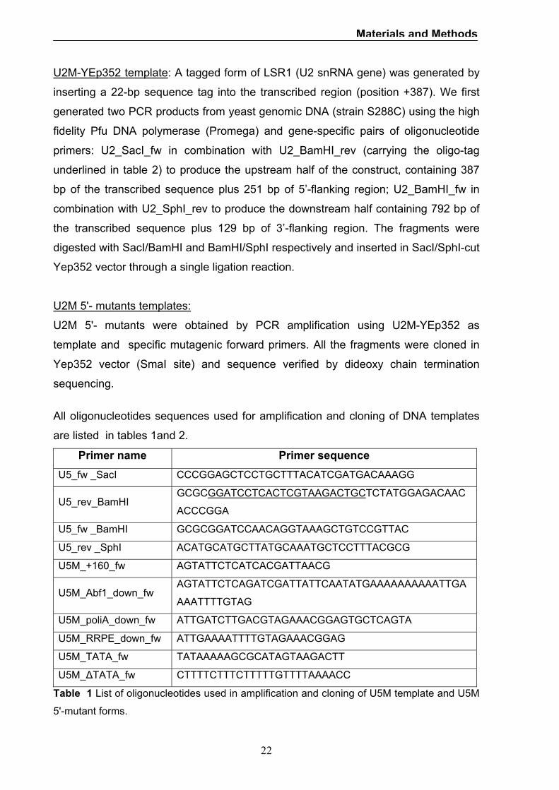
U2M-YEp352 template: A tagged form of LSR1 (U2 snRNA gene) was generated by
inserting a 22-bp sequence tag into the transcribed region (position +387). We first
generated two PCR products from yeast genomic DNA (strain S288C) using the high
fidelity Pfu DNA polymerase (Promega) and gene-specific pairs of oligonucleotide
primers: U2_SacI_fw in combination with U2_BamHI_rev (carrying the oligo-tag
underlined in table 2) to produce the upstream half of the construct, containing 387
bp of the transcribed sequence plus 251 bp of 5’-flanking region; U2_BamHI_fw in
combination with U2_SphI_rev to produce the downstream half containing 792 bp of
the transcribed sequence plus 129 bp of 3’-flanking region. The fragments were
digested with SacI/BamHI and BamHI/SphI respectively and inserted in SacI/SphI-cut
Yep352 vector through a single ligation reaction.
U2M 5'- mutants templates:
U2M 5'- mutants were obtained by PCR amplification using U2M-YEp352 as
template and specific mutagenic forward primers. All the fragments were cloned in
Yep352 vector (SmaI site) and sequence verified by dideoxy chain termination
sequencing.
All oligonucleotides sequences used for amplification and cloning of DNA templates
are listed in tables 1and 2.
Primer name Primer sequence U5_fw _SacI CCCGGAGCTCCTGCTTTACATCGATGACAAAGG
U5_rev_BamHI GCGCGGATCCTCACTCGTAAGACTGCTCTATGGAGACAAC
ACCCGGA
U5_fw _BamHI GCGCGGATCCAACAGGTAAAGCTGTCCGTTAC
U5_rev _SphI ACATGCATGCTTATGCAAATGCTCCTTTACGCG
U5M_+160_fw AGTATTCTCATCACGATTAACG
U5M_Abf1_down_fw AGTATTCTCAGATCGATTATTCAATATGAAAAAAAAAATTGA
AAATTTTGTAG
U5M_poliA_down_fw ATTGATCTTGACGTAGAAACGGAGTGCTCAGTA
U5M_RRPE_down_fw ATTGAAAATTTTGTAGAAACGGAG
U5M_TATA_fw TATAAAAAGCGCATAGTAAGACTT
U5M_∆TATA_fw CTTTTCTTTCTTTTTGTTTTAAAACC
Table 1 List of oligonucleotides used in amplification and cloning of U5M template and U5M
5'-mutant forms.

23
Materials and Methods
Primer name Primer sequence
U2_SacI_fw CCCGGAGCTCACACTTTCACTACGTGTATAACG
U2_BamHI_rev GCGCGGATCCTCACTCGTAAGACTGCGACAGGGAAGAGTATGAA
GC
U2_BamHI_fw GCGCGGATCCTTTCCGAGCCGTTTATGTCC
U2_SphI_rev ACATGCATGCCCAATTAGTGCACACACATAC
U2_Rap_fw GGGGTGTATGGGTGTGGGTGG
U2_Rpn4_fw GGTGGCAAAAAAAACCTAGCAAC
U2_poliT_fw CCGGAGCTCGCAACGCTCTATGTTTCTTTTC
U2_+127_fw CCGTTTCCGATGGGCCACTCG
Table 2 List of oligonucleotides used in amplification and cloning of U2M template and U2M
5'-mutant forms.
Yeast strains: Strain Genotype Source
BY4742 Matα, his3-∆1, leu2-∆0, lys2-∆0, ura3-
∆0
Yeast Knock Out Collection
(Open Biosystem)
BY4741 Matα, his3-∆1, leu2-∆0, met15-∆0,
ura3-∆0
Yeast TAP Fusion Collection
(Open Biosystem)
ABF1-TAP Matα, his3-∆1, leu2-∆0, met15-∆0,
ura3-∆0, ABF1-TAP-HIS3MX6
Yeast TAP Fusion Collection
(Open Biosystem)
STB3-TAP Matα, his3-∆1, leu2-∆0, met15-∆0,
ura3-∆0, STB3-TAP-HIS3MX6
Yeast TAP Fusion Collection
(Open Biosystem)
RAP1-TAP Matα, his3-∆1, leu2-∆0, met15-∆0,
ura3-∆0, RAP1-TAP-HIS3MX6
Yeast TAP Fusion Collection
(Open Biosystem)
RPN4-TAP Matα, his3-∆1, leu2-∆0, met15-∆0,
ura3-∆0, RPN4-TAP-HIS3MX6
Yeast TAP Fusion Collection
(Open Biosystem)
Table 3 Yeast strains used in this work.

24
Materials and Methods
In vivo RNA analyses: U5M-YEp352 transformants: Yeast cells (BY4742 strain) were transformed with the
different U5M-YEp352 templates and high-copy number plasmid YEp352 by the
lithium acetate procedure (Ito et al., 1983), and resulting transformants were selected
for uracil auxotrophy. RNA extraction was performed according to a previously
described procedure (Schmitt et al., 1990). Total RNA samples (10 µg) were
fractionated on a 6% polyacrylamide/7 M urea gel, then transferred to a positively
charged nylon membrane (Gene Screen Plus, Perkin Elmer). The filter was
hybridised with a 5'-labeled probe (5'-GGATCCTCACTCGTAAGACTGC)
complementary to the oligonucleotide inserted as previously described in the coding
region of SNR7 gene. Hybridization was carried out overnight at 28°C in 5X SSC, 5X
Denhardt’s solution , 0,1 mg/ml denatured salmon sperm DNA, 0,5 % (w/v) SDS,
followed by one 10-minute washing with 2X SSC solution containing 0,1% SDS and a
short washing in 1X SSC solution containing 0,1 % SDS. Hybridization products were
visualized and quantified by phosphorimaging. A tRNAAla specific primer
(Ala_AGC_probe: 5'-GGAGACCTCTCCCATGCTAAGGGAGCGCGC) was used for
normalization.
U2M-YEp352 transformants: Yeast cells (BY4742 strain) were transformed with the
different U2M-YEp352 templates and high-copy number plasmid YEp352 by the
lithium acetate procedure (Ito et al., 1983), and resulting transformants were selected
for uracil auxotrophy. RNA extraction was performed according to a previously
described procedure (Schmitt et al., 1990). Total RNA samples (10 µg) were
fractionated on a 1,2% agarose, 1,9% formaldehyde gel, then transferred to a
positively charged nylon membrane (Gene Screen Plus, Perkin Elmer). The filter was
hybridised with a 5'-labeled probe (5'-GGATCCTCACTCGTAAGACTGC)
complementary to the oligonucleotide inserted as previously described in the coding
region of LSR1 gene. Hybridization was carried out overnight at 28°C in 5X SSC, 5X
Denhardt’s solution , 0,1 mg/ml denatured salmon sperm DNA, 0,5 % (w/v) SDS,
followed by one 10-minute washing with 2X SSC solution containing 0,1% SDS and a
short washing in 1X SSC solution containing 0,1 % SDS. Hybridization products were
visualized and quantified by phosphorimaging. A tRNAAla specific primer
(Ala_AGC_probe: 5'-GGAGACCTCTCCCATGCTAAGGGAGCGCGC) was used for
normalization.

25
Materials and Methods
Chromatin immunoprecipitation: Cross-linked chromatin was prepared essentially as described (Kuras, 2004; Kuras
and Struhl, 1999). Yeast strains expressing tandem affinity purification (TAP) protein-
tagged version of Abf1, Stb3, Rap1, Rpn4 proteins were from the Yeast TAP Fusion
Collection (Open Biosystem) (Ghaemmaghami et al., 2003). BY4741 (table 3) was
used as nontagged control strain. Yeast cultures (200 ml) were grown exponentially
in glucose-containing medium to OD600 = 0,5. Cultures were treated with
formaldehyde (1% final concentration) for 10 minutes at room temperature, with
occasional swirling, and then quenched with glycine (240 mM final concentration) for
5 minutes at room temperature. Cells were collected, washed once with cold TBS (20
mM Tris-HCl, pH 7.5; 150 mM NaCl), once with FA-lysis buffer (50mM HEPES-KOH,
pH 7.5, 150 mM NaCl, 1 mM EDTA, 1% Triton X-100, 0,1% sodium dioxycholate,
0,1% SDS, 1 mM PMSF) and resuspended in 1ml of FA-lysis buffer containing 0.5%
SDS. An equal volume of glass beads (diameter 0,5 mm) was added and the cells
were disrupted with vortexing (15 min, 4 °C). Glass beads were removed and
samples were diluited into 8 ml of FA-lysis buffer. Chromatin was pelleted by
centrifugation for 20 minutes at 20000g, washed twice with 1,5 ml of lysis buffer for 1
h at 4 °C and sonicated to reduce DNA fragments to an average size of 300bp.
Sonicated samples were diluited to a volume of 4 ml, centrifugated for 20 min at
20000g and the supernatant was transferred to clean tubes in 800 µl aliquots. Before
proceeding with immunoprecipitation , 400 µl of solution were put aside and marked
as input samples. For immunoprecipitation of TAP-tagged proteins chromatin solution
was incubated over night at 4°C with IgG-Sepharose (GE Healthcare).
Immunoprecipitated DNA was purified as described (Kuras and Struhl, 1999). Beads
were washed twice for 4 min in 1,4 ml FA-lysis buffer with 275 mM NaCl, twice in 1,4
ml FA-lysis buffer with 500 mM NaCl, once in 1,4 ml of 10 mM Tris-HCl, pH 8.0,
0,25M LiCl, 1mM EDTA, 0,5% N-P40, 0,5% sodium deoxycholate, and once in 1,4 ml
TE (10 mM Tris-HCl, pH 8.0, 1 mM EDTA). DNA was eluted by heating the beads for
10 minutes at 65°C in 400 µl of elution buffer (50 mM Tris–HCl, pH 7.5, 10 mM
EDTA, 1% SDS). To reverse crosslinks, pronase (0,8 mg/ml) was added and
samples were incubated for 1 h at 42 °C and for 5h at 65 °C. After extraction with
phenol-chloroform-isoamyl alchol and chloroform, DNA was precipitated with ethanol-
sodium acetate in the presence of 20 µg of glycogen, and resuspended in TE buffer.

26
Materials and Methods
DNA Amplification: DNA samples were amplified by PCR using GoTaq polymerase (Promega) and
specific primer pairs (Table 4). Typically 1/100 of the immunoprecipitated DNA and
1/30000 of the total DNA Input were used. The specific primers for the intergenic
region ARS540 of Chr V (Intergenic_V1; Intergenic_V2bis) were used as internal
control. Reactions were carried out in 10 µl and contained 0,25 µM each primer, 0,1
mM dNTPs, 0,06 mCi/ml of α32P-dCTP (Perkin Elmer). PCR products were
fractionated on a 6% polyacrylamide gel, visualized and quantified by
phosphorimaging. The fraction of immunoprecipitated material for a specific fragment
was calculated by ratio of immunoprecipitated DNA over total DNA. Control
chromatin immunoprecipitation experiments were performed with the untagged
BY4741 strain.
The primers used in DNA amplification are listed in table 4.
Primer name Primer sequence
U2_rap_rpn4_fw ACACTTTCACTACGTGTATAACG
U2_rap_rpn4_rev CGAGTGGCCCATCGGAAACG
U5_Abf1_fw TAACTTCCTATTTGAGTTCGTGG
U5_Abf1_rev CATTTAACAAAAAGTCTTACTATGC
Intergenic_V1 GGCTGTCAGAATATGGGGCCGTA
Intergenic_V2bis GACCCGAGGGTATGGTTTTCACAAG
Table 4 List of oligonucleotides used as primers for the PCR on immunoprecipitated
chromatin. The amplicon length was set to 144 bp and 127bp for gene-specific primers U2
and U5, respectively, and to 112bp for intergenic control region.
27
Materials and Methods
Primer extension: Total RNA was purified from yeast cells transformed with U5M_YEp352 according to
a previously described procedure (Schmitt et al., 1990). Reverse transcription of the
isolated RNA was carried out using Superscript III reverse transcriptase (Invitrogen),
following the manufacturer's protocol by annealing with a specific labeled probe (5'-
GGATCCTCACTCGTAAGACTGC) complementary to the oligonucleotide inserted as
previously described in the coding region of SNR7 gene. The reactions were
performed at 55 °C for 1h and contained 3 µg of total RNA, 2 pmol 5’ labeled probe,
1X Superscript III buffer, 1 unit/µl of SUPERase-In (Ambion), 200 unit/µl of
Superscript III and RNase-free water to a final volume of 50 µl. The enzyme was
heat-inactivated for 15 minutes at 70°C. The extension products were precipitated
with ethanol/ammonium acetate, separated by electrophoresis on 7%
polyacrylamide/7 M urea gel and detected by phosphorimaging. The 5’ end was
mapped by direct comparison with dideoxy chain termination sequencing reactions
(Kit Thermo-Sequenase, GE Healthcare) run on the same gel. Total RNA from yeast
cells transformed with U5M_∆TATA_YEp352 was used as a negative control.

28
Results and discussion
Results and discussion
Phylogenetic Footprinting of snRNA genes promoter s in Saccharomyces.
To begin to define the Pol II-transcribed snRNA promoters architecture in
Saccharomyces cerevisiae, we used a comparative analysis approach to identify
putative functional elements upstream of snRNA genes. Because functional
sequences are maintained in evolution, these elements should stand out by the
characteristic of being highly conserved across related species. This kind of
approach, in fact, has been used recently with success (Cliften et al., 2003) (Kellis
et al., 2003) (McCutcheon and Eddy, 2003). We selected for the analysis members of
the Saccharomyces sensu strictu group, S. paradoxus , S. mikatae, S. bayanus and
S. kudriazevii, because these species, even if closely related to S. cerevisiae, have
sufficient sequence divergence to allow identification of motif with high degree of
conservation by a simple sequence alignment.
Through an homology search in the fungal genomes in the Saccharomyces Genome
Database (see methods and materials) we performed ClustalX-multiple alignment of
the region (400 bp) upstream the start site of Pol II-transcribed snRNA genes. The
phylogenetic footprints obtained are shown in fig. 1 (U1 snRNA), fig. 2 (U2 snRNA),
fig. 3 (U4 snRNA) and fig. 4 (U5 snRNA).
All S. cerevisiae snRNA genes are characterized by the presence of a highly
conserved TATA box (TATAAAT/A) in canonical position (-95;-90 upstream the start
site) that matches with the consensus obtained by Basehoar et al., 2004.
Apart from the TATA box the analysis identified conserved sequence patterns in
Saccharomyces snRNA promoters; some evolutionary conserved elements matched
the consensus binding site of known transcriptional proteins while others did not
match any known regulatory motif.
The most strikingly conserved region in the U1 snRNA promoter (figure 1) is located
157-149 base pair upstream of the transcriptional start site; the motif exactly match
the Reb1p binding sequence (CGGGTAA or TTACCCG). Reb1p is an essential DNA
binding protein that has been implicated in the activation of transcription by
polymerase II, in the termination of transcription by Pol I and in the organization of
nucleosomes.

29
Results and discussion
A T-rich sequence is located 10 bp downstream of the Reb1p site; a combined effect
of these two elements in nucleosome positioning has been observed previously in
yeast promoters (Angermayr, Oechsner et al.,2003).
Sequence alignment also revealed a conserved motif AAAtCCTC, whose location
just upstream the transcription start site could suggest an involvement in start site
selection (Kuehner and Brow, 2006)
The alignment of U2 snRNA promoter regions revealed a conserved motif at position
-206, -193 whose sequence conforms to the Rap1p binding site. The Repressor
Activator Protein RAP1 is well known for its involvement in gene activation and
repression, telomere structure, function and replication. In addition to these roles, this
protein can also participate in the formation of boundary elements, stimulate meiotic
recombination and transcriptional activation by opening chromatin (Morse, 2000).
Close to the Rap1p binding motif, the sequence GGTGGCAA stands out by his high
conservation; the motif exactly matches the Rpn4p binding site, also known as PACE
(Proteasome Associated Control Elements), a common motif in the promoters of
proteasome genes and other several genes for factors involved in cell wall synthesis,
protein folding, mRNA stability and processing (Mannhaupt et al., 1999).
The location of a poly(dT) sequence downstream of Rap1p site is conserved in
Saccharomyces group. T-tracts are abundant genomic DNA elements that operate
not by recruitment of specific transcription factors but rather by their intrinsic DNA
rigid structure that might affect nucleosome stability and enhance accessibility to
nearby sequences. A synergistic effect of Rap1p and T-rich elements in
transcriptional activation been studied in rp (ribosomal protein) gene promoters
(Goncalves et al., 1995).
An element located at position -127, showing an high degree of conservation, didn’t
match with any known transcriptional protein binding site.
The alignment identified also the conserved sequence TTAAATCCCC located just
upstream the start site, whose location suggests involvement in transcription start site
selection, as observed also in U1 snRNA promoter.
The alignment of U4 snRNA and U5 snRNA promoter regions revealed common
motifs. An Abf1p binding motif is located at similar positions (-144 and -150 in U4 and
U5 respectively) in the upstream regions of these genes and in both cases it is
coupled with a poly(dT-dA) sequence (see fig.3 and 4).

30
Results and discussion
The autonomously replicating sequence-binding factor 1 (ABF1) is known as a
multifunctional DNA binding protein involved in transcriptional regulation, DNA-
replication, chromatin remodelling and gene silencing. The promoter regions of
numerous yeast genes contain ABF1 binding site and these genes are involved in
diverse range of cellular functions, leading to the notion that Abf1p acts as a global
transcriptional regulator (Miyake et al., 2004). Also, the large number of Abf1p
consensus binding site in yeast genome argue for its global role in gene regulation.
The conserved positions emerging from the alignments of U4 and U5 promoter
regions match the Abf1p consensus TatCGTattgcaTGAT from Beinoraviciūte-Kellner
(2005).
The combined presence of Abf1p binding site and poly(dT-dA) elements has been
observed in many other yeast promoters; this protein, in fact, has a relatively weak
transcriptional activation potential on its own but synergize strongly with other
transcription factors. Direct observations show that ABF1 can remodel chromatin
near its binding site, often requiring a downstream T-rich element to create a
nucleosome-free region (Goncalves et al., 1995; Lascaris et al., 2000).
The comparative analysis in U5 snRNA promoters revealed a conserved motif at
position -121 whose sequence exactly matches a motif known as RRPE (ribosomal
RNA processing element), overrepresented in genes involved in ribosome
biogenesis. Liko et al (2007) identified Stb3 as an RRPE binding protein that would
mediate the inhibition of the transcriptional response to fresh glucose in RRPE
containing genes.
The conserved element AAaACtCC upstream the start site in U4 snRNA has recently
been identified as a functional initiator element by Kuehner and Brow (2006).
The conserved motifs identified by the comparative analysis of Pol II-transcribed
snRNA promoters are summarized in Table 5 .

31
Results and discussion
Table 5. Conserved motifs in snRNA gene promoter regions. Capital letters in
sequences stand for highly conserved positions.
snRNA gene Conserved motif Location Putative binding factor
U1 ATTACCCG -157 Reb1p
U1 AAAtCCTC -8
U2 GtgTaTGGGTGT -206 Rap1p
U2 GGTGGCAAA -192 Rpn4p
U2 TTTTTTTTTTTTT -142
U2 CGtTTCCGATGG -127
U2 TTAAATCCCC -10
U4 ATCGTGtaNAaTGA -144 Abf1p
U4 TTTTTTt -113
U4 AAaACtCC -8
U5 ATCACNNTNaACGA -150 Abf1p
U5 aaAAaAAA - 131
U5 TGAAAATTTT -121 RRPE element

32
Results and discussion
Figure.1 Four -way ClustalX alignment of 400 bp upstream of snR19 (U1 snRNA).
Black boxes stand for sequence invariance across all four species: S. cerevisiae (S.cer),
S. mikatae (S.mik), S. paradoxus (S.par), S. kudriazevii (S.kud).
33
Results and discussion
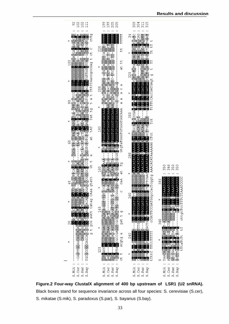
Figure. 2 Four -way ClustalX alignment of 400 bp upstream of L SR1 (U2 snRNA).
Black boxes stand for sequence invariance across all four species: S. cerevisiae (S.cer),
S. mikatae (S.mik), S. paradoxus (S.par), S. bayanus (S.bay).

34
Results and discussion
Fig. 3 Five-way ClustalX alignment of 400 bp upstream of SNR14 (U4 snRNA).
Black boxes stand for sequence invariance across all four species: S. cerevisiae (S.cer),
S. mikatae (S.mik), S. paradoxus (S.par), S. bayanus (S.bay), S. kudriazevii (S.kud).

35
Results and discussion
Fig. 4 Five-way ClustalX alignment of 400 bp upstream of SNR7 (U5 snRNA). Black
boxes stand for sequence invariance across all four species: S. cerevisiae (S.cer), S.
mikatae (S.mik), S. paradoxus (S.par), S. bayanus (S.bay), S. kudriazevii (S.kud).

36
Results and discussion
Generation of LRS1 and SNR7 reporter genes for in vivo expression analysis.
To better understand the involvement of the identified putative promoter elements in
transcription regulation we have chosen two snRNA genes LRS1 (coding for U2
snRNA) and SNR7 (coding for U5 snRNA), to be used as reporter genes for in vivo
expression analysis. Because snRNA genes are essential, a strain deleted for one of
these genes would be unable to survive.
In this work we prepared tagged versions of these genes, cloned into high-copy
vector, to identify their expression and monitor the effects on transcription of
changes in their promoter structures. We used as tag a 22 nucleotides sequence (5’-
GCAGTCTTACGAGTGAGGATCC-3’) whose BLAST analysis showed no significant
similarities in the S. cerevisiae genome.
The oligonucleotide was inserted within the coding regions with the aim to obtain
stable tagged-RNAs whose secondary structure were as similar as possible to RNAs
transcribed from wild type genes.
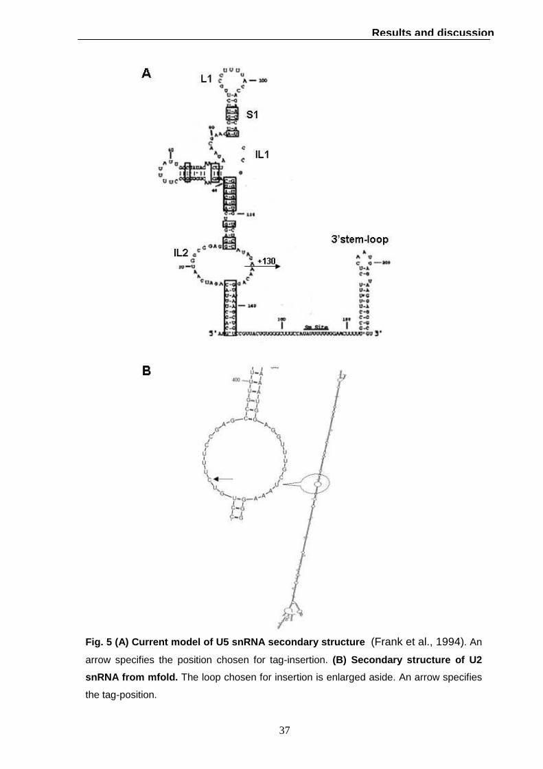
The secondary structure of U5 snRNA is characterized by a highly conserved stem-
loop (S1-L1) flanked by a moderately conserved internal loop (IL1); this 39-nucleotide
domain contains all U5-specific sequences essential for splicing activity (Frank et al.,
1994). We decided to insert the oligonucleotide at position +130 in the internal loop 2
(IL2) whose length is known to be quite variable in fungi and not required for U5
activity (Frank et al., 1994). The insertion of the tag within the U5 sequence is
represented in fig 5.
U2 snRNA is 1179 nucleotides long, six times larger than its mammalian counterpart
(188 nucleotides).; the 5’ domain is highly conserved (120 nucleotides) and essential
for viability while deletion of the central 945 nucleotides has no effect on growth rate
(Shuster and Guthrie, 1988). Since only the secondary structure of the 5’ end
domain has been studied, we used the structure prediction programme mfold
(http://mfold.bioinfo.rpi.edu/) to find the proper position for the tag. The secondary
structure is mainly characterized by a long stem; we have decided to insert the tag in
position +387 within a loop to reduce the disruption of base pairing (fig 5).
37
Results and discussion
Fig. 5 (A) Current model of U5 snRNA secondary stru cture (Frank et al., 1994). An
arrow specifies the position chosen for tag-insertion. (B) Secondary structure of U2
snRNA from mfold. The loop chosen for insertion is enlarged aside. An arrow specifies
the tag-position.

38
Results and discussion
Marked U5 snRNA and U2 snRNA genes produces stable RNAs.
The marked U5 snRNA gene (U5M) contains 248 bp of 5’-flanking region, a
transcribed region of 236 bp rather than 214 bp as in the wild type and 314 bp of 3’
flanking region. The marked U2 snRNA gene (U2M) contains 251 bp of 5’-flanking
region, transcribed region of 1201 bp rather than 1179 bp as in the wild type and 129
bp of 3’ flanking region(fig 6). Both templates were cloned into the Yep352 high-copy
vector (see methods and materials for details).
To verify whether stable RNAs could be generated from such templates, we tested
gene expression in vivo. Yeast cells (BY4741 strain) were transformed with U5M-
YEp352, U2M-YEp352 and empty vector Yep352 as a control. Total RNA was
extracted and probed with a radiolabeled oligonucleotide complementary to the
inserted tag.
As shown in fig. 6, the probe detected correctly transcribed marked RNAs from cells
transformed with U5M-YEp352 (lane 2,3) and U2M-YEp352 (lane 5,6) while no
specific signal was observed in cells transformed with the empty vector (lane 1,4).
The analysis showed that the insertion of the tag didn’t introduce significant
alterations in RNAs secondary structure since templates generates stable RNAs of
the expected length.
When cells were transformed with U5M templates, the specific probe detected two
marked RNAs of different length. This result can be explained by the fact that U5
snRNAs of S. cerevisiae are expressed in two forms, U5 short (U5S) and U5 long
(U5L), both products of the SNR7 gene. These forms differ for the presence of
absence of a stem-loop at their 3’ end due to alternative cleavage pathway of pre-
U5RNA (see fig 5A) (Patterson and Guthrie, 1987; Chanfreau et al., 1997).

39
Results and discussion
Fig. 6 (A) Schematic representation of U5M and U2M templates. Black boxes
indicates the position of the tag within the coding region. (B) In vivo expression of
marked U5M and U2M templates. Total RNA extracted from BY4741 transformed with
the empty YEp352 vector (lane 1,4), U5M-YEp352 (lane 2,3) or U2M-YEp352 (lane 5,6)
was gel fractionated and probed with a radiolabeled oligonucleotide complementary to
the inserted tag. The migration position of the two form of U5M RNA, large and small (L
and S) and of U2M RNA are indicated by an arrow. The asterisk indicates non-specific
hybridization.

40
Results and discussion
Marked U5 snRNA is correctly initiated in vivo.
Small nuclear gene transcription starts predominantly at a single position that
corresponds to a unique mature 5’ end. Since precise placement of the transcription
start site may be required to ensure a proper expression, we questioned if marked
RNAs were correctly initiated.
To determine the transcription start site of U5M RNA we used a primer extension
assay. The reaction was performed on total RNA extracted from cells transformed
with U5M-Yep352 template with a specific labeled probe complementary to the tag
inserted as previously described in the coding region of SNR7 gene. As shown in fig
7B (lane 5) the analysis gave rise to one major signal corresponding to initiation
position. The transcription initiation site was mapped by electrophoresing the primer
extension products adjacent to dideoxy chain termination sequencing reactions
performed with the same end-labeled oligonucleotide (Fig 7B, lane 1,2,3,4). Total
RNA from yeast cells transformed with U5M_∆TATA_YEp352, lacking all the
upstream region upstream necessary to transcription (fig 7A), was used as a
control(fig 7B, lane 6).
The result indicates that marked U5 RNA initiates transcription in vivo at the same A
residue as wild type. Since we have found a single start site, the two specific signal
found in the northern blot assay (fig 6B) have to differ in their 3’ end as supposed.

41
Results and discussion
Fig. 7 (A) Schematic representation of U5M and U5M_∆TATA templates. Black
boxes indicates the position of the tag within the coding region. (B) Primer extension.
Total RNA extracted from BY4741 transformed with U5M-Yep352 (lane 5) or U5M-
∆TATA-YEp352 (lane 6) was hybridized to [γ-32P]5’-labelad probe complementary to the
inserted tag. The adenosine residue at position +1(start site) is indicated by an arrow. On
the right side, the sequence surrounding the U5 transcription start site is shown. For
comparison, the U5M template was sequenced using the same end-labeled
oligonucleotide (lane 1, 2, 3, 4).

42
Results and discussion
SNR7 promoter region contains positive cis-acting elements.
To investigate in more detail the conserved elements found in U5 promoter region,
we began to generate progressive deletions in 5’ flanking region of U5M gene. A
schematic representation of templates is shown in fig 8A.
Plasmids were transformed into BY4741 strain and the expression level was
analyzed by northern blot (fig 8B).
Deletion of sequences upstream the Abf1 binding site caused a slight decrease in
expression (fig 8B, lane 3), thus indicating that this region is required for optimal
transcription, even if phylogenetic footprinting didn’t reveal any conserved element. A
significant drop in transcription was observed when the region containing the
conserved elements ABF1 and poly(dA) were eliminated (compare lane 3 and 5).
To verify the role of ABF1 element, we introduced point mutations in conserved
position of the Abf1p consensus motif (TCA→GAT; ACG→TTC); these mutations
diminished transcription (compare lane 3 and 4) and deletion of poli(dA) element
caused a further transcriptional decrease (compare lane 3, 4 and 5). Our results
suggest that U5 promoter-driven expression is positively influenced by both these
elements; their combined action has been already observed in many yeast promoter
in relation to chromatin organization (Goncalves et al., 1995; Lascaris et al., 2000).
RRPE site directed mutagenesis (TGAAAATTTT→TGATCTTGAC) revealed a
positive role of this element in transcription (compare lane 5 and 6); similar
requirement of an RRPE for basal snoRNA gene transcription has been observed in
our laboratory (Milena Preti, unpublished observations).
The presence of only a TATA box upstream of SNR7 ensures low, residual basal
transcription (lane 7), that is abolished when the TATA box is also removed (lane 8).

43
Results and discussion
Fig. 8 (A) Schematic representation of U5M 5'-mutant templates. Black boxes
indicates the position of the tag within the coding region. (B) In vivo expression of U5M
5'-mutant templates. Total RNA (10 µg) was extracted from BY4741 cells transformed
with the empty YEp352 vector (lane 1) and U5M 5'-mutant templates.(lane 2-8) was gel
fractionated and probed with a radiolabeled oligonucleotide complementary to the
inserted tag. The same blot was also probed for tRNAAla as an internal standard. The
histogram represents RNA quantitative value after normalization with the standard.
44
Results and discussion
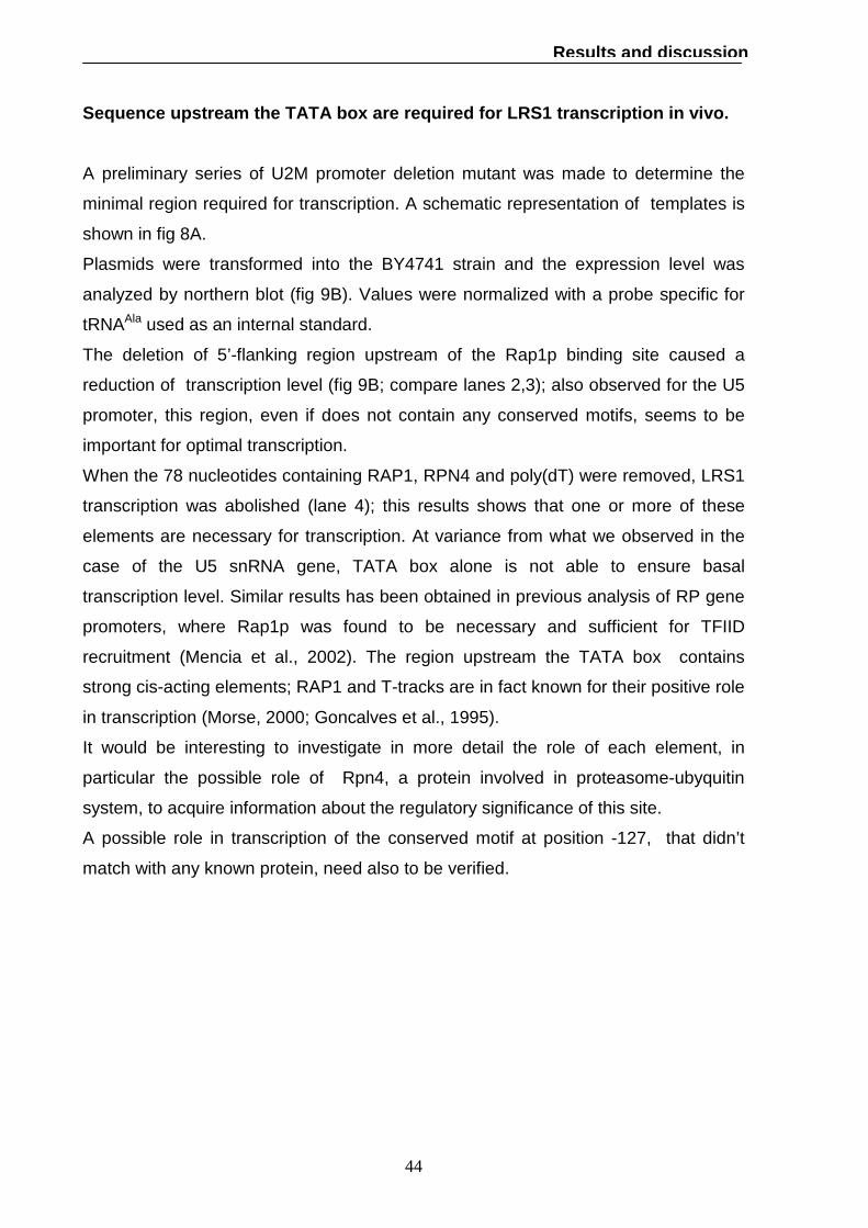
Sequence upstream the TATA box are required for LRS 1 transcription in vivo.
A preliminary series of U2M promoter deletion mutant was made to determine the
minimal region required for transcription. A schematic representation of templates is
shown in fig 8A.
Plasmids were transformed into the BY4741 strain and the expression level was
analyzed by northern blot (fig 9B). Values were normalized with a probe specific for
tRNAAla used as an internal standard.
The deletion of 5’-flanking region upstream of the Rap1p binding site caused a
reduction of transcription level (fig 9B; compare lanes 2,3); also observed for the U5
promoter, this region, even if does not contain any conserved motifs, seems to be
important for optimal transcription.
When the 78 nucleotides containing RAP1, RPN4 and poly(dT) were removed, LRS1
transcription was abolished (lane 4); this results shows that one or more of these
elements are necessary for transcription. At variance from what we observed in the
case of the U5 snRNA gene, TATA box alone is not able to ensure basal
transcription level. Similar results has been obtained in previous analysis of RP gene
promoters, where Rap1p was found to be necessary and sufficient for TFIID
recruitment (Mencia et al., 2002). The region upstream the TATA box contains
strong cis-acting elements; RAP1 and T-tracks are in fact known for their positive role
in transcription (Morse, 2000; Goncalves et al., 1995).
It would be interesting to investigate in more detail the role of each element, in
particular the possible role of Rpn4, a protein involved in proteasome-ubyquitin
system, to acquire information about the regulatory significance of this site.
A possible role in transcription of the conserved motif at position -127, that didn’t
match with any known protein, need also to be verified.

45
Results and discussion
Fig. 9 (A) Schematic representation of U 2M 5'-mutant templates. Black boxes
indicates the position of the tag within the coding region. (B) In vivo expression of U2M
5'-mutant templates. Total RNA (10 µg) was extracted from BY4741 transformed with
the empty YEp352 vector (lane 1) and U2M 5'-mutant templates.(lane 2-4) and gel
fractionated. A radiolabeled oligonucleotide complementary to the inserted tag was used
as probe. The same blot was hybridized with a probe specific for tRNAAla as an internal
standard.

46
Results and discussion
Rap1p interacts with LSR1 promoter region in vivo.
To gain insight into the involvement and mechanism of actions of Ab1p, Rap1p and
Rpn4p in snRNA gene transcriptional regulation, we verified physical association of
these proteins to their target promoters. We also checked SNR7 promoter for
occupancy by Stb3p, a recently discovered RRPE-binding protein (Liko et al.,2007).
We performed chromatin immunoprecipitation experiments from yeast strains
expressing tandem affinity purification (TAP) tagged version of Abf1, Stb3, Rap1 and
Rpn4 proteins. The untagged BY4741 strain was used as control.
Independent ChIP experiments were carried out, and immunoprecipitated DNA was
analyzed by PCR amplification. The results of ChIP analyses are reported in Fig. 10.
Occupancy values are relative to occupancy at intergenic region ARS540 Chr 5, that
was used as a reference.
As shown in fig 10A, Abf1p is not found specifically associated to SNR7 promoter
region; however we have demonstrated that promoter activity depends on Abf1
binding site (fig 10B). It is conceivable that regulations may not require stable binding
of Abf1p to the promoter region. This mode of action, defined “hit and run”, has been
previously suggested for Abf1p (Schroeder et al., 1998).
We tested if Stb3p could represent the protein binding to the SNR7 RRPE. ChIP
analysis didn’t show any enrichment of Stb3 at the SNR7 promoter; anyway not all
RRPE-containing genes are influenced by this protein, whose mode of action remain
unclear (Liko et al., 2007).
When we analyzed LRS1 promoter for enrichment we found a strong association of
Rap1p (fig 10B); since Rap1p binding is continuously required for enhancement, this
result is a further confirmation of Rap1 involvement in transcription (see fig 10B).
In vivo binding of Rpn4p showed only a slight enrichment; we can’t anyway exclude
that the low occupancy value obtained could be related to the extremely short half-life
of this protein (t1/2 <2 min).

47
Results and discussion
Fig. 10 In vivo occupancy at SNR7 ( A) and LRS1 (B) promoters. ChIP were performed
from yeast strains expressing tandem affinity purification (TAP) protein-tagged version of
Abf1, Stb3, Rap1and Rpn4 proteins. BY4741was used as non-tagged control strain.
Immunoprecipitated DNA was analyzed for enrichment by PCR. The asterisk indicates
the intergenic region ARS540 Chr 5, used as an internal standard.
Promoter are schematically represented; regions amplified by PCR in the ChIP
experiment are identified by arrow pairs.

48
Conclusion
Conclusion
Defining co-occurrence and spatial relationship of individual binding sites is an
important step in understanding the regulatory content of promoter regions.
The primary aim of this work was to identify putative regulatory elements in yeast Pol
II-transcribed snRNA gene promoters, starting from phylogenetic footprinting. The
comparative analysis showed elements conserved across Saccharomyces sensu
stricto group, a closely related set of species in which most of the genes and
regulatory elements are shared.
This approach gave a rich haul of information. The common pattern in all Pol II-
transcribed gene is the presence in their promoter region of General Regulatory
Factors (GRFs) Abf1p (U4 and U5 snRNAs), Reb1p (U1 snRNA) and Rap1p (U2
snRNA). These three abundant and essential transcription factors have many target
sites in yeast genome acting as multifunctional proteins. They can enhance both
activation and repression of transcription, but are also involved in silencing and
regulation of DNA replication initiation. The GRFs seems to share a common
mechanism of action; indeed the binding site for one GRF within a promoter can be
exchanged with another (Fourel et al, 2002).
Their binding motif usually has little intrinsic regulatory activity and is often found in
combination with poly(dT-dA) sequences, that operate not by recruitment of specific
transcription factors but rather by their intrinsic DNA rigid structure. It has been
hypothesized that these elements synergise in local opening of chromatin which then
permits increased binding of other transcription factors. With this respect, it is
remarkable that in snRNA gene promoters a T-rich or A-rich elements are found
downstream the GRF binding site.
To better understand the involvement of the identified putative promoter elements in
transcription regulation we have chosen two snRNA genes LRS1 (coding for U2
snRNA) and SNR7 (coding for U5 snRNA), to be used as reporter genes for in vivo
expression analysis. We have prepared tagged versions of these genes by inserting
an oligonucleotide within the transcribed region. Such marked templates generated
stable and corrected initiated RNAs, thus allowing in vivo promoter analysis.
In vivo expression analysis of 5’-mutated versions of marked genes allowed us to
identify the combined positive role of Abf1p-poly(dA) element and Rap1p-poly(dT)

49
Conclusion
element in SNR7 and LRS1 transcription respectively. The requirement for Rap1p-
poly(dT) element was absolute, since deletion of this region caused complete loss of
transcription; an analogous elimination of Abf1p-poly(dA) element caused a strong
reduction in SNR7 expression. An important role in regulation of Rap1p is confirmed
by its tight association to LRS1 promoter, revealed by ChIP analysis.
A high resolution atlas of nucleosome occupancy in yeast has been presented by
Lee et al (2007). Both SNR7 and LSR1 promoter are characterized by a free
nucleosome region that extends up to 100 bp upstream of the Abf1p and Rap1p
binding site respectively. Future work will investigate if these elements function as
chromatin-reorganizing factor preventing the deposition of nucleosomes in the region
close to their binding site, by analysing if the loss of Abf1p and Rap1p binding or
deletion of poly(dT-dA) elements are associated with changes in chromatin
structure.
GRFs usually amplify the effect of neighbouring regulatory sites; we have identified
the positive cis-acting element RRPE (ribosomal RNA processing element) in U5
snRNA promoter. It was interesting to find the same positive role of RRPE in snoRNA
transcription (data not shown); this could suggest a possible common regulatory
strategy of snRNA and snoRNA transcription. Further investigation will also be
directed toward understanding the strong conservation of Rpn4p in U2 snRNA
promoter and its possible relationship with the ubiquitin-proteasome pathway.

50
Characterization
of novel snRNA gene-like
transcriptional units
in the human genome

51
Abstract
The role of polymerase (Pol) III in eukaryotic transcription is commonly thought of as
being restricted to a small set of housekeeping, highly expressed non-protein-coding
(nc) RNA genes. Recent studies, however, have remarkably expanded the set of
known Pol III synthesized ncRNA. By means of computer search for upstream
promoter elements (proximal sequence element and distal sequence element) typical
of small nuclear RNA genes, we have identified in the human genome some putative
transcription units.
In this work we analyzed the in vitro transcription properties in HeLa and SKNBE
neuroblastoma cells nuclear extracts of some of these putative units, showing that
their promoter elements were actually able to support Pol III-dependent transcription.
In particular, we identified a novel 171 nt ncRNA, 17A, whose transcription was
driven by the presence of a PSE located at -59, which ensure efficient transcription,
and a TATA box located at -28, that directs start site selection. Indeed, in the
absence of the TATA box, another A-T rich element located downstream, not
affected by the presence of the PSE, could direct an efficient alternative initiation of a
shorter transcript. We have also defined the promoter architectures of two other
ncRNA genes, 38A and 29A, characterized by the presence of pol III-type3 elements,
PSE and TATA box, both necessary to ensure an efficient level of transcription.
Finally we identify a TATA less PSE-dependent promoter for 51A.
Chromatin immunoprecipitation experiments conducted in SKNBE and SHSY5Y
neuroblastoma cells using antibodies directed against SNAPc and other components
of the Pol III-type 3 transcription machinery, demonstrated its association with the
38A and 51A promoters, suggesting a Pol III-dependent in vivo expression of these
genes.
Some of the characterized transcriptional units (17A, 38A, 51A) are internal to known
protein-coding genes where alternative splicing events takes place, while another
(29A) was identified as an Alu.
Further studies will be required to better characterize these transcription units in vivo
and the function of their products, even though their location suggest a possible
regulatory role and an increasing complexity of the by pol III transcriptome.

52
Introduction
Introduction
RNA molecules are active participant in regulating, catalysis and controlling of many
fundamental cellular processes, role that was reserved for proteins just few years
ago; as happens for proteins the structure of these RNAs is crucial for their
functionality. These regulatory RNAs, that do not encode a protein, are referred as
non coding RNAs and fold into specific higher order structures that impart a function
to the molecule. They are characterized by different size, tissue specific expression
pattern and biological functions. Pol III is specialized in transcription of ncRNA, being
its role commonly restricted to small set of housekeeping, highly expressed non-
protein-coding (nc) RNA genes. Recent studies, however, have remarkably
expanded the set of known Pol III synthesized ncRNAs, suggesting that gene-
specific Pol III regulation is more common than previously appreciated. Newly
identified Pol III transcripts include small nucleolar RNAs, microRNAs, short
interspersed nuclear element-encoded or tRNA-derived RNAs. Recent advances in
mammalian genome studies are bringing to light the occurrence of a widespread
transcription of non coding regions devoted to the regulation of the protein coding
genome expression (Mattick, 2004). In addiction, the involvement of Pol III transcripts
in gene regulation and, more generally, in several important RNA dependent
functions makes it likely that alterations in their levels and activity will compromise
diverse cellular processes.
Comprehensive transcriptome analyses, genome wide location studies of
transcription factors and computational searches for ncRNA sequences and DNA
regulatory regions in whole genome are powerful tools in discovering new ncRNAs.
Pol III-transcribed genes are characterized by three promoter types called type 1-3.
Type 1 consist of an internal control region, which can be subdivided into A box,
intermediate element and C box. The type 2 promoter consist of an intragenic A and
B boxes. Type 3 promoter is totally extragenic. The best characterized type 3
promoters belong to snRNA U6, 7SK RNA, H1 RNA and RNase MRP RNA genes.
The sequences required for efficient basal transcription are a TATA element located
between -30 and -25, which acts as the major determinant of the polymerase
specificity, and a proximal sequence element (PSE) between -66 and -47. The PSE
recruits a stable protein complex, known as SNAPc or PTF, containing five subunits.

53
Introduction
Activated transcription is provided by the distal sequence element (DSE) located
between -260 and -190. DSEs are composed of many functional submotifs that can
be present either simultaneously or separately. Two of these are often the octamer
and the Staf motifs that binds respectively Oct-1, a homodomain transcriptional
activator, and Staf, a seven zinc finger protein (Hernandez, 2001).
Starting from the hypothesis the human genome might contain pol III transcription
units each specifically regulating one or more specific pol II genes, our laboratory
performed a screening in the human genome for pol III type-3 regulatory elements
and identified a set of about 30 novel putative pol III-transcribed units. A detail
investigation revealed that one of this novel non-coding genes, called 21A, played a
role in control of the proliferation of some tumor cells (Pagano et al, 2007). In these
work we selected four of these identified units (17A, 38A, 29A, 51A) that were
internal to known protein genes, to be characterized by experimental determinations,
showing that their promoter elements were actually able to support Pol III-dependent
in vitro transcription.

54
Materials and Methods
Materials and Methods Human genomic DNA isolation: Extraction of genomic DNA was performed as detailed below. Four volumes of PBS
(50mM potassium phosphate; 150 mM NaCl) were added to a sample of saliva (4 ml)
vortexed and then centifuged at 1800xg for 5 min. The supernatant was carefully
removed and the pellet rinsed using additional 2 ml of PBS, before centrifugation
again at 1800xg for 5 min. After removal of the supernatant, the pellet was
resuspended in 360 µl of PBS. An equal volume of lysis buffer (50 mM Tris-HCl, pH
8; 10% (w/v) SDS) was added and the mixture was incubated at 65 °C for 30 min.
Pronase was added to a final concentration of 1 mg/ml followed by incubation at 45
°C for 1h and 30 min and treatment with RNase (final concentration 75 µg/ml;
incubation at 37 °C for 45 min). After extraction with phenol-chloroform, DNA was
precipitated at RT with isopropyl alcohol in the presence of 20 µg of glycogen and
centrifugated for 10 min at 14000 rpm. The pellet was finally resuspended in 50 µl of
AE buffer (10 mM Tris-HCl, pH 8). DNA concentration was measured by
spectrophotometry at 260 nm. Purity of DNA was assessed using the ratio of
OD260/280 with a value of 1.8 -2.0 being of good purity.
Amplification and cloning of DNA templates: Some of the putative transcriptional units obtained by a computer analysis were
selected for experimental determinations. DNA fragments were PCR-amplified from
human genomic DNA using Taq DNA polymerase (Fermentas) and cloned into
pGEM –T easy vector (Promega). 12A, 14A, 17A, 22A, 24A, 29A, 31A, 38A, 41A,
42A, 43A and 51A templates were digested with SphI-SacI and subcloned into
pNEB193 vector. All constructs were verified by sequencing and purified with the
Qiagen Midi Kit according to manufacturer’s instructions before to be used for in vitro
transcription reactions. All oligonucleotides used in amplification and cloning of the
selected units are listed in table 1.

55
Materials and Methods
Primer name Primer sequence 12A_fw 12A_rev
GAAATAACTTATAAAACAGGTCATCC GGAAAATTAATGTATTATAAATGCAAAGG
14A_fw 14A_rev
ACTGATGTATGATTATATCTTATTTTGG TTTGTCTGTGGACAAATTGTCTCC
20A_fw 20A_rev
GGTAATGGATTAATTGATTCAGACC ACAGGAAAGTAGACAGCGGAGCTGG
22A_fw 22A_rev
AAACAATAATCATCCTTTGTAAAGAGC TATGCAGAAATCTAATACAGGAAGC
23A_fw 23A_rev
ATACCAGGTCTATAACACCTGAGC TACTATAACTACAATCCATTACTACAC
24A_fw 24A_rev
GCAACATAATACACAAGAAGAAGAAC CTTGCTATAAACATTTACTTGTGTGG
29A_fw 29A_rev
GGTATTTTGGTGTTTCAGCCTTTCC ACATTGAATCACCTTATGAG
31A_fw 31A_rev
CTTTCTTGAAATATTCTCATTGTTACC TAGTGACTTACTGGAGAGGATAGG
33A_fw 33A_rev
ATACGAAGGCATAGTAGAAGAAAGC CAGACTAATGTCTTCCTGAGAGG
34A_fw 34A_rev
TTTCTGCTTGTGCCCATTACAACC TACAGGCGTAGGCCACTACAGC
36A_fw 36A_rev
GCCCATTGCCAAGTCCTTCACG AAATCAAAAGTTGTCTCACTCAGG
37A_fw 37A_rev
AAAAGAAAAGAAACCATTGTTTAATCC TAACTTCTATCAAAATCTCTGCTGG
38A_fw 38A_rev
CTAGCAATAGCAATCAGACCAGAG TTCAGGGTGTCCTGTTGGTACC
41A_fw 41A_rev
TAGACACACATATGTAAATTCACTCC ATTGCTTACCATTTTGCATTATCACC
42A_fw 42A_rev
TCCAAGTTTACAAATAGGGTCTCC ATAAATCAAAAGTTGTCTCACTCAGG
43A_fw 43A_rev
AAAAGCCCAATGTTTAACATATCC AACCTGCGTGTTTAAAAAGAGTCC
44A_fw 44A_rev
GTTACATAAGCTTTCTATGCCTTGG AGCTGCACCCTGGCACTGTCC
45A_fw 45A_rev
TAATTGAGAAGCAACTCCAGCTCTAGC GGAACTCTGATAATTTCGTTTATAGG
48A_fw 48A_rev
GTCGGAGGGTCTTCTTGCATGG AACCCATTGAATGAAGGCCTAGC
51A_fw 51A_rev
ACAAACTCCATCTGCAATTCCTCG CAGGTATAGGAGGGGTGCAGC
Table 1 List of oligonucleotides used in amplification and cloning of putative transcriptional
units selected for experimental determinations.

56
Materials and Methods
17A templates: Constructs were obtained by PCR amplification using 17A_pNEB as
template (containing 90 bp of the 5’-flanking region and 34 bp of the 3’-flanking
region) and specific mutagenic forward primers. All the fragments were cloned in
pNEB193 vector (SmaI site), sequence verified and purified with the Qiagen Midi Kit.
38A templates:
Constructs were obtained by PCR amplification using 38A_pNEB as template
(containing 347 bp of the 5’-flanking region) and specific mutagenic forward primers.
38A_poliT template was generate by amplification from 38A template with a specific
reverse primer containing a sequence of 9T. All the fragments were cloned in
pNEB193 vector (SmaI site), sequence verified and purified with the Qiagen Midi Kit.
The fusion 38A_7SK DNA fragment was obtained by amplifying the 7SK coding
region with a specific primer containing the 38A upstream sequence from -68 bp to -
27 bp. The hybrid product was cloned into pNEB193 vector (SmaI site). The 5’-
mutant of 38A_7SK were obtained by PCR amplification with specific primers
forward. The fragments were cloned into the SmaI site of pNEB193 vector.
29 templates:
Constructs were obtained by PCR amplification using 29A_pNEB as template
(containing 522 bp of the 5’-flanking region) and specific mutagenic forward primers.
29A_poliT template was generate by amplification from 38A template with a specific
reverse primer containing a sequence of 9T. All the fragments were cloned in
pNEB193 vector (SmaI site), sequence verified and purified with the Qiagen Midi Kit.
The fusion 29A_7SK DNA fragment was realised generating two PCR products
amplifying from 29A_pNEB template and 7SK template using the high fidelity Pfu
DNA polymerase (Promega) and gene-specific pairs of oligonucleotide primers:
29A_SacI_fw in combination with 29A_BamHI_rev to realise the first insert containing
29A 5’-flanking region from positon – 513 to -25; 7SK_BamHI_fw in combination with
7SK_SphI_rev to realise the second insert containing 7SK the transcribed sequence
plus 18 bp of 5’-flanking region and 124 bp of 3’-flanking region. The fragments were
digested with SacI/BamHI and BamHI/SphI respectively and inserted in pNEB193
(sacI/ sphI sites). The 5’-mutant of 29A_7SK were obtained by PCR amplification with

57
Materials and Methods
specific primers forward. The fragments were cloned into the SmaI site of pNEB193
vector.
All oligonucleotides sequences used for amplification and cloning of 17A, 29A, 38A,
51A templates are listed in table 2.
Primer name Primer sequence
17A_CC_fw CCTCACCATAAAAGTGAAATAATG
17A_AA_fw CCTCAAAATAAAAGTGAAATAATGTTGC
17A_TATA1_fw AATAAATAGTGCAAAATATTAACAAAG
17A_TATA2_fw AAATATTAACAAAGACACAATTGAATA
38A_deltaDSE_fw TAATAACAACATATCTGAAAAAGACGC
38A_deltaTATA_fw GTTGAAGAAGACACATATAAATAGA
38A_DSE_fw TATTTGCATATAAAAATAGTTAGAAATAAATTTAACC
38A_poliT_rev AAAAAAAAACTATTTCTGTGATGCATATCCTTG
38A_7SK_fw TAACCATAAAGGTGAAATATTTGTATACCGATAACTAT
AAAGCTTGTGCGCCGCTTGG
7SK_Rev CGGGAGGTGGAGGTTACAGTGAGC
38A_7SK_TATA_fw ACTATAAAGCTTGTGCGCCGC
7SK_NP_fw GCTTGTGCGCCGCTTGGGTACC
29A_deltaDSE _fw GGAACTTTATGTCGCTACC
29A_ ∆TATA_fw AGACACTGAATTCTAACTAGACGC
29A_ FW_SacI CGCCGAGCTCGGTATTTTGGTGTTTCAGCCTTTCC
29A_ Rev_ BamHI TACTGGATCCTATTTATTGTGAGTTCTAGTAATTTC
7sK_ fw_ BamHI TATAGGATCCGCGCCGCTTGGGTACCTCGG
7sk _Rev_ SphI ACATGCATGCGGAGGTGGAGGTTACAGTGAGC
29A_710-1270 fw GGAACTTTATGTCGCTACC
29A_AA_7SK_ fw GGAACTTTATGTCGCTAAAATAAATG
29A_TATA_7SK AATAAATAGGATCCGCGCCGC
29A_deltaDSE _poliT_rev AAAAAAAAACCTGAGCTCAAGCAACCC
Table 2 List of oligonucleotides used in amplification and cloning 17A, 29A, 38A, 51A
templates.
58
Materials and Methods
Preparation of SKNBE extract: We prepared the extract according to Dignam et al (1983). We prepared 10 plates
(15 cm) of SKNBE cells grown to about 90% confluence. Each plate was washed
once with 10 ml of PBS; cells were trypsinized and collected by centrifugation at 4°C
for 3 min at 2000 rpm. The pellet was washed one with cold PBS (3 ml) and the
packed cell volume was determined. Cells were resuspended in 2.5 volumes of
hypotonic S100 buffer (20 mM Hepes/KOH, pH 7.9; 10 mM KCl; 1.5 mM MgCl2),
containing 0.2 % NP40, 3 mM DTT and 0.5 mM PMSF, and directly centrifuged at
12000 rpm for 30 sec at 4°C. The supernatant (cytoplasmic extract S100) was
carefully removed and dialyzed against a 10% glycerol containing buffer (20 mM
Hepes/KOH, pH 7.9; 60 mM KCl; 3 mM DTT; 0.5 mM PMSF). The pellet was
analysed under the microscope to verify the integrity of the nuclei then resuspended
in 1.5 volumes of nuclear extract buffer (20 mM Hepes/KOH, pH 7.9; 10 mM KCl; 1.5
mM MgCl2; 20% glycerol; 3 mM DTT; 0.5 mM PMSF). 0.5 volumes of the same buffer
containing 1.2 M KCL were added drop wise at 4°C to the nuclei-buffer slurry over a
period of half an hour, moderately vortexing. This mixture turn over-head at 4°C for
30 min then was centrifugated at 40000 g for 30 min at 4°C. The resulting
supernatant (nuclear extract) dialyzed against a 20% glycerol containing buffer (20
mM Hepes/KOH, pH 7.9; 60 mM KCl; 3 mM DTT; 0.5 mM PMSF; 0.2 mM EDTA).The
extract was frozen as aliquots in liquid nitrogen and stored at -80°C. The protein
concentration was 10 mg/ml.
In vitro transcription analyses: Transcription reactions were carried out in a final volume of 25 µl in the presence of 2
µg of template DNA and HeLa cell or SKNBE nuclear extract (100 µg) supplemented
with 50 ng of recombinant human TBP. The standard transcription mix contained: 5
mM creatine phosphate, 70 mM KCl , 5 mM MgCl2, 20 mM Tris/HCl pH 8, 1 mM
DTT, 2 µg/ml α-amanitin, 0.5 mM CTP,ATP,GTP, 25 µM/ 10 µCi UTP /[α-32P]UTP,
SUPERase IN (Ambion, 10 U), glycerol 10 % (v/v). The reactions were incubated for
1h at 30 °C. The products were phenol extracted and precipitated with ammonium
acetate. Radiolabeled transcripts were separated on 6% polyacrylamide/7 M urea

59
Materials and Methods
gel,quantified and visualized by phosphorimaging using a Personal Molecular Imager
FX (Bio-Rad).
We used as internal standard an RNA of 118 nt in length synthesized in vitro by T7
RNA polymerase (Amersham), following the manufacturer’s protocol. The RNA was
phenol extracted and precipitated within each sample.
Primer extension: A double scale transcription reaction was performed as described, without including
radiolabeled UTP. A parallel reaction ,no-DNA containing, was conducted as a
control.
Reverse transcription reactions were carried out using Superscript III reverse
transcriptase (Invitrogen), following the manufacturer's protocol. The purified
transcripts were resuspended in a final volume of 12 µl in the presence of 0.5 mM
dNTPs and 1 pmole of specific 5’-end-radiolabeled probe (table 6) . The mixture was
heated at at 65°C for 5 min and a mixture providing 50 mM Tris/HCl pH 8, 75 mM
KCl, 3 mM MgCl2,5 mM DTT, SUPERase IN (Ambion, 10 U) and 200 U Superscript
III reverse transcriptase (Invitrogen) was added to a final volume of 20 µl. The
reactions were incubated for 1h at 60 °C and subsequently for 15 min at 70°C to
inactivate the enzyme. The products were precipitated with ammonium acetate and
gel-fractionated. A parallel reaction was conducted as a control in absence of
Superscript III reverse transcriptase. The 5’ end was mapped by direct comparison with dideoxy chain termination
sequencing reactions (Kit Thermo-Sequenase, GE Healthcare) run on the same gel.
Oligonucleotides used as specific probe in primer extension reactions are listed in
table 3.
Primer name Primer sequence
17A_probe GGACTTTCCAAGATTGCCCAGGG
38A_probe GGTTAATACAGACATTTTAACATTGTTA
29A_probe GGATTACACACAGGAAGCCACCG
51A_probe CCCTCATGGCACTTGGAGATTTG
Table 3 List of oligonucleotides used as specific probe in primer extension analyses.

60
Materials and Methods
Chromatin immunoprecipitation: Cross-linked chromatin was prepared essentially as describe (Kurdistani S, 2003).
Human cells (SKNBE and SHSY5Y line) were grown to approximately 75%
confluence (two plates) and then crosslinked with formaldeyde for 30 min. Cells were
washed twice in cold PBS (each plate with 10 ml) complemented with protease
inhibitors (Complete, Roche: 1 tablet). Cells were resuspended in 500 µl of PBS,
scraped into microcentrifuge tubes and pelleted for 2 min at 10000 rpm at 4°C. The
pellet was then suspended in 400 µl of lysis buffer (1% SDS; 50 mM Tris/HCl, pH 8;
20 mM EDTA; protease inhibitors). Cells were incubated 10 min on ice and sonicated
to reduce DNA fragments to an average size of 300 bp. The lysate was clarify by
centrifugation for 10 min at maximum speed at 4°C. The supernatant was transferred
to clean tube. Before proceeding with immunoprecipitation, 100 µl of solution were
put aside as input samples, diluted to 1 ml with buffer DB (1.67 mM Tris/HCl, pH 8;
0.001 % SDS; 0.1 % Triton X-100; 0.12 mM EDTA; 16.7 mM NaCl). Beads (30 µl,
slurry 50%) were added, followed by incubation of the tubes on a nuotator at 4°C for
1h.
For immunoprecipitation chromatin solution was incubated over night at 4°C with 10
µl of antibody (α-SNAPc or α-Brf2 or α-pol III). Fifty microliters of a 50% (v/v)
suspension of protein A –Sepharose beads was added and incubated for 2 h at 4°C.
The protein A beads were pelleted at room temperature for 3 min at 8000 rpm. The
supernatant was removed and the beads were resuspended in 200 µl of wash buffer
(50 mM HEPES, pH 7.9, 0.1% SDS, 1% Triton X-100, 0.1% deoxycholate, 1 mM
EDTA, 140 mM NaCl) and transferred to a 0.45-µm filter unit (Millipore Ultrafree-MC).
The tubes were rinsed with another 200 µl of the wash buffer, and the rmaining
beads were added to the filter unit. The filter was rotated 15 min at 4°C and spun at
8000 rpm for 3 min. The flow-through fraction was discarded. The washing step was
repeated once with the wash buffer containing 500 mM NaCl, and twice with LiCl
Buffer (20 mM Tris-HCl, pH 8, 0.5% NP-40, 0.5% deoxycholate, 1 mM EDTA, 250
mM LiCl) and TE (10 mM Tris-HCl, pH 8.0, 1 mM EDTA). To elute chromatin from the
beads 100 µl of elution buffer (50 mM Tris-HCl, pH 8.0, 1 mM EDTA, and 1% SDS)
was added to the filter unit which was then incubated at 65°C for 10 min and spun at
13000 rpm for 3 min. Elution was repeated once more. The flow-through were
combined and incubated at 65 °C overnight. The sample was treated with RNase

61
Materials and Methods
(conc 0.04 mg/ml, incubation at 37°C for 30 min) and proteinase K ((conc 0.08
mg/ml, incubation at 56°C for 2 h). After extraction with phenol-chloroform, DNA was
precipitated overnight at -20 °C with ethanol-sodium acetate in the presence of 20 µg
of glycogen, and resuspended in TE buffer (10 µl). For input samples 5 µl of
sonicated crosslinked chromatin were added to 245 µl TE with 1% SDS and
incubated overnight at 65 °C. These input samples were then treated with Proteinase
K, phenol/chloroform extracted, ethanol precipitated and dissolved in 100 µl TE.
DNA Amplification: DNA samples were amplified by PCR using GoTaq polymerase (Promega) and
specific primer pairs (Table 4). Typically 1 µl of the immunoprecipitated DNA and 1µl
of the total DNA Input were used. The specific primers for the GAPDH exon 2 were
used as internal control. Reactions were carried out in 10 µl and contained 0,25 µM
primers, 0,1 mM dNTPs, 0,06 mCi/ml di α32P-dCTP (Perkin Elmer). PCR products
were fractionated on a 6% polyacrylamide gel, visualized and quantified by
phosphorimaging. The fraction of immunoprecipitated material for a specific fragment
was calculated by ratio of immunoprecipitated DNA over total DNA.
The primers used in DNA amplification are listed in table 4.
Primer name Primer sequence
38A_fw_deltaDSE TAATAACAACATATCTGAAAAAGACGC
38A_rev_ChIP GTGTCTTCTTCAACTTTTTTTATC
51A_fw ACAAACTCCATCTGCAATTCCTCG
51A_rev_ChIP GAGATTTGAAAGGACTGCAGG
U6_fw GTACAAAATACGTGACGTAGAAAG
U6 _rev GGTGTTTCGTCCTTTCCACAAG
GAPDH _fw AGGTCATCCCTGAGCTGAAC
GAPDH_rev CCACCTGGTGCTCAGTGTAG
Table 4 List of oligonucleotides used as primers for the PCR on imunoprecipitated
chromatin. The amplicon length was set to 143 bp, 151 bp, 150 bp for gene-specific primers
38A, 51A, U6 respectively and to 189 bp for GAPDH control region.

62
Results and discussion
Results and discussion
RNA polymerase III-dependent transcription of putative transcription units
identified by computational analysis.
In a previously work from our laboratory, we identified a novel set of ncRNA
screening the human genome for upstream promoter elements typical of type 3 -pol
III promoters (proximal sequence element and distal sequence element). In particular
we tested H1 PSE as query sequence for the search of similar elements in the
human genome by using the BLAST algorithm (http://www.ncbi.nlm.nih.gov/BLAST)
and selected among the sequences obtained those that contained a DSE sequence
element within a distance of 1000 bp upstream of the PSE. We further investigated if
those units had features compatible a pol III promoter structure: a TATA-like element
downstream of the PSE and a termination signal (run of at least four T residues). To
complete the definition of the transcriptional unit, we assumed the transcriptional
start site about 30 bp downstream the TATA box. We finally selected 31 novel
putative units with pol III transcribed snRNA gene features (Pagano et al.,2007).
When these non coding sequences were used to challenge the human genome
database, it was found that some of them were internal to known or predicted
protein-coding genes.
We have already investigated 21A transcriptional unit (table 5) and its regulatory
activity (Pagano et al.,2007).
To test if those units were actually transcribed, we selected for experimental
determination those characterized by a PSE-TATA distance close to the canonical
distance of 20 bp. These units were PCR amplified from genomic human DNA and
inserted in pGEM-T easy vector. After a preliminary in vitro transcription assay, we
decided to continue the analysis with the templates generating the most interesting
transcription patterns (data not shown). The selected units (12A, 14A, 17A, 22A, 24A,
29A, 31A, 38A, 41A, 42A, 43A and 51A) were subcloned in pNEB193, a vector
producing a lower non-specific transcription background, and in vitro transcription
reactions were performed. The reactions were carried out in HeLa extract in the
presence of α-amanitin (2 µg/ml), to ensure pol III specific transcription. We used
transcription of the empty vector pNEB193 as a control.

63
Results and discussion
Table 5. Novel putative snRNA gene-like transcriptional units. Units selected for
experimental determinations are written in bold.
Tr.Unit PSE-TATA
distance Tr.Length(nt) Hum.Gen.Map
BLAST
Human Genome
12A 25 99 2p24.3 -
14A 19 148 3p12 -
17A 24 159 9q22-q31 GPR51(intron3) Sense
20A 21 547 14q22.1 -
21A 76 333 8q24.1 CENPF(intron7,14,18)Antisense
22A 23 235 6q16-q21 -
23A 24 200 Xq21.3 -
24A 28 307 12q21 -
27A 51 91 7q22 -
29A 15 360 11p15 ASCL3(intron1)Sense
30A - 258 Xp11.4 -
31A 25 231 12q21 -
32A - 140 17q21 -
33A 31 210 1q32.2 -
34A 17 33 5q15 -
35A 50 351 8p11.2 -
36A 16 122 3p12 -
37A 17 49 14q13 -
38A 17 354 4p15.31 KCNIP4(intron1)Antisense
39A 78 76 Xp11.3 FLJ22843(intron10)Antisense
40A 55 484 11p15 -
41A 28 79 2q31 -
42A 17 122 3p12.3 -
43A 13 65 4q34.3 -
44A 27 218 4q13.3 -
45A 21 78 4p14 APBB2(intron1)Antisense
47A 58 48 2q22.1 -
48A 20 405 11q24.2 -
50A 6 156 21q21 -
51A 17 273 11q23.2 SORL1(intron1)Sense
52A 50 142 8p11.2 -

64
Results and discussion
Comparison of the transcription profiles with the one obtained with the empty vector
(fig 1B, lane 9-10) allowed us to identify pol III RNAs selectively transcribed from the
inserted units.
In vitro transcription in HeLa extract of template 12A and 22A generated a group of
specific strong signals between 55-80 nt for the former and 90 nt for the latter (lane
1,3). Since putative transcript length was predicted to be 99 nt and 235 nt
respectively, this multiple-transcript pattern could be the result of either RNA
processing or RNA multiple initiation/termination. Other termination signals beside
the canonical oligo(T) sequences can in fact affect pol III termination (Thomann et al.,
1989).
An interesting result was obtained from 17A and 43A template transcription (lane
2,8). The analysis revealed the presence of signals of the expected length (table 1),
with the 17A transcript more strongly expressed than the 43A RNA. Both seemed to
be the specific result of transcription from these novel units, as the inserts did not
contain any other sequence beside the promoter and the putative coding regions (fig
1A). The same also for 51A template whose promoter is characterized only by the
presence of a PSE and a TATA box; the about 400 nt-sized RNA (lane 11), longer
than expected, could be the result of a termination signal positioned downstream the
one assigned in our previous analysis (Pagano et al., 2007).
Promoter region of the other units contains one or more DSE motifs upstream the
PSE element. The identification of specific signals in transcription of a wide
sequence could be complicated by the possible presence of non-specific products
from the 5’ flanking region. This was the case for 24A template whose analysis
revealed two main signals, about 55 and 200 nt in length (lane 4 and 15), 38A
template which generated differently sized RNAs (lane 12), 41A template with two
transcripts of 150 and 50 nt (lane 5) and 42A template with a strong band of 60 nt
(lane 7). None of the transcripts was of the expected length; were they specific
signals or not?
The analysis on 31A template identified a strong signal but the RNA size, more
longer than expected (lane 13) suggested a read-through transcript.
We have already identified 29A as a short interspersed element AluJb (see Pagano
et al., 2007); in addition its 5’ flanking region contains an AluSg. As expected, its
transcription resulted in abundant differently sized RNAs (lane 5 and 14); again, it
was not clear which DNA sequence directed transcription.

65
Results and discussion
Fig. 1 In vitro transcription of putative transcription units. (A) Schematic
representation of the transcription units selected for the transcription assay. (B) In vitro
transcription reactions were performed in HeLa nuclear extract supplemented with 50 ng
of recombinant human TBP, in the presence of α-amanitin (2 µg/ml). Transcription of the
empty vector pNEB193 (lane 9,10) was used as a control. Transcription products were
radiolabeled during synthesis, gel-fractionated and directly visualized. Radiolabeled RNA
size markers were loaded on the same gel; their migration positions and length (in nt) are
indicated on each side.

66
Results and discussion
Starting from these preliminary results, showing that polymerase III could transcribe
these transcription units in vitro, our next goal was to clarify if the identified type 3
promoter elements were actually able to support and regulate pol III-dependent
accurate transcription and in which way.
In this work we focused on transcriptional features of those units that were internal to
known protein genes (in sense or in antisense configuration) since this peculiar
location could be related to the existence of regulatory RNAs (Pagano et al.,2007).
Promoter region of 17A contains a positive cis-acting PSE and two functional
TATA boxes.
The putative transcription unit 17A maps to intron 3 of GPR51 gene, coding for the
GABA B receptor subunit 2 (GABA B2r)(fig 2). GPR51 is involved in inhibition of
neuronal activity through G protein-coupled second-messenger system, which
regulate the release of neurotransmitters and the activity of ion channels and
adenylyl cyclase.
In addition to the PSE, promoter region contains two A-T rich elements: one quite
close to the PSE (11bp downstream), the other one located at the canonical PSE-
TATA distance of 24 bp (fig 2A). Before proceeding in investigation of element’s
function, we tried to predict the putative transcribed region. We predicted the
transcription start site (the closest Py-Pu sequence about 30 bp downstream the
second A-T motif) and the termination signal (run of 5 consecutive T) according to
the general features of pol III transcription. Putative transcript length was set to 159
nt.
In vitro transcription analysis in HeLa extracts showed a pol III dependent production
of a single transcript (fig. 2D) whose accurate initiation was confirmed by primer
extension (fig. 2E, lane 4). Differently from what we predicted, the transcription
started at the A residue located 21 bp downstream the first A-T rich motif (TAAATA),
thus acts as a dominant TATA box. The ncRNA selectively transcribed from 17A unit
was 171 nt long. RNA secondary structure from the structure prediction programme
mfold (http://mfold.bioinfo.rpi.edu/) is shown in fig. 2C.
When we transcribed 17A in SKNBE neuroblastoma cells extract we also detected
also a shorter specific signal (fig 2D); since the sequence of 5 T seemed to be a
strong terminator, we postulated that these transcripts could differ in their 5’ ends.

67
Results and discussion
Figure.2 Transcription properties of the unit 17A. (A) nucleotide sequence of unit17A
inserted in pNEB193 vector. Transcribed sequence is in bold. (B) Schematic view of
GPR51locus. (C) Secondary structure of 17A RNA from mfold. (D) In vitro transcription of
17A in Hela and SKNBE extract. Empty vector pNEB193 was used as a control. RNA
size markers were loaded on the same gel; their migration positions and length (in nt) are
indicated on the left side. (E) Primer extension analysis: Lane 1, no reverse transcriptase
during primer extension; lane 2 no DNA during in vitro transcription. Lane 4, primer
extension product (indicated by an arrow). Lane 5-8, sequencing reactions.

68
Results and discussion
To identify in more detail the role of the identified promoter elements, we generated
progressive deletions in 5’ flanking region of unit 17A (fig 3A).
In vitro transcription activity of the different constructs was tested in HeLa extract
and the efficiencies were normalized to an internal standard synthesized using T7
RNA polymerase, phenol extracted and precipitated together with Pol III-synthesized
RNAs (fig 3B).
The deletion of the region upstream the PSE didn’t affect transcription (data not
shown) while further deletion of the PSE element caused a transcriptional decrease
of about 3.3 fold (fig.3B, lane 4). To gain insight into the mechanism of regulation,
we introduced a double point mutation at the two C residues in position 4 and 5 in the
PSE sequence (CC→AA). These residues are positionally invariant in human PSE
sequences both in pol II and pol III PSE-dependent genes (for PSE consensus see
Jawdekar and Henry, 2008). Also PSE consensus between human and mouse H1
RNA contains invariant CC (Carbon P, 2001). The constructs where PSE was
mutated or not were indicated as 17A_AA and 17A_CC. Mutation reduced
transcription of about 4 fold (fig 3B, lane 3). This result demonstrates that the two
residues in the PSE play a crucial role in transcription.
It’s interesting to observe that TATA-2 can replace TATA-1 in directing efficient
transcription; the construct containing only TATA-2 generated a shorter transcript
whose expression level was no affected by the loss of the PSE (fig 3C, lane 5). The
result shows that TATA box is determinant for the start site selection.
If In HeLa extract TATA-1 directed transcription was prevailing, in SKNBE extract
both TATA elements seemed functional (fig 2 D).

69
Results and discussion
Fig. 3 Transcriptional analysis of 17A promoter region. (A) Schematic representation
of 17A templates. (B) In vitro transcription analysis. Reactions were performed in HeLa
extract supplemented with 50 ng of recombinant human TBP, in the presence of α-
amanitin (2 µg/ml). Transcription of the empty vector pNEB193 (lane 1) was used as a
control. The histogram represents RNA quantitative value relative to 17A_CC value after
normalization with the RNA used as standard. (C) Alternative transcription supported by
the two TATA boxes; an arrow indicates the transcribed RNAs.

70
Results and discussion
PSE and TATA box support pol III dependent transcription of unit 38A.
The putative transcription unit 38A maps in intron 1 of KCNIP4 gene, encoding a
member of the family of voltage-gated potassium channel-interacting proteins
(KCNIPs). Members of the KCNIP family are small calcium binding proteins. They
may regulate neuronal excitability in response to changes in intracellular calcium.
Promoter region of this unit contains a putative DSE motif 195 bp upstream of the
PSE element (fig 4A). The preliminary in vitro transcription analyses identified three
differently sized specific RNAs, but none of the expected length of 354 nt. Since the
region cloned in the template was quite wide, the interpretation of the transcription
pattern was complicated by a possible presence of non specific-products.
To clarify the result of the in vitro transcription, we decided to generate a series of
deletion in the 5’-flanking region of 38A. We created also a template where the DSE
was positioned 23 bp upstream the PSE as in H1 RNA (Carbon P et al, 2001), to
test if this element could stimulate transcription. The templates shown in figure 4B
were tested in in vitro transcription assay both in Hela and SKNBE nuclear extracts
(fig 4C).
Comparing the transcription pattern of 38A_∆DSE template and of 38A_∆DSE
template, not containing any of the promoter elements, allowed us to identify a
unique specific signal, probably result of a read-through. It’s known in fact that other
termination signals beside the canonical oligo(T) sequences can affect pol III
termination (Thomann et al., 1989). Anyway when we added a strong termination
signal (9T) within the transcribed region, the cut RNA was well detected (fig 4E),
showing that PSE and TATA element could actually support transcription. Accurate
initiation was verified by primer extension analysis: the start site mapped at the A
residue located 23 bp downstream the TATA element (fig 4D).
The octamer motif recruits the transcription factor Oct-1, well known for its stimulatory
role (review: Hernadez N, 2001). At variance from what we expected, the insertion of
the DSE next to the PSE produced a dramatic decrease in transcription (fig 4D, lane
10). Further investigation are required to study the possible role of the DSE element
in transcriptional repression and the mechanism by which Oct-1 mediates its
repression function. The role of Oct-1 as transcriptional repressor has been
documented in other human gene promoters (Leung K et al., 2002).

71
Results and discussion
Figure.4 Transcription properties of the unit 38A. (A) nucleotide sequence of unit 38A
inserted in pNEB193 vector. Transcribed sequence is in bold. (B) Schematic
representation of 38A templates. (C) In vitro transcription of 38A in Hela and SKNBE
extracts. Empty vector pNEB193 was used as a control. RNA size markers were loaded
on the same gel; their migration positions and length (in nt) are indicated on the left side.
(D) Primer extension analysis: Lane 1, no DNA during in vitro transcription ; lane 2 no
reverse transcriptase during primer extension; lane 4,11 primer extension product
(indicated by an arrow). Lane 5-8, 13-16 sequencing reactions. (E) In vitro transcription
of 38A containing a poly T sequence as termination signal.

72
Results and discussion
To better characterize the promoter activity, we decided to investigate the
quantitative role of the PSE in transcription using 7SK as a reporter gene. 7SK RNA
is an abundant 331 nt ncRNA that is transcribed by pol III. In vitro transcription of the
7SK template is easy to monitor and dependent only on the presence of external
elements (Murphy S et al., 1987), ideal features for a reporter gene. We generated a
fusion construct attaching the 5’-flanking sequence of 38A from -68 bp to -27 bp to
the transcribed region of 7SK (fig 5A). This fusion template supported efficient
transcription of an RNA of the expected length. Primer extension analysis showed
that the initiation site was unchanged in the fusion template with respect to the
natural 7SK gene (lanes 5,7). Removal of the sequence upstream the TATA box
resulted in a fourfold drop in transcriptional efficiency (fig 5B, lane 3); anyway the
TATA box alone was able to direct a basal transcription level. Values were
normalized with an internal standard RNA synthesized using T7 RNA polymerase.
This result showed that in vitro the PSE acts as a positive regulator of transcription,
in agreement with studies demonstrating the requirement of this element for efficient
transcription of other well known pol III- type 3 genes. The factor binding to the PSE
element has been best characterized in the human system and is variously known as
PTF or SNAPc (Jawdekar and Henry, 2008). To determine whether SNAPc complex
directly occupies 38A promoter in vivo, ChIP experiments were performed with
SKNBE and SHSY5Y neuroblastoma cells and antibodies directed against SNAPc. In
addition, we tested the binding of other components of the Pol III-type 3 transcription
machinery, with antibodies directed against Brf2, one of the multiple subunits of the
pol III transcription initiator factor, or polymerase III itself. Recovered DNA segments
were analyzed by PCR with specific primer to the 38A promoter (see Matherials and
methods for details). A primer pairs specific to GAPDH exon 2 were used for internal
standard control. The pol III-type 3 U6 snRNA promoter was analysed as positive
control to verify the efficiency of the procedure. As shown in fig 5, 38A promoter
region was specifically immunoprecipitated with anti-SNAPc and anti-Brf2 antibodies
in both cell lines. In contrast promoter enrichment in the anti-Pol III
immunoprecipitated samples was higher than that observed for the GAPDH negative
control only in SKNBE cell line. Anyway also the enrichment for positive control U6
snRNA was lower than expected, suggesting an inefficient immnoprecipitation with
the anti-pol III antibody. The presence of pol III, SNAPc and Brf2 in the promoter
region of 38A is an important result to postulate active in vivo expression of this unit.

73
Results and discussion
Fig. 5 Cis-trans factor in transcriptional activity of 38A (A) Schematic representation
of 38A_7SK templates. (B) In vitro transcription in HeLa extract of 38A_7SK templates.
Natural 7SK (lane 1) was used as positive control. The histogram represents RNA
quantitative value relative to 38A_7SK value after normalization with the RNA used as
standard. (C) Primer extension analysis of 38A_7SK transcript. Natural 7SK (lane 7) was
used as positive control. (D) ChIP analysis of in vivo association of SNAPc, Brf2 and pol
III to 38A promoter region. ChIP experiments were performed with SKNBE and SHSY5Y
cell lines. Primer specific to GAPDH exon 2 were used as internal standard control. The
pol III-type 3 U6 snRNA promoter was analyzed to verify the efficiency of the procedure.
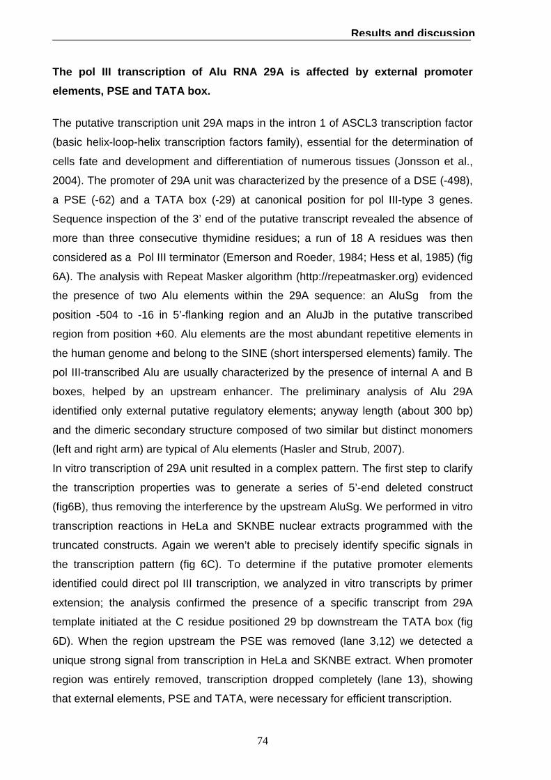
74
Results and discussion
The pol III transcription of Alu RNA 29A is affected by external promoter
elements, PSE and TATA box.
The putative transcription unit 29A maps in the intron 1 of ASCL3 transcription factor
(basic helix-loop-helix transcription factors family), essential for the determination of
cells fate and development and differentiation of numerous tissues (Jonsson et al.,
2004). The promoter of 29A unit was characterized by the presence of a DSE (-498),
a PSE (-62) and a TATA box (-29) at canonical position for pol III-type 3 genes.
Sequence inspection of the 3’ end of the putative transcript revealed the absence of
more than three consecutive thymidine residues; a run of 18 A residues was then
considered as a Pol III terminator (Emerson and Roeder, 1984; Hess et al, 1985) (fig
6A). The analysis with Repeat Masker algorithm (http://repeatmasker.org) evidenced
the presence of two Alu elements within the 29A sequence: an AluSg from the
position -504 to -16 in 5’-flanking region and an AluJb in the putative transcribed
region from position +60. Alu elements are the most abundant repetitive elements in
the human genome and belong to the SINE (short interspersed elements) family. The
pol III-transcribed Alu are usually characterized by the presence of internal A and B
boxes, helped by an upstream enhancer. The preliminary analysis of Alu 29A
identified only external putative regulatory elements; anyway length (about 300 bp)
and the dimeric secondary structure composed of two similar but distinct monomers
(left and right arm) are typical of Alu elements (Hasler and Strub, 2007).
In vitro transcription of 29A unit resulted in a complex pattern. The first step to clarify
the transcription properties was to generate a series of 5’-end deleted construct
(fig6B), thus removing the interference by the upstream AluSg. We performed in vitro
transcription reactions in HeLa and SKNBE nuclear extracts programmed with the
truncated constructs. Again we weren’t able to precisely identify specific signals in
the transcription pattern (fig 6C). To determine if the putative promoter elements
identified could direct pol III transcription, we analyzed in vitro transcripts by primer
extension; the analysis confirmed the presence of a specific transcript from 29A
template initiated at the C residue positioned 29 bp downstream the TATA box (fig
6D). When the region upstream the PSE was removed (lane 3,12) we detected a
unique strong signal from transcription in HeLa and SKNBE extract. When promoter
region was entirely removed, transcription dropped completely (lane 13), showing
that external elements, PSE and TATA, were necessary for efficient transcription.

75
Results and discussion
Fig.6 Transcription properties of the unit 29A. (A) nucleotide sequence of unit 29A
inserted in pNEB193 vector. Transcribed sequence is in bold. (B) Schematic
representation of truncated 29A templates. (C) In vitro transcription of 29A in Hela and
SKNBE extracts. Empty vector pNEB193 was used as a control. RNA size markers were
loaded on the same gel; their migration positions and length (in nt) are indicated on the
left side. (D) Primer extension analysis: primer extension product was indicated by an
arrow. Lane 4-7, 8-11 sequencing reactions. (E) In vitro transcription in HeLa extract of
29A containing a poly T sequence as termination signal.

76
Results and discussion
When we added a strong termination signal (9T) within the transcribed region, the
truncated RNA product was well detected (fig 6E), confirming that PSE and TATA
element could actually support transcription.
The role of the PSE in 29A expression was further investigated using 7SK RNA as a
reporter gene, as previously done for 38A. We generated a fusion construct attaching
the 5’-flanking sequence of 29A from -535 bp to -23 bp to the coding sequence of
7SK (fig 7A).
This fusion template supported efficient transcription of an RNA of the expected
length. Primer extension analysis verified that the initiation site was unchanged in the
fusion template respect to natural 7SK gene (7C lane 8,9). Transcription values were
normalized with an internal standard RNA synthesized using T7 RNA polymerase.
Removal of the sequence upstream the PSE resulted in enhancement of specific
transcription thus removing the interference by the upstream AluSg (compare lane
2,3). Further deletion of the PSE caused a fourfold drop in transcriptional efficiency
(fig 7B, lane 5): TATA box alone was thus able to direct a basal transcription level.
The involvement of the PSE was investigated more in detail introducing a double
point mutation at the two C residues in position 4 and 5 in the PSE sequence
(CC→AA) as already done in the analysis of 17A promoter. Transcription efficiency of
the mutated template 29A_AA_7SK was half respect to the wild type. Even if
transcription reduction caused by mutation and deletion of PSE wasn’t exactly
comparable, both these results suggest that in vitro the PSE acts as a positive signal
for transcription as it happens for other well known pol III- type 3 genes.

77
Results and discussion
Fig. 7 Regulation of 29A transcription by PSE element. (A) Schematic representation
of 29A_7SK templates. (B) In vitro transcription in HeLa extract of 29A_7SK templates.
Natural 7SK (lane 6) was used as positive control. The histogram represents RNA
quantitative value relative to 29A_∆DSE_7SK value after normalization with the RNA
used as standard. (C) Primer extension analysis of 29A_7SK transcript. Natural 7SK
(lane 9) was used as positive control.

78
Results and discussion
51A RNA transcription is supported by a TATA less PSE-dependent promoter.
Unit 51A maps in the intron 1 of SORL1, encoding a protein that belongs to the family
of vacuolar protein sorting 10 (VPS10) domain-containing receptor proteins. This
gene is strongly expressed in the central nervous system. The preliminary analysis of
51A promoter identified a TATA box located downstream the PSE (17 bp) (fig 8A). In
vitro transcription of 51A template resulted in a specific signal (fig 8B), that was much
stronger when transcription was carried out in nuclear extract from neuroblastoma
cells (SKNBE). Transcript length could be explained by a the read-through of
polymerase III across the first potential terminator, a T4 run, to the next available
downstream stop signal (T6). A secondary structure model of 51A RNA from the
structure prediction programme mfold is shown in fig 8D. Primer extension analysis
confirmed the efficient transcription of 51A both in HeLa and SKNBE cells nuclear
extract of a transcript initiated at the A residue upstream the TATA box (fig 8C, lane
4,12). This result suggested that in this particular case the PSE, located at position -
32 with respect to the TSS (the canonical position for TATA box), might act as the
main actor in selection of the start site. SNAPc binding to the PSE of snRNA gene
promoters is a prelude to the recruitment of the general transcription machinery; in
the case of pol III-transcribed snRNA genes SNAPc recruits TBP to the TATA box.
The SNAPc/TBP juxtaposition results in a recruitment of a TFIIB-related factor called
Brf2 (Jawdekar and Henry, 2008). To verify whether the pol III transcription
machinery interacts with the 51A promoter in vivo, we performed ChIP experiments in
SKNBE and SHSY5Y neuroblastoma cells using antibodies directed against SNAPc,
Brf2 or a subunit of RNA polymerase III. Recovered DNA fragments were analyzed
by PCR with primer specific for the 51A promoter region. A Primer pair amplifying a
region of GAPDH exon 2 were used as a specificity control. The pol III-type 3 U6
snRNA promoter was analysed as a positive control for interaction with the Pol III
machinery. As shown in fig. 9, pol III machinery components specifically
immunoprecipitated with 51A promoter region in both cell lines, suggesting a possible
PSE-dependent pol III active transcription in vivo. 51A transcribed region was finally
analyzed with Pol3scan program (Pavesi et al., 1994) to verify the presence of
putative internal regulatory elements; a B box-like motif (CTTTCAAATCT) was found
at position +74. Further investigation are required to test the possible cooperation of
extragenic and intragenic elements in transcriptional regulation of 51A RNA.

79
Results and discussion
Fig. 8 Transcription properties of the unit 51A. (A) nucleotide sequence of unit 51A
inserted in pNEB193 vector. Transcribed sequence is in bold. (B) In vitro transcription of
51A in Hela and SKNBE extract. Empty vector pNEB193 was used as a control. RNA
size markers were loaded on the same gel; their migration positions and length (in nt) are
indicated on the left side. (C) Primer extension analysis: Lane 3,10 no reverse
transcriptase during primer extension; lane 1,9 no DNA during in vitro transcription. In
lanes 4, 12 the primer extension product is indicated by an arrow. Lane 5-8, 13-16
sequencing reactions. (D) Secondary structure of 51A RNA from mfold
(http://mfold.bioinfo.rpi.edu/).

80
Results and discussion
Fig. 9 Occupancy at 51A promoter region. Cross-linked chromatin from SHSY5Y and
SKNBE cells was immunoprecipitated with anti-SNAPc, anti-Brf2 or anti-Pol III antibodies
and analyzed by PCR amplification for enrichment of 51A promoter. Primer specific to
GAPDH exon 2 were used as internal standard control. The pol III-type 3 U6 snRNA
promoter was also analyzed as a positive interaction control.

81
Conclusion
Conclusion
Pol III transcriptome appeared as a group of ncRNA, with a few well-known abundant
members and an unexplored realm of non abundant transcripts. In past few year
computational searches for ncRNA sequences and DNA regulatory regions in whole
genome has expanded the knowledge concerning the ncRNAs and their cellular
roles. Starting from these observations, our laboratory performed a screening in the
human genome for elements typical of snRNA gene promoters (PSE and DSE) and
identified a set of about 30 novel putative pol III-transcribed units (Pagano et al,
2007). In these work we selected four of these identified units (17A, 38A, 29A, 51A)
to be characterized by experimental determinations.
We first tested their transcription properties to verify if core promoter elements were
actually able to direct efficient pol III transcription. Though in vitro transcription and
primer extension analyses, we defined the boundary of two novel ncRNAs, 17A and
51A, 171 nt and 423 nt in length respectively. Correct in vitro termination required at
least a run of five T , as demonstrated in correctly-ended transcription of 17A . In 51A
the sequence of four T was not sufficient for termination by Pol III, that read-through
this sequence up to the next stop signal (6T). 38A and 29A RNAs were correctly
initiated in vitro but their 3’-end weren’t clearly identified; maybe alternative stop
signals instead of canonical T sequences were required for termination.
Since these units were the results of an homology research for H1 RNA -like PSE
elements within the human genome, all core promoters shared the presence of a
PSE. This element was located at similar position in 17A, 38A and 29A promoters,
upstream of TATA box elements located in canonical position for start site selection.
PSE-TATA distance was instead slightly shorter than expected (Myslinski et al.,
2001). As happens in other well known pol III type-3 genes, the PSE within promoter
region of 17A, 38A and 29A, acted as a positive regulator of transcription; mutation or
deletion of this element caused in fact a reduction of about four fold in expression of
17A natural transcript, or of an auxiliary reporter gene (7SK) attached at the
promoter region of 38A and 29A. 51A RNA transcription was supported by a TATA-
less PSE-dependent promoter, even if PSE position (-32 upstream the start site)
seemed more canonical for a TATA box rather than a PSE element.
Core promoter elements and their position are summarized in table 1.

82
Conclusion
The factor binding to the PSE element has been best characterized in the human
system and is variously known as PTF or SNAPc (see for a review Jawdekar and
Henry, 2008). Chip experiments confirmed the binding of SNAPc at 38A and 51A
promoters; this result, together with the observed occupancy of the same regions by
other component of pol III machinery (Brf2 and pol III itself) suggested an active in
vivo expression of these units.
Interestingly some of these novel units were internal to known protein-coding genes,
one being in sense (17A) and two in antisense (38A, 51A), in regions where
alternative splicing events takes place. A growing number of endogenous antisense
RNA transcripts have been reported in the last years in eukaryotic organism where
they exert control at various levels of gene expression, such as transcription, mRNA
processing, splicing, stability transport and translation (Knee and Murphy, 1997;
Hastings et al., 2000). In addiction recent evidence suggests that antisense intronic
ncRNA may play a key role in a range of human diseases (Larvogna et al., 2004).
The peculiar location of 17A (GPR51-intron3), 38A (KCNIP4-intron1) and 51A
(SORL1-intron1) RNAs could suggest an involvement in splicing of these proteins ,
all directly or indirectly associated to amyloid precursor protein (APP) processing and
therefore possibly to Alzheimer disease (AD).
As further intriguing observation the 29A RNA is located in a chromosomal region
associated to a tumor suppressor activity, and was identified as an Alu. Alu RNAs are
known as posttranscriptional regulator of gene expression, for example by affecting
protein translation, alternative splicing and mRNA stability (Hasler and Strub, 2007).
Further studies will be required to better characterize these transcription units in vivo
and the function of their products, that are suggestive of an increasing complexity of
regulation by pol III transcriptome.
Table 1. Core promoter elements and their position within 17A, 38A, 29A, and 51A
transcriptional units
Transcriptional unit PSE position TATA position PSE-TATA distance
17A -56 -27 11
38A -64 -23 15
29A -62 -23 17
51A -32 / /

83
References
References Abou Elela S, Ares M (1998) Depletion of yeast RNase III blocks correct U2 39 end formation and results in polyadenylated but functional U2 snRNA. EMBO J 17,3738–3746. Angermayr M, Oechsner U, Bandlow W. (2003) Reb1p-dependent DNA bending effects nucleosome positioning and constitutive transcription at the yeast profilin promoter. J Biol Chem. 278, 17918-26. Basehoar AD, Zanton SJ, Pugh BF. (2004) Identification and distinct regulation of yeast TATA box-containing genes. Cell. 116, 699-709.
Beinoraviciūte-Kellner R, Lipps G, Krauss G. (2005) In vitro selection of DNA binding sites for ABF1 protein from Saccharomyces cerevisiae. FEBS Lett. 579, 4535-4540.
Boyd D, Greger H, Murphy S (2002) In vivo footprinting studies suggest a role for chromatin in transcription of the human 7SK gene. Gene 247,33–44.
Chanfreau G, Elela SA, Ares M Jr, Guthrie C. (1997) Alternative 3'-end processing of U5 snRNA by RNase III. Genes Dev. 11, 2741-2751.
Cheng C, Yeung C, Hoo R, Chow B, Leung (2002) Oct-1 is involved in the transcriptional repression of the gonadotropin-releasing hormone receptor gene. Endocrinology. 143, 4693-4701.
Christov CP, Gardiner TJ, Szuts D, Krude T (2006) functional requirement of non coding Y RNAs for human chromosomal DNA replication. Mol Cell. Biol. 26, 6993-7004.)
Connelly S, Filipowicz W (1993) Activity of chimeric U small nuclear RNA (snRNA)/mRNA genes in transfected protoplasts of Nicotiana plumbaginifolia: U snRNA 39-end formation and transcription initiation can occur independently in plants. Mol Cell Biol 13:6403–6415. Cliften, P., Sudarsanam, P., Desikan, A., Fulton, L., Fulton, B., Majors, J., Waterston, R., Cohen, B.A. and Johnston, M. (2003) Finding functional features in Saccharomyces genomes by phylogenetic footprinting. Science, 301, 71-76. Dewang Z, Frendewey D, Lobo Rupper S (1999) Pac1p, an RNase homolog, is required for formation of the 3’ end of U2 snRNA in Schizosaccharomyces pombe. RNA 5, 1083-1098. Dignam JD, Lebovitz RM, Roeder RG. (1983) Accurate transcription initiation by RNA polymerase II in a soluble extract from isolated mammalian. Nucleic Acids Res. 11, 1475-1489. Dittmar, K.A. (2006) Tissue-specific differences in human transfer RNA expression. PLoS Genet. 2, e221

84
References
Egloff S, O'Reilly D, Chapman RD, Taylor A, Tanzhaus K, Pitts L, Eick D, Murphy S (2007) Serine-7 of the RNA polymerase II CTD is specifically required for snRNA gene expression. Science 318,1777–1779. Emerson B, Roeder R, (1984) DNA sequences and transcription factor interactions of active and inactive forms of mammalian 5 S RNA genes. J. Biol. Chem. 259, 7926-7935.
Eschenlauer JB, Kaiser MW, Gerlach VL, Brow DA. (1993) Architecture of a yeast U6 RNA gene promoter. Mol Cell Biol. 13, 3015-26.
Ford E, Strubin M, Herandez N (1998) The Oct-1 POU domain activates snRNA gene transcription by contacting a region in the SNAPc largest subunit that bears sequence similarities to the Oct-1 coactivator OBF-1. Genes Dev. 12, 3528-3540.
Fourel G, Miyake T, Defossez P, Li R, Gilson E. (2002) General regulatory factors as genome partitioners. J. Biol. Chem. 277, 41736–41743. Frank DN, Roiha H, Guthrie C. (1994) Architecture of the U5 small nuclear RNA. Mol Cell Biol., 14, 2180-2190. Ghaemmaghami S, Huh WK, Bower K, Howson RW, Belle A, Dephoure N, O'Shea EK, Weissman JS. (2003) Global analysis of protein expression in yeast. Nature, 425, 737-741. Goncalves PM, Griffioen G, Minnee R, Bosma M, Kraakman LS, Mager H, Planta RJ. (1995) Transcriptional activation of yeast ribosomal protein genes requires additional elements apart from binding sitee for Abf1p or Rap1p. Nucleic Acids Res, 23, 1475-1480. Goodrich JA, Kugel JE (2006) Non-coding-RNA regulators of polymerase II transcription. Nature 7, 612-616. Gridasova A, Henry RW (2000) The retinoblastoma tumor suppressor protein targets dinstinc general transcription factors to regulate RNA polymerase III gene expression. Mol. Cell. Biol. 20, 9182-9191. Gu L, Esselman WJ, Henry RW (2005) Cooperation between small nuclear RNA- activating protein complex (SNAPC) and TATA-box-binding protein antagonizes protein kinase CK2 inhibition of DNA binding by SNAPC. J. Biol. Chem. 280,27697–27704. Häsler J, Samuelsson T, Strub K. (2007) Useful 'junk': Alu RNAs in the human transcriptome. Cell Mol Life 64, 1793-800. Hastings M, Ingle H, Lazar M, Munroe S (2000) Post-transcriptional regulation of thyroid hormone receptor expression by cis-acting sequences and a naturally occurring antisense RNA. J. Biol. Chem. 275, 11507-11513.

85
References
Hernandez N. (2001) Small nuclear RNA genes: a model system to study fundamental mechanisms of transcription. J Biol Chem. 276,26733-26736.
Hernandez N, Lucito R (1988) Elements required for transcription initiation of the human U2 snRNA gene coincide with elements required for snRNA 3′ end formation, Embo. J. 7, 3125–3134.
Hess J, Perez-Stable C, Wu GJ, Weir B, Tinoco I Jr, Shen CK (1985) End-to-end transcription of an Alu family repeat. A new type of polymerase-III-dependent terminator and its evolutionary implication. J Mol Biol. 184,7-21.
Hinkley CS, Hirsh H, Gu L, Lamere B, Henry RW (2003) The small nuclear RNA-activating protein 190 Myb DNA binding domain stimulates TATA box-binding protein-TATA box recognition. J. Biol. Chem. 278, 18649-18657. Hirsch HA, Jawdekar GW, Lee KA, Gu L, Henry RW (2004) Distinct mechanisms for repression of RNA polymerase III transcription by the retinoblastoma tumor suppressor protein, Mol. Cell Biol. 24,5989–5999 Hu P, Samudre K, Wu S, Sun Y, Hernandez N (2004) CK2 phosphorylation of Bdp1 executes cell cycle-specific RNA polymerase III transcription repression. Mol. Cell 16, 81–92. Ito H, Fukuda Y, Murata K, Kimura A. (1983) Transformation of intact yeast cells treated with alkali cations. J Bacteriol.153,163-168. Jarrous N, Reiner R (2007) Human RNAse P: a tRNA processing enzyme and transcription factor. Nucleic Acids Res. 35, 3519-3524 Jawdekar G, Henry RW (2008) Transcriptional regulation of human small nuclear RNA genes. Biochimica et Biofisica acta. 1779, 295-305. Jawdekar G, Hanzlowsky A, Hovde S, Jelencic B, Feig M, Geiger JH, Henry RW (2006) The unorthodox SNAP50 zinc finger domain contributes to co-operative promoter recognition by human SNAPc. J. Biol. Chem. 281, 31050-31060.
Jensen RC, Wang Y, Hardin SB, Stumph WE. (1998) The proximal sequence element (PSE) plays a major role in establishing the RNA polymerase specificity of Drosophila U-snRNA genes. Nucleic Acids Res. 26, 616-22.
Jonsson M, Björntorp Mark E, Brantsing C, Brandner JM, Lindahl A, Asp J. (2004) Hash4, a novel human achaete-scute homologue found in fetal skin. Genomics. 84:859-66.
Kellis, M., Patterson, N., Endrizzi, M., Birren, B. and Lander, E.S. (2003) Sequencing and comparison of yeast species to identify genes and regulatory elements. Nature, 423, 241-254. Knee R, Murphy P (1997) Regulation of gene expression by natural antisense RNA transcripts. Neurochem. Int. 31, 379-392.

86
References
Kwek KY, Murphy S, Furger A, Thomas B, Kimura H, Proudfoot NJ, Akoulitchev A (2002) U1 snRNA associates with TFIIH and regulates transcriptional initiation. Nat. Struct. Biol. 9, 800-805. Kuehner, J.N. & Brow, D.A.(2006) Quantitative analysis of in vivo initiator selection by yeast polymerase II supports a scanning model J. Biol. Chem. 281, 14119–14128. Kuhlman TC, Cho H, Reinberg D, Hernandez N. (1999) The general transcription factors IIA, IIB, IIF, and IIE are required for RNA polymerase II transcription from the human U1 small nuclear RNA promoter.Mol. Cell. Biol. 19, 2130–2141. Kuras, L. (2004) Characterization of protein-DNA association in vivo by chromatin immunoprecipitation. Methods Mol. Biol., 284, 147-162. Kuras, L. and Struhl, K. (1999) Binding of TBP to promoters in vivo is stimulated by activators and requires Pol II holoenzyme. Nature, 399, 609-613. Kurdistani S, Grunstein M (2003) In vivo protein-protein and protein-DNA crosslinking for genomewide binding microarray. Methods. 31, 90-95. Lascaris RF, De Groot E, Mager WH, Planta RJ. (2000) Different roles for Abf1p and T-rich promoter element in nucleosome organization of the yeast RPS28A gene. Nucleic Acids Res. 28, 1390-1396. Lavorgna G, Dahary D, Lehner B, Sorek R, Sanderson C M, Casari G, (2004) In search of antisense. Trends Biochem. Sci. 29, 88-94. Lee W, Tillo D, Bray N, Morse RH, Davis R, Hughes T, Nislow C. (2007) A high-resolution atlas of nucleosome occupancy in yeast. Nature genetics 39, 1235-1244 Lin, Y. Brosius J, Tiedge H (2001) Neuronal BC1 RNA: co-expression with growth-associated protein-43 messenger RNA. Neuroscience 103, 465–479
Liko D, Slattery MG, Heideman W. (2007) Stb3 binds to ribosomal RNA processing element motifs that control transcriptional responses to growth in Saccharomyces cerevisiae. J Biol Chem. 282,26623-26628.
Lobo SM, Hernandez N (1989) A 7 bp mutation converts a human polymerase II snRNA promoter into an RNA polymerase III promoter. Cell 58, 55-67.
Mannhaupt G, Schnall R, Karpov V, Vetter I, Feldmann H. (1999) Rpn4 acts as a transcription factor by bindig PACE, a nonamer box found upstream of 26S proteasomal and other genes in yeast. FEBS Lett 450, 27-34.
Matera AG, Terns RM, Terns MP (2007) Non-coding RNAs: lessons from the small nuclear and small nucleolar RNAs. Nat Rev Mol Cell Biol. 8, 209-220.
Mattaj IW, Dathan NA, Parry HD, Carbon P, Krol A (1988) Changing the RNA polymerase specificity of U snRNA genes promoters. Cells 55, 435-442.

87
References
Mattai IW (1986) Cap trimethylation of U snRNA is cytoplasmic and dependent on U snRNP protein binding. Cell 46, 905-911.
Mattick JS (2004) RNA regulation: a new genetic? Nat. Rev. Genet. 5,316-323.
Mencía M, Moqtaderi Z, Geisberg JV, Kuras L, Struhl K. (2002) Activator-specific recruitment of TFIID and regulation of ribosomal protein genes in yeast. Mol Cell. 9, 823-833.
McCutcheon, J.P. and Eddy, S.R. (2003) Computational identification of non-coding RNAs in Saccharomyces cerevisiae by comparative genomics. Nucleic Acids Res, 31, 4119-4128. Mittal V, MA B, Hernandez N (1999) SNAPc: a core promoter factor with a built-in DNA-binding damper that is deactivated by the Oct-1 Pou domain. Genes Dev. 13, 1807-1821. Mittal V, Cleary A, Herr W, Hernandez N (1996) The Oct-1 POU-specific domain can stimulate small nuclear RNA gene transcription by stabilizing the basal transcription complex SNAPc, Mol. Cell Biol. 16, 1955–1965
Miyake T, Reese J, Loch CM, Auble DT, Li R. (2004) Genome-wide analysis of ARS (autonomously replicating sequence) binding factor 1(Abf1p)-mediated transcriptional regulation in Saccharomyces cerevisiae. J Biol Chem. 279,34865-34872.
Morse RH (2000) RAP, RAP, open up! New wrinkles for RAP1 in yeast. Trends Genet 16, 51-53.
Myslinski E, Amè J, Krol A, Carbon P. (2001) An unusually compact external promoter for RNA polymeras III transcription in the human H1RNA gene. Nucleic Acids Res. 29, 2502-2509.
Myslinski E, Krol A, Carbon P (1998) ZNF76 and ZNF143 are two human homologs of the transcriptional activator staf. J. Biol. Chem. 273, 21998–22006.
Murphy S, Liegro C, Melli M. (1987) The in vitro transcription of the 7SK RNA gene by RNA polymerase III is dependent only on the presence of an upstream promoter. Cell 51, 81-87.
Mus E, Hof P, Tiedge H (2007) Dendritic BC200 RNA in aging and in Alzheimer’s disease. Proc. Natl. Acad. Sci. 104, 10679–10684
Nguyen VT, Kiss T, Michels A, Bensaude O (2001) 7SK small nuclear RNA binds and inhibits the activity of CDK9/cyclyn T complexes. Nature 414, 317-322.
Nilsen TW (2003) The spliceosome: the most complex macromolecular machine in the cell? BioEssays 25, 1147-1149.

88
References
Pagano A, Castelnuovo M, Tortelli F, Ferrari R, Dieci G, Cancedda R. (2007) New small nuclear RNA gene-like transcriptional units as sources of regulatory transcripts. PLoS Genet. 3, 174-184.
Patterson B, Guthrie C. (1987) An essential yeast snRNA with a U5-like domain is required for splicing in vivo. Cell 49, 613-624. Pavesi A, Conterio F, Bolchi A, Dieci G, Ottonello S (1994) Nucleic. Acids Res. 22, 1247-1256. Sadowski CL, Henry RW, Kobayashi R, Hernandez N (1996) The SNAP45 subunit of the small nuclear RNA (snRNA) activating protein complex is required for RNA polymerase II and III snRNA gene transcription and interacts with the TATA box binding protein, Proc. Natl. Acad. Sci. 93,4289–4293. Schmitt, M.E., Brown, T.A. and Trumpower, B.L. (1990) A rapid and simple method for preparation of RNA from Saccharomyces cerevisiae. Nucleic Acids Res. 18, 3091-3092. Schroeder SC, Weil A. (1998) Genetic test of the role of Abf1p in driving transcription of the yeast TATA box binding protein-encoding gene, SPT15. J Biol Chem. 273,19884-19891. Schramm L, Pendergrast PS, Sun Y, Hernandez N (2000) Different human TFIIIB activities direct RNA polymerase III transcription from TATA-containing and TATA-less promoters. Genes Dev. 14,2650–2663. Schramm L, Hernandez N (2002) Recruitment of RNA polymerase III to its target promoters. Genes Dev. 16, 2593–2620. Segref A, Mattaj I, Ohno W (2001) The evolutionary conserved region of the U snRNA export mediator PHAX is a novel RNA-binding domain that is essential for U snRNA export. RNA 7, 351-360
Shuster EO, Guthrie C. (1988) Two conserved domains of yeast U2 snRNA are separated by 945 nonessential nucleotides. Cell. 55, 41-48.
SinghR, Gupta S, Reddy R. (1990) Capping of mammalian U6 snRNA in vitro is directed by a conserved stem-loop and AUAUAC sequence: conversion of a noncapped RNA into a capped RNA. Mol. Cell. Biol. 10, 939-946.
Sylvain E, Murphy S (2008) Role of the C-terminal domain of RNA polymerase II in expression of small nuclear RNA genes. Biochem. Soc. Trans. 36, 537-539.
Thomann H, Schmutzler C, Hudepohl U, Blow M, Gross HJ. (1989) Genes, variant genes and pseudogenes of the human tRNAval gene family expression and pre-tRNA maturation in vitro. J Mol. Biol 209,505-523.

89
References
Vilalta, A. et al. (1994) The rat vault RNA gene contains a unique RNA polymerase III promoter composed of both external and internal elements that function synergistically. J. Biol. Chem. 269, 29752–29759
Waibel F, Filipowicz W. (1990) RNA-polymerase specificity of transcription of Arabidopsis U snRNA genes determined by promoter element spacing. Nature 346, 199-202.
Wang KL, Warner JR. (1998) Positive and negative autoregulation of REB1 transcription in Saccharomyces cerevisiae. Mol Cell Biol., 18, 4368-76. Wendelburg BJ, Marzluff WF (1992) Formation of the 3’ end of sea urchin U1 small nuclear RNA occurs independently of the conserved 3’ box and on transcripts initiated from a histone promoter. Mol Cell Biol 12, 4132–4141. Wong MW, Henry RW, Ma B, Kobayashi R, Klages N, Matthias P, Strubin M, Hernandez N (1998) The large subunit of basal transcription factor SNAPc is a Myb domain protein that interacts with Oct-1, Mol. Cell. Biol.18, 368-377
Zamrod Z, Tyree CM, Song Y, Stumph WE. (1993) In vitro transcription of a Drosophila U1 small nuclear RNA gene requires TATA box-binding protein and two proximal cis-acting elements with stringent spacing requirements. Mol Cell Biol. 9,5918-27.
Zhao X, Pendergrast P, Hernandez N (2001) A positioned nucleosome on the human U6 promoter allows recruitment of SNAPc by the Oct-1 POU domain. Mol. Cell 7, 539–549.
Zhou D, Lobo-Ruppert SM. (2001) Transcription of the Schizosaccharomyces pombe U2 gene in vivo and in vitro is directed by two essential promoter elements. Nucleic Acids Res. 29, 2003-11.


![Levels at which eukaryotic gene expression is controlled Initiating or inhibiting Transcription [majority] –Transcriptional activators, coactivators, repressors](https://img.pdfslide.us/doc/110x75/5697bfc61a28abf838ca6f5a/levels-at-which-eukaryotic-gene-expression-is-controlled-initiating-or-inhibiting.jpg)