Embed Size (px)
Citation preview
Ia
Pa
b
c
d
a
ARRAA
KHNNGL
1
PtHviBesdota
ia
0d
Talanta 85 (2011) 1634–1641
Contents lists available at ScienceDirect
Talanta
journa l homepage: www.e lsev ier .com/ locate / ta lanta
dentification and quantification of nucleosides and nucleobases in Geosaurusnd Leech by hydrophilic-interaction chromatography
ei Chena,b, Wei Lia,∗, Qin Li c, Yinghua Wangb, Zhenguo Lid, Yefeng Nid, Kazuo Koikea
Faculty of Pharmaceutical Sciences, Toho University, Chiba 274-8510, JapanNingxia Institute for Drug Control, Yinchuan 750004, People’s Republic of ChinaLaboratory Center, The Fourth Affiliated Hospital, China Medical University, Shenyang 110005, People’s Republic of ChinaMudanjiang Youbo Pharmaceutical Co., Ltd., People’s Republic of China
r t i c l e i n f o
rticle history:eceived 9 April 2011eceived in revised form 22 June 2011ccepted 22 June 2011vailable online 29 June 2011
eywords:ydrophilic-interaction chromatographyucleosidesucleobaseseosaurus
a b s t r a c t
A simple hydrophilic-interaction chromatography (HILIC) method was developed for the identificationand quantification of 14 nucleosides and nucleobases, namely cytosine, uracil, cytidine, guanine, hypox-anthine, xanthine, uridine, thymine, inosine, guanosine, thymidine, 2′-deoxyadenosine, 2′-deoxyinosineand 2′-deoxyuridine in two traditional Chinese medicines, Geosaurus and Leech. The separation wasachieved on a TSKgel Amide-80 column (150 mm × 2.0 mm, 3.0 �m) with a mixture of acetonitrile and10 mM aqueous ammonium acetate as the mobile phase at a flow rate of 0.2 mL/min. The temperature wasset at 30 ◦C and UV detection wavelength was set at 260 nm. All calibration curves showed good linearity(R2 > 0.9957) within the test ranges. The overall intra- and inter-day RSD ranged from 0.4 to 3.4% and from0.7 to 3.3%, respectively. The LOD and LOQ were in the range of 0.07–30.49 ng/mL and 0.26–60.98 ng/mL,respectively. The repeatability of the method was in the range of 2.2–5.8% for Geosaurus and 1.4–5.5% for
eech Leech. The recoveries of the samples were in the range of 91.4–100.9% for Geosaurus, and 91.9–99.3% forLeech. The established method was applied successfully for the analysis of nucleosides and nucleobases in22 commercially available samples collected from different regions in China and Japan. Our data showedthat HILIC had advantages as a useful tool for the study of the bioactive components in Geosaurus andLeech as well as their quality control, and could therefore be used for the determination of the analytes
cts an
in pharmaceutical produ. Introduction
The annelids of Geosaurus (Pheretima aspergillum (E. Perrier),heretima vulgaris Chen, Pheretima guillelmi (Michaelsen), Phere-ima pectinifera Michaelsen) and Leech (Whitmania pigra Whitman,irudo nipponica Whitman, Whitmania acranulata Whitman) arealued traditional Chinese medicines (TCMs) which are listedn the Pharmacopoeia of the People’s Republic of China [1,2].oth Geosaurus and Leech preparations, especially their aqueousxtracts, have been used widely for medical purposes, and havehown to exert various effects such as blood-activating, stasis-issolving, antipyretic and diuretic effects base on a combinationf multiple mechanisms. However, the bioactive components ofhe medical products of Geosaurus and Leech and their relatedctivities are still not fully understood.
Recently, nucleobases and nucleosides have been proven asmportant bioactive compounds involved in multiple biologicalctivities such as anti-platelet aggregation, anti-arrhythmic and
∗ Corresponding author. Tel.: +81 47 4721161; fax: +81 47 4721404.E-mail address: [email protected] (W. Li).
039-9140/$ – see front matter © 2011 Elsevier B.V. All rights reserved.oi:10.1016/j.talanta.2011.06.056
d biological fluids.© 2011 Elsevier B.V. All rights reserved.
anti-seizure effects [3–6], and have also been used as markers inthe quality control of several TCMs, such as Ganoderma lucidumnand Cordyceps sinensis [7,8]. Therefore, the purpose of this studywas to quantitatively and qualitatively analyze the nucleobase andnucleoside compounds from the water extracts of Geosaurus andLeech.
The contents of nucleosides and nucleobases in biological flu-ids and herbal materials have been determined by a number ofanalytical methods including thin layer chromatography (TLC)[9], high-performance liquid chromatography (HPLC) [10–18],liquid chromatography–mass spectrometry (LC–MS) [19–23],ultra-performance liquid chromatography (UPLC) [24], capillaryelectrophoresis–mass spectrometry (CE–MS) [25,26], gas chro-matography (GC) [27,28], capillary zone electrophoresis (CZE)[29,30], capillary electrochromatography (CEC) [31,32] and micel-lar electrokinetic chromatography (MEKC) [33,34], but manyof these methods have disadvantages such as limited analytes[12,14,15,17–20,23,26–31,33], low sensitivity [9] or expensive
instrumentation [19–23,25,26]. The establishment of a simple,efficient and sensitive method is thus required for the iden-tification and quantification of nucleosides and nucleobases inTCMs.
ta 85
a[shpicb[
pntrcd2tcat
2
2
dtsapJpmCpU
GJsiwP
2
mt0tfo
2
sGgp2(
P. Chen et al. / Talan
The HILIC method was first developed by Alpert in 1990 as anlternative to reversed-phase liquid chromatography (RP-HPLC)35]. In contrast to RP-HPLC where a hydrophobic octadecyl (C18)tationary phase is used, HILIC separation is based on the strongydrophilic interaction of polar compounds with the hydrophilicolar stationary phase. It has consequently been shown that HILIC
s suitable for the separation of a broad spectrum of hydrophilicompounds, including peptides, amino acids, oligonucleotides, car-ohydrates and many other biologically important compounds35–37].
In this study, both RPLC and HILIC method were used and com-ared for the identification and quantification of nucleosides anducleobases in the samples of Geosaurus and Leech. In contrasto RP-HPLC, where the separation of hypoxanthine and guanineemains a problem [22], 14 nucleosides and nucleobases, includingytosine, uracil, cytidine, guanine, hypoxanthine, xanthine, uri-ine, thymine, inosine, guanosine, thymidine, 2′-deoxyadenosine,′-deoxyinosine and 2′-deoxyuridine could be identified and quan-ified simply and accurately by HILIC method. The investigatedompounds of the collected Geosaurus and Leech samples in theirqueous extracts could be satisfactorily separated, and their con-ents were also compared.
. Material and methods
.1. Materials and chemicals
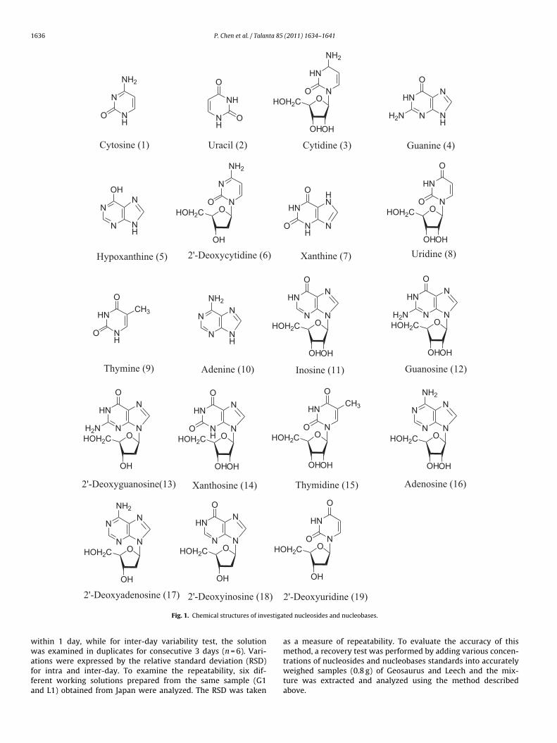
Nucleoside and nucleobase standards of cytosine, uracil, cyti-ine, guanine, hypoxanthine, 2′-deoxycytidine, xanthine, uridine,hymine, adenine, inosine, guanosine, 2′-deoxyguanosine, xantho-ine, thymidine, adenosine, 2′-deoxyadenosine, 2′-deoxyinosinend 2′-deoxyuridine (the structures are shown in Fig. 1) wereurchased from Wako Pure Chemical Industries, Ltd. (Osaka,
apan); ammonium acetate, acetic acid, monopotassium phos-hate, ammonia solution (25%), hydrochloric acid, HPLC gradeethanol and acetonitrile were also purchased from Wako Pure
hemical Industries, Ltd. (Osaka, Japan); Ultra-pure water wasrepared using a Milli-Q Plus system (Millipore, Bedford, MA,SA).
Seventeen samples of Geosaurus and Leech were collected fromuangdong, Jiangxi, Sichuan, Guangxi, Shandong, Hebei, Anhui,
iangsu Provinces, China, and 5 (G1, G8, L1, L4, L9) from Japan. Allamples were authenticated by one of our authors, W.L. accord-ng to their morphological characteristics. The voucher specimens
ere deposited in the Department of Pharmacognosy, Faculty ofharmaceutical Sciences, Toho University, Japan.
.2. Preparation of standard solutions for linearity studies
Nucleoside and nucleobase standards were dissolved inethanol, except for inosine, adenosine, hypoxanthine, xan-
hine, adenine and guanine, which were dissolved in water,.1 mol/L hydrochloric acid, 1.2% ammonia solution and concen-rated hydrochloric acid, respectively. The stock solutions wereurther diluted with methanol to obtain stocks at concentrationf 1.0 mg/mL, and stored in a refrigerator at 4 ◦C.
.3. Sample preparation
Nucleosides and nucleobases are generally extracted in wateroluble form [14,16,19–21,25,38]. In this study, samples ofeosaurus and Leech were first dried at 40 ◦C for 2 h before being
rinded into powder (approximately 20 meshes), and then pre-ared as 5% (g/mL) solution by dissolving 1 g of the powder with0 mL Milli-Q water. The solution was then ultrasonic extracted40 kHz, 240 W) for 2 h at room temperature followed by filtration.(2011) 1634–1641 1635
Five millilitres of the filtrate was vacuum-dried, and the residuewas dissolved in 3 mL of mixture of water/methanol (1:1). Aftercentrifugation at 3000 rpm for 10 min, the supernatant was thenfiltered through a 0.45 �m membrane filter prior to further analy-sis.
2.4. Instrumentation
Analysis were performed on a Waters Series 2950 (Waters Tech-nologies, USA) liquid chromatograph system comprising a vacuumdegasser, a quaternary pump, an autosampler, and a Photo-DiodeArray (PDA) system. Data was collected and analyzed by WatersChemStation software.
2.5. Chromatographic condition
For RP-HPLC, chromatograms were run on an YMC C18(4.6 mm × 250 mm, 5 �m) column, and two sets of elution bufferswere used as mobile phase. The system operated at 30 ◦C, and thePDA detection wavelength was set at 260 nm. The elution condi-tions were as follows:
(a) Mobile phase A = 20 mM monopotassium phosphate solution,B = acetonitrile, flow rate, 0.5 mL/min; a linear gradient of 1–6%B for the first 24 min; increased from 6 to 20% B from 24 to35 min; increased from 20 to 70% B from 35 to 45 min; 75% Bfrom 45 to 50 min; and a linear gradient of 75–50% B from 50to 52 min.
(b) Mobile phase A = acetate–ammonium acetate (pH 3.5),B = acetate–ammonium acetate (pH 3.5)/acetonitrile (90/10),flow rate, 0.7 mL/min; 75% B isocratic for the first 20 min; alinear gradient from 1 to 70% B from 20 to 30 min; increasedfrom 70 to 95% B from 30 to 40 min.
For HILIC, chromatograms were run on TSKgel Amide-80(2.0 mm × 150 mm, 3 �m) column. Mobile phase including (A)acetonitrile and (B) ammonium acetate (10 mM, pH 6.9) wasdegassed ultrasonically before use. The flow rate and sample injec-tion volume was 0.2 mL/min and 2 �L, respectively. The systemoperated at 30 ◦C, and the PDA detection wavelength was set at260 nm.
2.6. Calibration curves
Standard stock solutions of the reference compounds were pre-pared and diluted to a series of appropriate concentration forthe construction of calibration curves. At least 6 concentrationsof each reference compound solution were analyzed in tripli-cate, and then the calibration curves were constructed by plottingthe peak areas versus the concentration of each reference com-pound.
2.7. LOD and LOQ
The limits of detection (LOD) was defined as the lowest con-centration resulting in peak heights of three times the baselinenoise. The limits of quantification (LOQ) was defined as the low-est concentration resulting in peak heights of interest with S/Nratio higher than 10, with a precision of 15% and accuracy of80–120%.
2.8. Precision, repeatability and recovery
Intra- and inter-day variations were chosen to determinethe precision of the method. For intra-day variability test, themixed standards solution was analyzed for six replicates (n = 6)
1636 P. Chen et al. / Talanta 85 (2011) 1634–1641
HN
NO
O
OHOH2C
N
NH
NH2
O NH
NH
O
O
HN
NO
NH2
OHOH2C
OHOH
OHOH
HN
N
O
H2N
N
NH
N
N
OHN
NH
N
NO
NH2
OHOH2C
OH
HN
NH
O
O
HN
N
HN
NH
O
O
CH3 N
N
NH2N
NH
NOHOH2C
OHOH
N
N
HN
O
NOHOH2C
OHOH
N
N
HN
O
H2N
NOHOH2C
OH
N
N
HN
O
H2N NOHOH2C
OHOH
N
NH
HN
O
O
HN
NO
O
OHOH2C
OH
CH3
NOHOH2C
OHOH
N
N
N
NH2
NOHOH2C
OH
N
N
N
NH2
Cytosine (1) Uracil (2) Cytidine (3) Guanine (4)
Hypoxanthine (5) 2'-Deoxycytidine (6) Xanthine (7) Uridine (8)
Thymine (9) Adenine (10) Inosine (11) Guanosine (12)
2'-Deoxyguanosine(13) Xanthosine (14) Thymidine (15) Adenosine (16)
2'-Deoxyadenosine (17)
HN
NO
O
OHOH2C
OH
2'-Deoxyuridine (19)
NOHOH2C
OH
N
N
HN
O
2'-Deoxyinosine (18)
OH
estiga
wwaffa
Fig. 1. Chemical structures of inv
ithin 1 day, while for inter-day variability test, the solutionas examined in duplicates for consecutive 3 days (n = 6). Vari-
tions were expressed by the relative standard deviation (RSD)or intra and inter-day. To examine the repeatability, six dif-erent working solutions prepared from the same sample (G1nd L1) obtained from Japan were analyzed. The RSD was taken
ted nucleosides and nucleobases.
as a measure of repeatability. To evaluate the accuracy of thismethod, a recovery test was performed by adding various concen-
trations of nucleosides and nucleobases standards into accuratelyweighed samples (0.8 g) of Geosaurus and Leech and the mix-ture was extracted and analyzed using the method describedabove.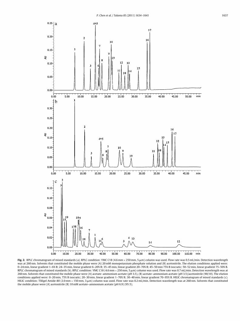
P. Chen et al. / Talanta 85 (2011) 1634–1641 1637
Fig. 2. RPLC chromatogram of mixed standards (a). RPLC condition: YMC C18 (4.6 mm × 250 mm, 5 �m) column was used. Flow rate was 0.5 mL/min. Detection wavelengthwas at 260 nm. Solvents that constituted the mobile phase were (A) 20 mM monopotassium phosphate solution and (B) acetonitrile. The elution conditions applied were:0–24 min, linear gradient 1–6% B; 24–35 min, linear gradient 6–20% B; 35–45 min, linear gradient 20–70% B; 45–50 min 75% B isocratic; 50–52 min, linear gradient 75–50% B.RPLC chromatogram of mixed standards (b). RPLC condition: YMC C18 (4.6 mm × 250 mm, 5 �m) column was used. Flow rate was 0.7 mL/min. Detection wavelength was at260 nm. Solvents that constituted the mobile phase were (A) acetate–ammonium acetate (pH 3.5), (B) acetate–ammonium acetate (pH 3.5)/acetonitrile (90/10). The elutionconditions applied were: 0–20 min, 75% B isocratic; 20–30 min, linear gradient 1–70% B; 30–40 min, linear gradient 70–95% B. HILIC chromatogram of mixed standards (c).HILIC condition: TSKgel Amide-80 (2.0 mm × 150 mm, 3 �m) column was used. Flow rate was 0.2 mL/min. Detection wavelength was at 260 nm. Solvents that constitutedthe mobile phase were (A) acetonitrile (B) 10 mM acetate–ammonium acetate (pH 6.9) (95:5).
1638 P. Chen et al. / Talanta 85 (2011) 1634–1641
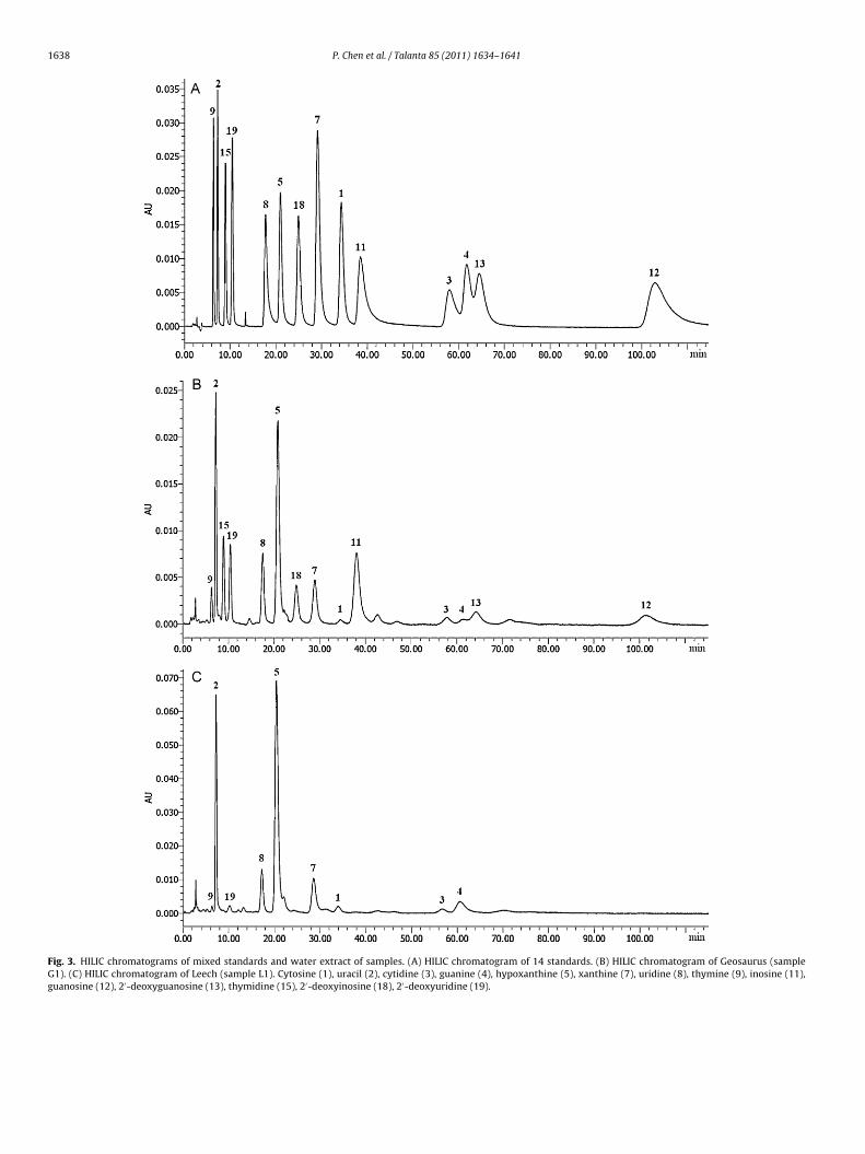
Fig. 3. HILIC chromatograms of mixed standards and water extract of samples. (A) HILIC chromatogram of 14 standards. (B) HILIC chromatogram of Geosaurus (sampleG1). (C) HILIC chromatogram of Leech (sample L1). Cytosine (1), uracil (2), cytidine (3), guanine (4), hypoxanthine (5), xanthine (7), uridine (8), thymine (9), inosine (11),guanosine (12), 2′-deoxyguanosine (13), thymidine (15), 2′-deoxyinosine (18), 2′-deoxyuridine (19).
P. Chen et al. / Talanta 85 (2011) 1634–1641 1639
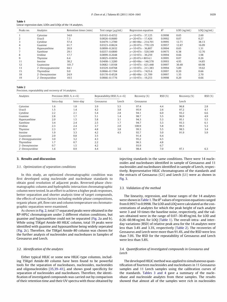
Table 1Linear regression date, LODs and LOQs of the 14 analytes.
Peaks no. Analytes Retention times (min) Test range (�g/mL) Regression equation R2 LOD (ng/mL) LOQ (ng/mL)
1 Cytosine 34.0 0.0163–0.4352 y = 1E+07x − 37,125 0.9998 0.65 2.602 Uracil 7.3 0.0026–0.0680 y = 2E+07x − 17,426 0.9992 0.07 0.273 Cytidine 57.4 0.0479–1.2780 y = 8E+06x − 214,701 0.9995 12.71 38.134 Guanine 61.7 0.0323–0.8624 y = 2E+07x − 770,129 0.9957 12.87 16.095 Hypoxanthine 20.9 0.0099–0.2632 y = 1E+07x − 36,807 0.9994 0.65 1.317 Xanthine 29.1 0.0257–0.6800 y = 2E+07x − 520,549 0.9975 6.38 12.768 Uridine 17.7 0.0099–0.2648 y = 1E+07x − 39,259 0.9994 0.66 1.989 Thymine 6.4 0.0025–0.0656 y = 2E+07x + 8014.1 0.9995 0.07 0.2611 Inosine 38.2 0.0498–1.3280 y = 6E+06x − 146,578 0.9993 4.95 14.8512 Guanosine 101.7 0.0682–1.8168 y = 1E+07x − 621,448 0.9997 30.49 60.9813 2′-Deoxyguanosine 64.1 0.0329–0.8768 y = 1E+07x − 211,383 0.9994 5.89 19.6215 Thymidine 9.0 0.0066–0.1784 y = 1E+07x − 7425.6 0.9997 0.20 0.6618 2′-Deoxyinosine 24.9 0.0170–0.4528 y = 8E+06x − 21,709 0.9997 1.35 2.7019 2′-Deoxyuridine 10.5 0.0066–0.1776 y = 1E+07x − 19,253 0.9998 0.20 0.66
Table 2Precision, repeatability and recovery of 14 analytes.
Analytes Precision (RSD, %, n = 6) Repeatability (RSD, %, n = 6) Recovery (%) RSD (%) Recovery (%) RSD (%)
Intra-day Inter-day Geosaurus Leech Geosaurus Leech
Cytosine 1.6 1.8 3.9 5.5 97.4 4.4 96.8 2.8Uracil 1.5 1.4 4.2 3.8 95.9 2.6 97.2 4.1Cytidine 2.2 2.1 5.4 2.8 94.1 6.0 93.2 6.6Guanine 2.8 1.7 5.1 1.4 98.7 5.5 96.9 4.9Hypoxanthine 2.0 1.5 5.8 3.1 94.3 5.3 95.1 5.5Xanthine 3.4 2.4 2.5 1.7 94.7 5.3 93.3 6.1Uridine 2.3 2.4 3.7 2.0 100.9 5.8 99.3 5.3Thymine 2.3 0.7 4.8 3.8 99.3 5.5 98.5 3.4Inosine 1.9 3.3 4.2 4.5 92.7 5.0 91.9 5.9Guanosine 2.0 0.8 3.5 91.4 6.42′-Deoxyguanosine 3.0 3.2 4.3 95.2 6.5
3.6
3
3
fiomcbtog
RgWi(tG
3
itasfio
Thymidine 1.7 0.7 2.22′-Deoxyinosine 0.7 1.5 4.22′-Deoxyuridine 1.6 0.9 4.4
. Results and discussion
.1. Optimization of separation conditions
In this study, an optimized chromatographic condition wasrst developed using nucleoside and nucleobase standards tobtain good resolution of adjacent peaks. Reversed-phase chro-atographic column and hydrophilic interaction chromatographic
olumn were tested. In an effort to achieve a higher peak responses,etter separation and shorter analysis time of target compounds,he effects of various factors including mobile phase compositions,rganic phase, pH, flow rate and column temperature on chromato-raphic separation were examined.
As shown in Fig. 2, total 17 separated peaks were obtained in theP-HPLC chromatogram under 2 different elution conditions, butuanine and hypoxanthine could not be separated (Fig. 2a and b).hile using TSKgel Amide-80 HILIC column, total 19 peaks were
dentified with guanine and hypoxanthine being widely seperatedFig. 2c). Therefore, the TSKgel Amide-80 column was chosen forhe further analysis of nucleosides and nucleobases in Samples ofeosaurus and Leech.
.2. Identification of the analytes
Either typical HILIC or some new HILIC-type columns, includ-ng TSKgel Amide-80 column have been found to be powerfulools for the separation of nucleobases, nucleosides, nucleotides
nd oligonucleotides [35,39–41], and shows good specificity foreparation of nucleosides and nucleobases. Therefore, the identi-cation of investigated compounds was carried out by comparisonf their retention time and their UV spectra with those obtained by96.3 4.893.9 6.798.4 7.0 97.1 6.3
injecting standards in the same conditions. There were 14 nucle-osides and nucleobases identified in sample of Geosaurus and 11nucleosides and nucleobases identified in sample of Leech, respec-tively. Representative HILIC chromatograms of the standards andthe extracts of Geosaurus (G1) and Leech (L1) were as shown inFig. 3.
3.3. Validation of the method
The linearity, regression, and linear ranges of the 14 analyteswere shown in Table 1. The R2 values of regression equations rangedfrom 0.9957 to 0.9998. The LOD and LOQ were calculated as the con-centrations of analytes for which the peak height of each analytewere 3 and 10 times the baseline noise, respectively, and the val-ues obtained were in the range of 0.07–30.49 ng/mL for LOD and0.26–60.98 ng/mL for LOQ (Table 1). The overall intra- and inter-day variations (RSD) of relative peak area for the 14 analytes wereless than 3.4% and 3.3%, respectively (Table 2). The recoveries ofGeosaurus and Leech were more than 91.4%, and the RSD were lessthan 7.0%. The RSD for the repeatability of Geosaurus and Leechwere less than 5.8%.
3.4. Quantification of investigated compounds in Geosaurus andLeech
The developed HILIC method was applied to simultaneous quan-tification of fourteen nucleosides and nucleobases in 11 Geosaurus
samples and 11 Leech samples using the calibration curves ofthe standards. Tables 3 and 4 gave a summary of the nucle-obase and nucleoside analytes from these samples. The resultsshowed that almost all of the samples were rich in nucleosides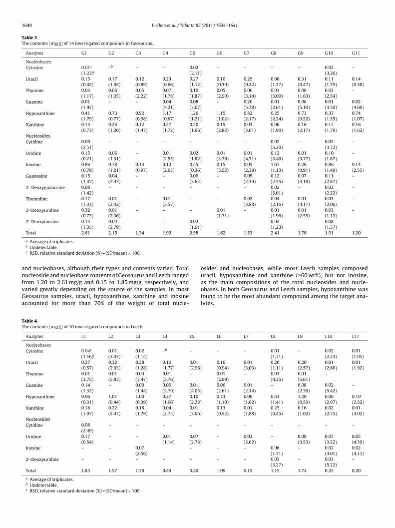
1640 P. Chen et al. / Talanta 85 (2011) 1634–1641
Table 3The contents (mg/g) of 14 investigated compounds in Geosaurus.
Analytes G1 G2 G3 G4 G5 G6 G7 G8 G9 G10 G11
NucleobasesCytosine 0.01a –b – – 0.02 – – – – 0.02 –
(1.23)c (2.11) (3.20)Uracil 0.15 0.17 0.12 0.23 0.27 0.10 0.29 0.06 0.31 0.11 0.14
(0.42) (1.04) (0.89) (0.66) (1.12) (0.39) (0.22) (1.37) (0.47) (1.75) (0.30)Thymine 0.03 0.06 0.05 0.07 0.10 0.05 0.06 0.01 0.06 0.03 –
(1.17) (1.35) (2.22) (1.78) (1.87) (2.90) (3.14) (3.09) (1.63) (2.54)Guanine 0.01 – – 0.04 0.08 – 0.20 0.01 0.08 0.01 0.02
(1.92) (4.21) (3.07) (5.38) (2.61) (5.10) (3.50) (4.60)Hypoxanthine 0.41 0.73 0.92 1.17 1.26 1.15 0.82 0.25 0.73 0.37 0.74
(1.79) (0.77) (0.98) (0.67) (1.21) (1.02) (2.17) (2.34) (0.52) (1.55) (1.07)Xanthine 0.13 0.25 0.12 0.27 0.20 0.15 0.03 0.06 0.16 0.12 0.16
(0.73) (1.20) (1.47) (1.72) (1.96) (2.82) (3.01) (1.90) (2.17) (1.79) (1.62)NucleosidesCytidine 0.09 – – – – – – 0.02 – 0.02 –
(2.51) (5.20) (3.72)Uridine 0.15 0.06 – 0.01 0.02 0.01 0.01 0.12 0.01 0.10 –
(0.21) (1.31) (2.55) (1.82) (3.76) (4.71) (3.46) (3.77) (1.87)Inosine 0.86 0.78 0.13 0.12 0.55 0.15 0.05 1.67 0.26 0.86 0.14
(0.78) (1.21) (0.97) (2.03) (0.36) (3.52) (2.38) (1.13) (0.91) (1.49) (2.55)Guanosine 0.15 0.04 – – 0.06 – 0.05 0.12 0.07 0.11 –
(1.32) (2.43) (3.62) (2.39) (2.55) (3.19) (2.87)2′-Deoxyguanosine 0.08 – – – – – – 0.02 – 0.02 –
(1.42) (5.01) (2.22)Thymidine 0.17 0.01 – 0.01 – – 0.02 0.04 0.01 0.03 –
(1.33) (2.42) (3.57) (3.88) (2.16) (4.17) (2.06)2′-Deoxyuridine 0.32 0.01 – – – 0.01 – 0.01 0.01 0.03 –
(0.73) (2.36) (1.71) (1.96) (2.55) (1.13)2′-Deoxyinosine 0.13 0.04 – – 0.02 – – 0.02 – 0.08 –
(1.35) (2.79) (1.91) (1.23) (1.57)Total 2.61 2.15 1.34 1.92 2.58 1.62 1.53 2.41 1.70 1.91 1.20
anfvGa
TT
a Average of triplicates.b Undetectable.c RSD, relative standard deviation (%) = (SD/mean) × 100.
nd nucleobases, although their types and contents varied. Totalucleoside and nucleobase contents of Geosaurus and Leech ranged
rom 1.20 to 2.61 mg/g and 0.15 to 1.85 mg/g, respectively, andaried greatly depending on the source of the samples. In mosteosaurus samples, uracil, hypoxanthine, xanthine and inosineccounted for more than 70% of the weight of total nucle-
able 4he contents (mg/g) of 10 investigated compounds in Leech.
Analytes L1 L2 L3 L4 L5
NucleobasesCytosine 0.04a 0.01 0.02 –b –
(1.16)c (3.03) (1.14)Uracil 0.27 0.32 0.38 0.10 0.01
(0.57) (2.03) (1.28) (1.77) (2.96)Thymine 0.01 0.01 0.04 0.01 –
(3.75) (5.83) (3.47) (3.76)Guanine 0.14 – 0.09 0.06 0.01
(1.32) (1.44) (2.79) (4.05)Hypoxanthine 0.96 1.01 1.00 0.27 0.10
(0.31) (0.44) (0.39) (1.96) (2.28)Xanthine 0.18 0.22 0.18 0.04 0.01
(1.07) (2.47) (1.79) (2.75) (3.06)NucleosidesCytidine 0.08 – – – –
(2.49)Uridine 0.17 – – 0.01 0.07
(0.54) (1.14) (2.78)Inosine – – 0.07 –
(2.56)2′-Deoxyuridine – – – – –
Total 1.85 1.57 1.78 0.49 0.20
a Average of triplicates.b Undetectable.c RSD, relative standard deviation (%) = (SD/mean) × 100.
osides and nucleobases, while most Leech samples composeduracil, hypoxanthine and xanthine (>60 wt%), but not inosine,
as the main compositions of the total nucleosides and nucle-obases. In both Geosaurus and Leech samples, hypoxanthine wasfound to be the most abundant compound among the target ana-lytes.L6 L7 L8 L9 L10 L11
– – 0.01 – 0.02 0.01(1.31) (2.23) (1.05)
0.16 0.01 0.20 0.20 0.01 0.01(0.94) (3.03) (1.11) (2.57) (2.86) (1.92)0.01 – 0.01 0.01 – –(2.99) (4.35) (5.61)0.06 0.01 – 0.08 0.02 –(2.61) (2.14) (2.36) (5.42)0.73 0.09 0.61 1.20 0.06 0.10(1.19) (1.62) (1.41) (0.59) (2.67) (2.32)0.13 0.01 0.23 0.16 0.02 0.01(0.52) (1.88) (0.45) (1.02) (2.75) (4.02)
– – – – – –
– 0.03 – 0.09 0.07 0.05(2.62) (3.53) (3.22) (4.39)
– – 0.06 – 0.02 0.02(1.71) (3.01) (4.11)
– – 0.03 – 0.03 –(3.27) (5.22)
1.09 0.15 1.15 1.74 0.25 0.20
ta 85
HndacTwsoeants
4
oao2tmtLwrcGtpL
R
[
[
[
[[
[
[
[[
[
[
[
[
[[[
[
[
[
[[
[[[[
[[
[
P. Chen et al. / Talan
Although the results showed that HILIC was superior to RP-PLC on the separation of nucleosides and nucleobases, however,o adenine, adenosine, 2′-deoxycytidine or 2′-deoxyadenosine wasetected in samples of Geosaurus and Leech, and the separation ofdenine and hypoxanthine standards was still a problem as theyould not be separated completely using the present condition.his was also observed by Marrubini et al. using HILIC method,ho found that 5 and 3 �m TSKgel Amide-80 columns demon-
trated great chemical and mechanical stability, but the separationf adenine and hypoxanthine also could not be achieved [35]. Nev-rtheless, HILIC showed its potentials and therefore could be useds an alternative method for the separation of nucleosides anducleobases, escepially for, guanine, 2′-deoxyuridine, uridine andhymidine, which cannot be separated by other analytical methodsuch as LC–MS, CE–MS [22,25] or RP-HPLC.
. Conclusion
In this study, quantitative and qualitative analysis of nucle-bases and nucleosides from the aqueous extracts of Geosaurusnd Leech was carried out using an HILIC method. Fourteen nucle-sides and nucleobases were identified and quantified from total2 samples with high sensitivity and selectivity. Uracil, hypoxan-hine, xanthine and inosine were quantitatively determined as the
ain nucleosides in most Geosaurus, which accounted for morehan 70% of the total nucleosides and nucleobases. While for mosteech samples, the content of uracil, hypoxanthine and xanthineere more than 60% of the total nucleosides and nucleobases. Our
esults in the present study clearly suggests that HILIC methodould be employed as an useful tool for the quality assessment ofeosaurus and Leech using certain nucleosides and nucleobases as
he markers. Setting a minimum limit to the amount of these com-ounds would be helpful for the quality control of Geosaurus andeech preparations.
eferences
[1] Pharmacopoeia of the People’s Republic of China, Chemical Industry Press,Beijing, 2010, p. 113.
[2] Pharmacopoeia of the People’s Republic of China, Chemical Industry Press,Beijing, 2010, p. 77.
[3] A.P. Schmidt, D.R. Lara, J.F. Maraschin, A.S. Perla, D.O. Souza, Brain Res. 864
(2000) 40–43.[4] G. Anfossi, I. Russo, P. Massucco, L. Mattiello, F. Cavalot, A. Balbo, M. Trovati,Thromb. Res. 105 (2002) 71–78.
[5] J.B. Conti, L. Belardinelli, D.B. Utterback, A.B. Curtis, Circulation 91 (1995)1761–1767.
[[[[
(2011) 1634–1641 1641
[6] J. Wang, Z.G. Huang, H. Cao, Y.T. Wang, P. Hui, C. Hoo, S.P. Li, J. Sep. Sci. 31 (2008)1173–1180.
[7] S.P. Li, F.Q. Yang, K.W.K. Tsim, J. Pharm. Biomed. Anal. 41 (2006) 1571–1584.[8] W.H. Carmen, D. Xin, J. Chromatogr. B 812 (2004) 241–257.[9] J.M. Liu, D. Liu, L.X. Yang, Y.R. Zhong, S.L. Cui, Chin. J. Chin. Mater. Med. 10 (1994)
615–616.10] S.P. Li, P. Li, C.M. Lai, Y.X. Gong, K.K.W. Kelvin, T.T.X. Dong, K.W.K. Tsim, Y.T.
Wang, J. Chromatogr. A 1036 (2004) 239–243.11] M.S. Shiao, Z.N. Wang, L.J. Lin, J.Y. Lien, J.J. Wang, Bot. Bull. Acad. Sin. 35 (1994)
261–267.12] S.K. Yan, G.A. Luo, Y.M. Wang, Y.Y. Cheng, J. Pharm. Biomed. Anal. 40 (2006)
889–895.13] L.S. Li, M. Liu, S.L. Da, Y.Q. Feng, Talanta 63 (2004) 433–441.14] Z.M. Qian, J.B. Wan, Q.W. Zhang, S.P. Li, J. Pharm. Biomed. Anal. 48 (2008)
1361–1367.15] R. Ikeda, M. Nishimura, Y. Sun, M. Wada, K. Nakashima, Biomed. Chromatogr.
22 (2008) 630–636.16] X.W. Cao, J. Li, S.B. Chen, X.B. Li, P.G. X, S.L. Chen, D.J. Yang, J. Sep. Sci. 33 (2010)
1587–1594.17] A. Ranogajec, S. Beluhan, Z. Smit, J. Sep. Sci. 33 (2010) 1024–1033.18] S. Wang, F.Q. Yang, K. Feng, D.Q. Li, J. Zhao, S.P. Li, J. Sep. Sci. 32 (2009)
4069–4076.19] F.Q. Guo, A. Li, L.F. Huang, Y.Z. Liang, B.M. Chen, J. Pharm. Biomed. Anal. 40
(2006) 623–630.20] L.F. Huang, Y.Z. Liang, F.Q. Guo, Z.F. Zhou, B.M. Chen, J. Pharm. Biomed. Anal. 33
(2003) 1155–1162.21] J.L. Gao, K.S.Y. Leng, Y.T. Wang, C.M. Lai, S.P. Li, L.F. Hu, G.H. Lu, Z.H. Jiang, Z.L.
Yu, J. Pharm. Biomed. Anal. 44 (2007) 807–811.22] H. Fan, S.P. Li, J.J. Xiang, C.M. Lai, F.Q. Yang, J.L. Gao, Y.T. Wang, Anal. Chim. Acta
567 (2006) 218–228.23] A. Brink, U. Lutz, W. Volkel, W.K. Lutz, J. Chromatogr. B 830 (2006) 255–261.24] F.Q. Yang, J. Guan, S.P. Li, Talanta 73 (2007) 269–273.25] F.Q. Yang, L. Ge, J.W.H. Yong, S.N. Tan, S.P. Li, J. Pharm. Biomed. Anal. 50 (2009)
307–314.26] X. Cahours, H. Dessans, P. Morin, M. Dreux, L. Agrofoglio, J. Chromatogr. A 895
(2000) 101–109.27] W.G. Stillwell, H.X. Xu, J.A. Adkins, J.S. Wishnok, S.R. Tannenbaum, Chem. Res.
Toxicol. 2 (1989) 94–99.28] A.J.R. Teixeira, J.H. Gommers-Ampt, G.V. deWerken, J.G. Westra, J.F.C. Stavenu-
iter, A.P.J.M. de Jong, Anal. Biochem. 214 (1993) 474–483.29] S.P. Li, P. Li, T.T.X. Dong, K.W.K. Tsim, Electrophoresis 22 (2001) 144–150.30] Y.X. Gong, S.P. Li, P. Li, J.J. Liu, Y.T. Wang, J. Chromatogr. A 1055 (2004)
215–221.31] T. Helboe, S.H. Hansen, J. Chromatogr. A 836 (1999) 315–324.32] F.Q. Yang, Sh.P. Li, P. Li, Y.T. Wang, Electrophoresis 28 (2007) 1681–1688.33] H.Y. Cheung, C.W. Ng, D.J. Hood, J. Chromatogr. A 911 (2001) 119–126.34] H.M. Liebich, R. Lehmann, G. Xu, H.G. Wahl, H.U. Haring, J. Chromatogr. B 745
(2000) 189–196.35] A.J. Alpert, J. Chromatogr. 499 (1990) 177–196.36] A.J. Alpert, M. Shukla, A.K. Shukla, L.R. Zieske, S.W. Yuen, M.A.J. Ferguson, A.
Mehlert, M. Pauly, R. Orlando, J. Chromatogr. A 676 (1994) 191–202.37] J.A. Boutin, A.-P. Ernould, G. Ferry, A. Genton, A.J. Alpert, J. Chromatogr. B 583
(1992) 137–143.
38] A.K. Leung, F. Gong, Y.Z. Liang, F.T. Chau, Anal. Lett. 33 (2000) 3195–3211.39] G. Marrubini, B.E. Castillo Mendoza, G. Massolini, J. Sep. Sci. 33 (2010) 803–816.40] G. Jin, Z. Guo, F. Zhang, X. Xue, Y. Jin, X. Liang, Talanta 76 (2008) 522–527.41] T. Ikegami, K. Tomomatsu, H. Takubo, K. Horie, N. Tanaka, J. Chromatogr. A 1184(2008) 474–503.