Embed Size (px)
Citation preview
Identification and Characterization of ZEL-H16 as a NovelAgonist of the Histamine H3 ReceptorYing Shi1, Rong Sheng2, Tingting Zhong1, Yu Xu2, Xiaopan Chen1, Dong Yang1, Yi Sun1, Fenyan Yang2,
Yongzhou Hu2*, Naiming Zhou1*
1College of Life Sciences, Zhejiang University, Hangzhou, Zhejiang Province, China, 2College of Pharmaceutical Sciences, Zhejiang University, Hangzhou, Zhejiang
Province, China
Abstract
The histamine H3 receptor (H3R) has been recognized as a promising target for the treatment of various central andperipheral nervous system diseases. In this study, a non-imidazole compound, ZEL-H16, was identified as a novel histamineH3 receptor agonist. ZEL-H16 was found to bind to human H3R with a Ki value of approximately 2.07 nM and 4.36 nM to ratH3R. Further characterization indicated that ZEL-H16 behaved as a partial agonist on the inhibition of forskolin-stimulatedcAMP accumulation (the efficacy was 60% of that of histamine) and activation of ERK1/2 signaling (the efficacy was 50% ofthat of histamine) at H3 receptors, but acted as a full agonist just like histamin in the guinea-pig ileum contraction assay.These effects were blocked by pertussis toxin and H3 receptor specific antagonist thioperamide. ZEL-H16 showed noagonist or antagonist activities at the cloned human histamine H1, H2, and H4 receptors and other biogenic amine GPCRs inthe CRE-driven reporter assay. Furthermore, our present data demonstrated that treatment of ZEL-H16 resulted in intensiveH3 receptor internalization and delayed recycling to the cell surface as compared to that of control with treatment ofhistamine. Thus, ZEL-H16 is a novel and potent nonimidazole agonist of H3R, which might serve as a pharmacological toolfor future investigations or as possible therapeutic agent of H3R.
Citation: Shi Y, Sheng R, Zhong T, Xu Y, Chen X, et al. (2012) Identification and Characterization of ZEL-H16 as a Novel Agonist of the Histamine H3 Receptor. PLoSONE 7(8): e42185. doi:10.1371/journal.pone.0042185
Editor: Joao B. Calixto, Universidad Federal de Santa Catarina, Brazil
Received April 23, 2012; Accepted July 2, 2012; Published August 1, 2012
Copyright: � 2012 Shi et al. This is an open-access article distributed under the terms of the Creative Commons Attribution License, which permits unrestricteduse, distribution, and reproduction in any medium, provided the original author and source are credited.
Funding: This work was supported by grants from the National Natural Science Foundation of China (No. 30670425), the Ministry of Science and Technology (No.2008AA02Z138) and the National Key Tech Project for Major Creation of New drugs (No. 2009ZX09501-003). The funders had no role in study design, datacollection and analysis, decision to publish, or preparation of the manuscript.
Competing Interests: The authors have declared that no competing interests exist.
* E-mail: [email protected] (NZ); [email protected] (YH)
Introduction
Histamine, a biogenic amine with multiple physiological effects,
exerts its biological activities through four distinct G-protein-
coupled receptors (GPCRs) known as the histamine H1, H2, H3,
and H4 receptors [1,2]. The histamine H1 and H2 receptors were
identified decades ago [3,4], and have been shown to be excellent
drug targets for the treatment of allergy and gastric ulcers,
respectively [5,6]. The histamine H4 receptor was discovered in
2000 [7], and it has been identified as a potential target for the
treatment of inflammatory diseases such as chronic allergies,
asthma, atopic dermatitis, and inflammatory bowel diseases [8].
The histamine H3 receptor (H3R) was first identified by Arrang
and colleagues in 1983 using a functional assay in which it was
found that histamine inhibits its own synthesis and release [9].
However, the cloning of the human histamine H3 receptor cDNA
in 1999 by Lovenberg and colleagues [10] prompted H3R
research in both academia and industry.
Although H3R mRNA is detectable in the heart, lung,
gastrointestinal tract, and endothelial cells [11], H3R is pre-
dominantly expressed in the central nervous system (CNS) and
peripheral nervous system. The highest levels of H3R are found in
the cerebral cortex, hippocampal formation, basal ganglia, and
hypothalamus [12,13]. It has been established that H3R associates
with the heterotrimeric Gi/o-protein, which leads to a decrease in
cAMP formation and PKA activation, and also causes the
activation of the Akt/GSK-3b axis and ERK1/2 pathways, the
inhibition of the Na+/H+ exchanger, and modulation of in-
tracellular calcium upon histamine stimulation [14]. The H3R was
first identified as a presynaptic autoreceptor that negatively
regulated the synthesis and release of histamine from histaminergic
neurons [9]. However, the H3R has also been shown to act as
a presynaptic heteroreceptor on non-histaminergic neurons,
inhibiting the release of other neurotransmitters such as acetyl-
choline, dopamine, norepinephrine, serotonin, and various
neuropeptides in both the central and peripheral nervous system
[15,16,17]. Therefore, the H3 receptor has long been recognized
as a promising target for the treatment of various central and
peripheral nervous system diseases. Antagonists and inverse
agonists of the H3 receptor have been proposed as potential
drugs for the treatment of attention-deficit hyperactivity disorders
(ADHD), Alzheimer’s disease, obesity and others [18,19,20,21],
whereas H3R agonists are suggested for the treatment of asthma,
migraine, and ischemic arrhythmias. However, increasing evi-
dence suggests H3R agonists could serve as potential therapeutics
for obesity, diabetes mellitus, and liver cholestasis [22,23,24]. BP
2–94, one of the earliest explored agonists of H3R, displayed anti-
inflammatory and anti-nociceptive properties in mice and was
recognized as a promising drug for the treatment of asthma,
migraines, related inflammatory diseases, and pain [25,26]. N-a-methylhistamine, another promising agonist of H3R, was tested in
PLoS ONE | www.plosone.org 1 August 2012 | Volume 7 | Issue 8 | e42185
a Phase III clinical study for migraine prophylaxis [23]. Therefore,
it is reasonable to believe that H3R agonists could hold therapeutic
value for the treatment of human diseases.
In the current study, ZEL-H16 was identified as a novel agonist
of H3R using CRE-luciferase assay and internalization assay with
HEK-293 cells stably expressing H3R. Affinities of ZEL-H16 for
hH3R and rH3R were measured by competition binding
experiments. We also investigated the ability of ZEL-H16 to
induce the phosphorylation of ERK1/2 in mouse cortical
neuronal cultures expressing endogenous H3R and the ability to
inhibit the contraction of the guinea-pig ileum. Another major
result of this study is that ZEL-H16 could induce intense
internalization and delay recycling of internalized H3R to the
cell surface compared with histamine. Our results clearly
demonstrate that ZEL-H16 is a potent, selective and non-
imidazole agonist of H3R that could serve as a useful pharma-
cological tool for future studies or as a possible therapeutic agent.
Results
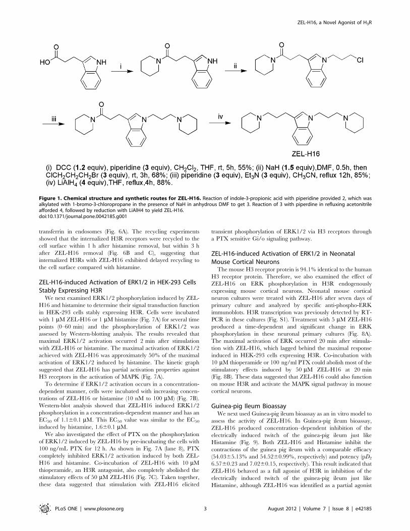
Characterization of ZEL-H16 as a Selective Partial H3RAgonist in CRE Reporter AssayWe used CRE-driven reporter assay as primary assay to screen
more than 300 antagonist compounds in 6 serials of structures,
and found several compounds with the activity in inhibition of
forskolin-induced luciferase activity. We then employed hH3R-
GFP redistribution assay as a secondary assay to confirm the
antagonistic activity. One of the compounds identified, ZEL-H16,
whose chemical structure and synthetic routes are shown in
Figure 1, triggered a significant increase in receptor internalization
as compared to the control compound histamine, behaving as an
agonist on H3R internalization. The agonist activity of ZEL-H16
was further confirmed in HEK-293 cell lines that stably express
the human H3R and a reporter gene consisting of the firefly
luciferase coding region that is under the control of minimal
promoter containing cAMP-response elements (CREs). The CRE-
driven reporter assay is widely used to measure the function of
GPCR agonists and antagonists. Changes of intracellular cAMP
could cause changes of CRE-driven report gene transcription. As
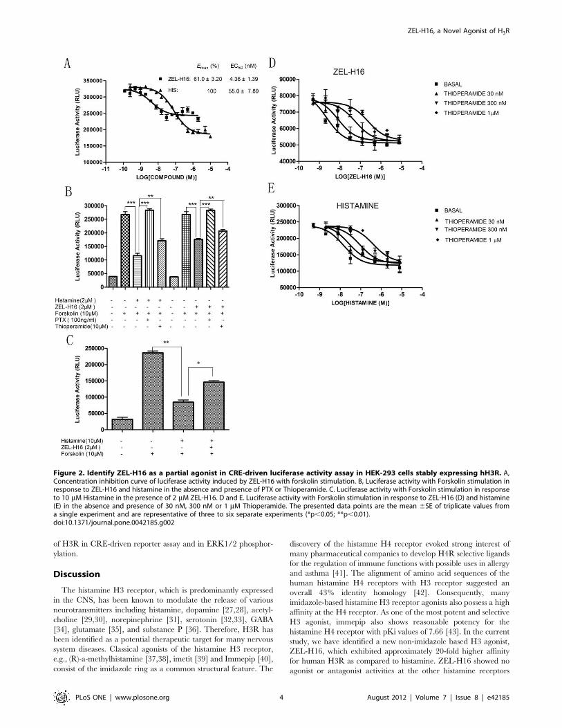
indicated in Figure 2A, compound ZEL-H16 has partial agonistic
properties and a low EC50 value for H3R as compared to
histamine (EC50[ZEL-H16] = 4.3661.39 nM, Emax[ZEL-H16]
= 61.063.20%, Mean 6 SEM, n= 6; EC50[histamine]
= 55.067.89 nM, Emax[histamine] = 100%, Mean 6 SEM, n=6).
The agonistic activity of ZEL-H16 could be disrupted by co-
incubation with PTX, a Gi inhibitor, and thioperamide, an
antagonist of H3R (Fig. 2B). Moreover, the inhibition of forskolin-
induced luciferase activity by Histamine could be reduced by co-
incubation with ZEL-H16 (Fig. 2C). We also performed the
experiments to determine the CRE-driven luciferase activity in the
response to ZEL-H16 and histamine in the presence of three
different concentrations of antagonist thioperamide (Fig. 2D and
2E), and obtained the Schild slopes 1.11660.256 for histamine,
1.14060.168 for ZEL-H16, that were not significantly different
from unity. The data suggested that it is likely for both ZEL-H16
and histamine to bind to the same binding site of H3 receptor.
We then examined the selectivity of ZEL-H16 versus various
histamine receptors by assaying intracellular Ca2+ flux and cAMP
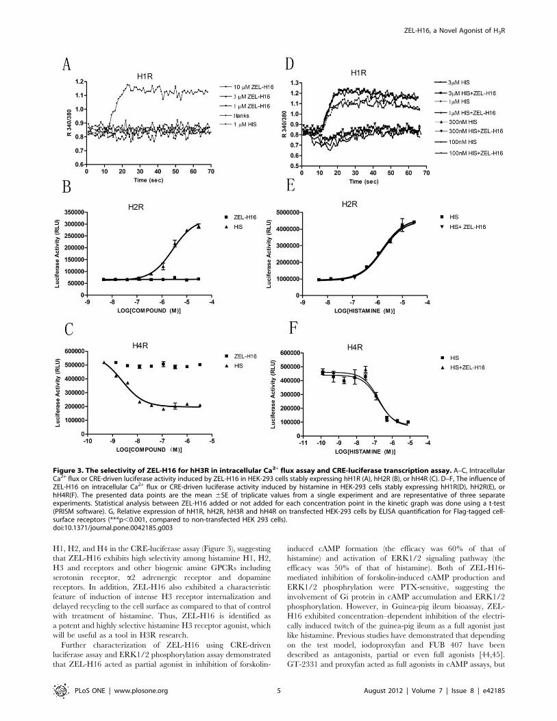
formation. As shown in Fig. 3A and 3D, ZEL-H16 did not induce
similar intracellular Ca2+ release in H1R-expressing HEK-293
cells compared to histamine, neither modulate the intracellular
Ca2+ release stimulated by histamine. In addition, no significant
responses were observed in the luciferase activity after stimulation
by ZEL-H16 in cells expressing H2R or H4R (Fig. 3B and 3C).
The addition of ZEL-H16 also did not significantly change the
luciferase activity curves induced by histamine in H2R or H4R-
expressing cells (Fig. 3E and 3F). The relative expression of H1R,
H2R, H3R and H4R on transfected cell membrane was shown in
Figure 3G. The selectivity of ZEL-H16 toward other biogenic
amine GPCRs, such as dopamine receptor DRD1 and DRD2,
serotonin receptor 5-HT1A, adrenergic receptor a2AR was also
examined by CRE-luciferase activity assay. ZEL-H16 showed no
agonist or antagonist activities to these receptors in the experi-
ments (Table 1). These results suggested that ZEL-H16 is a potent
and selective H3 receptor agonist.
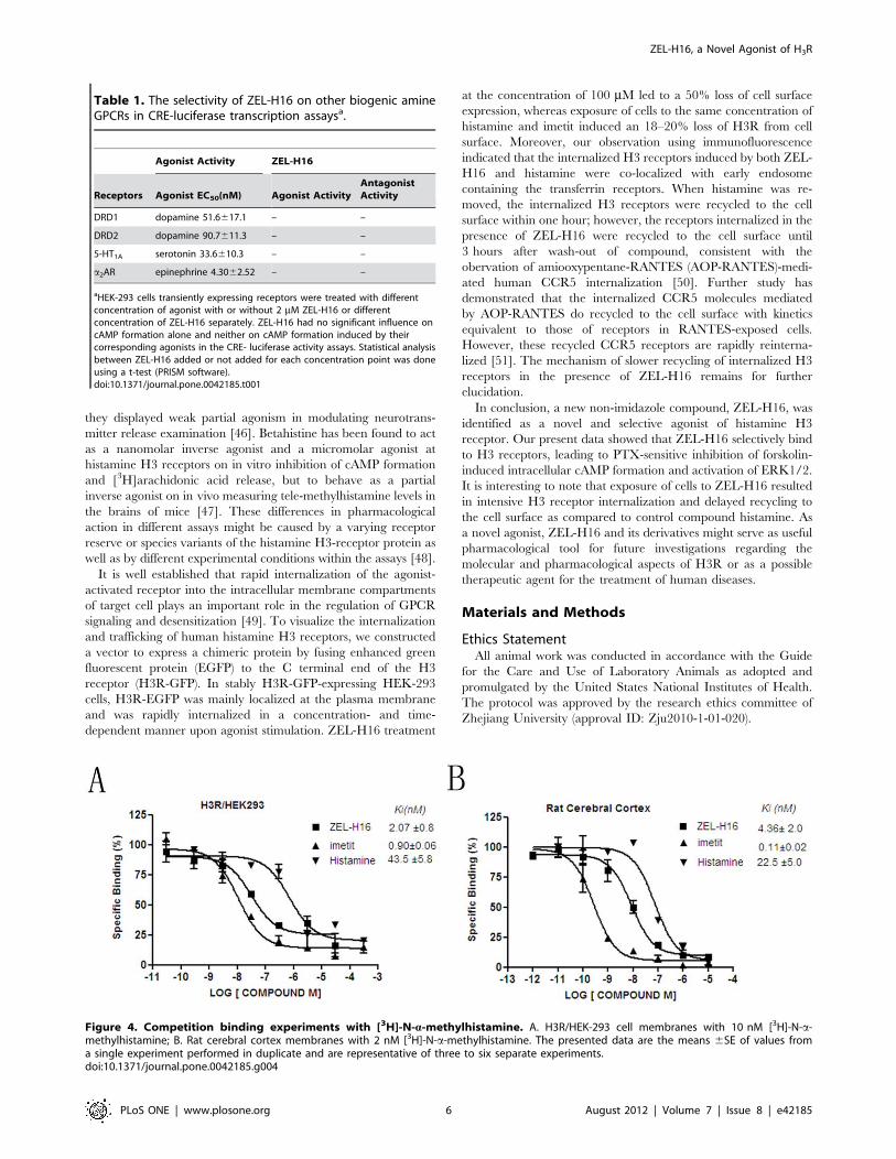
Direct Binding of ZEL-H16 to Human H3R and Rat H3RCompetitive binding experiments were conducted to assess
whether ZEL-H16 could directly bind to H3R. Scatchard analysis
of the saturation binding using [3H]N-a-methylhistamine against
human histamine H3 receptor revealed a KD value of 0.71 nM
and a Bmax value of 430 fmol/mg protein (data not shown). In the
competitive binding assays, binding of [3H]N-a-methylhistamine
to human H3R expressing HEK-293 cells was blocked by cold
histamine, ZEL-H16 and imetit, yielding 43.565.8, 2.0760.8 and
0.960.06 nM of the Ki values respectively (Fig. 4A), and binding
of [3H]N-a-methylhistamine to rat cerebral cortex was also
inhibited by cold histamine, ZEL-H16 and imetit, yielding
22.565.0, 4.3662.0 and 0.1160.02 nM of the Ki values re-
spectively (Bmax = 767 fmol/mg protein, KD=0.830 nM) (Fig. 4B).
The results showed that ZEL-H16 could directly bind to hH3R
and rH3R.
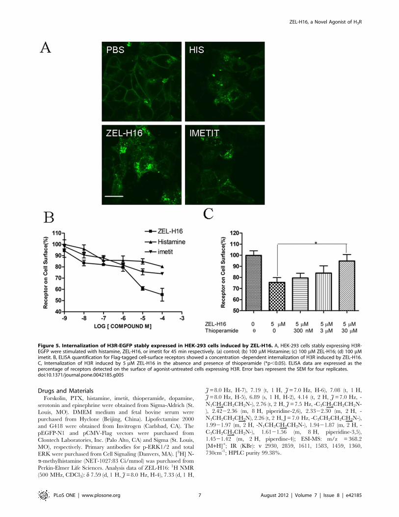
Internalization of H3R Induced by ZEL-H16 in HEK-293cells Stably Expressing H3RNext, we conducted internalization experiments to determine
the ability of ZEL-H16 to induce H3R internalization. HEK-293
cells stably transfected with H3R-EGFP were incubated with
100 mM ZEL-H16, histamine and imetit for 45 min at 37uCseparately and internalization was examined by confocal micros-
copy. As seen in Figure 5A (c), H3R internalized from the cell
surface into the cytoplasm with a punctuate distribution upon
activation by ZEL-H16. Moreover, the receptor internalization
induced by ZEL-H16 was more intense than the internalization
induced by histamine and imetit (Fig. 5A). This result was
confirmed by experiments that quantified the levels of receptors on
the surface of the cells following stimulation by compounds. HEK-
293 cells stably transfected with Flag-H3R were treated with
different concentrations of ZEL-H16 or histamine or imetit for
45 min at 37uC respectively, and the amount of H3R remaining
on the cell surface was quantitatively detected by ELISA.
Quantification by cell-surface ELISA showed a significant loss of
cell-surface receptors due to treatment with ZEL-H16 at
concentration ranging from 1–100 mM (Fig. 5B). Treatment with
100 mM ZEL-H16 caused a 50% loss in the expression of the
receptors on the cell surface, whereas the same concentration of
histamine or imetit only induced approximately 18–20% in-
ternalization of cell-surface receptors. This result suggested that
ZEL-H16 may be a powerful tool to investigate the internalization
behavior of H3R. In addition, H3R internalization induced by
5 mM ZEL-H16 could be blocked by pre-incubation with
thioperamide for 20 min at high concentration (Fig. 5C).
Furthermore, we used the endosome marker Alexa Fluor 546-
labeled transferrin to assess whether internalized H3Rs induced by
histamine or ZEL-H16 are generally recycled back to the plasma
membrane via early and recycling endosomes. Confocal micros-
copy analysis revealed that the internalized H3R receptors
induced by histamine or ZEL-H16 were both colocalized with
ZEL-H16, a Novel Agonist of H3R
PLoS ONE | www.plosone.org 2 August 2012 | Volume 7 | Issue 8 | e42185
transferrin in endosomes (Fig. 6A). The recycling experiments
showed that the internalized H3R receptors were recycled to the
cell surface within 1 h after histamine removal, but within 3 h
after ZEL-H16 removal (Fig. 6B and C), suggesting that
internalized H3Rs with ZEL-H16 exhibited delayed recycling to
the cell surface compared with histamine.
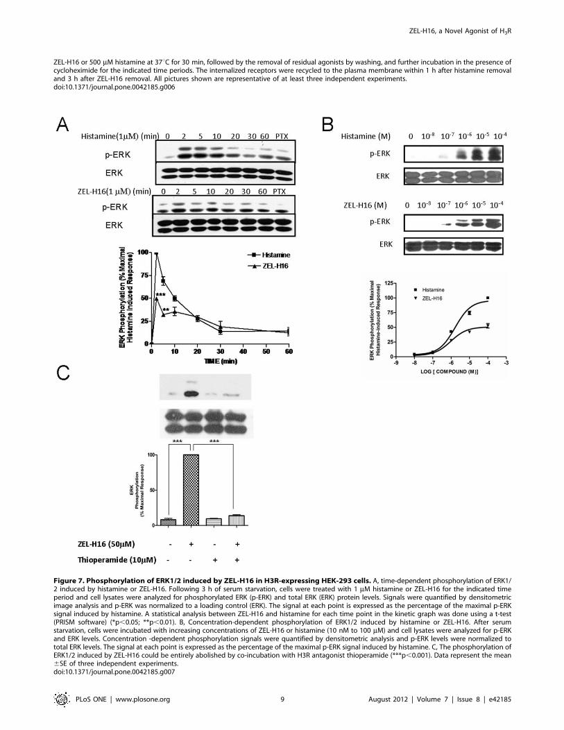
ZEL-H16-induced Activation of ERK1/2 in HEK-293 CellsStably Expressing H3RWe next examined ERK1/2 phosphorylation induced by ZEL-
H16 and histamine to determine their signal transduction function
in HEK-293 cells stably expressing H3R. Cells were incubated
with 1 mM ZEL-H16 or 1 mM histamine (Fig. 7A) for several time
points (0–60 min) and the phosphorylation of ERK1/2 was
assessed by Western-blotting analysis. The results revealed that
maximal ERK1/2 activation occurred 2 min after stimulation
with ZEL-H16 or histamine. The maximal activation of ERK1/2
achieved with ZEL-H16 was approximately 50% of the maximal
activation of ERK1/2 induced by histamine. The kinetic graph
suggested that ZEL-H16 has partial activation properties against
H3 receptors in the activation of MAPK (Fig. 7A).
To determine if ERK1/2 activation occurs in a concentration-
dependent manner, cells were incubated with increasing concen-
trations of ZEL-H16 or histamine (10 nM to 100 mM) (Fig. 7B).
Western-blot analysis showed that ZEL-H16 induced ERK1/2
phosphorylation in a concentration-dependent manner and has an
EC50 of 1.160.1 mM. This EC50 value was similar to the EC50
induced by histamine, 1.660.1 mM.
We also investigated the effect of PTX on the phosphorylation
of ERK1/2 induced by ZEL-H16 by pre-incubating the cells with
100 ng/mL PTX for 12 h. As shown in Fig. 7A (lane 8), PTX
completely inhibited ERK1/2 activation induced by both ZEL-
H16 and histamine. Co-incubation of ZEL-H16 with 10 mMthioperamide, an H3R antagonist, also completely abolished the
stimulatory effects of 50 mM ZEL-H16 (Fig. 7C). Taken together,
these data suggested that stimulation with ZEL-H16 elicited
transient phosphorylation of ERK1/2 via H3 receptors through
a PTX sensitive Gi/o signaling pathway.
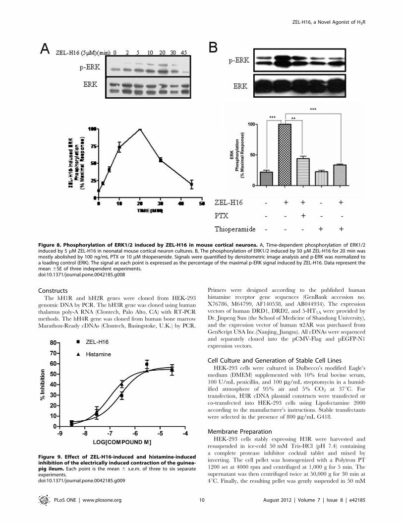
ZEL-H16-induced Activation of ERK1/2 in NeonatalMouse Cortical NeuronsThe mouse H3 receptor protein is 94.1% identical to the human
H3 receptor protein. Therefore, we also examined the effect of
ZEL-H16 on ERK phosphorylation in H3R endogenously
expressing mouse cortical neurons. Neonatal mouse cortical
neuron cultures were treated with ZEL-H16 after seven days of
primary culture and analyzed by specific anti-phospho-ERK
immunoblots. H3R transcription was previously detected by RT-
PCR in these cultures (Fig. S1). Treatment with 5 mM ZEL-H16
produced a time-dependent and significant change in ERK
phosphorylation in these neuronal primary cultures (Fig. 8A).
The maximal activation of ERK occurred 20 min after stimula-
tion with ZEL-H16, which lagged behind the maximal response
induced in HEK-293 cells expressing H3R. Co-incubation with
10 mM thioperamide or 100 ng/ml PTX could abolish most of the
stimulatory effects induced by 50 mM ZEL-H16 at 20 min
(Fig. 8B). These data suggested that ZEL-H16 could also function
on mouse H3R and activate the MAPK signal pathway in mouse
cortical neurons.
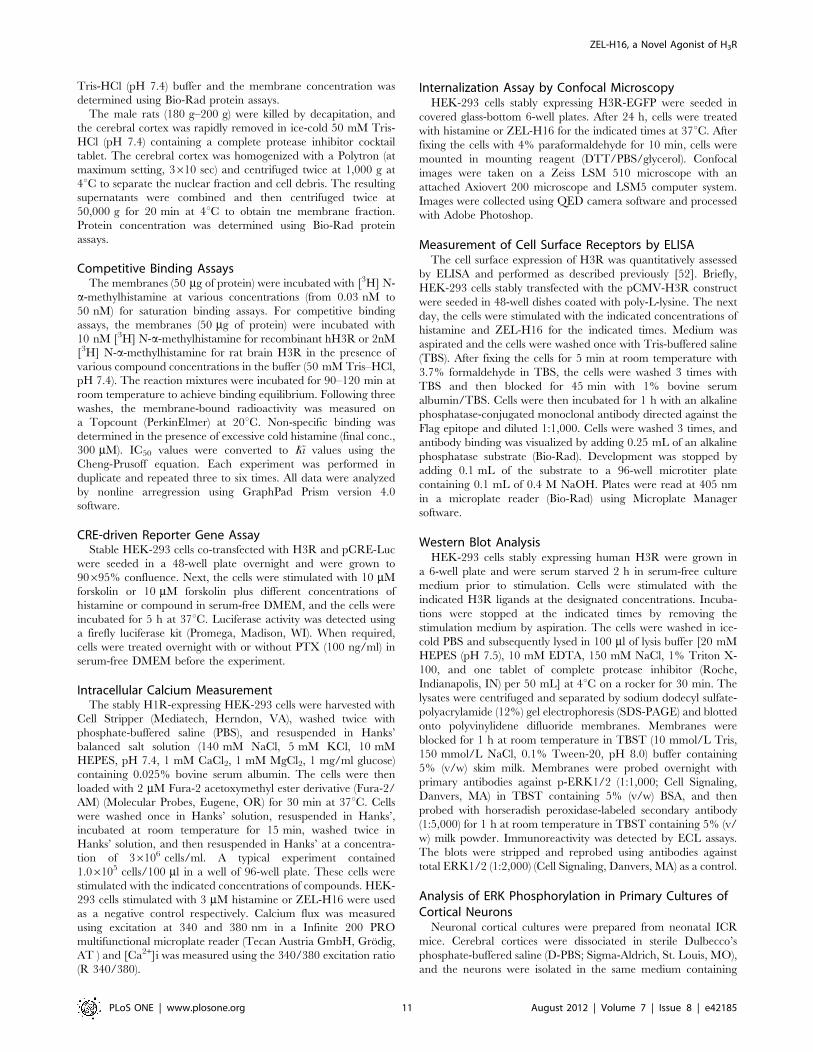
Guinea-pig Ileum BioassayWe next used Guinea-pig ileum bioassay as an in vitro model to
assess the activity of ZEL-H16. In Guinea-pig ileum bioassay,
ZEL-H16 produced concentration–dependent inhibition of the
electrically induced twitch of the guinea-pig ileum just like
Histamine (Fig. 9). Both ZEL-H16 and Histamine inhibit the
contractions of the guinea pig ileum with a comparable efficacy
(54.0365.13% and 54.5260.99%, respectively) and potency (pD2
6.5760.23 and 7.0260.15, respectively). This result indicated that
ZEL-H16 behaved as a full agonist of H3R in inhibition of the
electrically induced twitch of the guinea-pig ileum just like
Histamine, although ZEL-H16 was identified as a partial agonist
Figure 1. Chemical structure and synthetic routes for ZEL-H16. Reaction of indole-3-propionic acid with piperidine provided 2, which wasalkylated with 1-bromo-3-chloropropane in the presence of NaH in anhydrous DMF to get 3. Reaction of 3 with piperdine in refluxing acetonitrileafforded 4, followed by reduction with LiAlH4 to yield ZEL-H16.doi:10.1371/journal.pone.0042185.g001
ZEL-H16, a Novel Agonist of H3R
PLoS ONE | www.plosone.org 3 August 2012 | Volume 7 | Issue 8 | e42185
of H3R in CRE-driven reporter assay and in ERK1/2 phosphor-
ylation.
Discussion
The histamine H3 receptor, which is predominantly expressed
in the CNS, has been known to modulate the release of various
neurotransmitters including histamine, dopamine [27,28], acetyl-
choline [29,30], norepinephrine [31], serotonin [32,33], GABA
[34], glutamate [35], and substance P [36]. Therefore, H3R has
been identified as a potential therapeutic target for many nervous
system diseases. Classical agonists of the histamine H3 receptor,
e.g., (R)-a-methylhistamine [37,38], imetit [39] and Immepip [40],
consist of the imidazole ring as a common structural feature. The
discovery of the histamne H4 receptor evoked strong interest of
many pharmaceutical companies to develop H4R selective ligands
for the regulation of immune functions with possible uses in allergy
and asthma [41]. The alignment of amino acid sequences of the
human histamine H4 receptors with H3 receptor suggested an
overall 43% identity homology [42]. Consequently, many
imidazole-based histamine H3 receptor agonists also possess a high
affinity at the H4 receptor. As one of the most potent and selective
H3 agonist, immepip also shows reasonable potency for the
histamine H4 receptor with pKi values of 7.66 [43]. In the current
study, we have identified a new non-imidazole based H3 agonist,
ZEL-H16, which exhibited approximately 20-fold higher affinity
for human H3R as compared to histamine. ZEL-H16 showed no
agonist or antagonist activities at the other histamine receptors
Figure 2. Identify ZEL-H16 as a partial agonist in CRE-driven luciferase activity assay in HEK-293 cells stably expressing hH3R. A,Concentration inhibition curve of luciferase activity induced by ZEL-H16 with forskolin stimulation. B, Luciferase activity with Forskolin stimulation inresponse to ZEL-H16 and histamine in the absence and presence of PTX or Thioperamide. C. Luciferase activity with Forskolin stimulation in responseto 10 mM Histamine in the presence of 2 mM ZEL-H16. D and E. Luciferase activity with Forskolin stimulation in response to ZEL-H16 (D) and histamine(E) in the absence and presence of 30 nM, 300 nM or 1 mM Thioperamide. The presented data points are the mean 6SE of triplicate values froma single experiment and are representative of three to six separate experiments (*p,0.05; **p,0.01).doi:10.1371/journal.pone.0042185.g002
ZEL-H16, a Novel Agonist of H3R
PLoS ONE | www.plosone.org 4 August 2012 | Volume 7 | Issue 8 | e42185
H1, H2, and H4 in the CRE-luciferase assay (Figure 3), suggesting
that ZEL-H16 exhibits high selectivity among histamine H1, H2,
H3 and receptors and other biogenic amine GPCRs including
serotonin receptor, a2 adrenergic receptor and dopamine
receptors. In addition, ZEL-H16 also exhibited a characteristic
feature of induction of intense H3 receptor internalization and
delayed recycling to the cell surface as compared to that of control
with treatment of histamine. Thus, ZEL-H16 is identified as
a potent and highly selective histamine H3 receptor agonist, which
will be useful as a tool in H3R research.
Further characterization of ZEL-H16 using CRE-driven
luciferase assay and ERK1/2 phosphorylation assay demonstrated
that ZEL-H16 acted as partial agonist in inhibition of forskolin-
induced cAMP formation (the efficacy was 60% of that of
histamine) and activation of ERK1/2 signaling pathway (the
efficacy was 50% of that of histamine). Both of ZEL-H16-
mediated inhibition of forskolin-induced cAMP production and
ERK1/2 phosphrylation were PTX-sensitive, suggesting the
involvement of Gi protein in cAMP accumulation and ERK1/2
phosphorylation. However, in Guinea-pig ileum bioassay, ZEL-
H16 exhibited concentration–dependent inhibition of the electri-
cally induced twitch of the guinea-pig ileum as a full agonist just
like histamine. Previous studies have demonstrated that depending
on the test model, iodoproxyfan and FUB 407 have been
described as antagonists, partial or even full agonists [44,45].
GT-2331 and proxyfan acted as full agonists in cAMP assays, but
Figure 3. The selectivity of ZEL-H16 for hH3R in intracellular Ca2+ flux assay and CRE-luciferase transcription assay. A–C, IntracellularCa2+ flux or CRE-driven luciferase activity induced by ZEL-H16 in HEK-293 cells stably expressing hH1R (A), hH2R (B), or hH4R (C). D–F, The influence ofZEL-H16 on intracellular Ca2+ flux or CRE-driven luciferase activity induced by histamine in HEK-293 cells stably expressing hH1R(D), hH2R(E), orhH4R(F). The presented data points are the mean 6SE of triplicate values from a single experiment and are representative of three separateexperiments. Statistical analysis between ZEL-H16 added or not added for each concentration point in the kinetic graph was done using a t-test(PRISM software). G, Relative expression of hH1R, hH2R, hH3R and hH4R on transfected HEK-293 cells by ELISA quantification for Flag-tagged cell-surface receptors (***p,0.001, compared to non-transfected HEK 293 cells).doi:10.1371/journal.pone.0042185.g003
ZEL-H16, a Novel Agonist of H3R
PLoS ONE | www.plosone.org 5 August 2012 | Volume 7 | Issue 8 | e42185
they displayed weak partial agonism in modulating neurotrans-
mitter release examination [46]. Betahistine has been found to act
as a nanomolar inverse agonist and a micromolar agonist at
histamine H3 receptors on in vitro inhibition of cAMP formation
and [3H]arachidonic acid release, but to behave as a partial
inverse agonist on in vivo measuring tele-methylhistamine levels in
the brains of mice [47]. These differences in pharmacological
action in different assays might be caused by a varying receptor
reserve or species variants of the histamine H3-receptor protein as
well as by different experimental conditions within the assays [48].
It is well established that rapid internalization of the agonist-
activated receptor into the intracellular membrane compartments
of target cell plays an important role in the regulation of GPCR
signaling and desensitization [49]. To visualize the internalization
and trafficking of human histamine H3 receptors, we constructed
a vector to express a chimeric protein by fusing enhanced green
fluorescent protein (EGFP) to the C terminal end of the H3
receptor (H3R-GFP). In stably H3R-GFP-expressing HEK-293
cells, H3R-EGFP was mainly localized at the plasma membrane
and was rapidly internalized in a concentration- and time-
dependent manner upon agonist stimulation. ZEL-H16 treatment
at the concentration of 100 mM led to a 50% loss of cell surface
expression, whereas exposure of cells to the same concentration of
histamine and imetit induced an 18–20% loss of H3R from cell
surface. Moreover, our observation using immunofluorescence
indicated that the internalized H3 receptors induced by both ZEL-
H16 and histamine were co-localized with early endosome
containing the transferrin receptors. When histamine was re-
moved, the internalized H3 receptors were recycled to the cell
surface within one hour; however, the receptors internalized in the
presence of ZEL-H16 were recycled to the cell surface until
3 hours after wash-out of compound, consistent with the
obervation of amiooxypentane-RANTES (AOP-RANTES)-medi-
ated human CCR5 internalization [50]. Further study has
demonstrated that the internalized CCR5 molecules mediated
by AOP-RANTES do recycled to the cell surface with kinetics
equivalent to those of receptors in RANTES-exposed cells.
However, these recycled CCR5 receptors are rapidly reinterna-
lized [51]. The mechanism of slower recycling of internalized H3
receptors in the presence of ZEL-H16 remains for further
elucidation.
In conclusion, a new non-imidazole compound, ZEL-H16, was
identified as a novel and selective agonist of histamine H3
receptor. Our present data showed that ZEL-H16 selectively bind
to H3 receptors, leading to PTX-sensitive inhibition of forskolin-
induced intracellular cAMP formation and activation of ERK1/2.
It is interesting to note that exposure of cells to ZEL-H16 resulted
in intensive H3 receptor internalization and delayed recycling to
the cell surface as compared to control compound histamine. As
a novel agonist, ZEL-H16 and its derivatives might serve as useful
pharmacological tool for future investigations regarding the
molecular and pharmacological aspects of H3R or as a possible
therapeutic agent for the treatment of human diseases.
Materials and Methods
Ethics StatementAll animal work was conducted in accordance with the Guide
for the Care and Use of Laboratory Animals as adopted and
promulgated by the United States National Institutes of Health.
The protocol was approved by the research ethics committee of
Zhejiang University (approval ID: Zju2010-1-01-020).
Table 1. The selectivity of ZEL-H16 on other biogenic amineGPCRs in CRE-luciferase transcription assaysa.
Agonist Activity ZEL-H16
Receptors Agonist EC50(nM) Agonist ActivityAntagonistActivity
DRD1 dopamine 51.6617.1 – –
DRD2 dopamine 90.7611.3 – –
5-HT1A serotonin 33.6610.3 – –
a2AR epinephrine 4.3062.52 – –
aHEK-293 cells transiently expressing receptors were treated with differentconcentration of agonist with or without 2 mM ZEL-H16 or differentconcentration of ZEL-H16 separately. ZEL-H16 had no significant influence oncAMP formation alone and neither on cAMP formation induced by theircorresponding agonists in the CRE- luciferase activity assays. Statistical analysisbetween ZEL-H16 added or not added for each concentration point was doneusing a t-test (PRISM software).doi:10.1371/journal.pone.0042185.t001
Figure 4. Competition binding experiments with [3H]-N-a-methylhistamine. A. H3R/HEK-293 cell membranes with 10 nM [3H]-N-a-methylhistamine; B. Rat cerebral cortex membranes with 2 nM [3H]-N-a-methylhistamine. The presented data are the means 6SE of values froma single experiment performed in duplicate and are representative of three to six separate experiments.doi:10.1371/journal.pone.0042185.g004
ZEL-H16, a Novel Agonist of H3R
PLoS ONE | www.plosone.org 6 August 2012 | Volume 7 | Issue 8 | e42185
Drugs and MaterialsForskolin, PTX, histamine, imetit, thioperamide, dopamine,
serotonin and epinephrine were obtained from Sigma-Aldrich (St.
Louis, MO). DMEM medium and fetal bovine serum were
purchased from Hyclone (Beijing, China). Lipofectamine 2000
and G418 were obtained from Invitrogen (Carlsbad, CA). The
pEGFP-N1 and pCMV-Flag vectors were purchased from
Clontech Laboratories, Inc. (Palo Alto, CA) and Sigma (St. Louis,
MO), respectively. Primary antibodies for p-ERK1/2 and total
ERK were purchased from Cell Signaling (Danvers, MA). [3H] N-
a-methylhistamine (NET-1027:83 Ci/mmol) was purchased from
Perkin-Elmer Life Sciences. Analysis data of ZEL-H16: 1H NMR
(500 MHz, CDCl3): d 7.59 (d, 1 H, J = 8.0 Hz, H-4), 7.33 (d, 1 H,
J = 8.0 Hz, H-7), 7.19 (t, 1 H, J = 7.0 Hz, H-6), 7.08 (t, 1 H,
J = 8.0 Hz, H-5), 6.89 (s, 1 H, H-2), 4.14 (t, 2 H, J = 7.0 Hz, -
N1CH2CH2CH2N-), 2.76 (t, 2 H, J = 7.5 Hz, -C3CH2CH2CH2N-
), 2.4222.36 (m, 8 H, piperidine-2,6), 2.3322.30 (m, 2 H, -
N1CH2CH2CH2N), 2.26 (t, 2 H, J = 7.0 Hz, -C3CH2CH2CH2N-),
1.9921.97 (m, 2 H, -N1CH2CH2CH2N-), 1.9421.87 (m, 2 H, -
C3CH2CH2CH2N-), 1.6121.56 (m, 8 H, piperidine-3,5),
1.4521.42 (m, 2 H, piperdine-4); ESI-MS: m/z = 368.2
[M+H]+; IR (KBr): n 2930, 2859, 1611, 1583, 1459, 1360,
730cm-1; HPLC purity 99.38%.
Figure 5. Internalization of H3R-EGFP stably expressed in HEK-293 cells induced by ZEL-H16. A, HEK-293 cells stably expressing H3R-EGFP were stimulated with histamine, ZEL-H16, or imetit for 45 min respectively. (a) control; (b) 100 mM Histamine; (c) 100 mM ZEL-H16; (d) 100 mMimetit. B, ELISA quantification for Flag-tagged cell-surface receptors showed a concentration -dependent internalization of H3R induced by ZEL-H16.C, Internalization of H3R induced by 5 mM ZEL-H16 in the absence and presence of thioperamide (*p,0.05). ELISA data are expressed as thepercentage of receptors detected on the surface of agonist-untreated cells expressing H3R. Error bars represent the SEM for four replicates.doi:10.1371/journal.pone.0042185.g005
ZEL-H16, a Novel Agonist of H3R
PLoS ONE | www.plosone.org 7 August 2012 | Volume 7 | Issue 8 | e42185
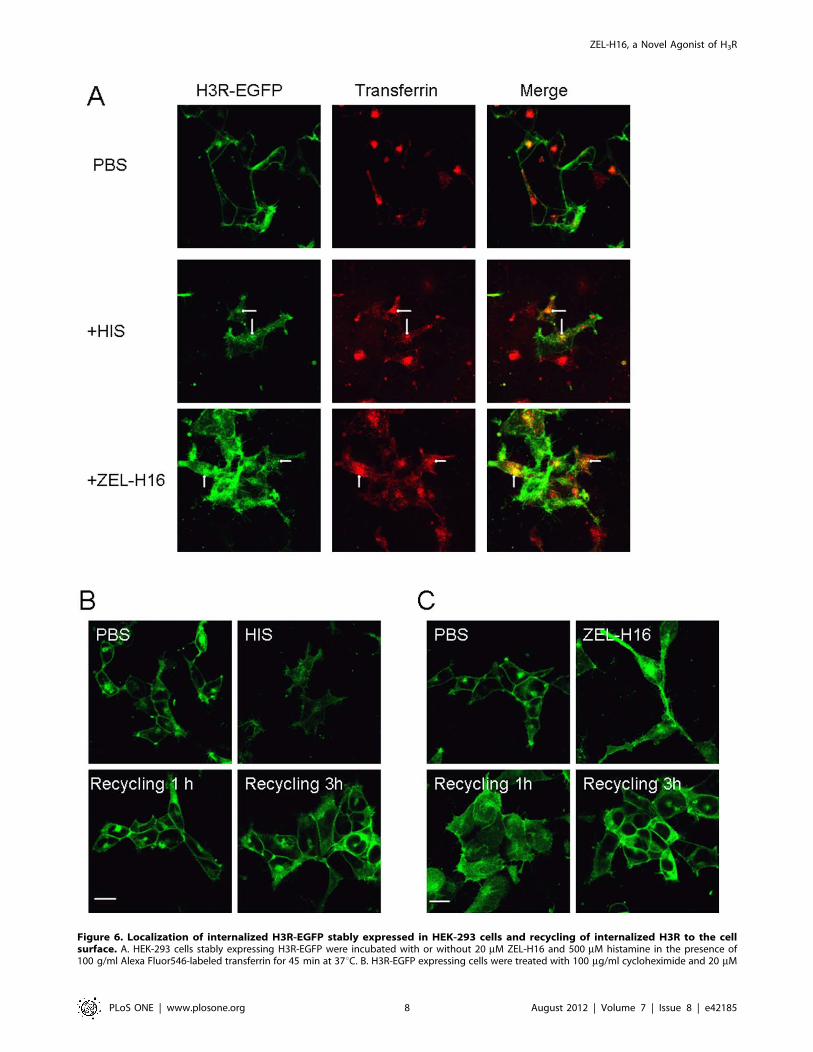
Figure 6. Localization of internalized H3R-EGFP stably expressed in HEK-293 cells and recycling of internalized H3R to the cellsurface. A. HEK-293 cells stably expressing H3R-EGFP were incubated with or without 20 mM ZEL-H16 and 500 mM histamine in the presence of100 g/ml Alexa Fluor546-labeled transferrin for 45 min at 37uC. B. H3R-EGFP expressing cells were treated with 100 mg/ml cycloheximide and 20 mM
ZEL-H16, a Novel Agonist of H3R
PLoS ONE | www.plosone.org 8 August 2012 | Volume 7 | Issue 8 | e42185
ZEL-H16 or 500 mM histamine at 37uC for 30 min, followed by the removal of residual agonists by washing, and further incubation in the presence ofcycloheximide for the indicated time periods. The internalized receptors were recycled to the plasma membrane within 1 h after histamine removaland 3 h after ZEL-H16 removal. All pictures shown are representative of at least three independent experiments.doi:10.1371/journal.pone.0042185.g006
Figure 7. Phosphorylation of ERK1/2 induced by ZEL-H16 in H3R-expressing HEK-293 cells. A, time-dependent phosphorylation of ERK1/2 induced by histamine or ZEL-H16. Following 3 h of serum starvation, cells were treated with 1 mM histamine or ZEL-H16 for the indicated timeperiod and cell lysates were analyzed for phosphorylated ERK (p-ERK) and total ERK (ERK) protein levels. Signals were quantified by densitometricimage analysis and p-ERK was normalized to a loading control (ERK). The signal at each point is expressed as the percentage of the maximal p-ERKsignal induced by histamine. A statistical analysis between ZEL-H16 and histamine for each time point in the kinetic graph was done using a t-test(PRISM software) (*p,0.05; **p,0.01). B, Concentration-dependent phosphorylation of ERK1/2 induced by histamine or ZEL-H16. After serumstarvation, cells were incubated with increasing concentrations of ZEL-H16 or histamine (10 nM to 100 mM) and cell lysates were analyzed for p-ERKand ERK levels. Concentration -dependent phosphorylation signals were quantified by densitometric analysis and p-ERK levels were normalized tototal ERK levels. The signal at each point is expressed as the percentage of the maximal p-ERK signal induced by histamine. C, The phosphorylation ofERK1/2 induced by ZEL-H16 could be entirely abolished by co-incubation with H3R antagonist thioperamide (***p,0.001). Data represent the mean6SE of three independent experiments.doi:10.1371/journal.pone.0042185.g007
ZEL-H16, a Novel Agonist of H3R
PLoS ONE | www.plosone.org 9 August 2012 | Volume 7 | Issue 8 | e42185
ConstructsThe hH1R and hH2R genes were cloned from HEK-293
genomic DNA by PCR. The hH3R gene was cloned using human
thalamus poly-A RNA (Clontech, Palo Alto, CA) with RT-PCR
methods. The hH4R gene was cloned from human bone marrow
Marathon-Ready cDNAs (Clontech, Basingstoke, U.K.) by PCR.
Primers were designed according to the published human
histamine receptor gene sequences (GenBank accession no.
X76786, M64799, AF140538, and AB044934). The expression
vectors of human DRD1, DRD2, and 5-HT1A were provided by
Dr. Jinpeng Sun (the School of Medicine of Shandong University),
and the expression vector of human a2AR was purchased from
GenScript USA Inc.(Nanjing, Jiangsu). All cDNAs were sequenced
and separately cloned into the pCMV-Flag and pEGFP-N1
expression vectors.
Cell Culture and Generation of Stable Cell LinesHEK-293 cells were cultured in Dulbecco’s modified Eagle’s
medium (DMEM) supplemented with 10% fetal bovine serum,
100 U/mL penicillin, and 100 mg/mL streptomycin in a humid-
ified atmosphere of 95% air and 5% CO2 at 37uC. For
transfection, H3R cDNA plasmid constructs were transfected or
co-transfected into HEK-293 cells using Lipofectamine 2000
according to the manufacturer’s instructions. Stable transfectants
were selected in the presence of 800 mg/mL G418.
Membrane PreparationHEK-293 cells stably expressing H3R were harvested and
resuspended in ice-cold 50 mM Tris-HCl (pH 7.4) containing
a complete protease inhibitor cocktail tablet and mixed by
inverting. The cell pellet was homogenized with a Polytron PT
1200 set at 4000 rpm and centrifuged at 1,000 g for 5 min. The
supernatant was then centrifuged twice at 50,000 g for 30 min at
4uC. Finally, the resulting pellet was gently suspended in 50 mM
Figure 8. Phosphorylation of ERK1/2 induced by ZEL-H16 in mouse cortical neurons. A, Time-dependent phosphorylation of ERK1/2induced by 5 mM ZEL-H16 in neonatal mouse cortical neuron cultures. B, The phosphorylation of ERK1/2 induced by 50 mM ZEL-H16 for 20 min wasmostly abolished by 100 ng/mL PTX or 10 mM thioperamide. Signals were quantified by densitometric image analysis and p-ERK was normalized toa loading control (ERK). The signal at each point is expressed as the percentage of the maximal p-ERK signal induced by ZEL-H16. Data represent themean 6SE of three independent experiments.doi:10.1371/journal.pone.0042185.g008
Figure 9. Effect of ZEL-H16-induced and histamine-inducedinhibition of the electrically induced contraction of the guinea-pig ileum. Each point is the mean 6 s.e.m. of three to six separateexperiments.doi:10.1371/journal.pone.0042185.g009
ZEL-H16, a Novel Agonist of H3R
PLoS ONE | www.plosone.org 10 August 2012 | Volume 7 | Issue 8 | e42185
Tris-HCl (pH 7.4) buffer and the membrane concentration was
determined using Bio-Rad protein assays.
The male rats (180 g–200 g) were killed by decapitation, and
the cerebral cortex was rapidly removed in ice-cold 50 mM Tris-
HCl (pH 7.4) containing a complete protease inhibitor cocktail
tablet. The cerebral cortex was homogenized with a Polytron (at
maximum setting, 3610 sec) and centrifuged twice at 1,000 g at
4uC to separate the nuclear fraction and cell debris. The resulting
supernatants were combined and then centrifuged twice at
50,000 g for 20 min at 4uC to obtain tne membrane fraction.
Protein concentration was determined using Bio-Rad protein
assays.
Competitive Binding AssaysThe membranes (50 mg of protein) were incubated with [3H] N-
a-methylhistamine at various concentrations (from 0.03 nM to
50 nM) for saturation binding assays. For competitive binding
assays, the membranes (50 mg of protein) were incubated with
10 nM [3H] N-a-methylhistamine for recombinant hH3R or 2nM
[3H] N-a-methylhistamine for rat brain H3R in the presence of
various compound concentrations in the buffer (50 mM Tris–HCl,
pH 7.4). The reaction mixtures were incubated for 90–120 min at
room temperature to achieve binding equilibrium. Following three
washes, the membrane-bound radioactivity was measured on
a Topcount (PerkinElmer) at 20uC. Non-specific binding was
determined in the presence of excessive cold histamine (final conc.,
300 mM). IC50 values were converted to Ki values using the
Cheng-Prusoff equation. Each experiment was performed in
duplicate and repeated three to six times. All data were analyzed
by nonline arregression using GraphPad Prism version 4.0
software.
CRE-driven Reporter Gene AssayStable HEK-293 cells co-transfected with H3R and pCRE-Luc
were seeded in a 48-well plate overnight and were grown to
90695% confluence. Next, the cells were stimulated with 10 mMforskolin or 10 mM forskolin plus different concentrations of
histamine or compound in serum-free DMEM, and the cells were
incubated for 5 h at 37uC. Luciferase activity was detected using
a firefly luciferase kit (Promega, Madison, WI). When required,
cells were treated overnight with or without PTX (100 ng/ml) in
serum-free DMEM before the experiment.
Intracellular Calcium MeasurementThe stably H1R-expressing HEK-293 cells were harvested with
Cell Stripper (Mediatech, Herndon, VA), washed twice with
phosphate-buffered saline (PBS), and resuspended in Hanks’
balanced salt solution (140 mM NaCl, 5 mM KCl, 10 mM
HEPES, pH 7.4, 1 mM CaCl2, 1 mM MgCl2, 1 mg/ml glucose)
containing 0.025% bovine serum albumin. The cells were then
loaded with 2 mM Fura-2 acetoxymethyl ester derivative (Fura-2/
AM) (Molecular Probes, Eugene, OR) for 30 min at 37uC. Cellswere washed once in Hanks’ solution, resuspended in Hanks’,
incubated at room temperature for 15 min, washed twice in
Hanks’ solution, and then resuspended in Hanks’ at a concentra-
tion of 36106 cells/ml. A typical experiment contained
1.06105 cells/100 ml in a well of 96-well plate. These cells were
stimulated with the indicated concentrations of compounds. HEK-
293 cells stimulated with 3 mM histamine or ZEL-H16 were used
as a negative control respectively. Calcium flux was measured
using excitation at 340 and 380 nm in a Infinite 200 PRO
multifunctional microplate reader (Tecan Austria GmbH, Grodig,
AT ) and [Ca2+]i was measured using the 340/380 excitation ratio
(R 340/380).
Internalization Assay by Confocal MicroscopyHEK-293 cells stably expressing H3R-EGFP were seeded in
covered glass-bottom 6-well plates. After 24 h, cells were treated
with histamine or ZEL-H16 for the indicated times at 37uC. Afterfixing the cells with 4% paraformaldehyde for 10 min, cells were
mounted in mounting reagent (DTT/PBS/glycerol). Confocal
images were taken on a Zeiss LSM 510 microscope with an
attached Axiovert 200 microscope and LSM5 computer system.
Images were collected using QED camera software and processed
with Adobe Photoshop.
Measurement of Cell Surface Receptors by ELISAThe cell surface expression of H3R was quantitatively assessed
by ELISA and performed as described previously [52]. Briefly,
HEK-293 cells stably transfected with the pCMV-H3R construct
were seeded in 48-well dishes coated with poly-L-lysine. The next
day, the cells were stimulated with the indicated concentrations of
histamine and ZEL-H16 for the indicated times. Medium was
aspirated and the cells were washed once with Tris-buffered saline
(TBS). After fixing the cells for 5 min at room temperature with
3.7% formaldehyde in TBS, the cells were washed 3 times with
TBS and then blocked for 45 min with 1% bovine serum
albumin/TBS. Cells were then incubated for 1 h with an alkaline
phosphatase-conjugated monoclonal antibody directed against the
Flag epitope and diluted 1:1,000. Cells were washed 3 times, and
antibody binding was visualized by adding 0.25 mL of an alkaline
phosphatase substrate (Bio-Rad). Development was stopped by
adding 0.1 mL of the substrate to a 96-well microtiter plate
containing 0.1 mL of 0.4 M NaOH. Plates were read at 405 nm
in a microplate reader (Bio-Rad) using Microplate Manager
software.
Western Blot AnalysisHEK-293 cells stably expressing human H3R were grown in
a 6-well plate and were serum starved 2 h in serum-free culture
medium prior to stimulation. Cells were stimulated with the
indicated H3R ligands at the designated concentrations. Incuba-
tions were stopped at the indicated times by removing the
stimulation medium by aspiration. The cells were washed in ice-
cold PBS and subsequently lysed in 100 ml of lysis buffer [20 mM
HEPES (pH 7.5), 10 mM EDTA, 150 mM NaCl, 1% Triton X-
100, and one tablet of complete protease inhibitor (Roche,
Indianapolis, IN) per 50 mL] at 4uC on a rocker for 30 min. The
lysates were centrifuged and separated by sodium dodecyl sulfate-
polyacrylamide (12%) gel electrophoresis (SDS-PAGE) and blotted
onto polyvinylidene difluoride membranes. Membranes were
blocked for 1 h at room temperature in TBST (10 mmol/L Tris,
150 mmol/L NaCl, 0.1% Tween-20, pH 8.0) buffer containing
5% (v/w) skim milk. Membranes were probed overnight with
primary antibodies against p-ERK1/2 (1:1,000; Cell Signaling,
Danvers, MA) in TBST containing 5% (v/w) BSA, and then
probed with horseradish peroxidase-labeled secondary antibody
(1:5,000) for 1 h at room temperature in TBST containing 5% (v/
w) milk powder. Immunoreactivity was detected by ECL assays.
The blots were stripped and reprobed using antibodies against
total ERK1/2 (1:2,000) (Cell Signaling, Danvers, MA) as a control.
Analysis of ERK Phosphorylation in Primary Cultures ofCortical NeuronsNeuronal cortical cultures were prepared from neonatal ICR
mice. Cerebral cortices were dissociated in sterile Dulbecco’s
phosphate-buffered saline (D-PBS; Sigma-Aldrich, St. Louis, MO),
and the neurons were isolated in the same medium containing
ZEL-H16, a Novel Agonist of H3R
PLoS ONE | www.plosone.org 11 August 2012 | Volume 7 | Issue 8 | e42185
0.5% trypsin at 37uC for 10 min. After centrifugation, dissociated
neurons were re-suspended in neurobasal medium (NBM; Gibco,
Carlsbad, CA) supplemented with 2% B-27 and 0.5 mmol/L-
glutamine (Gibco, Carlsbad, CA) and then plated on 48-well plates
at a density of approximately 1.36106 per well. After 7 days of
culture, the neurons were serum starved for approximately 3 h
prior to drug treatment. After the cells were stimulated by H3R
ligands, the neurons were harvested in lysis buffer and proteins
were separated using SDS-PAGE and then transferred onto PVDF
membranes. Immunoblotting was performed as previously de-
scribed.
Guinea-pig Ileum AssayThe procedure used was as described previously [53]. Adult
male guinea pigs (300–500 g) were killed by cervical dislocation.
The ileum was removed at a point 20 cm from the caecum and
flushed with and placed in modified K–H buffer of following
composition: 118 mM NaCl, 5.9 mM KCl, 1.2 mM CaCl2,
1.2 mM MgSO4, 1 mM Na2HPO4, 25 mM NaHCO3 and
10 mM D-glucose. Ileum segments (2.5–3 cm) were suspended
in 20 ml organ baths containing K–H buffer maintained at 37uCand gassed with 95% O2/5% CO2. Contractile activity under
stimulation (rectangular pulses of 15 V, 0.5 ms, and 0.1 Hz) was
recorded using isometric transducers (Grass FTO3). Concentra-
tion–response effects of histamine or ZEL-H16 were obtained in
different each tissue. Mepyramine (3 mM) and famotidine
(10 mM) (Sigma-Aldrich, St. Louis, MO) were added to the K–
H buffer to block postsynaptic H1 and presynaptic H2 receptors,
respectively.
Data AnalysisSigmoidal agonist concentration-response curves (in the pres-
ence and absence of antagonists) were created through computer-
assisted nonlinear regression using the GraphPad Prism program
(GraphPad Software, San Diego, CA, USA). Schild slopes (n) were
constructed from linear regression of the Schild equation as the
following:
Log(DR{1)~n log½B�{ log (KD)
DR is the dose-ratio as the EC50 in the presence of antagonist
divided by the EC50 in the absence of antagonist. B is the
concentration of the antagonist. These points were then fitted to
a straight line. A slope of 1 then indicates competitive antagonism
[54]. All data are presented as mean 6 SEM. Then in the text
refers to the number of separate experiments.
Supporting Information
Figure S1 A, the 7 th –day’s cortical neurons cultures of
neonatal mouse. B, RT-PCR detection of mouse H3R of the 7 th –
day’s cortical neurons cultures of neonatal mouse.
(TIF)
Acknowledgments
The authors of this paper would like to thank Ms. Aiping Shao, Mrs.
Hanmin Chen and Ming Ding for their technical assistance and equipment
usage. We are grateful to Dr. Jinpeng Sun for expression vectors of DRD1,
DRD2 and 5-HT1A.
Author Contributions
Conceived and designed the experiments: NZ YH. Performed the
experiments: Y. Shi TZ XC DY Y. Sun. Analyzed the data: Y. Shi NZ.
Contributed reagents/materials/analysis tools: RS FY YX. Wrote the
paper: NZ Y. Shi RS.
References
1. Hough LB (2001) Genomics meets histamine receptors: new subtypes, new
receptors. Mol Pharmacol 59: 415–419.
2. Leurs R, Smit MJ, Timmerman H (1995) Molecular pharmacological aspects of
histamine receptors. Pharmacol Ther 66: 413–463.
3. Gantz I, Schaffer M, DelValle J, Logsdon C, Campbell V, et al. (1991)
Molecular cloning of a gene encoding the histamine H2 receptor. Proc Natl
Acad Sci U S A 88: 429–433.
4. Yamashita M, Fukui H, Sugama K, Horio Y, Ito S, et al. (1991) Expression
cloning of a cDNA encoding the bovine histamine H1 receptor. Proc Natl Acad
Sci U S A 88: 11515–11519.
5. Ares JJ, Outt PE (1998) Gastroprotective agents for the prevention of NSAID-
induced gastropathy. Curr Pharm Des 4: 17–36.
6. Ciprandi G, Buscaglia S, Cerqueti PM, Canonica GW (1992) Drug treatment of
allergic conjunctivitis. A review of the evidence. Drugs 43: 154–176.
7. Oda T, Morikawa N, Saito Y, Masuho Y, Matsumoto S (2000) Molecular
cloning and characterization of a novel type of histamine receptor preferentially
expressed in leukocytes. J Biol Chem 275: 36781–36786.
8. Lim HD, Smits RA, Leurs R, De Esch IJ (2006) The emerging role of the
histamine H4 receptor in anti-inflammatory therapy. Curr Top Med Chem 6:
1365–1373.
9. Arrang JM, Garbarg M, Schwartz JC (1983) Auto-inhibition of brain histamine
release mediated by a novel class (H3) of histamine receptor. Nature 302: 832–
837.
10. Lovenberg TW, Roland BL, Wilson SJ, Jiang X, Pyati J, et al. (1999) Cloning
and functional expression of the human histamine H3 receptor. Mol Pharmacol
55: 1101–1107.
11. Korte A, Myers J, Shih NY, Egan RW, Clark MA (1990) Characterization and
tissue distribution of H3 histamine receptors in guinea pigs by N alpha-
methylhistamine. Biochem Biophys Res Commun 168: 979–986.
12. Drutel G, Peitsaro N, Karlstedt K, Wieland K, Smit MJ, et al. (2001)
Identification of rat H3 receptor isoforms with different brain expression and
signaling properties. Mol Pharmacol 59: 1–8.
13. Martinez-Mir MI, Pollard H, Moreau J, Arrang JM, Ruat M, et al. (1990) Three
histamine receptors (H1, H2 and H3) visualized in the brain of human and non-
human primates. Brain Res 526: 322–327.
14. Bongers G, Bakker RA, Leurs R (2007) Molecular aspects of the histamine H3receptor. Biochem Pharmacol 73: 1195–1204.
15. Coruzzi G, Bertaccini G, Schwartz JC (1991) Evidence that histamine H3receptors are involved in the control of gastric acid secretion in the conscious cat.
Naunyn Schmiedebergs Arch Pharmacol 343: 225–227.
16. Schlicker E, Kathmann M, Detzner M, Exner HJ, Gothert M (1994) H3
receptor-mediated inhibition of noradrenaline release: an investigation into theinvolvement of Ca2+ and K+ ions, G protein and adenylate cyclase. Naunyn
Schmiedebergs Arch Pharmacol 350: 34–41.
17. Schlicker E, Malinowska B, Kathmann M, Gothert M (1994) Modulation of
neurotransmitter release via histamine H3 heteroreceptors. Fundam ClinPharmacol 8: 128–137.
18. Bonaventure P, Letavic M, Dugovic C, Wilson S, Aluisio L, et al. (2007)Histamine H3 receptor antagonists: from target identification to drug leads.
Biochem Pharmacol 73: 1084–1096.
19. Celanire S, Wijtmans M, Talaga P, Leurs R, de Esch IJ (2005) Keynote review:
histamine H3 receptor antagonists reach out for the clinic. Drug Discov Today10: 1613–1627.
20. Esbenshade TA, Fox GB, Cowart MD (2006) Histamine H3 receptorantagonists: preclinical promise for treating obesity and cognitive disorders.
Mol Interv 6: 77–88, 59.
21. Wijtmans M, Leurs R, de Esch I (2007) Histamine H3 receptor ligands break
ground in a remarkable plethora of therapeutic areas. Expert Opin InvestigDrugs 16: 967–985.
22. Francis H, Franchitto A, Ueno Y, Glaser S, DeMorrow S, et al. (2007) H3histamine receptor agonist inhibits biliary growth of BDL rats by downregulation
of the cAMP-dependent PKA/ERK1/2/ELK-1 pathway. Lab Invest 87: 473–
487.
23. Millan-Guerrero RO, Isais-Millan R, Benjamin TH, Tene CE (2006) Nalpha-methyl histamine safety and efficacy in migraine prophylaxis: phase III study.
Can J Neurol Sci 33: 195–199.
24. Yoshimoto R, Miyamoto Y, Shimamura K, Ishihara A, Takahashi K, et al.
(2006) Therapeutic potential of histamine H3 receptor agonist for the treatment
of obesity and diabetes mellitus. Proc Natl Acad Sci U S A 103: 13866–13871.
25. Rouleau A, Garbarg M, Ligneau X, Mantion C, Lavie P, et al. (1997)
Bioavailability, antinociceptive and antiinflammatory properties of BP 2–94,
ZEL-H16, a Novel Agonist of H3R
PLoS ONE | www.plosone.org 12 August 2012 | Volume 7 | Issue 8 | e42185
a histamine H3 receptor agonist prodrug. J Pharmacol Exp Ther 281: 1085–
1094.26. Rouleau A, Stark H, Schunack W, Schwartz JC (2000) Anti-inflammatory and
antinociceptive properties of BP 2–94, a histamine H(3)-receptor agonist
prodrug. J Pharmacol Exp Ther 295: 219–225.27. Schlicker E, Malinowska B, Kathmann M (1993) CGP 35348 blocks
noradrenaline-release-inhibiting GABAB receptors in the pig retina, rat venacava and pithed rat vasculature. Pharmacology 47: 111–116.
28. Ferrada C, Ferre S, Casado V, Cortes A, Justinova Z, et al. (2008) Interactions
between histamine H3 and dopamine D2 receptors and the implications forstriatal function. Neuropharmacology 55: 190–197.
29. Cecchi M, Giorgetti M, Bacciottini L, Giovannini MG, Blandina P (1998)Increase of acetylcholine release from cortex of freely moving rats by
administration of histamine into the nucleus basalis magnocellularis. InflammRes 47 Suppl 1: S32–33.
30. Kraus MM, Fischer H, Tran MH, Philippu A, Prast H (2001) Modulation of
acetylcholine release by histamine in the nucleus accumbens. Inflamm Res 50Suppl 2: S74–75.
31. Korotkova TM, Sergeeva OA, Ponomarenko AA, Haas HL (2005) Histamineexcites noradrenergic neurons in locus coeruleus in rats. Neuropharmacology
49: 129–134.
32. Fink K, Schlicker E, Neise A, Gothert M (1990) Involvement of presynaptic H3receptors in the inhibitory effect of histamine on serotonin release in the rat
brain cortex. Naunyn Schmiedebergs Arch Pharmacol 342: 513–519.33. Threlfell S, Cragg SJ, Kallo I, Turi GF, Coen CW, et al. (2004) Histamine H3
receptors inhibit serotonin release in substantia nigra pars reticulata. J Neurosci24: 8704–8710.
34. Korotkova TM, Haas HL, Brown RE (2002) Histamine excites GABAergic cells
in the rat substantia nigra and ventral tegmental area in vitro. Neurosci Lett 320:133–136.
35. Garduno-Torres B, Trevino M, Gutierrez R, Arias-Montano JA (2007) Pre-synaptic histamine H3 receptors regulate glutamate, but not GABA release in rat
thalamus. Neuropharmacology 52: 527–535.
36. Ohkubo T, Shibata M, Inoue M, Kaya H, Takahashi H (1995) Regulation ofsubstance P release mediated via prejunctional histamine H3 receptors.
Eur J Pharmacol 273: 83–88.37. Arrang JM, Garbarg M, Lancelot JC, Lecomte JM, Pollard H, et al. (1987)
Highly potent and selective ligands for histamine H3-receptors. Nature 327:117–123.
38. Arrang JM, Garbarg M, Schwartz JC (1987) Autoinhibition of histamine
synthesis mediated by presynaptic H3-receptors. Neuroscience 23: 149–157.39. Garbarg M, Arrang JM, Rouleau A, Ligneau X, Tuong MD, et al. (1992) S-[2-
(4-imidazolyl)ethyl]isothiourea, a highly specific and potent histamine H3receptor agonist. J Pharmacol Exp Ther 263: 304–310.
40. Vollinga RC, de Koning JP, Jansen FP, Leurs R, Menge WM, et al. (1994) A
new potent and selective histamine H3 receptor agonist, 4-(1H-imidazol-4-ylmethyl)piperidine. J Med Chem 37: 332–333.
41. Liu C, Ma X, Jiang X, Wilson SJ, Hofstra CL, et al. (2001) Cloning and
pharmacological characterization of a fourth histamine receptor (H(4)) expressed
in bone marrow. Mol Pharmacol 59: 420–426.
42. Nguyen T, Shapiro DA, George SR, Setola V, Lee DK, et al. (2001) Discovery
of a novel member of the histamine receptor family. Mol Pharmacol 59: 427–
433.
43. Kitbunnadaj R, Zuiderveld OP, De Esch IJ, Vollinga RC, Bakker R, et al.
(2003) Synthesis and structure-activity relationships of conformationally
constrained histamine H(3) receptor agonists. J Med Chem 46: 5445–5457.
44. Schlicker E, Kathmann M, Bitschnau H, Marr I, Reidemeister S, et al. (1996)
Potencies of antagonists chemically related to iodoproxyfan at histamine H3
receptors in mouse brain cortex and guinea-pig ileum: evidence for H3 receptor
heterogeneity? Naunyn Schmiedebergs Arch Pharmacol 353: 482–488.
45. Sasse A, Stark H, Reidemeister S, HuE ls A, Elz S, et al. (1999) Novel partial
agonists for the histamine H(3) receptor with high in vitro and in vivo activity.
J Med Chem 42: 4269–4274.
46. Krueger KM, Witte DG, Ireland-Denny L, Miller TR, Baranowski JL, et al.
(2005) G protein-dependent pharmacology of histamine H3 receptor ligands:
evidence for heterogeneous active state receptor conformations. J Pharmacol
Exp Ther 314: 271–281.
47. Gbahou F, Davenas E, Morisset S, Arrang JM (2010) Effects of betahistine at
histamine H3 receptors:mixed inverse agonism/agonism in vitro and partial
inverse agonism in vivo. J Pharmacol ExpTher 334: 945–954.
48. Sasse A, Stark H, Ligneau X, Elz S, Reidemeister S, et al. (2000) (Partial)
agonist/antagonist properties of novel diarylalkyl carbamates on histamine H3
receptors. Bioorg Med Chem 8: 1139–1149.
49. Ferguson SS (2001) Evolving concepts in G protein-coupled receptor
endocytosis: the role in receptor desensitization and signaling. Pharmacol Rev
53: 1–24.
50. Mack M, Luckow B, Nelson PJ, Cihak J, Simmons G, et al. (1998)
Aminooxypentane-RANTES induces CCR5 internalization but inhibits recy-
cling: a novel inhibitory mechanism of HIV infectivity. J Exp Med 187: 1215–
1224.
51. Signoret N, Pelchen-Matthews A, Mack M, Proudfoot AE, Marsh M (2000)
Endocytosis and recycling of the HIV coreceptor CCR5. J Cell Biol 151: 1218–
1294.
52. Orsini MJ, Parent JL, Mundell SJ, Marchese A, Benovic JL (1999) Trafficking of
the HIV coreceptor CXCR4. Role of arrestins and identification of residues in
the c-terminal tail that mediate receptor internalization. J Biol Chem 274:
31076–31086.
53. Ligneau X, Lin J, Vanni-Mercier G, Jouvet M, Muir JL, et al. (1998)
Neurochemical and behavioral effects of ciproxifan, a potent histamine H3-
receptor antagonist. J Pharmacol Exp Ther 287: 658–666.
54. Arunlakshana O, Schild HO (1959) Some quantitative uses of drug antagonists.
Br J Pharmacol Chemother 14: 48–58.
ZEL-H16, a Novel Agonist of H3R
PLoS ONE | www.plosone.org 13 August 2012 | Volume 7 | Issue 8 | e42185


![Phoenix Times - H16 - First Edition[1]](https://img.pdfslide.us/doc/110x75/577d1f181a28ab4e1e8fdcc2/phoenix-times-h16-first-edition1.jpg)

