Embed Size (px)
Citation preview
L E T T E R S
Retinitis pigmentosa is an untreatable, inherited retinal diseasethat leads to blindness. The disease initiates with the loss ofnight vision due to rod photoreceptor degeneration, followedby irreversible, progressive loss of cone photoreceptor1–3. Coneloss is responsible for the main visual handicap, as cones areessential for day and high-acuity vision4. Their loss is indirect,as most genes associated with retinitis pigmentosa are notexpressed by these cells. We previously showed that factorssecreted from rods are essential for cone viability5–8. Here weidentified one such trophic factor by expression cloning andnamed it rod-derived cone viability factor (RdCVF). RdCVF isa truncated thioredoxin-like protein specifically expressed byphotoreceptors. The identification of this protein offers newtreatment possibilities for retinitis pigmentosa.
We used a viability assay based on cone-enriched primary culturesfrom chicken embryos9 for expression cloning. Unlike those of mam-mals, bird retinas are cone-dominated. Cones represent 60–80% of thetotal population in cultured cells8. Once cultured, these cells degener-ate over a few days, but adding conditioned medium from wild-typemouse retinal explants delays this loss8. We carried out a screen to iso-late factors that could support cone survival.
We constructed a cDNA expression library from neural retinas of5-week-old wild-type mice and we purified plasmid DNA from poolsof 100 individual clones and used them to transfect COS-1 cells. Weadded conditioned medium from transfected COS cells to chickenretinal cultures seeded in 96-well plates. After 7 d of culture, we car-ried out an automated viability assay and we screened 2,100 pools,corresponding to 210,000 individual clones. Pool 939 contained twiceas many living cells as the negative controls (Fig. 1). We isolated clone939.09.08 by limiting dilution and found that it contained a 502-bpinsert with an open reading frame encoding a putative polypeptide of109 amino acids. We named this protein rod-derived cone viabilityfactor (RdCVF, international patent no. PCT/EP 02/03810;Supplementary Fig. 1 online).
We then expressed and purified RdCVF as a fusion protein withgluthatione-S-transferase (GST; Fig. 2a). We incubated cone-enrichedcultures with increasing amounts of GST-RdCVF or GST alone.Adding 10 µg ml–1 of purified GST-RdCVF doubled the number of liv-ing cells per plate. The rescue activity of this factor increased withhigher protein concentration (Fig. 2b).
We next cultured retinal explants from 5-week-old rd1 mice (lackingrods) for 7 d with COS cells transfected with cDNA encoding RdCVF(from clone 939.09.08). We labeled cone cells with cone-specific peanutagglutinin (PNA) and counted them using stereological methods7. Thenumber of cones per field was higher in retinal explants incubated withcells transfected with RdCVF than in controls (Fig. 2c). The amplitudeof the rescue effect was 40% when comparing the number of cones lostduring the 1-week period, a range of efficacy similar to that observedwhen wild-type retinas were used as a source of RdCVF activity7.RdCVF was thus able to slow cone degeneration in both chick andmouse models8. We cultured retinal explants from 10-d-old rd1 mice(with rods) for 7 d with COS cells transfected with cDNA encodingRdCVF and measured the thickness of the outer retina. We observed nostatistically significant differences in the thickness of the outer retinabetween mice transfected with RdCVF and controls, indicating that theprotective effects of RdCVF might not extend to degenerating rods(Fig. 2d).
We next tested the ability of RdCVF antibodies to block endoge-nous activity. We prepared conditioned medium from retinalexplants of 5-week-old wild-type mice and immunodepleted themwith antibodies to RdCVF or control antibodies. We incubated retinalexplants of 5-week-old rd1 mice (lacking rods) for 7 d with condi-tioned medium and then labeled and counted the cones. The numberof cones was higher when rd1 explants were incubated with condi-tioned medium preincubated with control polyclonal antibodies thanwhen they were incubated with medium alone (Fig. 2e). The condi-tioned medium did not stimulate cone survival in rd1 explants whenRdCVF was specifically removed by immunodepletion. These resultsindicate that RdCVF is required for cone rescue in cultured explants.
1Laboratoire de Physiopathologie Cellulaire et Moléculaire et de la Rétine, Inserm U592, Université Pierre et Marie Curie, Hôpital St-Antoine, 184 rue du FaubourgSt-Antoine, 75571, Paris cedex 12, France. 2Institut de Génétique et de Biologie Moléculaire et Cellulaire, 1 rue Laurent Fries, 67404 Illkirch, France. 3NovartisPharma AG Ophthalmology Research WKL-127.1.04 CH-4002 Basel, Switzerland. 4Institute of Ophthalmology, University College of London, UK. 5These authorscontributed equally to this work. Correspondence should be addressed to T.L. ([email protected]).
Published online 27 June 2004; doi:10.1038/ng1386
Identification and characterization of rod-derived coneviability factorThierry Léveillard1,5, Saddek Mohand-Saïd1, Olivier Lorentz1, David Hicks1, Anne-Claire Fintz1,Emmanuelle Clérin1, Manuel Simonutti1, Valérie Forster1, Nükhet Cavusoglu1, Frédéric Chalmel2, Pascal Dollé2,Olivier Poch2, George Lambrou3 & José-Alain Sahel1,4,5
NATURE GENETICS VOLUME 36 | NUMBER 7 | JULY 2004 755
©20
04 N
atur
e P
ublis
hing
Gro
up
http
://w
ww
.nat
ure.
com
/nat
ureg
enet
ics

L E T T E R S
When antibodies to RdCVF were injected into the subretinal space ofthe wild-type mouse, the number of cones decreased 7 d after theinjection, as compared with mice similarly injected with control anti-bodies (data not shown), but the difference was not statistically signif-icant. Complete blocking of RdCVF signaling in vivo requires geneinactivation by homologous recombination.
To assess the effect of RdCVF in vivo, we injected 1 µl (100 ng) ofpurified GST-RdCVF in the subretinal space of 35-d-old rd1 mice and
repeated the injection 7 d later. We killed themice at 7 weeks of age (Fig. 2f). The averagenumber of cones per mm2 was higher in miceinjected with the RdCVF fusion protein(4,500) than in mice injected with either GSTalone (3,800) or with phosphate-bufferedsaline (PBS; 3,500). Therefore, injection ofRdCVF can prevent 40% of cones fromdegenerating in the rd1 mouse over a periodof 2 weeks.
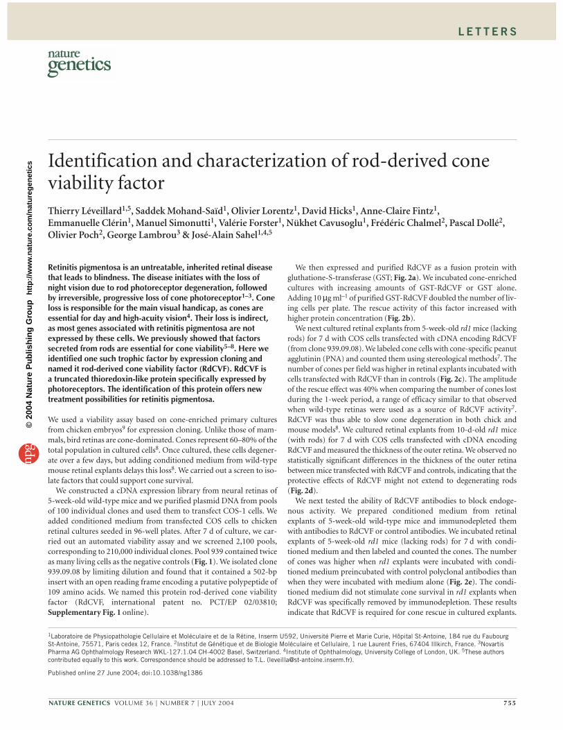
Northern-blot analysis detected two dis-tinct RdCVF mRNAs of 2.3 and 2.7 kb in onlythe retina and not in any other tissue tested(Fig. 3a). The pattern of RdCVF mRNAexpression shown by real-time RT-PCRmatched that of rhodopsin, largely increasingduring maturation of photoreceptors in thewild-type mouse. In the rd1 mouse, however,rod degeneration, as indicated by decreasedrhodopsin mRNA levels, was accompaniedby a marked decrease in RdCVF expression(Fig. 3b). The two RdCVF mRNAs are proba-bly a result of alternative splicing. Whentested by real-time RT-PCR, the most abun-dant mRNA encoded the factor that we iso-lated (Fig. 3c). The level of expression of
RdCVF in the whole retina and in the photoreceptor layer (isolated bysectioning the mouse retina with a vibratome10) were similar. Thissuggests that RdCVF expression is mainly restricted to the outernuclear layer containing the rods and cones (Fig. 3d). RdCVF is alsoexpressed in cultures of pure photoreceptors (Fig. 3e).
Polyclonal antibodies raised against the N-terminal and C-terminalregions of the RdCVF sequence (RdCVF-N and RdCVF-C, respec-tively) detected the same two bands (17 and 34 kDa, Fig. 4a). In vitro,translated RdCVF consists only of a 17-kDa band, possibly resulting
756 VOLUME 36 | NUMBER 7 | JULY 2004 NATURE GENETICS
Cell counting
Division of the selected pools into subpools:
of 10 for second round isolated clones for third round
Selection of candidate pools
Plasmid puri fication by pools of 100 clones
Collection of con ditioned medium
Transfection in COS-1 cells
Cone-enriched cultures
Live cells Dead cells
1 1 1 1 2 C 2 2 2 3…
Mouse retinal cDNA lib rary
RdCVF
0.0
0.4
0.8
1.2
1.6
2.0
921
922
923
924
925
926
927
928
929
930
931
932
933
934
935
936
937
938
939
940
Rel
ativ
e to
con
trol
0102030405060
Cel
l co
unt
a
0
200
400
600
800
1,000
1,200
1,400
Live
cel
ls
b36
13
GSTGST-R
dCVF
RdCVF
GST
GST-RdC
VFGST
GST-RdC
VF
5 µg/ml 10 µg/ml
P < 0.05
Con
es/m
m2
Con
es/m
m2
Con
es/m
m2
c
C RdCVF1,500
2,000
2,500
3,000
3,500
4,000
4,500
5,000P < 0.05
e
CContro
l
RdCVF-N
f
CGST
GST-RdC
VF1,500
1,700
1,900
2,100
2,300
2,500
1,500
2,000
2,500
3,000
3,500
4,000
4,500
5,000
0
5
10
15
20
25
30
35
40
C RdCVF
Out
er n
ucle
ar la
yer
thic
knes
s (
µm)
d
P < 0.05
P < 0.05
P < 0.05
Figure 1 Schematic representation of the expression cloning strategy. Cell count indicates the numberof live cells. ‘Relative to control’ is the ratio of live cells from the indicated clone to live cells in wellstransfected with pcDNA3.
Figure 2 Viability activity of RdCVF. (a) Gelanalysis of purified GST (lane 1), GST-RdCVF(lane 2) and thrombin-cleaved and purifiedRdCVF (lane 3). Arrows on the left indicatethe RdCVF proteins; the open arrowheadindicates GST. Molecular weight markers(kDa) are shown on the right. (b) Activity ofGST and GST-RdCVF in chicken cone-enrichedcultures. The assay represents the sum of eightindependent experiments. (c) Effect of cDNAencoding RdCVF transfected into COS-1 cellson cone viability in rd1 mouse retinal explants.C, pcDNA3; RdCVF, pcDNA-RdCVF. (d) Effectof cDNA encoding RdCVF transfected intoCOS-1 cells on rod viability in rd1 mouseretinal explants. C, pcDNA3; RdCVF, pcDNA-RdCVF. (e) Immunodepletion with antibodiesto RdCVF-N. C, chemically defined medium;control, conditioned medium from wild-typeretinal explants after immunodepletion withcontrol antibodies; RdCVF-N, conditionedmedium from wild-type retinal explants afterimmunodepletion with RdCVF antibodies.(f) Subretinal injection of 1 µl of PBS (C),GST (100 ng) and GST-RdCVF (100 ng).
©20
04 N
atur
e P
ublis
hing
Gro
up
http
://w
ww
.nat
ure.
com
/nat
ureg
enet
ics
L E T T E R S
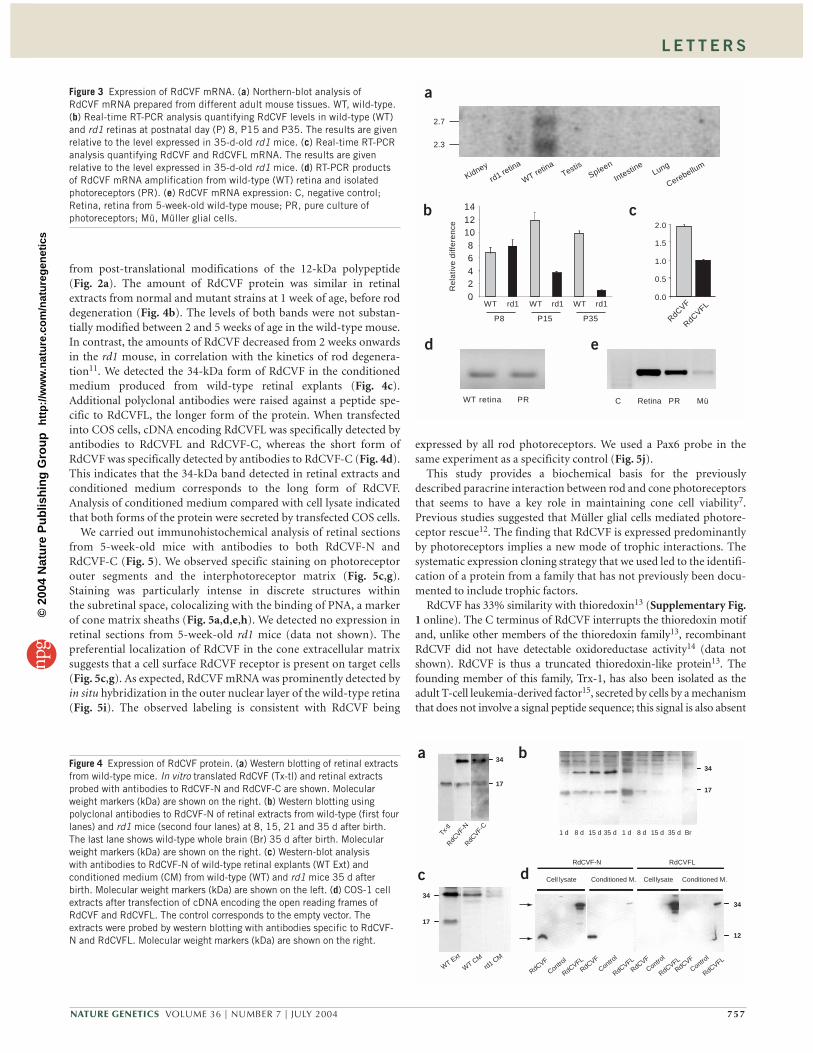
from post-translational modifications of the 12-kDa polypeptide(Fig. 2a). The amount of RdCVF protein was similar in retinalextracts from normal and mutant strains at 1 week of age, before roddegeneration (Fig. 4b). The levels of both bands were not substan-tially modified between 2 and 5 weeks of age in the wild-type mouse.In contrast, the amounts of RdCVF decreased from 2 weeks onwardsin the rd1 mouse, in correlation with the kinetics of rod degenera-tion11. We detected the 34-kDa form of RdCVF in the conditionedmedium produced from wild-type retinal explants (Fig. 4c).Additional polyclonal antibodies were raised against a peptide spe-cific to RdCVFL, the longer form of the protein. When transfectedinto COS cells, cDNA encoding RdCVFL was specifically detected byantibodies to RdCVFL and RdCVF-C, whereas the short form ofRdCVF was specifically detected by antibodies to RdCVF-C (Fig. 4d).This indicates that the 34-kDa band detected in retinal extracts andconditioned medium corresponds to the long form of RdCVF.Analysis of conditioned medium compared with cell lysate indicatedthat both forms of the protein were secreted by transfected COS cells.
We carried out immunohistochemical analysis of retinal sectionsfrom 5-week-old mice with antibodies to both RdCVF-N andRdCVF-C (Fig. 5). We observed specific staining on photoreceptorouter segments and the interphotoreceptor matrix (Fig. 5c,g).Staining was particularly intense in discrete structures withinthe subretinal space, colocalizing with the binding of PNA, a markerof cone matrix sheaths (Fig. 5a,d,e,h). We detected no expression inretinal sections from 5-week-old rd1 mice (data not shown). Thepreferential localization of RdCVF in the cone extracellular matrixsuggests that a cell surface RdCVF receptor is present on target cells(Fig. 5c,g). As expected, RdCVF mRNA was prominently detected byin situ hybridization in the outer nuclear layer of the wild-type retina(Fig. 5i). The observed labeling is consistent with RdCVF being
expressed by all rod photoreceptors. We used a Pax6 probe in thesame experiment as a specificity control (Fig. 5j).
This study provides a biochemical basis for the previouslydescribed paracrine interaction between rod and cone photoreceptorsthat seems to have a key role in maintaining cone cell viability7.Previous studies suggested that Müller glial cells mediated photore-ceptor rescue12. The finding that RdCVF is expressed predominantlyby photoreceptors implies a new mode of trophic interactions. Thesystematic expression cloning strategy that we used led to the identifi-cation of a protein from a family that has not previously been docu-mented to include trophic factors.
RdCVF has 33% similarity with thioredoxin13 (Supplementary Fig.1 online). The C terminus of RdCVF interrupts the thioredoxin motifand, unlike other members of the thioredoxin family13, recombinantRdCVF did not have detectable oxidoreductase activity14 (data notshown). RdCVF is thus a truncated thioredoxin-like protein13. Thefounding member of this family, Trx-1, has also been isolated as theadult T-cell leukemia-derived factor15, secreted by cells by a mechanismthat does not involve a signal peptide sequence; this signal is also absent
NATURE GENETICS VOLUME 36 | NUMBER 7 | JULY 2004 757
2.7
2.3
a
b
0.2.4.6.8.
10.12.14.
Re
lativ
e d
iffe
ren
ce
d
WT retina PR
Kidney
rd1 retina
WT retina
TestisSpleen
IntestineLung
Cerebellum
WT rd1WT rd1 WT rd1
c
C Retina PR Mü
e
0.0
0.5
1.0
1.5
2.0
RdCVF
RdCVFL
P8 P15 P35
Figure 3 Expression of RdCVF mRNA. (a) Northern-blot analysis ofRdCVF mRNA prepared from different adult mouse tissues. WT, wild-type.(b) Real-time RT-PCR analysis quantifying RdCVF levels in wild-type (WT)and rd1 retinas at postnatal day (P) 8, P15 and P35. The results are givenrelative to the level expressed in 35-d-old rd1 mice. (c) Real-time RT-PCRanalysis quantifying RdCVF and RdCVFL mRNA. The results are givenrelative to the level expressed in 35-d-old rd1 mice. (d) RT-PCR productsof RdCVF mRNA amplification from wild-type (WT) retina and isolatedphotoreceptors (PR). (e) RdCVF mRNA expression: C, negative control;Retina, retina from 5-week-old wild-type mouse; PR, pure culture ofphotoreceptors; Mü, Müller glial cells.
a
17
34 b
17
34
c
WT CM
rd1 CMWT Ext
17
34
d
12
34
RdCVFLRdCVF-N
RdCVF
RdCVFL
Control
RdCVF
RdCVFLContro
l
RdCVF
RdCVFLContro
l
RdCVF
RdCVFLContro
l
Cell lysate Conditioned M. Cell lysate Conditioned M.
RdCVF-
N
Tx-tl
RdCVF-
C
1 d 8 d 15 d 35 d 1 d 8 d 15 d 35 d Br
Figure 4 Expression of RdCVF protein. (a) Western blotting of retinal extractsfrom wild-type mice. In vitro translated RdCVF (Tx-tl) and retinal extractsprobed with antibodies to RdCVF-N and RdCVF-C are shown. Molecularweight markers (kDa) are shown on the right. (b) Western blotting usingpolyclonal antibodies to RdCVF-N of retinal extracts from wild-type (first fourlanes) and rd1 mice (second four lanes) at 8, 15, 21 and 35 d after birth.The last lane shows wild-type whole brain (Br) 35 d after birth. Molecularweight markers (kDa) are shown on the right. (c) Western-blot analysiswith antibodies to RdCVF-N of wild-type retinal explants (WT Ext) andconditioned medium (CM) from wild-type (WT) and rd1 mice 35 d afterbirth. Molecular weight markers (kDa) are shown on the left. (d) COS-1 cellextracts after transfection of cDNA encoding the open reading frames ofRdCVF and RdCVFL. The control corresponds to the empty vector. Theextracts were probed by western blotting with antibodies specific to RdCVF-N and RdCVFL. Molecular weight markers (kDa) are shown on the right.
©20
04 N
atur
e P
ublis
hing
Gro
up
http
://w
ww
.nat
ure.
com
/nat
ureg
enet
ics
L E T T E R S
in RdCVF16. An alternatively spliced form of RdCVF mRNA results in alonger protein with a C-terminal extension and could have oxidoreduc-tase activity (Supplementary Fig. 1 online). RdCVF might be a newexample of a bifunctional protein17, with one extracellular forminvolved in cone viability and the other (with an extended C-terminalsequence) having oxidoreductase activity.
METHODSMice. Care and handling of mice in these studies conformed to the Associationfor Research on Vision and Ophthalmology Resolution on the use of animals inresearch.
Expression cloning. We constructed a directional cDNA plasmid library usingthe neural retina mRNAs of 35-d-old wild-type (C57BL/6@N) mice. We puri-fied pools of 100 plasmids and used them to transfect COS-1 cells18. Weremoved the serum and, 48 h after transfection, collected the conditionedmedium from COS-transfected cells and incubated it for 7 d with primaryretina cultures from chicken embryos9 (stage 29) in 96-well black tissue-cultureplates (Corning). We included 14 negative control wells (conditioned mediumfrom COS cells transfected with pcDNA3 vector alone). We used a live/deadassay (Molecular Probes) to monitor cell viability. For acquisition and cellcounting, we developed an algorithm based on the Metamorph software(Universal Imaging Corporation). We read plates under an inverted fluores-cence microscope (TE 200, Nikon) equipped with a mercury epifluorescentlamp with two excitation filters (485 and 520 nm), two emission filters (520 and635 nm), a 10× objective, a computer-driven motorized scanning stage(Märzhäuser) and a CCD camera (Fig. 1). For the first round of screening, wecompared numbers of live cells with the mean number of live cells in the nega-tive controls. For the second and third rounds, the screening for sub-poolsincluded positive controls (conditioned medium from wild-type retina).
We tested RdCVF activity on cones of rd1 retinal explants by transfectingCOS cells with pcDNA-RdCVF or the empty vector. We incubated COS-trans-fected cells for 72 h in coculture chambers containing retinal explants from 35-d-old rd1 mice. We repeated the transfection and coculture steps for anadditional 72 h. We tested RdCVF activity on rods of 10-d-old rd1 mice usingthe same experimental protocol. We assessed the number of rods by measuringthe thickness of the outer nuclear layer on four different areas spanning the totalwidth of the explants, one central and one peripheral region on each side of theoptic nerve. For in vivo injections, we injected 100 ng of purified GST-RdCVFand unfused GST into the subretinal space of 35-d-old rd1 mice. We labeledcones with PNA (50 µg ml–1) and counted them using a stereological method7.
DNA sequencing and data mining. We sequenced isolated cDNAs using the T7primer and used the DNA sequences to screen databases using BLAST19.
Northern blotting and real-time RT-PCR. For northern-blot analysis we used2 µg of poly(A)+ mRNA. We produced cDNAs by random priming and nor-malized them to glucose-6-phosphate dehydrogenase mRNA. We amplifiedfirst-strand cDNA (0.2 µl) in triplicate using 2 µM of the specific primers forRdCVF and for the long form on a lightcycler (Roche). Primer sequences areavailable on request. Results are expressed as difference relative to the lowestexpressing sample.
Recombinant protein production and purification. We cloned the open read-ing frame of RdCVF into pGex2TK (Amersham), produced the fusion proteinat 30 °C in Escherichia coli BL21 pLysS (Promega) and purified it using standardprocedures.
In vitro translation. We incubated pcDNA-RdCVF (2 µg) with TNT reticulo-cyte lysate (Promega) and 35S methionine according to standard procedures.
Polyclonal antibody production and purification. We prepared rabbit poly-clonal antibodies with the following peptides: for RdCVF-N, IRNNSDQDE-VETEAELSRRLEN; for RdCVF-C, SQDPTEEQQDLFLRDMPE; and forRdCVFL, RKYRVDRDVGRERGRNGRD. We purified immunoglobulins fromimmune serum and affinity-purified specific antibodies onto immobilized epi-topes. We produced polyclonal antibodies against blue cone pigment using thepeptide CGPDWYTVGTKYRSE and purified them as described above.
Western blotting, immunohistochemistry, in situ hybridization and immun-odepletion. For western blotting, we resolved 40 µg of whole-cell extract fromneural retina or conditioned medium from mouse retinal explants by 15%SDS-PAGE and transferred it onto nitrocellulose. We saturated the membraneand incubated it for 3 h at 20 °C with affinity-purified polyclonal antibodies toRdCVF (0.1 µg ml–1).
For immunohistochemistry, we dissected eyes from 5-week-old wild-typemice (C57BL/6@N), fixed them in 4% paraformaldehyde in PBS at 4 °C for12 h, cryoprotected them and embedded them in OCT. We cut serial cryostatsections (10 µm). We rinsed coverslips and slides in PBS and blocked them withblocking buffer (PBS containing 0.1% Tween 20 and 0.1% bovine serum albu-min) for 30 min and then incubated them with the diluted RdCVF antibodies(2 µg ml–1) overnight at 4 °C. We rinsed the slides in PBS (ten washes over atotal of 1 h) and incubated them with antibodies to goat IgG conjugated toAlexaFluor 488 (Molecular Probes). Finally, we washed slides extensively inPBS and mounted them in PermaSave. We observed cellular distribution usinga confocal laser scanning microscopy (Zeiss LSM 510 v2.5) scanning devicemounted on a Zeiss Axiovert 100 inverted microscope.
For immunodepletion, we maintained retinal explants from 35-d-old wild-type (C57BL/6@N) neural retinas for 48 h at 37 °C in a 5% CO2 atmosphere in achemically defined medium9 (1 explant per 0.75 ml). We covalently coupled
758 VOLUME 36 | NUMBER 7 | JULY 2004 NATURE GENETICS
a
b
h
e
d
c
f
PNA
Nomarski
RdCVF + PNA
RdCVF
RdCVF- N RdCVF- C ISH
RdCVF
Pax6
i
j
ONL
INL
GC
ONL
INL
GC
g
Figure 5 RdCVF localization in 35-d-old wild-typemouse retina. (a,e) PNA labeling. (b,f) Nomarskiimage of immunolabeled sections. (c,g) Stainingwith an affinity-purified polyclonal antibodyto RdCVF-N (c) and RdCVF-C (g). (d,h) Mergeof PNA and RdCVF staining. (i,j) In situhybridization (ISH) of RdCVF (i) and Pax6 (j)riboprobes on adjacent sections of 5-week-oldwild-type mouse retina. GC, ganglion cells; INL,inner nuclear layer; ONL, outer nuclear layer.
©20
04 N
atur
e P
ublis
hing
Gro
up
http
://w
ww
.nat
ure.
com
/nat
ureg
enet
ics
L E T T E R S
affinity-purified polyclonal antibodies (2.5 mg) to Sepharose beads using pro-tein A. We incubated 6 ml of conditioned medium overnight at 4 °C with immo-bilized polyclonal antibodies, control antibodies raised against the blue conevisual pigment and antibodies to RdCVF-N. We incubated the retinal explantsprepared from 35-d-old mice over a period of 7 d with conditioned medium. Welabeled cones with PNA. We mounted labeled explants on glass slides and placedthem on a computer-driven motorized scanning stage (Märzhäuser). Wecounted cone cells using a specially designed procedure and Metamorph soft-ware (Universal Imaging Corporation). We generated a composite image of theexplants to calculate the explant area. Within this area, we acquired five imagesat the focal depth for 40× magnification after excitation at 520 nm and emissionat 635 nm with automatic exposure times. We deconvoluted these stacks ofimages to count cone numbers in the depth of the explants. The automationgives a density of cones that is reproducibly lower by a factor of 2 than the den-sity obtained by stereological methods, due to the filtering of the images. We car-ried out in situ hybridization on cryosections of eyes from 5-week-old wild-typemice (C57BL/6@N) essentially as described20, using riboprobes labeled withdixoxigenin-UTP (Roche).
Enzymatic assay. We carried out enzymatic assays as described14. We moni-tored the enzymatic activity of E. coli thioredoxin as a positive control.
Statistical analysis. Statistical analysis of the results is based on different non-parametric tests: Mann-Whitney t-test for paired comparisons and Bonferronimethod for multiple comparisons21. We used variance analysis (ANOVA) andFischer t-test to analyze the dose-response effect.
URLs. The Retinal Information Network is available at http://www.sph.uth.tmc.edu/Retnet/.
Note: Supplementary information is available on the Nature Genetics website.
ACKNOWLEDGMENTSWe thank A. Gluck, J. Ravey, D. Thiersé, M. Simonutti, V. Forster, N. Hanotteau,G. Millet-Puel, G. Lucchi, P. Oberlin and G. Tarlet for technical assistance;A. Dolemeyer, C. Grolleau, E. Scherbeck and P.-Y. Boelle for help; P. Chambon forsupport; and E. Borelli, O. Goureau, V. Heidinger, A. Triller and D. Zack for criticalreading of the manuscript. This work was financed by Novartis, Inserm, Ministèrede la Recherche, the Association Française contre les Myopathies, the Fédération desAveugles de France, Retina France, Foundation Fighting Blindness (USA), IPSENFoundation and the European Community (PRO-AGE-RET program).
COMPETING INTERESTS STATEMENTThe authors declare competing financial interests (see the Nature Genetics websitefor details).
Received 6 November 2003; accepted 17 May 2004Published online at http://www.nature.com/naturegenetics/
1. Rosenfeld, P.J. et al. A null mutation in the rhodopsin gene causes rod photoreceptordysfunction and autosomal recessive retinitis pigmentosa. Nat. Genet. 1, 209–213(1992).
2. McLaughlin, M.E., Sandberg, M.A., Berson, E.L. & Dryja, TP. Recessive mutations inthe gene encoding the beta-subunit of rod phosphodiesterase in patients with retinitispigmentosa. Nat. Genet. 4, 130–134 (1993).
3. Kajiwara, K., Berson, E.L. & Dryja, T.P. Digenic retinitis pigmentosa due to mutationsat the unlinked peripherin/RDS and ROM1 loci. Science 264, 1604–1608 (1994).
4. Dowling, J.E. The retina: an approachable part of the brain. in Retinal Cells andProcessing (Harvard University Press, Cambridge, Massachusetts, 1987).
5. Mohand-Saïd, S. et al. Photoreceptor transplants increase host cone survival in theretinal degeneration (rd) mouse. Ophthalmic Res. 29, 290–297 (1997).
6. Mohand-Saïd, S., Hicks, D., Léveillard, T., Dreyfus, H. & Sahel, J. Selective trans-plantation of rods delays cone loss in a retinitis pigmentosa model. Arch. Ophthalmol.118, 807–811 (2000).
7. Mohand-Saïd, S. et al. Normal rod photoreceptors increase cone survival in the retinaldegeneration (rd) mouse. Proc. Natl. Acad. Sci. USA 95, 8357–8362 (1998).
8. Fintz, A.C. et al. Partial characterization of retina-derived cone neuroprotection in twoculture models of photoreceptor degeneration. Invest. Ophthalmol. Vis. Sci. 44,818–825 (2003).
9. Adler, R. & Hatlee, M. Plasticity and differentiation of embryonic retinal cells afterterminal mitosis. Science 243, 391–393 (1989).
10. Fontaine, V., Kinkl, N., Sahel, J., Dreyfus, H. & Hicks, D. Survival of purified rat pho-toreceptors in vitro is stimulated directly by fibroblast growth factor-2. J. Neurosci.18, 9662–9672 (1998).
11. Carter-Dawson, L.D., La Vail, M.M. & Sidman, R.L. Differential effect of the rd muta-tion on rods and cones in the mouse retina. Invest. Ophthalmol. Vis. Sci. 17,489–498 (1978).
12. Zack, D. Neurotrophic rescue of photoreceptors: are Müller cells the mediator of sur-vival? Neuron 26, 285–286 (2000).
13. Arner, E.S. & Holmgren, A. Physiological functions of thioredoxin and thioredoxinreductase. Eur. J. Biochem. 267, 6102–6109 (2000).
14. Holmgren, A. Thioredoxin catalyzes the reduction of insulin disulfides by dithiothre-itol and dihydrolipoamide. J. Biol. Chem. 254, 9627–9632 (1979).
15. Wakasugi, N. et al. Adult T-cell leukemia-derived factor/thioredoxin, produced byboth human T-lymphotropic virus type I- and Epstein-Barr virus-transformed lympho-cytes, acts as an autocrine growth factor and synergizes with interleukin 1 and inter-leukin 2. Proc. Natl. Acad. Sci. USA 87, 8282–8286 (1990).
16. Nickel, W. The mystery of nonclassical protein secretion. A current view on cargo pro-teins and potential export routes. Eur. J. Biochem. 270, 2109–2119 (2003).
17. Jefferey, C.J. Moonlighting proteins. Trends Biochem. Sci. 24, 8–11 (1999).18. Chen, C. & Okayama, H. High-efficiency transformation of mammalian cells by plas-
mid DNA. Mol. Cell. Biol.7, 2745–2752 (1987).19. Altschul, S.F. et al. Gapped BLAST and PSI-BLAST: a new generation of protein data-
base search programs. Nucleic Acids Res. 25, 3389–3402 (1997).20. Cau E., Gradwohl G., Fode C. & Guillemot F. Mash1 activates a cascade of bHLH reg-
ulators in olfactory neuron progenitors. Development 124, 1611–1621 (1997).21. Dixon, W.J. & Massey, F.J. Jr. Statistical test for significance. Introduction to
Statistical Analysis (McGraw-Hill, New York, 1969).
NATURE GENETICS VOLUME 36 | NUMBER 7 | JULY 2004 759
©20
04 N
atur
e P
ublis
hing
Gro
up
http
://w
ww
.nat
ure.
com
/nat
ureg
enet
ics


![INDEX [] · 155 II5I503008 Vibration Damper 7/9 SWG No. 30 156 II5I503009 Vibration Damper 7/10 SWG No. 30 160 II6I601001 Armour Rod/ Jumpher Cone/ Plate/ Bird Guard Armour Rod Moose](https://img.pdfslide.us/doc/110x75/608a804c8492e74dbe78a98c/index-155-ii5i503008-vibration-damper-79-swg-no-30-156-ii5i503009-vibration.jpg)