Embed Size (px)
Citation preview

Identification and characterisation of hemicellulases from
thermophilic Actinomycetes
Lesley-Ann A. Matthews
A thesis submitted in fulfilment of the requirements for the degree of Magister Scientae at the
University of the Western Cape.
Supervisor: Prof. D.A. Cowan
Co-Supervisors: Dr. R. Bauer, Dr. S. Easton and Dr. I.M. Tuffin
November 2010

iii
DECLARATION
I, the undersigned, hereby declare that “Identification and characterisation of hemicellulases
from thermophilic actinomycetes” is my own work that has not been submitted for any
degree or examination at any other university and that all the sources I have used or quoted
have been indicated and acknowledged by complete references.
…………………………........... ................................
Lesley-Ann A. Matthews Date
iv
ABSTRACT
To ensure the sustainability of bioethanol production, major attention has been directed to
develop feedstocks which provide an alternative to food-crop biomass. Lignocellulosic (LC)
biomass, which is chiefly composed of industrial plant residues, is a carbon-rich reservoir
that is presently attracting much attention. However LC material is highly recalcitrant to
bioprocessing and requires a mixture of physical and enzymatic pretreatment in order to
liberate fermentable sugars. Thermostable enzymes are extremely desirable for use in
thermophilic fermentations due to their inherent stability. Hemicellulose, a core constituent
of LC, requires a cascade of hemicellulases to stimulate the depolymerisation of its xylan
backbone. α-L-arabinofuranosidase (AFase) increases the rate of lignocellulose
biodegradation by cleaving arabinofuranosyl residues from xylan thereby increasing the
accessibility of other hemicellulases. Twenty thermophilic Actinomycete isolates were
screened for AFase activity using pnp-arabinofuranoside as the substrate. Three strains (ORS
#1, NDS #4 and WBDS #9) displayed significant AFase activity and were identified as
Streptomyces species with 16S rRNA gene sequence analysis. Genomic DNA was isolated
from these strains and a cosmid library constructed in the shuttle vector pDF666. Subsequent
functional and PCR-based screening revealed no positive clones. Strain ORS #1 was analysed
for its AFase activity by determining the rate of enzyme production when cultured in the
presence of 1 % Birchwood xylan. Optimal production was obtained after three days of
growth and more than 90 % of AFase activity was detected in the cell lysate. Cellular
proteins were precipitated to 80 % ammonium sulphate saturation and separated via ion
exchange chromatography. SDS-PAGE analysis revealed seven prominent protein bands in
the active protein fraction, five of which correspond to the reported sizes of AFases as
described in literature. Trypsin digestion and MALDI-TOF Mass spectrometry separation
identified a 42 kDa protein as xylose isomerase with homologues in Streptomyces. Partially
purified AFase displayed optimum activity at pH7 and at 50 ºC. The enzyme was stable at
pH7 for at least 24 hours at 4 °C, and for one hour at 40 ºC. Vmax and Km values were found
to be 3958U.mg-1
and 8.223 mM, respectively. Preliminary enzyme characterization data
suggests that the AFase isolated in this study is suitable for use in mesophilic rather than
thermophilic fermentations.
Keywords: Bioethanol, lignocellulose, thermophilic, Streptomyces, α-L-arabinofuranosidase,
sequencing, purification

v
This thesis is dedicated to my Mommy for the selfless sacrifices,
endless determination and enduring courage.
“The only people who achieve much are those who want knowledge so badly
that they seek it while the conditions are still unfavourable. Favourable
conditions never come.”
- C.S Lewis

vi
ACKNOWLEDGEMENTS
I would like to thank my principal investigator, Prof Donald Cowan for affording me the
opportunity to attain my MSc as well as the funding. To Dr Iris Marla Tuffin, for her
constant assistance as well as babysitting me during my supervisor-free period: a BIG thank
you. To Dr Rolene Bauer, for putting up with what could be described as “the most high
maintenance student in the history of IMBM”. I really appreciate your input. Dr Samantha
Easton, for being an alarmingly efficient supervisor: I’m sure that nobody reviews
manuscripts quicker than you do. Your frank, no nonsense, goal-oriented attitude was
refreshing and at times extremely entertaining. You deserve very many gold stars. To my
unofficial supervisor, William Mavengere, I am gravely indebted. Without you constant
assistance, I’d probably still be floating in a pool of protein or have broken your baby in
which case, I’d be in hiding. Dr Heide Goodman, for her words of encouragement and
exerting her influence at important places like UWC finance and HR.
To Bench Inc
(Leila, Tamni, Deantjie, Meggie and Gab) for the hours of entertainment and
endless laughs without which I may not have managed to cling to my sanity. Timna
January, for helping me “get it all” every morning. To my mother for her endless support
and encouragement without whom I would not have made it this far. Darren Bear Jeftha, for
holding me together while the walls were crumbling in around me yet never enabling a pity
party: you’re the best boyfriend a girl could ask for. To Aunty Lynette and Uncle Stevan,
for the support and writing space: I really appreciate it. Janine Miles, for listening attentively
when I need to unload even when she doesn’t understand the terminology. Such is a great
friend. Last but definately not least, I would like to thank God for His bounty of blessings
and grace.

vii
PREFACE
This thesis is presented as a compilation of five chapters. All research conducted is
represented in a single chapter.
Chapter 1 General Introduction and Project Aims
Chapter 2 Literature Review
Bioethanol production through lignocellulose depolymersation
Chapter 3 Research Results
Detection, isolation and partial characterisation of α-L-
arabinofuranosidases from thermophilic Actinomycete isolates
Chapter 4 General Discussion and Conclusion
Chapter 5 References

viii
CONTENTS
Declaration................................................................................................ II
Abstract...................................................................................................... III
Dedication...................................................................................................................... IV
Acknowledgements........................................................................................................ V
Preface............................................................................................................................ VI
List of Figures and Tables.............................................................................................. X
CHAPTER 1: GENERAL INTRODUCTION AND PROJECT AIMS 1
1.1
Introduction..........................................................................................................
2
1.2 Project Aims......................................................................................................... 2
CHAPTER 2: LITERATURE REVIEW 3
2.1
Bioethanol............................................................................................................
4
2.1.1 Fuel ethanol production.................................................................. 5
2.2 Lignocellulose....................................................................................... 6
2.2.1 Structural composition of lignocelluloses........................................ 7
2.2.2 Hemicellulose.............................................................................. 8
2.3 From lignocellulose to fermentable sugars............................................... 10
2.4 α-L-arabinofuranosidase........................................................................ 12
2.4.1 Classification systems for α-L-arabinofuranosidase activity............... 12
2.4.2 Physio-chemical characteristics of α-L-arabinofuranosidase.................... 14
2.4.3 α-L-arabinofuranosidase production: location, isozymes and enzyme
variation...............................................................................................
16

ix
CHAPTER 3: RESEARCH RESULTS 17
3.1
Materials and Methods......................................................................................
19
3.1.1 Screening for α-L-arabinofuranosidase activity......................................... 19
3.1.1.1 Strain acquisition and growth conditions ......................................... 19
3.1.1.2 Screening for xylanolytic activity................................................... 19
3.1.1.3 Biomass production and preparation of cellular proteins.................... 19
3.1.1.4 Screening for crude α-L-arabinofuranosidase activity........................ 20
3.1.2 Genebank assembly and screening............................................................. 20
3.1.2.1 Cosmid library construction.......................................................... 20
3.1.2.2 Library screening for α-L-arabinofuranosidase activity...................... 20
3.1.3 Gene amplification and sequencing.......................................................... 21
3.1.3.1 Primers and PCR Parameters................................................................... 21
3.1.3.2 16S rRNA characterisation............................................................ 23
3.1.3.3 Amplification of the α-L-arabinofuranosidase gene........................... 23
3.1.3.4 Cloning and sequencing of the α-L-arabinofuranosidase gene............. 24
3.1.4 Protein purification and characterisation.................................................. 24
3.1.4.1 Growth curves and protein production ............................................ 24
3.1.4.2 Ammonium sulphate protein precipitation and desalting.................... 24
3.1.4.3 Ion-exchange chromatography (IEC)....................................................... 25
3.1.4.4 Hydrophobic interaction chromatography......................................... 25
3.1.4.5 Gel electrophoresis........................................................................ 25
3.1.4.6 Activity staining ........................................................................... 25
3.1.4.7 Protein sequencing............................................................................... 26
3.1.5 Characterisation of the precipitated protein................................................... 26
3.1.5.1 pH optimum and stability....................................................................... 26
3.1.5.2 Temperature optimum and stability......................................................... 27
3.1.5.3 Enzyme Kinetics...................................................................................... 27

x
3.2 Results................................................................................................................. 27
3.2.1 Substrate depolymerisation........................................................................ 27
3.2.2 Library screening for α-L-arabinofuranosidase activity............................... 29
3.2.3 Gene sequencing........................................................................................ 30
3.2.3.1 16S rRNA gene analysis of ORS #1, NDS #4 and WBDS #9.......... 30
3.2.3.2 Amplification of the putative AFase gene...................................... 32
3.2.4 ORS #1: AFase purification....................................................................... 33
3.2.4.1 Cellular growth and α-L-arabinofuranosidase production................. 33
3.2.4.2 Protein separation and activity assays............................................ 35
3.2.4.3 Protein Sequencing..................................................................... 37
3.2.5 Characterisation of the crude α-L-arabinofuranosidase............................... 38
3.2.5.1 pH characterisation..................................................................... 38
3.2.5.2 Temperature constraints.............................................................. 39
3.2.5.3 Kinetic parameters..................................................................... 40
CHAPTER 4: GENERAL DISCUSSION AND CONCLUSION 43
4.1
General Discussion...............................................................................................
42
4.1.1 Identification of isolates with α-L-arabinofuranosidase activity................ 42
4.1.2 α-L-arabinofuranosidase gene discovery.................................................... 43
4.1.3 Purification of the α-L-arabinofuranosidase............................................... 45
4.1.4 Preliminary characterisation........................................................................ 46
4.2 Conclusion and future work.................................................................................. 48
CHAPTER 5: REFERENCES 49

xi
LIST OF TABLES AND FIGURES
LIST OF FIGURES
CHAPTER 2
Figure 2.1 Conventional feedstocks versus lignocellulosic feedstocks 6
Figure 2.2 The composition of lignocellulose 7
Figure 2.3 Hemicellulose constituents 8
Figure 2.4 The structure of arabinoxylan 9
Figure 2.5 Enzymatic action of α-L-arabinofuranosidase 12
CHAPTER 3
Figure 3.1 Intracellular AFase specific activity at different temperatures 29
Figure 3.2 Methods and controls used to screen a Cosmid library for α-L-
arabinofuranosidase (AFase) activity
30
Figure 3.3 Gel electrophoresis of 16SrRNA amplicons on a 0.8% agarose gel 31
Figure 3.4 Homology tree of strain ORS #1 32
Figure 3.5 PCR amplicons of the AFase gene obtained with primers AF2F1
and AF2R1, visualised on a 1% agarose gel
32
Figure 3.6 M13 colony PCR 33
Figure 3.7 Localization of AFase activity in ORS #1 34
Figure 3.8 Changes in intracellular protein profile of ORS #1 over four days 34
Figure 3.9 The effect of the flow rate on protein separation 35
Figure 3.10 Enzyme activity determination of the ion exchange peak fractions 36
Figure 3.11 SDS-polyacrylamide gel electrophoresis of protein from ORS #1 37
Figure 3.12 Sequence identification of protein band number five 38
Figure 3.13 pH optimum of the AFase 38
Figure 3.14 pH stability at 4˚C for 24 hours 39
Figure 3.15 AFase activity over a range of temperatures 39
Figure 3.16 Stability of the AFase over a temperature range of 30˚C- 70˚C 40
Figure 3.17 The Michaelis-Menten Curve for the kinetic activity of
α-L-arabinofuranosidase on pnp- α-L-arabinofuranoside
40

xii
6.2 LIST OF TABLES
CHAPTER 2
Table 2.1 Global bioethanol production for 2007 5
Table 2.2 Chemical composition of various lignocellulosic materials 8
Table 2.3 Classification of hemicellulolytic enzymes 11
Table 2.4 Comparative characteristics of microbial α-L-
arabinofuranosidases
15
CHAPTER 3
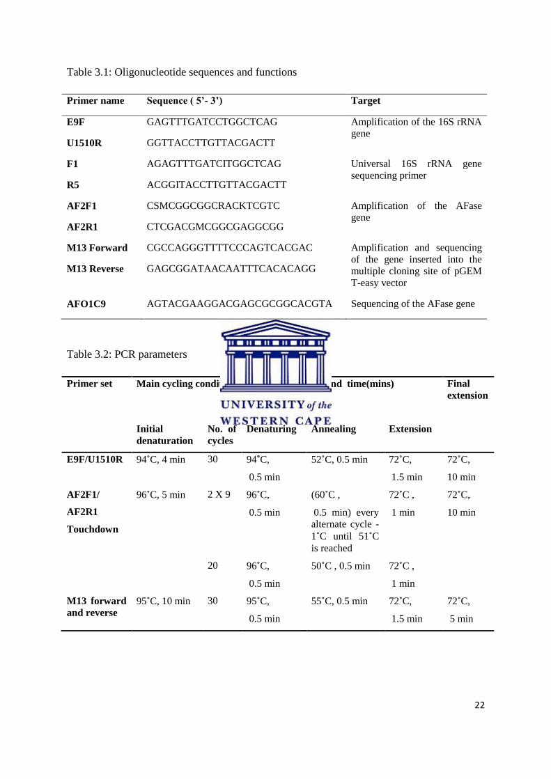
Table 3.1 Oligonucleotide sequences and functions 22
Table 3.2 PCR parameters 22
Table 3.3 Screening for α-L-arabinofuranosidase (AFase) activity: xylan
degradation and pnp-α-L-arabinofuranoside hydrolysis by
Actinobacteria
28
Table 3.4 BLAST results for the 16S rRNA gene 31
Table 3.5 Purification table for the sub-cellular AFase produced by ORS #1 36
Table 2.6 : Mascot results obtained for protein band 5 confirming its
identity and essential properties.
37

CHAPTER 1
GENERAL INTRODUCTION AND PROJECT AIMS

2
GENERAL INTRODUCTION
1.1 Introduction
In recent years a trend has developed which aims to make bioethanol production less reliant
on food crops such as maize and cassava and redirects its focus onto more renewable,
abundant feedstocks. Of these feedstocks lignocellulose has generated the most interest
despite the fact that it is a highly recalcitrant polymer that requires extensive pretreatment in
order to liberate the simple sugars required for bioethanol fermentation (Perez et al., 2002).
Methodologies for the enzymatic hydrolysis of the core polymers, cellulose, lignin and
hemicellulose, have been developed for the liberation of these simple sugars. Enzymatic
hydrolysis is advantageous as it avoids the formation of by-products that may inhibit
downstream processing (Hahn-Hagerdal, 2006). Lignocellulytic enzymes that function at
elevated temperatures are extremely desirable for thermophilic industrial fermentations as
their catalytic activity is unaltered by the high temperatures employed (Blumer -Schutte et
al., 2008).
Hemicelluloses and xylan in particular represent 20-35% of lignocellulosic biomass. Several
xylan degrading enzymes are required for the complete biodegradation of hemicelluloses
(Betts et al., 1991). Xylanolytic enzymes have frequently been isolated from Actinobacteria
genera including Streptomyces, Rhodococus, Bifidobacterium and Arthrobacter (Numan and
Bhosle, 2006). Among these enzymes, α-L-arabinofuranosidases (AFases) hydrolyse
arabinose residues present in arabino-glucuronoxylan and other xylo-oligomers. In addition,
AFases have been shown to have a synergistic effect in the enzymatic hydrolysis process and
are therefore of commercial value (De Vries et al., 2000).
1.2 Project Aims
The objectives of this study are summarized below:
i. Screening thermophilic Actinobacteria isolates for AFase activity
ii. 16S rRNA gene analysis of isolates that display the highest AFase activity
iii. Construction and screening of a cosmid library for positive clones
iv. Determination of the gene and protein sequence of the enzyme
v. Preliminary characterisation of the AFase enzyme

3
CHAPTER 2
LITERATURE REVIEW
BIOETHANOL PRODUCTION THROUGH LIGNOCELLULOSE
DEPOLYMERSATION

4
CHAPTER 2
2.1 Bioethanol
As sources of fossil fuels near depletion and fuel prices fluctuate, biofuels have regained
popularity as an alternative fuel source. Biofuels are derived from the fermentation of
biological matter, such as starch and sucrose, to produce secondary energy carriers, which
may then be used to fuel the industrial and commercial processes that rely solely on fossil
fuels (Monique et al., 2003; Saha, 2003; Gray et al., 2006; Lin and Tanaka, 2006; Antoni et
al., 2007). Biofuels are an attractive substitute for fossil fuels as they not only reduce
greenhouse emissions produced by biomass waste (the main source material for biofuels), but
also contribute to sustainable development whilst minimizing the global dependence on non-
renewable resources (Lynd, 1996; Gray et al., 2006). A variety of biofuels such as
biomethanol, biobutanol and biodiesel are commercially available, although the most
commonly used fuel is bioethanol (Balat et al., 2008).
The ethanol produced from biomass, also referred to as bioethanol, is an oxygenated fuel
source that is conventionally produced through the fermentation of various food crops such as
corn, sugar cane, wheat, barley rice and cassava. Microorganisms such as Saccharomyces
cerevisae and Zymomonas mobilis are used to ferment these carbon sources into bioethanol
(Sanchez and Cardona, 2008). Since the first commercial production of bioethanol in the
1860’s its popularity oscillated, peaking in the 1970’s as a direct result of the global oil crisis.
This crisis sparked a renewed interest in the use of bioethanol as a fuel source (Zaldivar et al.,
2001; Galbe and Zacchi, 2002; Antoni et al., 2007; Balat et al., 2008). The appealing
characteristics of bioethanol include renewability of the substrate as well as the catalyst,
enhanced automobile performance and an estimated 80% reduction in CO2 emissions when
compared to fossil fuel combustion (Wyman, 1996; Brown et al., 1998). The commercial
value of bioethanol requires an increase in the supply to meet future demand.
5
2.1.1 Fuel ethanol production
In the United States of America, Brazil and some European countries such as Spain, Russia
and Germany, concerns regarding diminishing fossil fuel reserves resulted in an increase in
bioethanol production (von Sivers and Zacchi, 1996; Hahn-Hagerdal et al., 2006). Smaller
countries, enticed by the concept of reduced oil imports, a boost in the rural economy and an
increase in overall air quality, also developed smaller scale bioethanol industries (Galbe and
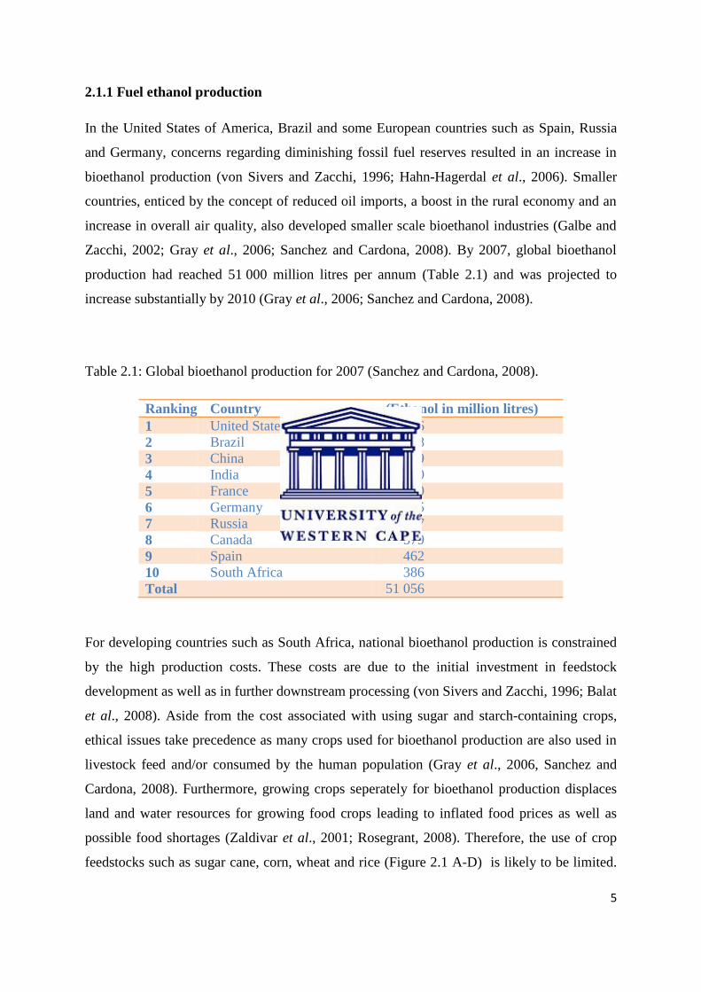
Zacchi, 2002; Gray et al., 2006; Sanchez and Cardona, 2008). By 2007, global bioethanol
production had reached 51 000 million litres per annum (Table 2.1) and was projected to
increase substantially by 2010 (Gray et al., 2006; Sanchez and Cardona, 2008).
Table 2.1: Global bioethanol production for 2007 (Sanchez and Cardona, 2008).
Ranking Country (Ethanol in million litres)
1 United States of America 18 376
2 Brazil 16 998
3 China 3 849
4 India 1 900
5 France 950
6 Germany 765
7 Russia 647
8 Canada 579
9 Spain 462
10 South Africa 386
Total 51 056
For developing countries such as South Africa, national bioethanol production is constrained
by the high production costs. These costs are due to the initial investment in feedstock
development as well as in further downstream processing (von Sivers and Zacchi, 1996; Balat
et al., 2008). Aside from the cost associated with using sugar and starch-containing crops,
ethical issues take precedence as many crops used for bioethanol production are also used in
livestock feed and/or consumed by the human population (Gray et al., 2006, Sanchez and
Cardona, 2008). Furthermore, growing crops seperately for bioethanol production displaces
land and water resources for growing food crops leading to inflated food prices as well as
possible food shortages (Zaldivar et al., 2001; Rosegrant, 2008). Therefore, the use of crop
feedstocks such as sugar cane, corn, wheat and rice (Figure 2.1 A-D) is likely to be limited.
6
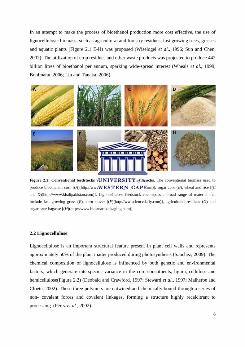
In an attempt to make the process of bioethanol production more cost effective, the use of
lignocellulosic biomass such as agricultural and forestry residues, fast growing trees, grasses
and aquatic plants (Figure 2.1 E-H) was proposed (Wiselogel et al., 1996; Sun and Chen,
2002). The utilization of crop residues and other waste products was projected to produce 442
billion litres of bioethanol per annum, sparking wide-spread interest (Wheals et al., 1999;
Bohlmann, 2006; Lin and Tanaka, 2006).
Figure 2.1: Conventional feedstocks versus lignocellulosic feedstocks. The conventional biomass used to
produce bioethanol: corn [(A)(http://www.moosecrossingardencenter.com)], sugar cane (B), wheat and rice [(C
and D)(http://www.khalipakistan.com)]. Lignocellulose feedstock encompass a broad range of material that
include fast growing grass (E), corn stover [(F)(http://ww.sciencedaily.com)], agricultural residues (G) and
sugar cane bagasse [(H)(http://www.biosmartpackaging.com)]
2.2 Lignocellulose
Lignocellulose is an important structural feature present in plant cell walls and represents
approximately 50% of the plant matter produced during photosynthesis (Sanchez, 2009). The
chemical composition of lignocellulose is influenced by both genetic and environmental
factors, which generate interspecies variance in the core constituents, lignin, cellulose and
hemicellulose(Figure 2.2) (Deobald and Crawford, 1997; Steward et al., 1997; Malherbe and
Cloete, 2002). These three polymers are entwined and chemically bound through a series of
non- covalent forces and covalent linkages, forming a structure highly recalcitrant to
processing (Perez et al., 2002).

7
Figure 2.2: The composition of lignocellulose. A layer of lignin (yellow) surrounds the cellulose (blue) and
hemicellulose (green) portions. These three structures are bound within the cell by ester (pink) linkages
(Morrison, 2008).
Initial estimates in 1999 suggested that harvesting lignocellulosic biomass may result in a 255
million ton per year reduction in global CO2 output by the year 2010 (Grassi, 1999). The use
of lignocellulosic material circumvents the conflict surrounding the diversion of agricultural
land for feedstock production (Hahn-Hagerdal et al., 2006; Sanchez and Cardona, 2008).
Lignocellulosic biomass is less expensive to produce than conventional feedstocks and also
requires less energy, pesticides and fertilizers (Chum and Overend, 2001; Balat et al., 2008).
2.2.1 Structural composition of lignocellulose
Angiosperms, gymnosperms and grasses have been identified as ideal substrates for
bioethanol production (Zaldivar et al., 2001). Variance in composition between lignin,
cellulose and hemicellulose in subsets of flora representative of these classes is presented in
Table 2.2. When considering the angiosperms (flowering plants) and gymnosperms (plants
that produced unenclosed seeds), it is interesting to note that these plants are fundamentally
composed of cellulose with hemicellulose as the second most common polymer (McKendry,
2002). The class of plants referred to as grasses are primarily composed of hemicellulose
(approximately 50%) followed closely by cellulose and much lower quantities of lignin (Betts
et al., 1991; Thomas, 1993).
8
Table 2.2: Chemical composition of various lignocellulosic materials (Betts et al., 1991)
Plant material Lignin % Cellulose % Hemicellulose %
Grasses 10-30 25-40 25-50
Angiosperms 25- 35 45- 50 25-35
Gymnosperms 18- 25 45- 55 24- 40
Biomass containing high quantities of cellulose is the primary candidate for bioethanol
production as cellulose is comprised purely of the hexose sugar, glucose (Zaldivar et al.,
2001). Biomass rich in hemicellulose is an attractive alternative because of its higher sugar
content.
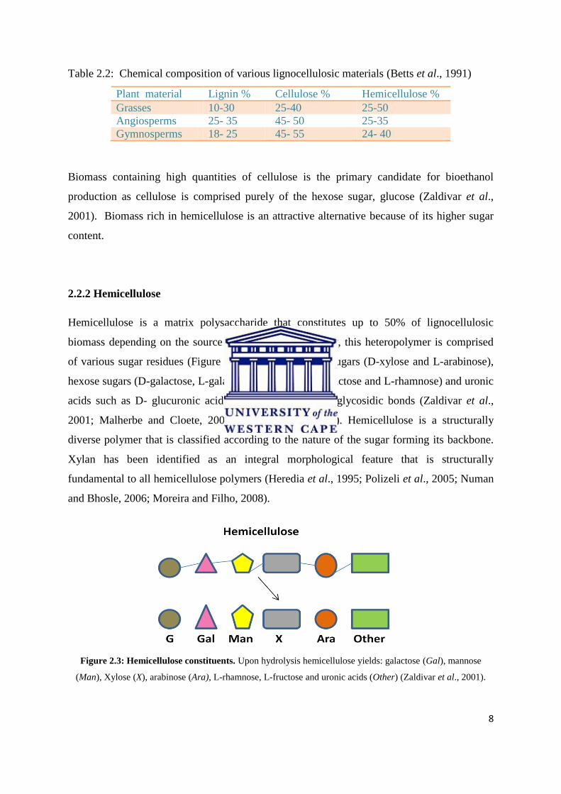
2.2.2 Hemicellulose
Hemicellulose is a matrix polysaccharide that constitutes up to 50% of lignocellulosic
biomass depending on the source (Table 2.2). Structurally, this heteropolymer is comprised
of various sugar residues (Figure 2.3) including pentose sugars (D-xylose and L-arabinose),
hexose sugars (D-galactose, L-galactose, D-mannose, L-fructose and L-rhamnose) and uronic
acids such as D- glucuronic acid, linked through β-1, 4 glycosidic bonds (Zaldivar et al.,
2001; Malherbe and Cloete, 2002; Polizeli et al., 2005). Hemicellulose is a structurally
diverse polymer that is classified according to the nature of the sugar forming its backbone.
Xylan has been identified as an integral morphological feature that is structurally
fundamental to all hemicellulose polymers (Heredia et al., 1995; Polizeli et al., 2005; Numan
and Bhosle, 2006; Moreira and Filho, 2008).
Figure 2.3: Hemicellulose constituents. Upon hydrolysis hemicellulose yields: galactose (Gal), mannose
(Man), Xylose (X), arabinose (Ara), L-rhamnose, L-fructose and uronic acids (Other) (Zaldivar et al., 2001).

9
Xylans
Xylans are heteropolysaccharides, composed of D-xylopyranose units bound by β1, 4 -
linkages in the homopolymeric backbone (De Vries and Visser, 2001; Saha, 2003).
Arabinoxylans, prominent in graminaceous plants, contain α-L-arabinofuranose side chains
on the second and third oxygen molecules in the xylan backbone (Izydroczyk and Biliaderis,
1995; Adams et al., 2004; Numan and Bhosle, 2006). Further branch substitutions (Figure
2.4) include the β1,2 and β1,4 xylose linked D-galactose and glucuronic acid, while the
arabinose portion is bound by 4- O- methyl ether, p- coumaric, ferulic and acetic acids joined
by β1,5 linkages (Smith and Hartley, 1983; Adams et al., 2004). The frequency and
occurrence of side-chain substitutions vary between xylans and are therefore designated
according to the sugar most frequently incorporated into its backbone (Saha, 2000; Saha,
2003).
Figure 2.4: The structure of arabinoxylan. Represented here is the xylan backbone and prominent arabinose
(Ara) substitutions. All other sugar side chains are clearly visible: xylose (Xyl), galactose (Gal), glucuronic acid
(Glca), 2-ο-acetyl ester (AcE) and ferulic acid (FeA) linked through their relative β-glycosidic bonds. Each
number represents the site where xylanolytic enzymes will cleave their substrates. Numbers 1 (xylanase) and 2
(xylanosidase) act on xylose-xylose linkages to produce xylose monomers. Number 3 (ferulic acid esterase)
hydrolyses ferulic acid from arabinofuranoside chains while number 4 (acetyl xylan esterase) liberates 2-ο-
acetyl ester from the xylan backbone. The arabinofuranosidase, number 5, exclusively cleaves arabinofuranose
from the xylan backbone (Adams et al., 2004; Numan and Bhosle, 2006).
The function of xylan has been determined via in situ studies. The covalent linkage of xylan
to lignin as well as its non-covalent association with cellulose plays an important role in

10
sustaining the structural integrity of cellulose by defending the fibres against microbial
cellulase degradation (Uffen, 1997; Beg et al., 2001).
2.3 From lignocellulose to fermentable sugars
The recalcitrant nature of lignocellulose biomass means that it requires significant processing
to relinquish sugars sufficient for fermentation to bioethanol (Mosier et al., 2005). These
processes involve polymer deconstruction through physical/ chemical or physiochemical
pretreatment methods. This step is succeeded by a chemical or enzymatic hydrolysis step
wherein all polysaccharides present are depolymerised to liberate simple fermentable sugars
(Galbe and Zacchi, 2002; Balat et al., 2008).
Polysaccharide depolymerisation through acidic hydrolysis is a highly effective technique but
produces inhibitory by-products that affect downstream processing, thus affecting
commercial viability (Hahn-Hagerdal et al., 2006). Enzymatic hydrolysis of polysaccharide
substrates renders substrate-specific conversions, which eliminate the risk of by-product
formation and ensures the complete conversion of lignocellulose (Hahn-Hagerdal et al.,
2006). The use of thermostable enzymes ensures total hydrolysis of the substrate as its
catalytic activity is unaffected by the high temperatures employed during industrial
fermentation (Blumer-Schutte et al., 2008; Sanchez and Cardona, 2008). These enzymes are
produced by thermophilic bacteria (active at 45˚C- 80˚C) including Clostridium,
Thermoanaerobacterium and multiple members of the Actinobacteria family (Sommer et al.,
2004). Actinobacteria are known for their ability to degrade recalcitrant polymers such as
lignocellulose. Species such as Streptomyces, Nocardia, Arthrobacter and Rhodococus
produce multiple thermostable hemicellulases, cellulases and lignases that play a pivotal role
in lignocellulose degradation (Goodfellow and William, 1983; MacKenzie et al., 1987;
Tuncer et al., 1999; Khanderparker et al., 2008).
The hydrolysis of hemicellulose by hemicellulases is of utmost importance in biofuel
production as this polysaccharide is widely distributed throughout the lignocellulosic

11
structure and contains a large amount of fermentable sugars. The complete depolymerisation
of hemicellulose requires a cascade of hemicellulases (Table 2.3) that either display glycoside
hydrolase (GH) activity or carbohydrate esterase (CE) activity. Due to its particular
importance in the degradation of xylan, the hemicellulase α-L-arabinofuranosidase (AFase)
will be the focus of this study.
Table 2.3: Classification of hemicellulolytic enzymes (adapted from Shallom and Shoham,
2003).
EC number Enzyme Substrate Family
3.2.1.8 Endo- β 1-4-xylanase β 1-4-xylan GH 5,8,10,43
3.2.1.37 Exo-β 1-4-xylosidase Β1-4-xylooligomers
xylobiose
GH 3,39,43,52,54
3.1.2.55 α-L-arabinofuranosidase α-L-arabinofuranosyl
(1,2) or (1,3) xylooligomers
α-L-arabinan
GH 3,43,51,54,62
3.2.1.99 Endo α-1,5-arabinase 1,5-arabinan GH 43
3.2.1.139 α-glucuronidase 4-O-methyl-α-glucuronic
acid, (1,2) xylooligomers
GH 67
3.2.1.78 Endo-β1,4-mannase β1,4-mannan GH 5, 26
3.2.1.25 Exo-β1,4- mannosidase β1,4-manooligomers
mannobiose
GH 1,2,5
3.2.1.22 α-galactosidase α-galactopyranoside
(1,6) mannoloigomers
GH 4,27,36,57
3.2.1.21 β-glucosidase β glucosidase GH 1, 3
3.2.1.89 Endo- galactanase β-1,4 galactan GH 53
3.1.1.72 Acetyl xylan esterase 2- or 3-O-acetyl xylan CE 1,2,3,4,5,6,7.
3.1.1.6 Acetyl mannan esterase 2- or 3-O-acetyl mannan
3.1.1.73 Ferulic and p-cumaric
acid esterases
Ferulic acid, p-cumaric acid CE 1.

12
2.4 α-L-arabinofuranosidase
α-L-arabinofuranosidase (AFase) is an auxiliary enzyme that cleaves α-L-arabinofuranosyl
linkages from arabinose-rich polysaccharides such as arabinan, arabinoxylans
arabinogalactan and pectin (Figure 2.5) (Margolles-Clark et al., 1996). AFases display
synergism with other hemicellulases, accelerating the rate at which their glycosidic bonds are
hydrolysed by 1017
fold (Rye and Withers, 2000; Shallom et al., 2002). For this reason
AFases have been described as one of the most efficient catalysts available (Numan and
Bhosle, 2006). Synergy ensures the rapid and efficient hydrolysis of hemicellulose to render
soluble substrates that are easily assimilated by the AFase producer (Saha, 2000; Takao et al.,
2002; Numan and Bhosle, 2006).
Figure 2.5: Enzymatic action of α-L-arabinofuranosidase. α-L-arabinofuranosidase acts on the xylan
backbone, cleaving α-L-arabinofuranose residues from the second oxygen molecule on the xylose subunit
(adapted from Kumar et al., 2008).
Industrial applications of α-L-arabinofuranosidases include enhancements in the aroma of
wine, the quality of bread, the efficacy of juice clarification and pulp treatment, synthesis of
anti-metastatic and anti-carcinogenic compounds, bioethanol and oligosaccharides (Numan
and Bhosle, 2006).
2.4.1 Classification systems for α-L-arabinofuranosidase activity
AFase was first described in 1928 by Ehrlich and Schubert as an enzyme that was capable of
liberating arabinose from arabinan beet (Uesaka et al., 1978). Subsequent purification and
13
crystallisation studies on the enzyme isolated from Aspergillus niger demonstrated that the
enzyme was an α-L-arabinofuranosidase (Kaji and Tagawa, 1964, 1970; Kaji et al., 1967;
Kaji et al., 1969).
The classification system developed by Kaji (1984), grouped AFases according to their origin
and substrate specificity. Beldman et al (1997) elaborated on Kaji’s system by including the
mode of action of the enzyme in the classification criteria. According to this scheme, two
types of AFase exist. The first group, Arafur A, displayed little or no activity toward
arabinose-containing polysaccharides while the second group, Arafur B, was capable of
hydrolysing L-arabinofuranosyl residues from polymers. This classification system proved to
be insufficient as its criteria were too broad to define the substrate specificity of all AFases.
The problem was further compounded by the isolation of AFases that displayed novel
mechanisms of action. These factors lead to the subdivision of group Arafur B into three
subclasses: (I) AXHB-md 2, 3 (II) AXHB-m 2, 3 and (III) AXHd3 (Numan and Bhosle,
2006).
The primary subclass (AXBH-md 2, 3) is capable of liberating arabinose from single as well
as di-substituted xylose residues. In addition, this enzyme is able to cleave α-L-
arabinofuranoside from p-nitrophenyl (pnp) α-L-arabinofuranoside at rates that parallel those
obtained for oligosaccharide substrates (Ferre et al., 2000). The second (AXHBm 2, 3) and
third (AXHd3) subclasses were isolated from Bifidobacterium adolescentis (Van Laere et al.,
1997; Van Laere et al., 1999). AXHB-m 2, 3 describes a class of enzymes that are capable of
hydrolysing arabinose residues with α-1, 2 as well as α-1, 3 linkages to mono-substituted
xylose residues. AXHd3 are α-L-arabinofuranosidases that exclusively cleave α-1, 3
glycosidic bonds in order to liberate arabinose. Neither of these two classes is able to
hydrolyse pnp α-L-arabinofuranoside. This implies that the pnp assays (the most common
screening tool for the detection of AFase activity) alone may be insufficient for the detection
of α-L-arabinofuranosidases.

14
Despite all attempts to classify AFases, the current classification does not accommodate the
more recently characterised AFases. Birgisson et al (2004) and Miyazaki (2005) described
an AFase that hydrolyses not only the internal α-1, 5 backbone but also the α-1, 3 side chains
of arabinan and debranched arabinan. This enzyme also actively hydrolyses pnp-α-L-
arabinofuranoside. Wagschal et al (2009) described a novel AFase isolated from a compost
starter mixture that displays dual xylosidase activity on a number of natural and artificial
substrates. Instead of revising the current classification system, AFase may alternatively be
grouped according to GH families as this system is less complicated and can accommodate
all known AFases according to their mechanism of action.
2.4.2 Physio-chemical characteristics of α-L-arabinofuranosidases
The biochemical properties of AFases vary greatly between species. The interspecies
differences in enzyme structure and tolerance to environmental stresses ensure α-L-
arabinofuranosidase production and activity in the harshest of environments. AFases function
over a pH range of 2 – 9 and temperature range of 30˚C – 80˚C (Table 2.4).

15
Table 2.4: Comparative characteristics of microbial α-L-arabinofuranosidases
Organism Molecular
Weight
Optimal
Temperature
Optimal
pH
Reference
Anoxybacillus
kestanbolensis
57 kDa 65˚C 5.5 Canakci et al (2008).
Arthrobacter sp.
MTCC5214
97 kDa 80˚C 8 Khanderparker et al
(2008)
Aureobasidium pullans 210 kDa 75˚C 4 Saha and Bothast
(1998)
Bacillus pumilus ARA 56 kDa 60˚C 6.4 Pei and Shao (2008)
B. stearothermophilus T-6 256 kDa 70˚C 5.5 Gilead and Shoham
(1995)
Corticium rolfsii - - 2.5 Kaji and Yoshihara
(1971)
Penicillium chrysogenum 79 kDa
and 52
kDa
50˚C 4- 6.6
3.3- 5
Sakamoto and
Kawasaki (2003)
Rhodotorula flava - 30˚C 2 Uesaka et al (1978)
Streptomyces
thermoviolaceus OPC- 520
37 kDa 60˚C 5 Tsujibo et al (2002)
Streptomyces avermitilis
NBRC 14893
- 30 ˚C 5- 6.5 Ichinose et al (2008)
Thermomonospora fusca 92 kDa 65 ˚C 9 Tuncer and Ball (2003)
2.4.3 α-L-arabinofuranosidase production: location, isozymes and enzyme variation
The majority of AFases produced by microbes are secreted into the culture media and
therefore play an active role in polysaccharide degradation (Matuso et al., 2000). Some
AFases, such as those found in S.lividans, S.avermitilis NBRC 14893 and Pseudomonas
flourescens are strictly intracellular enzymes (Kellett et al., 1990; Manin et al., 1994;

16
Ichinose et al., 2008). These enzymes lack the gene associated with the production of the
signalling peptide that is required for exportation to the extracellular environment (Manin et
al., 1994). Matsumura et al (2004) noticed that AFases secreted in high quantities are
predominantly found in monomeric configurations. Most organisms struggle to produce and
export high concentrations of multimeric AFases which results in total or partial enzymatic
inactivity.
Isozymes of AFases have been identified in S.diastaticus and Penicillium chrysogenum
(Tajana et al., 1992; Sakamoto and Kawasaki 2003). These isozymes have the same catalytic
mechanism but differ in their physical response to environmental stresses such as pH and
temperature variations, thus ensuring the proficient hydrolysis of arabinose- rich structures
(Ahmed et al., 2001). Aspergillus awamori IFO4033 and Streptomyces chartreusis GS901
have been shown to simultaneously produce both AFase A and B, as classified according to
the Beldman et al. (1997) system (Kaneko et al., 1998; Matsuo et al., 2000). This imparts on
the microbe the ability to assimilate both arabinose containing oligosaccharides and
polysaccharides, and results in the total hydrolysis of arabinofuranosyl linked structures.
Organisms that co-produce α-L-arabinofuranosidase A and B are therefore of more
commercial potential than organisms that produce only one form of the enzyme.

17
CHAPTER 3
RESEARCH RESULTS
DETECTION, ISOLATION AND PARTIAL CHARACTERISATION OF α-
L-ARABINOFURANOSIDASES FROM THERMOPHILIC
ACTINOMYCETE ISOLATES
18
CHAPTER 3
Detection, isolation and partial characterisation of α-L-arabinofuranosidases from
thermophilic Actinomycete isolates
Introduction
The degradation of xylan is a key step in the production of bioethanol from lignocellulosic
(LC) material, as xylan constitutes 25-50% of LC biomass (Betts et al., 1991). Structurally,
xylan is composed of a xylose backbone that is linked to various sugar moieties, the most
important of which is arabinose. Arabinose residues stabilize the plant cell wall through
cross–linkages with lignin and cellulose, while simultaneously inhibiting the action of
xylanolytic enzymes (Aristidou and Penttila, 2000; De Vries et al., 2000; Saha B.C. 2000).
The cleavage of arabinose moieties from the xylan backbone is thus a pivotal step in xylan
hydrolysis. AFase (E.C 3.2.1.55) is an auxillary enzyme that cleaves terminal non-reducing
α-L-arabinofuranosyl linkages to liberate terminal monosaccharide units (Shallom and
Shoham, 2003). AFases have been shown to have a synergistic effect on the catalytic activity
of other lignocellulolytic enzymes including xylanase, ferulic acid esterase, acetyl xylan
esterase and xylosidase, and are therefore widely employed in various industrial processes
(Tuncer and Ball, 2003; Numan and Bhosle, 2006). Moreover, thermostable AFases are of
greater commercial value as their high degree of specificity ensures efficient catalysis at
elevated temperatures (Haki and Rakshit, 2003; Pei and Shao, 2008).
AFases have been isolated from Aspergillus spp. (De Vries et al., 2000; Matsumura et al.,
2004), Trichoderma reesei (Nogawa et al., 1999), Bacillus stearothermophilis (Gilead and
Shoham, 1995), Bacillus subtilis (Inacio et al., 2008), Thermomonospora fusca
(Buchmannand McCarthy, 1991; Tuncer, 2000), and Actinobacteria such as Arthrobacter
spp. (Khanderparker et al., 2008), Streptomyces spp. (Kaji et al., 1981; Vincent et al., 1997;
Matsuo et al., 2000; Tsujibo et al., 2003) and Bifidobacterium spp.(Shin et al., 2003). A
number of techniques, ranging from genomic library construction to enzyme purification are
used to isolate AFases (Margolles-Clark et al., 1996; Sakamoto and Kawasaki, 2003). In this
manuscript we report on an α-L-arabinofuranosidase produced by a Streptomyces strain ORS
#1. The enzyme was partially purified and preliminary characterisation data provided.

19
3.1 MATERIALS AND METHODS
3.1.1 Screening for α-L-arabinofuranosidase activity
3.1.1.1 Strain acquisition and growth conditions
Thermophilic Actinomycetes were isolated from environmental samples obtained from the
Namib (NDS), Omaruru (ORS) and Walvis Bay (WBDS) regions of Namibia as well as the
Gwisho (GS) and Bwanda (BS) hot springs in Zambia. Isolates were cultivated on Desert
minimal agar [0.5g glucose, 0.5g yeast extract, 0.5g NaCl, 0.5g MgSO4.7H2O, 1g K2HPO4
15g agar and 1ml Trace elements solution (0,1g FeSO4.7H2O, 0.1g MnCl2.4H2O and 0.1g
ZnSO4.7H2 in 100ml dH2O)] and 172F agar (10g glucose, 5g yeast extract, 10g soluble
starch, 2.5g tryptone, 2.5g casamino acids, 2.5g MgSO4.7H2O, 1g CaCl2.2H2O, 15g NaCl),
respectively and incubated at 45˚C for four days. The isolates were then Gram stained,
transferred to the corresponding broth and grown for a further four days at 45˚C, with
subsequent maintenance in 20% (v/v) glycerol.
3.1.1.2 Screening for xylanolytic activity
The presence of xylanolytic activity was determined through carbon utilization assessment
(Shirling and Gottlieb, 1966) using the basal media ISP9 supplemented with 1% (w/v)
Birchwood xylan (Sigma). Inocula were incubated at 45˚C for four days.
3.1.1.3 Biomass production and preparation of cellular proteins
The isolates were cultivated in 10 ml cultures of ISP9 medium supplemented with 0.1%
Birchwood xylan at 45˚C for three days, and centrifuged for 10 minutes at 10 000 rpm
(Eppendorf 5810R). The supernatant was retained and the pellets washed in 50 mM
potassium phosphate buffer (pH 7). Cell pellets were resuspended in 50 mM potassium
phosphate buffer (pH 7) and sonicated at 50% power for six cycles of 30 seconds each.
Intracellular proteins were collected via centrifugation as previously described for
supernatant isolation. Both intracellular and supernatant extracts were stored on ice.

20
3.1.1.4 Screening for crude α-L-arabinofuranosidase activity
AFase activity was determined by spectrophotometric measurement (410 nm) as para-
nitrophenyl (pnp) released from pnp-α-L-arabinofuranoside (Carbosynth) and pnp-α-L-
arabinopyranoside (Sigma). The AFase assay was adapted from Birigisson et al. (2004) and
Pei and Shao (2008). The 200 µl reaction contained 50 mM potassium phosphate buffer (pH
7), 10 mM pnp-α-L-arabinofuranoside (pnpAraf) or pnp-α-L-arabinopyranoside (pnpArap)
and 10 µl of crude enzyme. The reaction was incubated at 49˚C for 10 minutes, stopped with
the addition of 550 mM Na2CO3 and an absorbance reading taken. All samples that rendered
an absorbance reading higher than 0.15 were re-assayed at 60˚C to check thermodurability.
Protein concentrations were determined via the Bradford assay (Bradford, 1976) using bovine
serum albumin (BSA) as the standard and AFase activity expressed as µmol p-nitrophenol
released per mg of protein in one minute (U.mg).
3.1.2 Genebank assembly and screening
3.1.2.1 Cosmid library construction
A cosmid library was constructed with high molecular weight genomic DNA extracted from
strains ORS #1, NDS #4 and WBDS #9. The DNA was end–repaired (End-It, Epicentre
Biotechnology), ligated to the vector pFD666 and packaged using the MaxPlax Lambda
Packaging Extracts kit (Epicentre Biotechnology). Packaged DNA was transfected into E.coli
Genehog (recA-), and incubated overnight at 37˚C on Luria-Bertani agar (LBA) containing
25µg/ml Kanamycin. Glycerol stocks were prepared as follows: a single colony was
transferred to a microtitre plate well that contained 200 µl LB and 25µg/ml Kanamycin, and
incubated at 37 ˚C overnight. Cultures were then divided between two microtitre plates, 20 %
glycerol added, and stored at -80˚C for future screening.
3.1.2.2 Library screening for α-L-arabinofuranosidase activity
The cosmid library was screened using three different methods as described below. These
methods were first tested on the positive control, Bacillus subtilis 168, which has known
AFase activity (Inacio et al., 2008). E.coli Genehog containing the vector pDF666 lacking a
DNA insert was used as a negative control.

21
Solid-phase screening with pnp-arabinofuranosidase. This screen required the solubilisation
of 50 mg of pnp- arabinofuranoside per 100 ml LB agar. Following inoculation of the clones
onto the plates, the plates were sealed with foil and incubated overnight at 37˚C.
Liquid-phase screening. AFase activity was induced in LB media containing 1 mM pnp-α-L-
arabinofuranoside as described by Margolles-Clark et al. (1996) and incubated at 37˚C.
Amendments made to the protocol included an increase in sampling on days one, three, four
and six.
Solid-phase screening that incorporates cell lysis. The protocol devised by Lee et al (2009)
was amended as follows. The microtitre plates containing the library were pooled, and
incubated overnight at 37 ˚C in LB/kanamycin. The overnight culture was inoculated onto a
LBA/kanamycin plate and grown overnight at 37˚C. The colonies formed were transferred
via a membrane from the LBA plate to a 1% agarose plate (50 mg Lysozyme, 1 mM pnp-
arabinofuranoside, 50 mM potassium phosphate buffer pH 7) and incubated at 30˚C
overnight.
3.1.3 Gene amplification and sequencing
3.1.3.1 Primers and PCR parameters
The oligonucleotide primers utilized in this study are represented in Table 3.1. The cycling
parameters for the individual polymerase chain reactions are defined in Table 3.2.
22
Table 3.1: Oligonucleotide sequences and functions
Primer name Sequence ( 5’- 3’) Target
E9F GAGTTTGATCCTGGCTCAG Amplification of the 16S rRNA
gene U1510R GGTTACCTTGTTACGACTT
F1 AGAGTTTGATCITGGCTCAG Universal 16S rRNA gene
sequencing primer R5 ACGGITACCTTGTTACGACTT
AF2F1 CSMCGGCGGCRACKTCGTC Amplification of the AFase
gene AF2R1 CTCGACGMCGGCGAGGCGG
M13 Forward CGCCAGGGTTTTCCCAGTCACGAC Amplification and sequencing
of the gene inserted into the
multiple cloning site of pGEM
T-easy vector
M13 Reverse GAGCGGATAACAATTTCACACAGG
AFO1C9 AGTACGAAGGACGAGCGCGGCACGTA Sequencing of the AFase gene
Table 3.2: PCR parameters
Primer set Main cycling conditions : temperature (˚C) and time(mins) Final
extension
Initial
denaturation
No. of
cycles
Denaturing Annealing Extension
E9F/U1510R 94˚C, 4 min 30 94˚C,
0.5 min
52˚C, 0.5 min 72˚C,
1.5 min
72˚C,
10 min
AF2F1/
AF2R1
Touchdown
96˚C, 5 min 2 X 9 96˚C,
0.5 min
(60˚C ,
0.5 min) every
alternate cycle -
1˚C until 51˚C
is reached
72˚C ,
1 min
72˚C,
10 min
20 96˚C,
0.5 min
50˚C , 0.5 min 72˚C ,
1 min
M13 forward
and reverse
95˚C, 10 min 30 95˚C,
0.5 min
55˚C, 0.5 min 72˚C,
1.5 min
72˚C,
5 min

23
3.1.3.2 16S rRNA characterisation
DNA was extracted from strains ORS #1, NDS #4 and WBDS #9 via the Wang method
(Wang et al., 1996). The 16S rRNA gene was amplified using primers E9F and U1510R
(Table 3.1) with PCR parameters as described in Table 3.2. The 16S rRNA amplicons were
purified using the Illustra GFX PCR DNA and gel band purification kit (GE Healthcare).
Sequencing reactions were carried out by the University of Stellenbosch sequencing facility
with primers F1 and R5 (Table 3.1).
3.1.3.3 Amplification of the α-L-arabinofuranosidase gene
Six Streptomyces α-L-arabinofuranosidase protein sequences that represent the GH families
62, 43 and 51 were obtained from the NCBI (National Center for Biotechnology Information)
database and aligned using DNAman. Although the Streptomyces AFases were similar on the
protein level, there was a high degree of degeneracy at the nucleotide level. For this reason,
the degenerate primers AF2F1 and AF2R1 were designed to flank an 825 base region that
represented 55% of the AFase gene.
Touchdown PCR (Table 3.2) was performed in a 50 µl reaction with 200 ng of genomic
DNA from strain ORS #1, NDS #4 and WBDS #9. Each reaction contained 200 µM of each
DNTP, 0.5 µM of the degenerate primers, AF2F1 and AF2R1 (Table 3.1), 0.5 U of Robust
Taq polymerase and 1 X GC buffer (Kappa Biosystems) as directed for high GC template
DNA. A magnesium chloride (Fermentas) gradient of 1.5 mM to 3.5 mM was included.
Troubleshooting: Additional PCR’s were conducted with 200 ng of WBDS #9 and NDS #4
DNA. These PCR’s included all current optimisation available for the Robust PCR kit i.e. the
use of 1 X GC buffer with 4% DMSO (Sigma) and also 1 X buffer A instead of the GC buffer
combined with enhancer A and 5% DMSO. For high fidelity, 0.5 U PrimeSTARTM
HS DNA
polymerase (Takara) was employed along with 1X PrimeSTAR buffer, 200 µM dNTP
mixture containing 1 mM MgCl2+
and 0.3 µM of primers AF2F1 and AF2R1. All amplicons
where visualised on a 1% agarose gel.
24
3.1.3.4 Cloning and sequencing of the α-L-arabinofuranosidase gene
With reference to the molecular weight marker, a band approximately 825 base pairs in size
was excised from a 2% agarose gel and the PCR product purified using the Nucleospin kit
(Macherey-Nagel). The gene was ligated using T4 ligase (NEB) to pGEM T-Easy (Promega),
transformed into E.coli Genehog by electroporation and screened using blue/white colony
selection.
DNA for colony PCR was prepared by immersing a single colony in sdH2O followed by a ten
minute incubation step at 96˚C. Cellular debris was collected via centrifugation at 8000 rpm
and the supernatant retained. The insert was confirmed through M13 PCR using the M13
forward and reverse primer set (Table 3.1). Plasmid DNA (pDNA) was isolated from the
selected clones using the alkaline lysis mini prep method for pDNA isolation (Ish-Horowicz
and Burke, 1981) and sequenced with the M13 and AFO1C9 oligonucleotides (Table 2.1).
3.1.4 Protein purification and characterisation
3.1.4.1 Growth curves and protein production
Strain ORS #1 was inoculated into 1L ISP 9 media supplemented with 1% (w/v) Birchwood
xylan and incubated at 45˚C for 30 days. Aliquots (100 ml) were removed daily and cell free
extracts assayed for intracellular and extracellular AFase activity. A growth curve was
generated and used to determine the optimal growth stage for highest AFase production.
3.1.4.2 Ammonium sulphate protein precipitation and desalting
Cell free extract (Section 3.1.1.3) was suspended in 50 mM potassium phosphate buffer (pH
7) and the proteins precipitated to 80% ammonium sulphate saturation at 4˚C. The proteins
were pelleted at 10 000 rpm for 40 minutes and resuspened in 50 mM phosphate buffer. The
precipitated protein was desalted by dialysis against 100 mM potassium phosphate buffer pH
7 at 4˚C overnight.

25
3.1.4.3 Ion-exchange chromatography (IEC)
Dialysed protein was applied to a Q-sepharose column (Amersham Bioscience) equilibrated
with potassium phosphate buffer (pH7). The bound protein was eluted against a 0-1 M NaCl
linear gradient with a flow rate of 1.5 ml/minute and peak fraction volume of 1 ml. The
fractions were screened for β-D-xylosidase activity on a 1% agarose plate containing 0.5
mM 4-methylumberferyl-xyloside [MUX (Sigma)] and α-L-arabinofuranosidase activity
using the standard assay (section 3.1.1.4). Active peaks were pooled, diluted in 0.5 volumes
of sdH2O and reapplied to the Q-sepharose column. The flow rate was further decreased to
1.25 ml/min and the gradient set at 70% 1 M NaCl. The separation efficiency was determined
through SDS-PAGE analysis.
3.1.4.4 Hydrophobic interaction chromatography
The peak containing the AFase protein was precipitated to 30% ammonium sulphate
saturation and applied to a Phenyl-sepharose column (Amersham Pharmacia Biotech)
equilibrated with 50 mM potassium phosphate buffer (pH 7) containing 1M ammonium
sulphate. The bound proteins were eluted with 50 mM potassium phosphate buffer (pH 7) at a
flow rate of 2 ml/min.
3.1.4.5 Gel electrophoresis
Purified proteins were prepared by boiling the sample with equal volumes of sample buffer
[1M (v/v) Tris pH 6.8, 20% (v/v) glycerol, 10% (v/v) SDS, 10 mM DTT, 0.01%
bromophenol blue (BPB)] prior to protein separation in the first dimension (Laemmli, 1970).
Bands were visualised by staining with Coomassie Brilliant Blue R-250 [CBB (Sigma)]. The
molecular weight was estimated according to the PageRuler (Fermentas).
3.1.4.5 Activity staining
SDS gels were washed in 250 ml sdH2O with gentle shaking for 30 minutes to remove all
traces of the SDS buffer. The proteins were refolded in R-buffer (25 mM Tris-HCl pH 7.5,
1% (v/v) Triton X-100) for 14- 18 hours at 37˚C whilst gently shaking (Ostile et al., 2002).
The gel was washed with 400 ml of 400 mM Tris-HCl (pH 6.5) and sandwiched against a 1%
26
(w/v) agarose gel containing 10 mM pnp-arabinofuranoside. The gels were bound in cling
wrap and incubated at 50˚C for one hour (Buchmann and McCarthy, 1991).
The in-gel assay for β-D-xylosidase activity was performed according to the protocol
established by Watson et al. (2009) with minor amendments. The SDS-PAGE was washed
twice with sdH2O and incubated in 30 ml refolding buffer (20 mM PIPES [Pipreazine-N,N’-
bis [2-ethanesulphonic acid] buffer (pH 6.8), 2.5% (v/v) Triton X-100, 2 mM DTT
[Dithiothreitol], 2.5 mM CaCl2) at 20ºC for one hour. Fresh refolding buffer was applied and
the gel incubated overnight at 4ºC. The gel was rinsed twice with 20 mM PIPES (pH 6.8) and
incubated at 37ºC for 12 hours in fresh buffer. β-D-xylosidase activity was determined by
incubating the renatured protein in 50 mM potassium phosphate (pH7) containing 0.5 mM
MUX at 45 ºC for one hour.
3.1.4.6 Protein sequencing
Purified protein was sent for sequencing at the Proteomics Research Group (UWC) where it
was subjected to trypsin in-gel digestion and peptide separation via MALDI- TOF mass-
spectrometry. The peptide mass fingerprints produced were queried in the MASCOT
database (Matrix Science). Proteins with a MOWSE score greater than 83 were considered to
be significant.
3.1.5 Characterisation of the precipitated protein
3.1.5.1 pH optimum and stability
All characterisation was performed on the desalted ammonium sulphate precipitate (section
3.1.4.2). The pH optimum was determined by incubating 10 µl of protein with 10 mM pnp-α-
L-arabinofuranoside (pnp) and 50 mM buffer at 49˚C for 10 minutes. The reaction was
quenched by the addition of 550 mM Na2CO3, and an absorbance reading taken at a
wavelength of 410 nm. The buffers utilized were as follows: citrate phosphate (pH 3-6),
potassium phosphate (pH 6-8) and Tris-HCl (pH 8-9). The pH stability of the enzyme was
determined by incubating the enzyme in each of these buffers at 4˚C for 24 hours and
subsequently assaying for residual AFase activity.

27
3.1.5.2 Temperature optimum and stability
The temperature optimum of the enzyme was established by assaying the effect that
temperature has on the catalytic ability of the AFase. The enzyme was assayed for 10 minutes
in 50 mM potassium phosphate (pH7) buffer over the temperature range of 30˚C- 80˚C. The
temperature stability of the AFase was examined by incubating the AFase in 50 mM
potassium phosphate (pH7) buffer over the predetermined temperature range for 19 hours.
Aliquots were removed at 30 minute intervals over a period of two hours and at 19 hours, and
assayed for residual AFase activity at 49˚C.
3.1.5.3 Enzyme Kinetics
The kinetic parameters of the α-L-arabinofuranosidase were determined by assaying its
activity over a range of substrate concentrations (0.5 mM- 25 mM) monitored for ten minutes
at a wavelength of 410 nm by the Cary 300 Win/ UV spectrophotometer. Vmax and Km were
deduced through non-linear regression fitting the data to equation 1, in GraphPad Prism 4
(GraphPad software).
Equation 1: Y= Vmax*X/ (Km+X)
3.2 RESULTS
3.2.1 Substrate depolymerisation
Twenty Actinomycete isolates were grown on carbon utilization media (ISP9) in the presence
of Birchwood xylan. All isolates listed in Table 3.3, with the exception of WBDS #11, BSII
#7 and BSII #1, were able to depolymerise xylan as indicated by sporadic growth. Screening
with pnpAraf at 49°C rendered five isolates (ORS #1, ORS #2, NDS #4, NDS #9 and WBDS
#9) with considerable intracellular and extracellular AFase activity.

28
Table 3.3: Screening for α-L-arabinofuranosidase (AFase) activity: xylan degradation and
pnp-α-L-arabinofuranoside hydrolysis by Actinobacteria.
Isolate 1% (w/v)
Birchwood
Xylan
Intracellular AFase
activity (pnpAraf,
U.mg)
Extracellular
AFase activity
(pnpAraf, U.mg)
BSII #1 - - -
BSII #7 - - -
GSIV #1 + - -
NDS #4 + 1.4 14.6
NDS #5 + * *
NDS #9 + 0.343 -
NDS #10 + * *
NDS #11 + - -
NDS’#14 + - -
NDS #15 + - -
ORS #1 + 1.4 0.2
ORS #2 + 0.4 -
ORS #3 + - -
ORS #10 + * *
ORS #13 + - -
WBDS #6 + - -
WBDS #8 + - -
WBDS #9 + 13.3 4.6
WBDS #10 + - -
WBDS #11 - * *
pnpAraf pnp-α-L-arabinofuranoside, + positive, - negative, * trace levels,
Screening for AFase activity in intracellular cell free extract at 60˚C indicated that an
increase in assay temperature by 11ºC decreased the specific activity of the protein (Figure
3.1). Isolate WBDS #9 displayed highest specific activity (30.2 U.mg).

29
Figure 3.1: Intracellular AFase specific activity at different temperatures.
3.2.2 Library screening for α-L-arabinofuranosidase activity
The cosmid library was constructed from genomic DNA extracted from ORS #1, NDS #4 and
WBDS #9. All methods utilised in cosmid library screening proved to be highly effective in
the detection of AFase activity displayed by the positive control, B. subtilis 168. Hydrolysis
of pnp-Araf was confirmed by the production of a yellow product (Figure 3.2). These
methods were, however, unable to detect AFase activity within the cosmid library.
0
10
20
30
40
50
NDS #9 ORS #2 NDS #4 ORS #1 WBDS #9
Spe
cifi
c A
ctiv
ity
(U.m
g-1)
Actinomycete Isolates
60 DEGREES
49 Degrees

30
Figure 3.2: Methods and controls used to screen a Cosmid library for α-L-arabinofuranosidase (AFase)
activity. E.coli (negative control) displayed no activity toward the substrate. B.subtilis 168 (positive control)
displayed AFase activity as indicated by the formation of a bright yellow colour in the media. A, pnp-Araf/LBA
method: (A1) E.coli and (A2) B.subtilis 168. B, pnp/LB method: B.subtilis on the left and E.coli on the right. C,
Colony transfer method visualised on an agarose plate containing 15mg/ml of lysozyme and 15mg/ml pnp-Araf:
E.coli on the left and B.subtilus on the right.
3.2.3 Gene sequencing
3.2.3.1 16S rRNA gene analysis of ORS #1, NDS #4 and WBDS #9
The 16S rRNA gene was amplified from isolates ORS #1, NDS #4 and WBDS #9 (Figure
3.3).

31
Figure 3.3: Gel electrophoresis of 16SrRNA amplicons on a 0.8% agarose gel.
Sequencing analysis indicated that all isolates belonged to the genus Streptomyces (Table
3.4). The 1414 base pair amplicon obtained for ORS#1 shares 97% sequence similarity with
its closest documented relative, Streptomyces albogriseolus (Figure 3.4).
Table 3.4: BLAST results for the 16S rRNA gene.
Isolate Closest known relative Identity (%)
NDS#4 Streptomyces collinus 99
ORS#1 Streptomyces alborgriseolus 97
WBDS#9 Streptomyces radiopugnans 99

32
Figure 3.4: Homology tree of strain ORS #1. The diagram indicates the degree of relatedness (%) between
ORS #1 and other Streptomyces species (constructed in DNAman).
3.2.3.2 Amplification of the putative AFase gene
Amplification of the AFase gene from ORS #1 with degenerate primers resulted in the
visualisation of four distinct bands (Figure 3.5). The most prominent band was of the desired
size (825bp).
Figure 3.5: PCR amplicons of the AFase gene obtained with primers AF2F1 and AF2R1, visualised on a
1% agarose gel.
Cloning of the 825 bp amplicon into E.coli Genehog was successful as indicated by the
presence of white colonies on the LB/ampicillin/IPTG/Xgal agar plates. Colony PCR
rendered the appropriately sized band (Figure 3.6).
Actinomycetales bac
S.sp M1037
S.alborgriseolus
S.thermoluteus
S.viridodiastaticus
ors#1
100% 95%

33
Figure 3.6: M13 colony PCR. The amplicons were resolved on a 0.8% agarose gel. The AFase insert was
identified in the range of 1093 base pairs. 1: Lambda Pst molecular weight marker. 2: negative control. 3: pUC
19 M13 positive. 4: pGEMT with insert. 5: pGEM lacking insert. 6: O1C1. 7: O1C2. 8: O1C3. 9: O1C4. 10:
O1C5. 11: O1C10. 12: O1C9. 13: O1C11. 14: O1C15 (within the blue oval).
Sequencing of clone O1C9 resulted in an 843 base product. BLASTx
(http://www.ncbi.nlm.gov/BLAST/) analysis showed 41% DNA sequence homology with a
recombinase gene from Streptomyces sp. SPB74. Alignment of the sequencing product with
the Streptomyces AFase’s used to design the degenerate primers revealed only 6.1%
homology.
3.2.4 ORS #1: α-L-arabinofuranosidase purification
3.2.4.1 Cellular growth and α-L-arabinofuranosidase production
The rate at which ORS #1 produces α-L-arabinofuranosidase was monitored daily by
examining the correlation of the number of enzyme units produced with total cellular protein.
Highest specific AFase activity was displayed on day three (Figure 3.7). Total protein
content was also highest by day three (Figure 3.8). Although low levels of activity were

34
detected in extracellular samples (Figure 3.7), no proteins were visualised by SDS-PAGE in
these fractions (data not shown).
Figure 3.7: Localization of AFase activity in ORS #1.
Figure 3.8: Changes in intracellular protein profile of ORS #1 over four days. Subtle changes in the
proteins expressed were observed for days one and two. A notable change in the banding pattern was visualised
for day three and four with a clear up-regulation of proteins in the range of 40 kDa- 100 kDa.

35
3.2.4.2 Protein separation and activity assays
Intracellular proteins were separated by IEC. The initial run was conducted at a flow rate of
1.5 ml per minute and resulted in the spectrophotometric detection of nine peak fractions
(Figure 3.9.A). The sixth peak tested positive for AFase activity. Further separation (1.25
ml/min) of proteins in this peak resulted in the resolution of ten peaks (Figure 3.9.B) of which
one was positive for both AFase and Xylosidase activity (Figure 3.10). Active fractions were
pooled and subjected to hydrophobic interaction chromatography but could not be recovered.
A
B
Figure 3.9: The effect of the flow rate on protein separation. A, Protein separation at 1.5 ml/min resulted in
the resolution of nine peak fractions. B, Separation of the active peak at 1.25 ml/min resulted in major
separation with ten visible peaks. AFase active peaks are denoted by the pink circles.

36
Figure 3.10: Enzyme activity determination of the ion exchange peak fractions. β-D-xylosidase activity
(left) was detected on MUX-agarose plates while AFase activity (right) was identified with pnp-Araf in
fractions 93- 109.
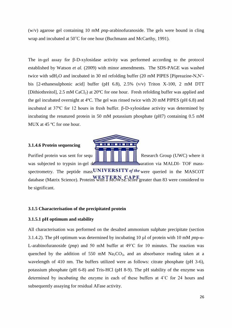
The efficacy of IEC separation was determined through the construction of a purification
table (Table 3.5) and SDS- PAGE analysis (Figure 3.11. A). Overall AFase yield achieved
was 1.2 % with a purification factor of 15.3. Purification of the AFase to homogeneity for
sequencing purposes was however not obtained. SDS-PAGE analysis revealed numerous
faint bands. Zymogram development for AFase and Xylosidase activity was unsuccessful.
The sizes of the proteins obtained in the active fraction was compared to the documented
molecular weights of AFases and appropriately sized bands excised from the SDS- PAGE
and sequenced (Figure 3.11. B).
Table 3.5: Purification table for the sub-cellular AFase produced by ORS #1.
Fraction Volume
(ml)
Total
protein
(mg)
Activity
(U)
Total
activity
(U)
Specific
activity
(U/mg)
Purification
(fold )
Yield
(%)
Crude 70 12.7 1700 119000 133.9 1 100
Ammonium
sulphate
precipitation
6 0.74 3559 2135.4 480.9 3.6 1.8
Q-sepharose 2.5 0.28 572.6 1431.5 2045 15.3 1.2
37
Figure 3.11: SDS-polyacrylamide gel electrophoresis of protein from ORS#1. A: Purification of the AFase.
1 and 7: Prestained PageRuler. 2: cell lysate. 3: 80% ammonium sulphate saturation. 4: Ion exchange
chromatography resulted in the visualisation of 7 distinct bands. 5: Nested AFase fraction separation. 6: IEC
fraction with dominant xylosidase activity. B: diagrammatic representation of the potential AFase bands excised
and sent for sequencing. 1: 10 µl protein. 2: 5µl protein. 3: Unstained PageRuler.
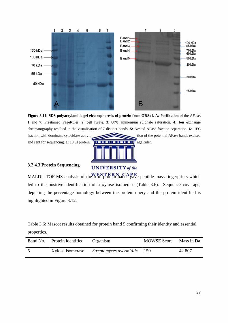
3.2.4.3 Protein Sequencing
MALDI- TOF MS analysis of the fifth protein band gave peptide mass fingerprints which
led to the positive identification of a xylose isomerase (Table 3.6). Sequence coverage,
depicting the percentage homology between the protein query and the protein identified is
highlighted in Figure 3.12.
Table 3.6: Mascot results obtained for protein band 5 confirming their identity and essential
properties.
Band No. Protein identified Organism MOWSE Score Mass in Da
5 Xylose Isomerase Streptomyces avermitilis 150 42 807

38
1 MNYQPTPEDR FTFGLWTVGW QGRDPFGDAT RRALDPVETV QRLAGLGAHG
51 VTFHDDDLIP FGSSDTERES HIKRFRQALD ATGMAVPMAT TNLFTHPVFK
101 DGAFTANDRD VRRYALRKTI RNIDLAAELG AKTYVAWGGR EGAESGAAKD
151 VRVALDRMKE AFDLLGEYVT AQGYDLRFAI EPKPNEPRGD ILLPTVGHAL
201 AFIERLERPE LYGVNPEVGH EQMAGLNFPH GIAQALWAGK LFHIDLNGQS
251 GIKYDQDLRF GAGDLRAAFW LVDLLESAGY EGPKHFDFKP PRTEDLDGVW
301 ASAAGCMRNY LILKERTAAF RADPEVQEAL RAARLDELAQ PTAGDGLTAL
351 LADRTAFEDF DVEAAAARGM AFEQLDQLAM DHLLGARG
Figure 3.12: Mascot results obtained for protein band 5 confirming its identity and essential properties. Alignment of polypeptide composition in the Mascot database illustrates the 33 % sequence coverage of band 5
where the highlighted red text depict the sequence similarities it shares with the xylose isomerase protein.
3.2.5 Characterisation of the crude α-L-arabinofuranosidase
Partially purified AFase active fractions obtained with IEC lost all activity within 24 hrs.
Characterisation studies were therefore performed with crude protein extract.
3.2.5.1 pH characterisation
AFase activity was optimum at pH 6 to 7 (Figure 3.13). No significant activity was observed
below pH 5 or above pH 9. At 4°C AFase activity was stable at pH 6 and pH 7 and retained
more than 98% of its activity for at least 24 hours (Figure 3.14).
Figure 3.13: pH optimum of the AFase. The α-L-arabinofuranosidase produced by ORS #1 function optimally
at pH7.
0
500
1000
1500
2000
2500
3000
3500
4000
5 6 7 8 9
Spe
cifi
c ac
tivi
ty in
µm
ol.
min
.mg
pH
pH optimum

39
Figure 3.14: pH stability at 4˚C for 24 hours. The enzyme is highly stabile at pH six and seven for 24 hours.
3.2.5.2 Temperature constraints
The AFase displayed a temperature optimum of 50˚C (Figure 3.15) and was stable at 40˚C for
up to 1.5 hours. AFase activity was rapidly lost with an increase in temperature, with no
residual activity detected after 30 minutes at 70˚C (Figure 3.16).
Figure 3.15: AFase activity over a range of temperatures. AFase activity was found to be optimal at 50ºC
-20
0
20
40
60
80
100
120
4 5 6 7 8 9
Re
lati
ve a
ctiv
ity
(%)
pH
pH stability
0
500
1000
1500
2000
2500
3000
3500
4000
4500
5000
30 40 50 60 70 80
Spe
cifi
c ac
tivi
ty (
µm
ol.
min
.mg)
Temperature (˚C)
temperature optimum

40
Figure 3.16: Stability of the AFase over a temperature range of 30˚C- 70˚C. The AFase is stable at 40ºC for
up to one hour and thereafter gradually starts to lose activity.
3.2.5.3 Kinetic parameters
The kinetic parameters were determined through non linear regression. Data was applied to
the Michaelis-Menten equation to generate the Michaelis-Menten curve (Figure 3.17). The α-
L-arabinofuranosidase displayed a Vmax of 3958 U.mg and a Km of 8.2 mM.
Figure 3.17: The Michaelis-Menten Curve for the kinetic activity of α-L-arabinofuranosidase on pnp-α-L-
arabinofuranoside.
-20
0
20
40
60
80
100
120
0.5 1 1.5 2 19
Re
lati
ve a
ctiv
ity
(%)
Time (hours)
30 Degrees
40 degress
50 Degress
60 Degrees

41
CHAPTER 4
GENERAL DISCUSSION AND CONCLUSION

42
CHAPTER 4
4.1 General Discussion
4.1.1 Identification of isolates with α-L-arabinofuranosidase activity
α-L-arabinofuranosidases (AFases) are hemicellulases which cleave arabinofuranosyl
residues from arabinose-rich polysaccharides and are industrially significant for the
synergistic effect they have on other hemicellulases and are therefore frequently isolated and
purified (Saha, 2000; Numan and Bhosle, 2006). Considering that Actinobacteria are
ubiquitous in terrestrial environments these organisms were expected to dominate Namibian
and Zambian soil isolates. Actinobacteria are well characterised for their biodegradative
capabilities (McCarthy and Williams, 1992) and are therefore a perfect reservoir for
lignocellulytic enzymes. Hemicellulose is comprised of many different forms of xylan to
which arabinofuranosyl residues are covalently linked. Screening for xylanolytic activity with
xylan as the inducer is thus instrumental in the isolation of AFases (Khanderparker et al.,
2008; Pei and Shao, 2008). Screening of the soil isolates for xylan degradation on the carbon
utilization media, ISP9, rendered seventeen positives. AFase activity is identified by the
cleavage of arabinofuranoside from the chromogenic substrate pnp-arabinofuranoside (pnp-
Araf) and is detected as a yellow product. Four isolates were able to hydrolyse pnp-Araf of
which three (ORS#1, NDS #4 and WBDS #9) with highest AFase activity were selected as
potential candidates for genebank construction pending phylogenetic analysis.
Because of its ubiquity and conservation of function, the 16S rRNA gene is the most
common housekeeping gene employed in determining bacterial phylogeny (Janda and
Abbott, 2007). Approximately 94% of the 1.5kb 16S rRNA gene was sequenced for all
isolates. Sequence analysis of WBDS #9 and NDS #4 revealed a 99 % homology with
Streptomyces radiopuqnans and Streptomyces collinus respectively. The 16S rRNA
sequence of ORS #1 shared 97 % homology with Streptomyces alborgriseolus, suggesting
that ORS #1 may be a novel Streptomyces species. The strain’s 16S rRNA gene falls just
short of the 97.5 % threshold value that delineates a bacterial species (Wayne et al., 1987,
Bauer et al., 2009). Novel organisms, especially those isolated from thermophilic
environments, are exciting as they are more likely to harbour unknown gene products and
enzymes with new catalytic mechanisms which are particularly interesting for

43
industrialization (Ferrer et al., 2007). In order to confirm that ORS#1 is truly a novel
organism, DNA-DNA hybridization studies would need to be conducted.
4.1.2 α-L-arabinofuranosidase gene discovery
Streptomyces species are well-documented for their ability to produce AFases and have
previously been isolated from genomic libraries through functional screening (Margolles-
Clark et al., 1996; Matsumura et al., 2004). In this project, functional screening of the cosmid
library containing the AFase-producing strains required an array of techniques, all of which
proved to be unsuccessful in the identification of an AFase. The liquid-phase screening
method that incorporated the substrate pnp-Araf was shown to be effective in the
identification of AFases from a yeast (Trichoderma reesei) expression library (Margolles-
Clarke et al., 1996). The adaptable solid-phase screen method devised by Lee et al (2009)
proved to be a robust method for detecting AFase activity under the amended conditions as
displayed by Bacillus subtilus 168 (Figure 1.4 C). This method also ensures the detection of
any AFase that may have been internalized by the host organism. Ineffective functional
screening experienced with the cosmid library therefore implies that no functional AFase was
translated by the E.coli host. Problems associated with heterologous expression of
Streptomyces genes may be the contributing factor. Differences in codon usage, regulatory
signals for transcription and post translational modification (PTM) between expression host
and the organism from which the gene originates is a major hurdle in the expression of
Actinobacteria genes (Connell, 2001). Transcription efficiency is determined by the
specificity of the interaction between RNA polymerase’s sigma (σ) factor with the promoter
region of a gene and is dependent on G+C content. The high G+C content of Actinobacteria
genes adversely affects the promoter recognition by prokaryotic bacterial σ factors resulting
in a limited degree of transcription (Helmann and Chamberlin, 1988; Patek et al., 1996).
Furthermore, α- L-arabinofuranosidase requires PTM in the form of glycosylation in order to
be active (Axamawaty et al., 1990; Filho et al., 1996). In most instances, heterologous host
systems lack the cellular machinery to perform these modifications (Nakashima et al., 2005).
Although the library was constructed in a Streptomyces-E.coli shuttle vector pDF666 in
anticipation of the problems associated with high G+C % genes, a great deal of fundamental
research is still necessary to render Streptomyces systems competitive for library screening.

44
A PCR screen based on consensus primers designed to specific AFase genes may be used to
confirm the presence of the AFase gene within the construct. This approach has had previous
success with an AFase from a metagenomic library (Wagschal et al., 2007). Considering this
approach, we set out to design consensus primers and subsequently used the gene product as
a probe during library screening. High levels of degeneracy were encountered at the DNA
level, and degenerate primers were designed based on DNA sequence of a few Streptomyces
strains. PCR amplification of the putative AFase gene was complicated due to the high G+C
content inherent to Actinomycete DNA. Initial amplification reactions were conducted under
conventional and gradient PCR conditions but with no success. Touchdown PCR, wherein the
template DNA anneals with primer over a gradient of ten degrees Celsius for eighteen cycles,
was employed and resulted in the amplification of a DNA fragment that correlated with the
estimated product size of 825 base pairs from Streptomyces isolate ORS #1 using the
KappaRobust system. The same sized band could however not be amplified from isolates
NDS #4 and WBDS #9 with any of the optimization parameters suggested by the
manufacturer. The PrimeStar high fidelity system produced faint bands with the expected size
for both these isolates but was irreproducible. The PrimeStar dNTP’s frequently hybridized
with the primer to produce DNA fragments of up to 803 base pairs evident in the negative
control, suggesting a problem with the PCR reagents. New reagents also produced no
amplification. PrimeStar has however been shown to effectively amplify an AFase from
Fibrobacter succinogenes (Yoshida et al., 2010) and a xylanase from Penicullium
griseofulvum (Tison et al., 2009).
Cloning of the approximate 825 base pair fragment from ORS #1 into E. coli produced an
approximate 1000 base pair fragment upon colony PCR. This indicates that the gene was
successfully integrated into the pGEMT backbone. Sequencing of the gene rendered an 843
base pair product which displayed 41% homology with a recombinase gene from
Streptomyces sp. SPB74 and 6.10% similarity with the Streptomyces AFases used to design
the degenerate primers. This suggests that the partial gene obtained is unlikely to be an
AFase. Successful amplification of the AFase gene from Streptomyces has been achieved by
Vincent et al. (1997), Tsujibo et al. (2002), Wagschal et al. (2007), and Ichinose et al.
(2008). Consensus primers were based on the sequence of specific GH families. This
approach was not considered as there was no way of determining which GH families our
isolates housed. Moreover, a literature survey revealed that Streptomyces chartreusis GS901

45
and Aspergillus awamori IFO 4033 are capable of producing multiple, functional AFases that
belong to different GH families (Kaneko et al., 1998; Matsuo et al., 2000).
4.1.3 Purification of the α-L-arabinofuranosidase
AFases have routinely been purified from both the culture filtrate and the cell free extract of
culturable microorganisms (Tajana et al., 1992; Sakamoto and Kawasaki, 2003; Pei and
Shao, 2008). In the current study, the growth rate of ORS #1 could not be reliably determined
through the conventional OD measurement method, as Streptomyces growth is filamentous
and the strain grew in the form of balls. A growth curve was therefore derived though
examination of the relationship between AFase activity and cellular proteins. In the current
study, intracellular protein concentration gradually increased over days one and two then
greatly increased on days three and four. This protein profile suggests that the organism
progresses from a lag phase (days 1 and two) into the exponential phase by day three, while
day four possibly denotes the stationary growth phase as suggested thereafter by the constant
decrease in AFase activity and the unaltered total protein concentration (data not shown).
Growth of ORS #1 in media containing xylan rendered optimal AFase synthesis on day three
and is consistent with the rate of AFase production displayed by Streptomyces lividans 66
(Manin et al., 1994). Other Streptomyces species, which include S. chartreusis GS901, S.
flavogriseus ATCC 33331 and S. olivochromogenes NBRC 2258, attained optimal AFase
levels on day four and in the case of Streptomyces sp. PC22, in five days (MacKenzie et al.,
1987; Matsuo et al., 2000; Raweesri et al., 2008). This suggests that ORS #1 is an efficient
AFase producer. Low levels of AFase activity was detected in the culture supernatant
although no protein was detected through CBB staining. The presence of the AFase in the
culture media may be a result of cell lysis as postulated by Manin et al. (1994).
Ion exchange chromatography separates proteins on the basis of ionic interactions and has
been described as the most frequently utilized technique for protein separation. This method
is routinely employed for its versatility, high protein–binding capacity, high resolving power
and efficacy (Karlsson and Ryden, 1998). Many AFases have been isolated using this
method as the first step in protein fractionation (Kaji et al., 1981; Gilead and Shoham, 1995;
Kaneko et al., 1998; Saha and Bothast, 1998; Matsuo et al., 2000; Sakamoto and Kawasaki,
2003; Shin et al., 2003; Matsumura et al., 2004). The use of the Q-Sepharose ion exchange

46
column in this work required some optimization to ensure thorough protein separation. By
decreasing the flow rate protein bands present in the active peak were reduced. AFase was
however not purified to homogeneity by ion exchange alone. Active fraction was applied to a
hydrophobic interaction column, from which the enzyme could not be recovered. This
phenomenon was unexpected as hydrophobic interaction is routinely used as either a primary
or secondary step in AFase purification (Nogawa et al., 1999; Birgisson et al., 2004). The
prominent protein bands revealed by SDS-PAGE were all treated as potential AFases as the
enzyme has been shown to have molecular weights of 108 kDa, 85 kDa, 70 kDa, 55 kDa, and
40 kDa (Hespell and O’Bryan, 1992; Bezalel et al., 1993; Beylot et al., 2001; Numan and
Bhosle, 2006; Fritz et al., 2008). Sequencing of the most prominent protein band in the
AFase active fraction revealed a protein with similarity to a 42 kDa xylose isomerase in the
MASCOT database. Xylose isomerase is an enzyme that plays a pivotal role in the pentose
phosphate cycle by converting D-xylose into D-xylulose, the initial step in xylose
assimilation (Sanchez and Smiley, 1975; Liu et al., 1996). This enzyme has not been well
documented though it is significant to note that a few xylose isomerases are bifunctional
enzymes that also display β-D-xylosidase activity (Belfaquih and Penninchx, 2000).
Coincidentally, many β-D-xylosidases have also been shown to display AFase activity
(Poutanen and Puls, 1988; Utt et al., 1991; Shao and Wiegel, 1992; Wagschal et al., 2007).
Xylose isomerases are not routinely tested for AFase activity so it is possible that this single
protein is responsible for all three activities. Fractions active for AFase activity were shown
to also display xylosidase activity. Xylose isomerase activity was however not confirmed.
All attempts at activity staining of the SDS-PAGE for AFase and Xylosidase functionality
through zymogram development were unsuccessful. The four remaining prominent bands
were also subjected to MALDI-TOF (results pending).
4.1.4 Preliminary characterisation
Determining the temperature and pH parameters of an enzyme is vital as these parameters
dictate the industrial viability of the enzyme. At temperatures higher than the optimal defined
for a given enzyme, the protein will become inactive thereby rendering the fermentation
process inefficient (Peterson et al., 2007). Fermentations conducted under thermophilic
conditions (45ºC- 80ºC) are highly desirable as the risk of contamination by omnipresent

47
mesophilic organisms is reduced, thereby ensuring maximal product yield (Haki and Rakshit,
2003). For their robust nature, thermostable enzymes are highly sought after to drive these
industrial processes. The thermoactivity of the AFase isolated in this study was optimal at
50ºC which corresponds to the thermoactivity reported for various AFase from Aspergillus
species and Penicillium chrysogenum (Van der Veen et al., 1991; Le Clinche et al, 1997;
Sakamoto and Kawasaki, 2003). This is surprisingly low considering that most known
thermophilic AFases display optimal activity between 60- 75ºC (Saha and Bothast, 1998;
Gilead and Shoham, 1995; Tsujibo et al., 2002; Tuncer and Ball, 2003; Canakci et al., 2008;
Pei and Shao, 2008). The AFase was most stable at 40ºC and retained 30% of its activity after
19 hours. The enzymes produced by Trichoderma reesei and Aspergillus niger are the only
other documented AFases that display optimal thermostability at 40ºC (Nogawa et al., 1999;
Yang et al., 2006). Other microbial AFases display thermostability ranging from 45ºC- 90ºC
(Tajana et al., 1992; Debeche et al., 2000; Margolles and de los Reyes-Gavilan., 2003;
Canacki et al., 2007; Morana et al., 2007; Canacki et al., 2008). The temperature profile of
AFase produced by Streptomyces isolate ORS #1 suggests that the enzyme is not an ideal
candidate for industrial thermophilic fermentations above 40°C. This AFase may be an ideal
candidate for use in mesophilic fermentation processes at 40°C (Numan and Bhosle, 2006).
It is imperative that the pH parameters of an enzyme be defined prior to its industrialization.
The pH of the environment affects the affinity of the enzyme for the substrate by altering the
charge on both the substrate and the enzymes active site, thus hindering catalytic activity
(Dixon, 1953). The partially purified AFase displayed an optimum range of pH 6-7 and
retained 100% of its activity at pH 7, whilst at 4ºC for at least 24 hours. This coincides with
the AFases purified from Streptomyces sp. PC22, (Raweesri et al., 2008), Streptomyces
lividans 66 (Manin et al., 1994) and Streptomyces avermitilis NBRC 14893 (Ichinose et al.,
2008). Some microbial AFases function optimally over a pH range of 2-8 (Uesaka et al.,
1978; Khanderparker et al., 2008). Many industrially utilized microorganisms function
optimally at neutral pH. The pH stability of this AFase ensures that it can be utilised in
mesophilic bioethanol fermentation wherein simultaneous saccharification is employed.
The catalytic activity of the AFase was determined by measuring the rate of hydrolysis of
pnp-arabinofuranoside under optimal conditions (40ºC, pH 7), over a substrate concentration
range of 5-25 mM. The Vmax and Km of the AFase was estimated to be 3958 U.mg and 8.223
mM, respectively. This is the second highest Vmax documented for an AFase and the highest

48
for a partially purified enzyme. The highest recorded Vmax for a purified AFase is 6610 U.mg,
isolated from Hordeum vulgare (Lee et al., 2003).
4.2 Conclusion/future work
α-L-arabinofuranosidase is a key enzyme in hemicellulose deconstruction for its cleavage of
arabinofuranosyl residues and the catalytic effect it has on other hemicellulases. This study
reports on an AFase isolated from a Streptomyces strain. The enzyme was partially purified
and characterized. Future work includes gene amplification and cloning into a protein
expression system.

49
CHAPTER 5
REFERENCES
50
CHAPTER 5
Adams, E.L., Kroon, P.A., Williamson, G., Gilbert, H.J. and Morrisa, V.J. 2004. Inactivated
enzymes as probes of the structure of arabinoxylans as observed by atomic force microscopy.
Carbohydrate Research. 339: 579- 590.
Ahmed, Z., Banu, H., Rahman, M.M., Akhter, F. and Haque, M.S. 2001. Microbial activity
on the degradation of lignocellulosic polysaccharides. Journal of Biological Sciences. 1: 993-
997.
Antoni. D., Zverlov, V.V. and Schwarz, W.H. 2007. Biofuels from microbes. Applied
Microbiology and Biotechnology. 77: 23- 35.
Aristidou, A. and Penttila, M. 2000. Metabolic engineering applications to renewable
resource utilization. Current Opinions in Biotechnology. 11: 187- 198.
Axamawaty, M.T., Fleet, G.W., Hannah, K.A., Namgoong, S.K. and Sinnott, M.L. 1990.
Inhibition of alpha-L-arabinofuranosidase III of Monilinia fructigena by 1,4-dideoxy-1,4-
imino-L-threitol and 1,4-dideoxy-1,4-imino-L-arabitol. Biochemistry, 266: 245-249.
Balat, M., Balat, H. And Oz, C. 2008. Progress in bioethanol processing. Progress in Energy
and Combustion Science. 34: 551- 573.
Buchmann, S.L. and McCarthy, A.J. 1991. Purification and co-operative activity of enzymes
constituting the xylan-degrading system of Thermomonospora fusca. Applied Microbiology
and Biotechnology. 57: 2121- 2130.
Bauer, R., Bekker, J.P., van Wyk, N., du Toit, C., Dicks, L.M.T. and Kossmann, J.
Exopolysaccharide production by lactose-hydrolyzing bacteria isolated from traditionally
fermented milk. International Journal of Food Microbiology. 131: 260- 264.
Beg, Q.K., Kapoor, M., Mahajan, L. and Hoondal, G.S. 2001. Microbial xylanases and their
industrial applications: a review. Applied Microbiology and Biotechnology. 56: 326- 338.
Belfaquih, N. And Penninckx, M.J. 2000. A bifunctional β-D-xylosidase–xylose isomerase
from Streptomyces sp. EC 10. Enzyme and Microbial Technology. 27: 114-121.

51
Beldman, G., Schols, H.A., Pitson, S.M., Searle-van Leeuwen, M.J.F. and Voragen, A.G.J.
1997. Arabinans and arabinose degrading enzymes. Advances in Macromolecules
Carbohydrate Research. 1: 1- 64.
Betts, W.B., Dart, R.K., Ball, A.S. and Pedlar, S.L. 1991. Biosynthesis and structure of
lignocellulose. Berlin, Germany. Springer-Verlag.
Beylot, M.H., Emani, K., McKie, V.A., Gilbert, H.J. and Pell, G. 2001. Pseudomonas
cellulosa expresses a single membrane-bound glycoside hydrolase family 51
arabinofuranosidase. Biochemistry Journal. 385: 599-605.
Bezalel, L., Shoham, Y. and Rosenberg, E. 1993. Characterization and delignification activity
of a thermostable alpha-L-arabinofuranosidase from Bacillus stearothermophilus. Applied
Microbiology and Biotechnology. 40: 57-62.
Birgisson, H., Fridjonsson, O., Bahrani-Mougeot, F.K., Hreggvidsson, G.O., Krisjansson,
J.K. and Mattiasson, B.A. 2004. New thermostable α-L-arabinofuranosidase from a novel
thermophilic bacterium. Biotechnology Letters. 26: 1347- 1351.
Blumer-Schutte, S.E., Kataeva, I., Westphiling, J., Adams, M.W.W. and Kelly, R.M. 2008.
Extreme thermophilic micro-organisms for biomass conversion: status and prospects. Current
Opinion in Biotechnology. 19: 210- 217.
Bohlmann, G.M. 2006. Process economic considerations for production of ethanol from
biomass feedstocks. Industrial Biotechnology. 2: 14- 20.
Bradford M.M. 1976. A rapid and sensitive for the quantitation of microgram quantities of
protein utilizing the principle of protein-dye binding. Analytical Biochemistry. 72: 248- 254.
Brown, M.A., Levine, M.D., Romm, J.P.R.A.H. and Koomey, J.G. 1998. Engineering
economic studies of energy technologies to reduces green house gas emissions: opportunities
and challenges. Annual Reviews in Energy and Environment. 23: 31- 39.
Canakci, S., Belduz, A.O., Saha, B.C., Yasar, A., Ayaz, F.A. and Yayli, N. 2007. Purification
and characterization of a highly thermostable alpha-L-arabinofuranosidase from Geobacillus
caldoxylolyticus TK4. Applied Microbiology and Biotechnology. 75: 813- 820.
Canakci, S., Kacagan, M., Inan, K., Osman Belduz, A. and Saha, B.C. 2008. Cloning,
purification and characterisation of a thermostable α-L-arabinofuranosidase from

52
Anoxybacillus kestanbolensis AC26Sari. Applied Microbiology and Biotechnology. 81: 61-
68.
Chum, H.L. and Overend, R.P. 2001. Biomass and renewable fuels. Fuel Processing and
Technology. 71: 187- 195.
Connell, N. D. 2001. Expression systems for use in Actinomycetes and related organisms.
Current Opinion in Biotechnology. 12: 446- 449.
Debeche, T., Cummings, N., Connerton, I., Debeire, P. and O'Donohue, M.J. 2000. Genetic
and biochemical characterization of a highly thermostable alpha-L-arabinofuranosidase from
Thermobacillus xylanilyticus. Applied Environmental Microbiology. 66: 1734- 1736.
De Vries, R.P., Kester, H.C.M., Poulsen, C.H., Benen, J.A.E. and Visser, J. 2000. Synergy
between enzymes from Aspergillus involved in the degradation of plant cell wall
polysaccharides. Carbohydrate Research. 327: 401- 410.
Deobald, L.A. and Crawford, D.L. 1997. Lignocellulose biodegradation. Washington DC.
ASM Press
De Vries, R.P. and Visser, J. 2001. Aspergillus enzymes involved in degradation of plant cell
wall polysaccharides. Microbiology and Molecular Biology Reviews. 65: 497- 522.
Dixon, M. 1953. The Effect of pH on the Affinities of Enzymes for Substrates and Inhibitors.
Biochemistry Journal. 55: 61- 70.
Ferre, H., Broberg, A., Duus, J.O. and Thomsen, K.K. 2000. A novel type of arabinoxylan
arabinofuranohydrolase isolated from germinated barley. Analysis of substrate preference and
specificity by nano-probe NMR. European Journal of Biochemistry.267: 6633- 6641.
Ferrer, M., Golyshina, O., Beloqui, A. and Golyshina, P.N. 2007. Mining enzymes from
extreme environments. Current Opinion in Microbiology. 10: 207- 214.
Filho, E. X., Puls, J. and Coughlan, M.P. 1996. Purification and characterisation of
arabinofuranosidases from solid-substrate cultures of the fungus Penicillium capsulatum.
Applied Environmental Microbiology. 62: 168- 178.

53
Fritz, M., Ravanal, M.C., Braet, C. and Eyzaguirre, J. 2008. A family 51 alpha-l-
arabinofuranosidase from Penicillium purpurogenum: purification, properties and amino acid
sequence. Mycology Research. 11: 933- 942.
Galbe, M. and Zacchi, G. 2002. A review of the ethanol production from softwood. Applied
Microbiology and Biotechnology. 59: 618- 628.
Gallagher, S.R. 1999. One-Dimensional-Electrophoresis using non-denaturing conditions.
Current Protocols in Molecular Biology Supplement. 47: 2B1- 11.
Gilead, S. and Shoham, Y. 1995. Purification and characterisation of a α-L-
arabinofuranosidase from Bacillus stearothermophilus T-6. Applied Environmental
Microbiology. 61: 170- 174.
Goodfellow, M. and Williams, S.T. 1983. Ecology of Actinomycetes. Annual Reviews in
Microbiology. 37: 189-216.
Grassi, G. 1999. Modern Bioenergy in the European Union. Renewable Energy. 16: 985- 990.
Gray, K.A., Zhao, L. and Emptage, M. 2006. Bioethanol. Current Opinion in Chemistry and
Biology. 10: 141- 146.
Hahn-Hagerdal, B., Galbe, M., Gorwa-Grauslund, M.F., Liden, G. and Zacchi, G. 2006.
Bioethanol- the fuel of tomorrow from the residues of today. Trends in Biotechnology. 24:
549- 556.
Haki, G.D. and Rakshit, S.K. 2003. Developments in industrially important thermostable
enzymes: a review. Bioresource Technology. 83: 17- 34.
Helmann, J.D. and Chamberlin, M.J. 1988. Structure and function of bacterial σ factors.
Annual Reviews in Biochemistry. 57: 839- 872.
Heredia, A., Jimenez, A. and Guillen, R. 1995. Composition of plant cell walls. Z Lebensm
Unters Forsch. 200: 24- 31.
Inacio, J.M., Correia, I.L. and de Sa-Nogueira, I. 2008. Two distinct arabinofuranosidases
contribute to arabino-oligosaccharide degradation in Bacillus subtilis. Microbiology. 154:
2719- 2729.

54
Inchinose, H., Yoshida, M., Fujimoto, Z. and Kaneko, S. 2008. Characterisation of a modular
enzyme of exo-1,5-α-L-arabinofuranosidase and an arabinan binding module from
Streptomyces avermitilis NBRC 14893. Applied Microbial Biotechnology. 80: 399- 408.
Ish-Horowicz, D. and Burke, J.F. 1981. Rapid and efficient cosmid cloning. Nucleic Acid
Research. 9: 2989- 2998.
Izydroczyk, M.S. and Biliaderis, C.G. 1995. Cereal arabinoxylans: advances in structure and
physiochemical properties. Carbohydrate Polymers. 28: 33- 48.
Janda, M.J. and Abbott, S.L. 2007. 16S rRNA gene sequence for bacterial identification in
the diagnostic laboratory. Journal of Clinical Microbiology. 45: 2761- 2764.
Janson, J-C. and Ryden, L. 1998. Ion Exchange chromatography. In Protein purification:
Principals, high resolution methods and applications. Second edition. New York: Wiley-
VCH, 145- 206.
Kaji, A. 1984. L-arabinosidases. Advances in Carbohydrate Chemistry and Biochemistry. 42:
382- 394
Kaji, A. and Tagawa, K. 1964. Studies on the enzymes acting on arabinan.V. Action of
arabinase producing Aspergillus niger. Nippon Nogei Kagaku Kaishi. 38: 580- 584.
Kaji, A., Tagawa, K. and Matsubara, K. 1967. Studies on enzymes acting on arabinan. VIII.
Purification and properties of arabinases produced by Aspergillus niger. Agriculture, Biology
and Chemistry. 31: 1023- 1028.
Kaji, A., Tagawa, K. and Ichimi, T. 1969. Properties of purified α-L-arabinofuranosidase
from Aspergillus niger. Biochimica et Biophysica Acta. 171: 186- 188.
Kaji, A. and Tagawa, K. 1970. Purification, crystallisation and amino acid composition of α-
L-arabinofuranosidase from Aspergillus niger. Biochimica et Biophysica Acta. 207: 456-464.
Kaji, A. and Yoshihara, O. 1971. Properties of purified α-L-arabinofuranosidase from
Corticium rolfsii. Biochimica et Biophysica Acta 250: 367– 71.
Kaji, A., Sato, M. and Tsutsui, Y. 1981. An α-L-arabinofuranosidase produced by wild type
Streptomyces sp. No. 17-1. Agriculture, Biology and Chemistry. 45: 925- 931.

55
Kaneko, S., Arimoto, M., Ohba, M., Kobayashi, H., Ishii, T. and Kusakabe, I. 1998.
Purification and substrate specificities of two α-L-arabinofuranosidases from Aspergillus
awamori IFO4033. Applied and Environmental Microbiology. 64: 4021- 4027.
Kellett, L.E., Poole, D.M., Ferreira, L.M.A, Durrant, A.J., Hazelwood, G.P. and Gilbert, H.J.
1990. Biochemistry Journal. 272: 369- 376.
Khanderparker, R., Numan, M.T.H., Mukherjee, B., Satwekar, A. and Bholse, N.B. 2008.
Purification and characterisation of α-L-arabinofuranosidase from Arthrobacter sp. MTCC
5214 in solid state fermentation. Process Biochemistry. 43: 707- 712.
Kumar, R., Singh, S. and Singh, O.V. 2008. Bioconversion of lignocellulose biomass:
biochemical and molecular prospective. Journal of Industrial Microbiology and
Biotechnology. 35: 377- 391
Laemmli, U.K. 1970. Cleavage of structural proteins during the assembly of the head of
bacteriophage T4. Nature. 227: 680- 685.
Le Clinche, F., Pinaga, F., Ramon, D. and Valles, S. 1997. alpha-L-arabinofuranosidases
from Aspergillus terreus with potential application in enology: Induction, purification, and
characterization. Journal of Agriculture and Food Chemistry. 45: 2379- 2383.
Lee, R.C., Hrmova, M., Burton, R.A., Lahnstein, J. and Fincher, G.B. 2003. Bifunctional
family of 3 glycoside hydrolases from barley with alpha-L-arabinofuranosidas and beta-D-
xylosidase activity. Journal of Biology and Chemistry. 278: 5377- 5387.
Lee, C.C., Wagschal, K., Kibblewhite-Accinelli, R.E., Orts, W.J., Robertson, G.H. and
Wong, D.W.S. 2009. An α-Glucuronidase enzyme activity assay adaptable for solid phase
screening. Applied Biochemistry and Biotechnology. 155: 314- 320.
Lin, Y. And Tanaka, S. 2006. Ethanol fermentation from biomass resources: current state and
prospect. Applied Microbiology and Biotechnology. 69: 627- 642.
Liu, S-Y., Wiegel, J. and Gherardini, FC. 1996. Purification and cloning of a thermostable
xylose (glucose) isomerase with an acidic pH optimum from Thermoanaerobacterium strain
JW/SL-YS 489. Journal of Bacteriology. 178: 5938–5945.

56
Lynd, L.R. 1996. Overview and evaluation of fuel ethanol production from cellulosic
biomass: technology, economics and the environment, a policy. Annual Reviews in Energy
and Environment. 403- 465.
MacKenzie, R.C., Bilous, D., Schineider, H. and Johnson, K.G. 1987. Induction of
cellulolytic and xylanolytic enzyme systems in Streptomyces spp. Applied Environmental
Microbiology. 53: 2853- 2839.
Malherbe, S. and Cloete, T.E. 2002. Lignocellulosic Biodegradation: fundementals and
applications. Reviews in Environmental Science and Biotechnology. 1: 105: 114.
Manin, C., Shareek, F., Morosoli, R. and Kleupfel, D. 1994. Purification and characterisation
of an α-L-arabinofuranosidase from Streptomyces lividans 66 and DNA sequence of the gene
(abfA). Biochemistry Journal. 302: 443- 449.
Margolles-Clark, E., Tenkanen, M., Nakari-Setala, T. and Penttila, M. 1996. Cloning of
genes encoding α-L-arabinofuranosidase and β-xylosidase from Trichoderma reesei by
Saccharomyces cerevisiae. Applied and Environmental Microbiology. 62: 3840- 3846.
Margolles, A. and de los Reyes-Gavilan, C.G. 2003. Purification and functional
characterisation of a novel α-L-arabinofuranosidase from Bifidobacterium longum B667.
Applied and Environmental Microbiology. 69: 5096- 5103.
Matsumura, K., Obata, H., Hata, Y., Kawato, A., Abe, Y. and Akita, O. 2004. Isolation and
characterisation of a novel gene encoding α-L-arabinofuranosidase from Aspergillus oryzae.
Journal of Bioscience and Bioengineering. 98: 77- 84.
Matuso, N., Kaneko, Kuno, A., Kobayashi, H. and Kusakabes, I. 2000. Purification,
characterisation and gene cloning of two α-L-arabinofuranosidase from Streptomyces
chartreusis GS901. Biochemical Journal. 34: 9- 15.
McCarthy, A.J. and William S.T. 1992. Actinomycetes as agents of biodegradation in the
environments- a review. Gene. 115: 189- 192.
McKendry, P. 2002. Energy production from biomass: overview of biomass. Bioresources
Technology. 83: 37- 43.

57
Miyazaki, K. 2005. Hyperthermophilic α-L-arabinofuranosidase from Thermotoga maritima
MSB8; molecular cloning, gene expression and characterisation of the recombinant protein.
Extremophiles. 9: 399- 406.
Monique, H., Fajj, A., Van den Broek, R., Berndes, G., Gielen, D. and Turkenburg, W. 2003.
Exploration of the ranges of global potential for biomass energy. Biomass Energy. 25: 119-
133.
Morana, A., Paris, O., Maurelli, L., Rossi, M. and Cannio, R. 2007. Gene cloning and
expression in Escherichia coli of a bi-functional beta-D-xylosidase/alpha-L-arabinosidase
from Sulfolobus solfataricus involved in xylan degradation. Extremophiles. 11: 123- 132.
Moreira, L.R.S. and Filho, E.X.F. 2008. An overview of mannan structure and mannan
degrading enzyme systems. Applied Microbiology and Biotechnology. 79: 165- 178.
Morrison, L. 2008. Second-generation biofuels may offer a way forward. Taylor.L. (ed).
www.engineeringnews.co.za. Accessed: 7 May 2009.
Mosier, N., Wyman,C., Dale, B., Elander, R., Lee, Y.Y., Holtzapple, M. and Ladisch, M.
2005. Features of promising technologies for pretreatment of lignocellulosic biomass.
Bioresource Technology. 96: 673- 686.
Nakashima, N. Mitani, Y. and Tamura, T. 2005. Actinomycetes as host cells for production
of recombinant proteins. Microbial Cell Factories. 4: 1- 5.
Nogawa, M., Yatsui, K., Tomioka, A., Okada, H. and Morikawa, Y. 1999. An α-L-
arabinofuranodidase from Trichoderma reesi containing a non catalytic xylan-binding
domain. Applied and Environmental Microbiology. 65: 3964- 3968.
Numan, M.T.H. and Bholse, N.B. 2006. α-L-arabinofuranosidases: the potential applications
in biotechnology. Journal of Industrial Microbiology and Biotechnology. 33: 247- 260.
Ostile, H.M., Vergarud, G. and Langsrud, T. 2002. Autolysis of Propionibacteria: detection
of autolytic enzymes by renaturing SDS-PAGE and additional buffer studies. International
Journal of Food Microbiology. 117: 167- 174.
Patek, M., Eikmanns, B.J., Patek, J. and Sahm, H. 1996. Promoters from Corynebacterium
glutamicum: cloning, molecular analysis and search for a consensus motif. Microbiology.
142: 1297- 1309.

58
Pei, J. and Shao, W. 2008. Purification and characterisation of an extra-cellular α-L-
arabinosidase from novel Bacillus pumilus ARA and its over expression in Eschericia coli.
Applied Microbiology and Biotechnology. 78: 115- 121.
Peterson, M.E., Daniel, R.M., Danson, M.J. and Eisenthal, R. 2007. The dependence of
enzyme activity on temperature: determination and validation of parameters. Biochemistry
Journal. 402: 331-337.
Perez, J., Munoz-Dorado, J., De La Rubia, T. and Martinez, J. 2002. Biodegradation and
biological treatments of cellulose, hemicellulose and lignin: an overview. International
Microbiology. 5: 53- 63.
Polizeli, M.L.T., Rizatti, A.C.S., Monti, R., Terenzi, H.F., Jorge, J.A. and Amorim, D.S.
2005. Xylanases from fungi: properties and industrial applications. Applied Microbiology and
Biotechnology. 67: 577- 591.
Poutanen, K. and Puls, J. 1988. Characteristics of Trichoderma reesei β-xylosidase and its
use in the hydrolysis of solubilised xylans. Applied Microbiology and Biotechnology. 28:
425- 432.
Raweesri, P., Riangrungrojana, P. And Pinphanichakarn, P. 2008. α-L-arabinofuranosidase
from Streptomyces sp. PC22: Purification, characterisation and its synergistic action with
xylanolytic enzymes in the degradation of xylan and agricultural residues. Bioresource
Technology. 99: 8981- 8986.
Rosegrant, M.W. 2008. Biofuels and grain prices: impacts and policy response. International
Food Policy Research Institute. Washington DC, USA.
Rye, C.S. and Withers, S.G. 2000. Glycosidase mechanisms. Current Opinion in Chemical
Biology. 4: 573- 580.
Saha, B.C. and Bothast, R.J. 1998. Purification and characterisation of a novel thermostable
α-L-arabinofuranosidase from a color-variant strain of Aureobasidium pullulans. Applied and
Environmental Microbiology. 64: 216- 220.
Saha, B.C. 2000. α-L-arabinofuranosidases: biochemistry, molecular biology and application
in biotechnology. Biotechnology Advances. 18: 403- 423.

59
Saha, B.C. 2003. Hemicellulose Bioconversion. Journal of Industrial Microbiology and
Biotechnology. 30: 279- 291.
Saha, B.C. and Bothast, R. 1998. Purification and characterisation of a novel thermostable α-
L-arabinofuranosidase from a colour-variant strain of Aureobasidium pullulans. Applied and
Environmental Microbiology. 60: 216- 220.
Sakamoto, T. and Kawasaki, H. 2003. Purification and properties of two type-B α-L-
arabinofuranosidase produced by Penicillium chrysogenum. Biochimica et Biophysica Acta.
1621: 204- 210.
Sanchez, C. 2009. Lignocellulose residues: biodegradation and bioconversion by fungi.
Biotechnology Advances. 27: 185- 194.
Sanchez, S and Smiley, K.L. 1975. Properties of D-Xylose Isomerase from Streptomyces
albus. Applied Microbiolgy. 29: 745-750.
Sanchez, O.J. and Cardona, C.A. 2008. Trends in biotechnological production of fuel ethanol
from different feedstocks. Bioresource Technology. 99: 5270- 5295.
Shallom, D. and Shoham, Y. 2003. Microbial hemicellulases. Current Opinion in
Microbiology. 6: 219- 228.
Shallom, D., Belakhov, V., Solomon, D., Gilead-Gropper, S., Baasov, T., Shoham, G. and
Shoham, Y. 2002. Identification of the acid-base catalyst of α-L-arabinofuranosidase from
Geobacillus stearothermophilus T-6, a family 51 glycoside hydrolase. FEBS Letters. 514:
163- 167.
Shao, W. and Wiegel, J. 1992. Purification and characterisation of a thermostable β-
xylosidase from Thermoanaerobacter ethanolicus. Journal of Bacteriology. 174: 5848- 5853.
Shin, H-Y., Park, S-Y., Sung, J.H. and Kim, D-H. 2003. Purification and characterisation of
α-L-arabinopyranosidase and α-L-arabinofuranosidase from Bifidobacterium breve K-110, a
human intestinal anaerobic bacterium metabolizing Ginsenoside Rb2 and Rc. Applied and
Environmental Microbiology. 69: 7116- 7123.
Shirling, E.B. and Gottlieb, D. 1966. Methods for characterisation of Streptomyces species.
International Journal of Systematic Bacteriology. 16: 313- 340.
60
Smith, M.M. and Hartley, R.D. 1983. Occurrence and nature of ferulic acid substitution of
cell wall polysaccharides in graminaceous plants. Carbohydrate Research. 118: 65- 80.
Sommer, P., Georgieva, T. and Ahring, B.K. 2004. Potential for using thermophilic anaerobic
bacteria for bioethanol production from hemicellulose. Biochemical Society Transactions. 32:
282- 288.
Steward, D., Azzini, A. and Morrison, I. 1997. Sisal fibres and their constituent non-
cellulosic polymers. Industrial Crop Production. 6: 17- 26.
Sun, Y. And Chen, J. 2002. Hydrolysis of lignocellulosic material for ethanol production: A
review. Bioeresource Technology. 83: 1- 11.
Tajana, E., Feichter, A. and Zimmermann, W. 1992. Purification and characterisation of two
α-L-arabinofuranosidases from Streptomyces diastaticus. Applied and Environmental
Microbiology. 58: 1447- 1450.
Takao, M., Akiyama, K. and Sakai, T. 2003. Purification and characterisation of the
thermostable endo-1,5- α-L-arabinase from strains of Bacillus thermodentrificans. Applied
Environmental Microbiology. 68: 1639- 1646.
Thomas, J.A. 1993. Molecular biology of xylan degradation. FEMS Microbiology Reviews.
10: 65- 82.
Tison, M.C., Andre-Leroux, G., Lafond, M. Georis, J., Juge, N., and Berrin J-G. 2009.
Molecular determinants of substrate and inhibitor specificities of Penicillium griseofulvum
family 11 xylanases. Biochemica et Biophysica Acta- Proteins and Proteomics. 1804: 438-
445.
Tsujibo, H., Takada, C., Wakamatsu, Y., Kosaka, M., Tsuji, A., Miyamoto, K. and Inamori,
Y. 2002. Cloning and expression of an α-L-arabinofuranosidase gene (stxIV) from
Streptomyces thermoviolaceus OPC- 520. Bioscience, Biotechnology and Biochemistry. 66:
434- 438.
Tuncer, M. 2000. Characterisation of β-xylosidase and α-L-arabinofuranosidase activities
from Thermomonospora fusca BD25. Turkish Journal of Biology, 24: 753-767.

61
Tuncer, M., Ball, A.S., Rob, A. and Wilson, M.T. 1999. Optimization of extracellular
lignocellulolytic enzyme production by the thermophilic Actinomycete Thermomonospora
fusca BD25. Enzyme Microbiology and Technology. 25: 38- 47.
Tuncer, M. and Ball, A.S. 2003. Co-operative actions and degradation analysis of purified
xylan-degrading enzymes from Thermomonospora Fusca BD25 on oat-spelt xylan. Journal
of Applied Microbiology. 94: 1030- 1035.
Uesaka, E., Sato, M., Raiju, M. and Kaji. 1978. α-L-arabinofuranosidase from Rhodotorula
flava. Journal of Bacteriology. 133: 1073- 1077.
Uffen, R.L. 1997. Xylan degradation: a glimpse of microbial diversity. Journal of Industrial
Microbiology and Biotechnology. 19: 1- 6.
Utt, E.A, Eddy, C.K., Keshav, K.F. and Ingram, L.O. 1991. Sequencing and expression of
Butyrivibrio firisolvens XylB gene encoding a novel bifunctional protein with β-D-xylosidase
and α-L-arabinofuranosidase activities. Applied Environmental Microbiology, 57: 1227-
12234.
Van der Veen, P., Flipphi, M.J.A., Voragen, A.G.J. and Visser, J. 1991. Induction,
purification and characterisation of arabinases produced by Aspergillus niger. Archives of
Microbiology. 15: 23- 28.
Van Laere, K.M.J., Beldman, G. and Voragen, A.G.J. 1997. A new arabinofuranohydrolase
from Bifidobacterium adolescentis able to remove the arabinosyl residue from double
substituted xylose units in arabinoxylan. Applied Microbiology and Biotechnology. 47: 231-
235.
Van Laere, K.M.J., Voragen, C.H.L., Kroef, T., Van den Broek, L.A.M., Beldman, G. and
Voragen, A.G.J. 1999. Purification and mode of action of two different arabinoxylan
arabinofuranohydrolases from Bifidobacterium adolescentis DSM 20083. Applied
Microbiology and Biotechnology. 51: 606- 613.
Vincent, P., Shareck, F., Dupont, C., Morosoli, R. and Kluepfel, D. 1997. New α-L-
arabinofuranosidase produced by Streptomyces lividans: cloning and DNA sequencing of the
afB gene and characterisation of the enzyme. Biochemistry Journal. 322: 845- 852.

62
von Sivers, M. and Zacchi, G. 1996. Ethanol from Lignocellulosics; a review of the economy.
Bioresource Technology. 56: 131- 140.
Wagschal, K., Heng, C., Lee, C.C. and Wong, D.W.S. 2009. Biochemical characterisation of
a novel dual function arabinofuranosidase/ xylosidase isolated from a compost starter
mixture. Applied Microbiology and Biotechnology. 81: 855- 863.
Wang, Y., Zhang, Z. And Ruan, J. 1996. A proposal to transfer Microbispora bispora
(Lechevalier 1965) to a new genus, Thermobispora gen.nov., as Thermobispora bispora
comb nov. International Journal of Systematic Bacteriology. 46: 933- 938.
Watson, B. J., Zhang, H., Longmire, A. G., Moon, Y. H. and Hutcheson, S.W. 2009.
Processive endoglucanases mediate degradation of cellulose by Saccharophagus degradans.
Journal of Bacteriology. 191: 5697– 5705.
Wayne, L.G., Brenner, D.J., Cowell, R.R., Grimont, P.A.D., Kandler, O., Krichevsky, M.I.,
Moore, H.L., Moore, W.E.C., Murray, R.G.E., Stackebrandt, E., star, M.P. and Trupper, H.
G. 1987. Report of the ad hoc committee on reconciliation of of approaches to bacterial
systematic. International Journal of systematic Bacteriology. 29: 463- 464.
Wheals, A.E., Basso, L.C., Alves, D.M.G. and Amorim, H.V. 1999. Fuel ethanol after 25
years. Trends in Biotechnology. 17: 482- 487.
Wiselogel, A., Tyson, J. and Johnsson, D. 1996. Biomass feedstock resources and
composition. In Handbook on bioethanol: production and utilization Wyman, C.E. (ed).
Washington DC: Taylor and Frances, 19- 30.
Wyman, C.E. 2003. Potential synergies and challenges in refining cellulosic biomass to
biofuels, chemicals and power. Biotechnology Progress. 19: 254-262.
Yang, H., Ichinose, H., Nakajima, M., Kobayashi, H. and Kaneko, S. 2006. Synergy between
and alpha-L-arabinofuranosidase from Aspergillus oryzae and an endo-arabinanase from
Streptomyces coelicolor for degradation of arabinan. Food Science Technology Research. 12:
43- 49.
Yoshida, S., Hespen, C.W., Beverly, R.L., Mackie, R.I. and Cann I.K.O. 2010. Domain
analysis of a modular α-L-arabinofuranosidase with a unique carbohydrate binding strategy
from the fibre degrading bacterium Fibrobacter succenogenes. Journal of Bacteriology. 585:
483- 493.

63
Zaldivar, J., Neilsen, J. and Olsson, L. 2001. Fuel ethanol production from lignocellulose: a
challenge for metabolic engineering. Applied Microbiology and Biotechnology. 56: 17- 34.
Internet resources
http://www.biosmartpackaging.com. Accessed: 7 May 2009
http://www.khalipakistan.com/trade/html Accessed: 7 May 2009
http://www.moosecrossingardencenter.com/store Accessed: 7 May 2009
http://www.sciencedaily.com/releases/2007/07/070709103138.htm Accessed: 7 September
2009