Embed Size (px)
Citation preview
Identification and annotation of non-protein coding
RNAs.
Kristin Reiche, Katharina Schutt, Kerstin Ullmann, Friedemann Horn,
Jorg Hackermuller
December 14, 2009
1 Introduction
Of the 3.3 billion bases of the human genome, only about 2% code for proteins. Since
very recently, the remaining 98% have been considered to be ’junk’ and functionless. How-
ever, large transcriptomic studies like ENCODE (ENCyclopedia Of DNA Elements) (1) or
FANTOM (The Functional Annotation Of the Mammalian Genome) (2) have shown that
around 90% of the genome is actively transcribed into RNA. The pilot phase of the ENCODE
Project was focused on 1% of the human genome sequence and was organized as an interna-
tional consortium of computational and laboratory-based scientists working to develop and
apply high-throughput approaches for detecting all sequence elements that confer biological
function. The studies advanced the collective knowledge about human genome function in
several major areas. The ENCODE project provided convincing evidence that the genome
is pervasively transcribed and the systematic examination of transcriptional regulation has
yielded new understanding about transcription start sites, including their relationship to spe-
cific regulatory sequences and features of chromatin accessibility and histone modification.
Beside this, the studies offered a more sophisticated view on chromatin structure and inter-
and intra-species sequence comparison with respect to mammalian evolution has yielded
new mechanistic and evolutionary insights concerning the functional landscape of the hu-
1
man genome (3). These findings stay in contrast to the so called ’central dogma of molecular
biology’, stating that DNA is transcribed into mRNA, which is then translated into protein,
based on the assumption that only DNA that encodes a protein is transcribed into RNA.
The potential of RNA as regulatory molecules has been revealed decades ago. Many classes
of RNA regulators in species ranging from viruses to mammalian have been discovered being
involved in the control of RNA stability, gene expression, tissue and cellular development,
RNA modification, chromatin organization, alternative splicing, sub-cellular localization of
proteins, heat shock sensing and other processes (4). RNAs, in particular non-coding RNAs
and other functional RNAs, are characterized by their base sequence and higher order struc-
tural motifs. Short- and long-range base pair interactions that organize the molecules into
domains are the main characteristics of those RNAs and provide a framework for functional
interactions. RNA molecules fold into characteristic secondary and tertiary structures that
are the base for their diverse functional activities. The RNA secondary structure consists
of nucleotides that are either paired or unpaired, where pairing includes all base-base inter-
actions. Most base pairings are adjacent and anti parallel with other base pairings to form
secondary structures (5; 6). On the one hand secondary structures of RNAs are responsi-
ble for their diverse functional activities and help finding a certain functional classification
for RNAs. On the other hand RNA structure, in particular secondary structures and their
search and prediction algorithms, may help to classify RNAs without knowing functional
details. Ongoing studies provide more and more evidence that the ’junk’ non-protein cod-
ing RNAs (ncRNA) have important regulatory function and could explain the complexity of
higher organisms, as the amount of ncRNAs increases dramatically during evolution, whereas
the number of protein-coding genes remains relatively constant. Additionally, compared to
mRNAs, ncRNAs are exceedingly cell-type specific expressed and regulated (1). A limited
number of trans-acting small ncRNAs have been described in prokaryotes that appear mainly
to regulate mRNA translation or stability. However, ncRNAs do not dominate genomic out-
put in prokaryotes, representing only a small fraction of their genomes, which are generally
dominated (80-95%) by protein-coding sequences (7; 8). In humans, and other eukaryotes,
the number of identified functional ncRNAs has increased tremendously during the last few
2

years, but most ncRNAs and especially their function still remain to be elucidated. Tak-
ing their functional repertoire into account, ncRNAs can be divided into two classes: (1)
housekeeping and (2) regulatory ncRNAs. Housekeeping RNAs are transfer RNAs (tRNAs)
and ribosomal RNAs (rRNAs) involved in mRNA translation, small nuclear (snRNAs) in-
volved in splicing, small nucleolar (snoRNAs) involved in the modification of rRNAs, RNase
P RNAs, so called Ribozymes, telomerase RNA and others. Whereas housekeeping ncRNAs
are generally constitutively expressed and are required for normal cell functions, the class of
regulatory ncRNAs or riboregulators are expressed at certain stages of development, during
cell proliferation or as a response to external stimuli (9). During the past few years mi-
croRNAs, ∼21nt long RNAs, which regulate mRNAs at the post-transcriptional level, have
become extremely popular, as they seem to be involved in crucial biological processes, like
development, differentiation and apoptosis (10). Initially, discovered in C.elegans (11; 12),
they are today known to be expressed in plants and throughout the animal kingdom, in-
cluding humans, and many of them are evolutionary conserved (13; 14). MiRNAs bind to
partially complementary target sites in the 3’UTR of their target mRNAs which leads either
to mRNA degradation or translational repression, depending on the grade of complementary.
Another class of small ncRNAs, are the Piwi-RNAs (piRNAs). PiRNAs form RNA-protein
complexes through interactions with Piwi proteins. These piRNA complexes have been
linked to transcriptional gene silencing of retro-transposons and other genetic elements in
germ line cells, particularly those in spermatogenesis (15). In contrast to the various classes
of small ncRNAs, the long ncRNAs, with a length >200nt, lack satisfactory classifications.
The broad functional repertoire of long ncRNAs includes epigenetic mechanisms, as well as
transcriptional and post-transcriptional regulation. The large non-coding RNA Xist is the
master regulator of X chromosome inactivation. Xist is negatively regulated by its antisense
transcript Tsix. This repressive antisense transcription across Xist operates at least in part
through the modification of the chromatin environment of the locus (16). Long-ncRNAs
can act as co-factors, by modulating transcription factor activity, they can also effect global
changes by interacting with basal components of the RNA polymerase II (RNAPII) depen-
dent transcription machinery, and they can regulate RNAPII activity, e.g. by influencing
3
promoter choice. For example, in humans an ncRNA transcribed from an upstream region of
the dihydrofolate reductase (DHFR) locus forms a triplex in the major promoter of DHFR
to prevent the binding of the transcriptional co-factor TFIID (17). Most mammalian genes
express antisense transcripts, which might constitute a class of ncRNA that is particularly
adept at regulating mRNA dynamics. They have been shown to direct the alternative splic-
ing of mRNA isoforms, for example Zeb2 (18), or alternatively the annealing of ncRNA
can target protein effector complexes to the sense mRNA transcript in a manner analogous
to the targeting of the RNA-induced silencing complex (RISC) to mRNAs by siRNAs (19).
Computational and experimental identification as well as functional classification of ncRNAs
will be described in the following sections.
2 ncRNA datasets
In this section we will briefly review available datasets of ncRNAs. In the field of non-coding
RNAs computational prediction of functional ncRNAs and experimental identification of
expressed RNAs were neck-to-neck to each other. Bioinformatic prediction delivered putative
RNAs that showed signs of stabilizing selection, i.e. of functionality but lacked knowledge of
expression. Experimental approaches delivered numerous expressed ncRNAs without being
able to delineate their functional relevance. We will therefore present datasets of predicted
RNAs as well as datasets of large scale experimental efforts for their identification. Due to
the dynamics of this field which has recently developed, this review cannot be comprehensive
and we mainly focus on the complex datasets derived for mammalian species, despite the
many interesting reports on functional ncRNAs in prokaryotes and more basal eukaryotic
model organisms.
2.1 Datasets of computationally predicted ncRNAs
Prediction of structured ncRNAs. Many of the long known, ’house-keeping’ ncRNAs,
like tRNAs, snRNAs or snoRNAs, exhibit pronounced secondary structure features which
are under stabilizing selection, e.g. because a particular structure is required for interaction
4
with protein complexes. It was therefore suggested that functional RNA elements should
have a secondary structure that is energetically more stable than expected by chance (20).
Thermodynamic stability alone did, however, not prove to be statistically significant enough
for ncRNA detection (21). Comparative approaches, in contrast, aimed to detect conserved
RNA secondary structure in sequence alignments. qrna, which was the first successful al-
gorithm of this type, compares which of three models describes a given pairwise sequence
alignment best. A pair stochastic context free grammar (SCFG) is used to model RNA sec-
ondary structure evolution. A pair Hidden Markov Model describes protein coding sequence
evolution and another pair HMM represent the null model of an unconstrained sequence
(22). An up-to-date extension of qrna to multiple sequence alignments (MSAs) is evofold,
which combines SCFGs to model RNA secondary structure with a phylogenetic tree to model
substitution rates along the branches of the tree (23). Human genome wide predictions of
structured RNAs using evofold are provided as a track in the UCSC genome browser.
An alternative strategy to the prediction of RNAs with secondary structure under sta-
bilizing selection is RNAz which is based on thermodynamic RNA folding. It determines a
structure conservation index (SCI) obtained by comparing folding energies of the individ-
ual sequences with the predicted consensus folding of a multiple sequence alignment as one
classification criterion and a z-score measuring thermodynamic stability of the individual se-
quences as a second. Both measures are combined by a support vector machine that detects
conserved and stable RNA secondary structures with high sensitivity and specificity (24).
RNAz has been applied to detect ncRNAs in the human genome (25), data are available
as bed files at http://www.tbi.univie.ac.at/papers/SUPPLEMENTS/ncRNA/, for Urochor-
dates (26), Nematodes (27), Drosophilids (28), yeast (29), Plasmodia (30) and teleost fishes
(31). Estimation of false discovery rates (FDR) for alignment based methods like RNAz is
intricate as it requires the randomization of alignments which is easily biased. A solution to
this issue is SISSIz, which generates random alignments satisfying various constraints and
has been used for more accurate FDR estimates of RNAz (32). RNAz 2.0 has been recently
released (33) and a web server is available for small scale predictions (34). Setting up your
own RNAz screen is described in (35; 36) and may require computation of dedicated multiple
5
sequence alignments for which NcDNAlign provides a solution adjusted to the needs of RNAz
(37).
Sequence alignment independent approaches. The above described approaches have
the disadvantage that no reliable multiple sequence alignments may be available, because
only distantly related genomes are available, or due to the rapid evolution of non-coding
sequences. This can be overcome by relying on structural alignments, however, at the cost
of a huge computational effort. Variants of the Sankoff algorithm, which solves RNA folding
and sequence alignment simultaneously (38), have been used for ncRNA prediction e.g.:
a classifier based on Dynalign (39), or a screen using foldalign to compare sequences
between human and mouse which are not alignable on primary sequence level (40). While
these and similar approaches can be used for ncRNA screens, a genome wide application is
usually not feasible or only when outstanding computational resources are available. Apart
from ncRNA identification, these approaches are useful for classification of ncRNAs, see the
following chapter.
Secondary structure independent prediction of ncRNAs. While the different ap-
proaches for detecting structured ncRNAs delivered a plethora of potentially functional ncR-
NAs, not all to date known ncRNAs have easily detectable structural features or contain
only few structured domains in a longer unstructured sequence, which is in particular true
for most so far identified mRNA-like ncRNAs. Also, the overlaps between ncRNAs detected
experimentally in ENCODE and those predicted by evofold and RNAz were surprisingly
small (41). A recently published approach that successfully identified novel unstructured
ncRNAs in Drosophila melanogaster relied on the detection of conserved intron positions in
otherwise largely non-conserved sequence context (42).
2.2 Datasets of experimentally identified ncRNAs
We briefly introduce large scale studies that led to the identification of non-coding tran-
scripts. Approaches aimed at finding individual ncRNAs cannot be covered, but we refer
6
to the databases listed in Section 4 which collect these transcripts. An experimental iden-
tification of ncRNAs has one important prerequisite: methods need to be unbiased, i.e.
detection is not restricted to a particular subset of RNAs, like polyadenylated transcripts.
Also, as many ncRNAs are – compared to mRNAs – expressed at low levels, approaches
for identifying novel ncRNAs benefit from increased sensitivity. Over the last years, unbi-
ased transcriptomics using tiling arrays and sequencing of cDNA and EST libraries have been
most successful in finding new ncRNAs. More recently, transcriptome sequencing (RNAseq),
approaches identifying RNAs bound to proteins and identification of ncRNAs based on epi-
genetic patterns proved to be of value.
ncRNA identification by transcriptomics approaches. Microarrays have been used
since the 1990s for quantifying the expression of known transcripts. Phil Kapranov (43),
Eric Schadt (44), Paul Bertone (45) and Viktor Stolc (46) and colleagues pioneered the
application of tiling arrays to identify novel transcripts. Tiling arrays are oligonucleotide
microarrays that unlike other arrays do not interrogate the sequences of specific transcripts
but rather tile the genome of interest. I.e., probes are spaced in regular intervals throughout
the genome. Typically, probes with low complexity or repetitive sequences are removed as
their signal cannot be attributed to a specific genomic location. Apart from that and the
variation of probe lengths for designs relying on longer probes, no probe design is possible for
this type of arrays and hybridization free energies vary strongly between probes. Together
with cross-hybridization effects this leads to rather noisy signals which require a stringent
processing. Therefore, Kampa et. al. (47) proposed to use a stringent cutoff for considering
individual probes as expressed and to require at least three consecutive probes to have signal
above this cutoff to designate a region as expressed. With a probe pitch of 35nt in many
whole genome tiling experiments, this results in the efficient detection of transcripts which
are at least approximately 110 nt in size. In (48) arrays with a resolution of 5nt were used
to extend the approach to the detection of small RNAs. However, with the need of using
around 180 arrays per sample to accommodate the resulting number of probes this approach
is hardly practical for whole genome applications. Different tiling array approaches have been
7
a mainstay of the pilot phase of the ENCODE project (49). Based on this data, ENCODE
concluded that more than 90 % of the human genome is transcribed into RNA on at least one
strand. Also, comparing expression data of several cell lines and tissues it was shown that on
average expression patterns of ncRNAs are far more specific than those of mRNAs. Datasets
of the ENCODE pilot phase and ongoing ENCODE studies are most easily accessible via
the UCSC genome browser. A disadvantage of tiling arrays is the locality of information,
i.e. transcript starts and ends can be inferred only indirectly based on segmentation of the
expression signal into blocks of similar expression. Tiling array data are therefore ideally
complemented by techniques like Cap-analysis gene expression (CAGE) tags, or paired end
tags (PET).
With the rapid development of second generation sequencing techniques transcriptome
sequencing (RNAseq) became a viable alternative to array based approaches, in particular
for small RNAs (50). Compared to tiling arrays RNAseq methods have the advantage
that sequences of any size that can be reliably mapped to the genome or assembled can
be identified. On the other hand, library preparation for RNAseq may more biased than
labeling and amplification for tiling arrays. Also, the capacity of today’s sequencers may
still be limiting for identifying differential expression of long ncRNAs in complex samples.
See chapter XX for details on RNA-seq.
NcRNAs identified from cDNA clones. FANTOM, the functional annotation of the
mouse transcriptome project initially identified numerous ncRNAs from sequencing full
length cDNA clones (51; 52). Subsequent FANTOM studies supplemented cDNA data by
massive identification of transcription start and termination sites (53) and most recently
combining CAGE (Cap-analysis gene expression) with deep sequencing (deepCAGE) (54).
In particular the latter technique alleviates one of the major down-sides of purely clone based
approaches, which hardly capture differential expression.
Epigenetics based approaches. Recently Mitchell Guttman and colleagues introduced
an approach for identifying ncRNAs that is orthogonal to transcriptomics (55): Studying the
profiles of specific histone modifications in and around protein coding genes, they developed
8
an epigenetic signature of actively transcribed genes and used these patterns to identify a
series of novel transcripts called lincRNAs.
3 Methods for ncRNA annotation
While genome databases offer a wealth of information about known and putative functions
for protein-coding genes, functional information for novel non-coding RNAs (ncRNA) genes
is almost non-existent. This is mainly explained by the lack of established software tools
to efficiently identify the function and evolutionary origin of ncRNAs. Cellular functions of
ncRNAs are either defined by structure motifs, sequence motifs or a combination of both,
which is a consequence of their action in RNA-(m)RNA or RNA-protein complexes. In or-
der to preserve the capability of interaction, evolutionary selection of ncRNA genes ensures
conservation of these motifs. NcRNAs of the same evolutionary origin are defined to belong
to the same RNA family. Members of the same ncRNA family do in general share strong
sequence similarity, which decreases for ancestral homologies, while secondary structure may
still be conserved. RNA families sharing the same evolutionary origin but no sequence sim-
ilarity are defined to be members of the same RNA clade. Lastly, in case RNA families did
not evolve from the same origin, but their similar cellular functions converged to similar
secondary structures they are seen as members of the same RNA class.
To classify new ncRNA genes (either retrieved by genome-wide high-throughput experi-
mental transcriptomics, or genome-wide computational screens) one may follow the outline
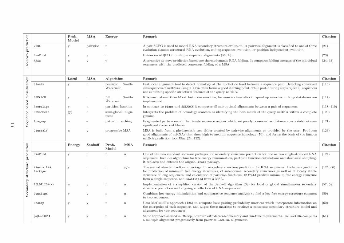
described here. A selection of the most important software tools which address the problem
of classifying ncRNA genes is given in table 1.
Sequence-based classification searches for ncRNAs that are conserved in their pri-
mary sequence and, thus, are ncRNAs exhibiting close homology as well as similar cellular
functionality. With standard local sequence alignment tools like blast , good results can be
achieved, already. However, sequence conservation in ncRNAs is mostly restricted to rather
short regions of sequence motifs being functional that are interrupted by structure motifs.
9
The most promising approaches to detect homologous ncRNAs by sequence conservation
are searches for fragmented patterns that do not require a conserved substring of sufficient
length as seed region for the local alignment (56). Regions of poor conservation are simply
treated as distance constraints between well conserved blocks.
Secondary structure-based classification, in contrast, resolves more distant homol-
ogous ncRNAs, because ancestral copies of the same ncRNA are likely to exhibit only low
sequence conservation, but still recognizable conservation in secondary structure. Further-
more, classes of ncRNAs likely to be involved in the same cellular processes can be identified
by structure-based classification, as they are expected to share secondary structure motifs.
Such classification problems have in common that structural similarity must be evaluated in
an efficient and reliable way among a large set of candidate structures.
Prerequisites for classification by secondary structure are reliable secondary structure
models that reflect structure and sequence motifs that are central for the functional con-
straints of an ncRNA. Predicting the secondary structure from a set of evolutionary re-
lated RNA sequences outperforms predictions from single RNA sequences. RNA structures
being functionally important are expected to evolve much more slowly than their under-
lying sequences. Exploiting patterns of sequence co-variations according to structure vari-
ation improves secondary structure prediction considerably. Most reliable predictions can
be achieved by the Sankoff algorithm that optimizes sequence and structure conservation
of related RNAs simultaneously (38). However, this is computationally too expensive for
widespread application. Implementations of the Sankoff algorithm that are tractable for
realistic input sizes are FOLDALIGN (57), FoldalignM (58), Dynalign (59), PMcomp (60), and
LocARNA (61). Probabilistic approaches to simultaneously align and predict secondary struc-
tures are usually based on stochastic context free grammars (SCFGs) like CMfinder (62),
SimulFold (63), and Consan (64). Efficient alternatives to those Sankoff-like methods are
approaches that evaluate co-variations in sequence and structure on the basis of a given mul-
tiple sequence-alignment. Implementations of such methods are RNAalifold which predicts
the minimum free energy secondary structure of a global sequence-alignment (65; 66), Pfold
which combines an evolutionary model of the RNA sequences with a probabilistic model
10
of the secondary structures (67; 68), and PETfold which simultaneously uses evolutionary
and energetic information (69). Predicting the common secondary structure from a set of
pre-aligned RNA sequences has the problem that the pure sequence alignment must reflect
sequence as well as structural conservation which is problematic for ncRNAs exhibiting se-
quence conservation less than 60% (70). For evolutionary distant ncRNAs hand-curation of
the multiple-sequence alignments is still necessary to retrieve reliable consensus secondary
structure predictions.
Specialized classification methods provide secondary structure models specifically de-
signed for particular ncRNA families/classes. They focus on specific structure and sequence
motifs occurring explicitly in the ncRNA family/class of interest. An alternative to prede-
fined models are descriptor based approaches, that provide description languages allowing
users to define combined sequence/structure models for particular ncRNA families/classes
and to search for instances of the defined patterns in databases. Such methods mostly
connect a pattern language with user-defined approximate rules, which rank the results ac-
cording to their distance to the motif. The construction of specialized models is in general
time-consuming and requires extensive expert knowledge about the RNA family/class of
interest.
General purpose classification tools are, in contrast, not restricted to a specific ncRNA
family/class, but detect new members for any predefined secondary structure motif. The
most common tools require as input a sequence alignment in combination with structural
annotation as a training set to automatically learn a statistical model. A recent evaluation of
such methods is presented by Freyhult et. al. (71). The most common tools, Infernal (72)
and CMfinder are based on covariance models, which are the stochastic context free grammar
analogue of profile hidden markov models. They are specifically designed to model base pair
interactions, and search in a database of candidate sequences for high-scoring matches to the
RNA model. Infernal requires an alignment of the set of RNA sequences, while CMfinder
is able to learn the covariance model from an unaligned set of RNA sequences. These
approaches are extremely time-consuming, and pre-filtering steps, as provided by RaveNnA,
are required. However, the recent version of Infernal might make pre-filtering unnecessary.
11
An alternative approach is followed by ERPIN (73). It takes a structure-annotated multiple
alignment as input to construct an RNA descriptor in terms of log-odds-score profiles.
In contrast to descriptor based search methods the user has little effort with generating
the statistical model, but, in consequence to that, cannot easily improve the model by
incorporating expert knowledge.
Secondary structure clustering, on the other hand, identifies among a diverse set of
ncRNAs those being members of the same secondary structure profile. It allows not only to
assign candidates for functional ncRNAs to known RNA families, but also to reveal novel
ncRNA families. These approaches use, in general, an efficient sequence-structural alignment
method to compute all pairwise alignments in order to reveal pairwise structural similarities
among the input set. Subsequently, all sequences are clustered according to a distance matrix
derived from pairwise alignments, using, for example, an agglomerative clustering approach.
Lastly, multiple alignments are computed for all clusters and the optimal number of clusters
is retrieved (61; 58; 74).
Interaction-based classification focuses on computational identification of binding
partners, i.e. the regulatory co-workers of ncRNAs. The majority of known ncRNAs are
regulatory active by either base pairing with a target (m)RNA, or binding to protein com-
plexes. Computational prediction of target (m)RNAs or target proteins, especially for novel
ncRNA families, is essential in order to reveal their regulatory role and signal transduction
pathways they may be involved in. Despite the importance of these facts, with exception of
miRNAs, only few efficient software tools are available to predict RNA interaction targets
with high sensitivity and specificity.
RNA target prediction either identifies RNA targets that simply show sense-antisense
base pair interactions, which is mainly observed for small RNAs and their target mRNAs,
or for targets exhibiting more complex interaction structures like co-folding of two long
RNA molecules. Hybridization energies, structural accessibility of the target sites, as well as
perfect Watson-Crick base pairing of short consecutive seed regions are important for sense-
antisense base pair predictions. Software tools designed for this task can be separated into
approaches that completely avoid internal base-pairing in both RNA strands (75; 76), and
12
tools that follow a more sophisticated approach which restricts base-pairing to sub-regions
that are likely to remain unpaired in the internal secondary structure of both RNA molecules
(77; 78). These approaches model general types of RNA-RNA interaction structures and do
not specifically consider hybridization characteristics that are typical for the RNA classes
under consideration. Specialized target prediction tools have mainly been developed for
miRNAs, while little effort has been put into methods for other regulatory RNA classes. An
exception is RNAsnoop a specialized target prediction tool for H/ACA box snoRNAs (? ).
First attempts to predict complex RNA-RNA interactions concatenate both RNA molecules
to one single strand and predict its minimum free energy structure. The drawbacks of this
approach is that it is not able to predict important RNA-RNA interaction motifs, like kiss-
ing hairpin loops. Recently, two efficient methods have been published that compute the
whole ensemble of interaction structures that are known from in-vivo RNA-RNA complexes
(79; 80). Their implementations follow an energy minimization algorithm for RNA-RNA
joint secondary structures proposed by Alkan et. al. (81).
MicroRNA target prediction: Though several hundred miRNAs have been validated
in humans and other animals, only a small fraction of verified miRNA:target pairs exist,
mainly due to lacking high-throughput experimental identification methods. However, sev-
eral methods to predict miRNA targets computationally have been developed. The major
challenge for such an in-silico target identification approach is the lack of complete comple-
mentarity between the miRNA and its target sequence, containing imperfect characteristics
like mismatches, gaps and G:U wobbles. The seed match, almost complete complemen-
tary between nucleotides 2 and 7 of the miRNA, evolutionary conservation of the miRNA
and target binding site, as well as binding of the miRNA to the 3’UTR of its target gene
are features most of the prediction programs have in common. Most of these methods, like
miRanda (now termed Microcosm) (82; 83), PicTar (84), PITA (85), TargetScan/TargetScanS
(86; 87), DIANA microT (88), MicroInspector (89) and miRtarget2 (90) primarily use se-
quence complementarity, thermodynamic stability calculations and evolutionary conserva-
tion among species to determine the likelihood of a productive miRNA:mRNA duplex for-
13
mation. Variations in these algorithms include for example the number of miRNA binding
sites in one target, the accessibility of the target (miRTarget2, PITA), binding sites outside
the 3’UTR (CDS+5’UTR) (miRanda (Microcosm), microInspector), as well as addition-
ally conserved nucleotides beside the seed region (TargetScanS). In addition, several target
prediction methods are available, that do not rely on sequence conservation (rna22 (91),
miTarget, (92) NbmiRTar (93)), or the seed match only (NBmiRTar). Rna22, in contrast to
all other programs, first identifies the putative target site in a given mRNA, before it then
identifies the targeting sequence. Future knowledge gained from experimentally validated
miRNA:target pairs is required for optimizing existing and implementing new algorithms
and to lower the false positive rates of the programs (∼30%).
Identification of protein binding partners for an ncRNA is one important step towards
understanding the cellular role of an RNA molecule. Computationally, this task is solved
by either predicting protein-binding motifs of a given ncRNA, or predicting RNA-binding
sites of a given protein. In both cases the corresponding binding partner is not known in
general, and thus, the searching for a binding motif in either an RNA molecule or protein
is only based on the sequence and structure information of the RNA or protein, respec-
tively. Two main types of protein-RNA interactions are known (94; 95): (1) Interactions
with the backbone of double-stranded RNA molecules and (2) interactions of single-stranded
RNA bases that are accommodated in the protein binding pockets. While little knowledge
is available to identify interactions concerning the backbone, different approaches exist to
predict binding interactions with single stranded RNA bases (96). Many RNA-binding pro-
teins contain domains, often also in multiple copies, that bind specifically to single-stranded
RNA. Examples for RNA binding domains of proteins are the K homology domain (KH),
the RNA recognition motif (RRM), the pumilio homology domain (PUF-HD) and the Zinc-
finger binding domains (96; 97; 98; 99; 100; 101). RNA molecules interacting with protein
domains contain sequence-specific motifs with a general structural property, such as being
located within single-stranded regions of an arbitrary secondary structure, as for example
the trans-activation response element (102) and the U1A polyadenylation inhibition element
(103).
14
Software tools designed to predict RNA-binding domains in proteins can be separated in
approaches incorporating knowledge about the tertiary structure of the protein (94), and
in approaches that identify those nucleotides that may be located at the RNA-interaction
interface from the amino-acid sequence of the protein of interest, while both the structure
of the protein and the sequence of the target RNA is unknown. Examples for the latter
approach are RNAProB (104), BindN (105), RNABindR (106), PPRint (107) and PRINTR (108).
While most of the research focuses on predicting RNA-binding domains in proteins, only lit-
tle research is done to predict protein-binding motifs in RNA sequences, with the exception
of the works by Westhof and colleagues (109; 110; 111). Mostly, standard sequence motif
search tools like MEME or Gibbs sampler (112; 113; 114) are utilized. The only tool that
also includes information about the secondary structure of the RNA is MEMERIS (115). It
guides motif search in RNA sequences towards single-stranded regions by considering infor-
mation about paired and unpaired regions as a requirement for putative start positions for
reasonable binding-motifs.
15
De-n
ovo
predic
tion Prob.
ModelMSA Energy Remark Citation
QRNA y pairwise n A pair-SCFG is used to model RNA secondary structure evolution. A pairwise alignment is classified to one of threeevolution classes: structural RNA evolution, coding sequence evolution, or position-independent evolution.
(21)
EvoFold y y n Extension of QRNA to multiple sequence alignments (MSA). (23)
RNAz n y y Alternative de-novo prediction based one thermodynamic RNA folding. It compares folding energies of the individualsequences with the predicted consensus folding of a MSA.
(24; 33)
Sequence
base
dcla
ssifi
cation Local MSA Algorithm Remark Citation
blastn y n heuristic Smith-Waterman
Fast local alignment tool to detect homology at the nucleotide level between a sequence pair. Detecting conservedsubsequences of ncRNAs using blastn often forms a good starting point, while post-filtering steps reject all sequencesnot exhibiting specific structural features of the query ncRNA.
(116)
SSEARCH y n full Smith-Waterman
It is much slower than blast but more sensitive, because no heuristics to speed up searches in large databases areimplemented.
(117)
Probalign y n partition function In contrast to blast and SSEARCH it computes all sub-optimal alignments between a pair of sequences. (118; 119)
GotohScan (y) n semi-global align-ment
Interprets the problem of homology searches as identifying the best match of the query ncRNA within a completegenome.
(120)
fragrep n - pattern matching Fragmented pattern search that treats sequence regions which are poorly conserved as distance constraints betweensignificant conserved blocks.
(121)
ClustalW n y progressive MSA MSA is built from a phylogenetic tree either created by pairwise alignments or provided by the user. Producesgood alignments of ncRNAs that show high to medium sequence homology (70), and forms the basis of the famousncRNA prediction tool RNAz (24; 122).
(123)
Secondary
structu
re
predic
tion Energy Sankoff Prob.
ModelMSA Remark Citation
UNAFold y n n n One of the two standard software packages for secondary structure prediction for one or two single-stranded RNAsequences. Includes algorithms for free energy minimization, partition function calculations and stochastic sampling.It replaces and extends the original mfold package.
(124)
Vienna RNA
Package
y n n y/n The second standard software package for secondary structure prediction for RNA sequences. Includes algorithmsfor prediction of minimum free energy structures, of sub-optimal secondary structures as well as of locally stablestructure of long sequences, and calculation of partition functions. RNAfold predicts minimum free energy structurefrom a single sequence, and RNAalifold from a MSA.
(125; 66)
FOLDALIGN(M) n y n n Implementation of a simplified version of the Sankoff algorithm (38) for local or global simultaneous secondarystructure prediction and aligning a collection of RNA sequences.
(57; 58)
Dynalign y y n n Combines free energy minimization and comparative sequence analysis to find a low free energy structure commonto two sequences.
(59)
PMcomp y y n n Uses McCaskill’s approach (126) to compute base pairing probability matrices which incorporate information onthe energetics of each sequence, and aligns these matrices to retrieve a consensus secondary structure model andalignment for two sequences.
(60)
(m)LocARNA y y n n Same approach as used in PMcomp, however with decreased memory and run-time requirements. (m)LocARNA computesa multiple alignment progressively from pairwise LocARNA alignments.
(61)
16
Pfold n n y y Combines an evolutionary model of the RNA sequences with a probabilistic model for the secondary structures. (67; 68)
PETfold y n y y Extends Pfold such that energetic information is incorporated into the structure prediction, too. (69)
Consan n y y n Use pair stochastic context-free grammars (pairSCFGs) as a unifying framework for scoring pairwise alignment andfolding.
(64)
SimulFold n y y n Predicts secondary structures (including pseudoknots), multiple sequence alignments, and an evolutionary tree fromunaligned RNA input sequences simultaneously.
(63)
Secondary
structu
re
base
dcla
ssifi
cation Known class Motif Prob.
ModelModel Remark Citation
tRNAscan-SE n y CM Standard software tool for scanning genomes for tRNA genes. It detects tRNA pseudogenes as well as unusualtRNA homologues such as selenocysteine tRNAs.
(127)
RNAmicro n n SVM An SVM that recognizes conserved microRNA precursors in multiple sequence alignments. (128)
SnoReport n n SVM An SVM that recognizes two major classes of snoRNAs, box C/D and box H/ACA snoRNAs, in multiple sequencealignments. Classification of the multiple alignment is solely based on information about conserved sequence boxesand secondary structure constraints. It does not require any target information.
(129)
Snoscan n y CM Implementation of a deterministic search algorithm and a probabilistic gene model to scan genomic sequences forC/D box snoRNA genes. It requires the sequence of the query, i.e. the snoRNA candidate, as well as the sequenceof the target, i.e. RNAs that may base-pair and be modified by the query snoRNA sequence, as input.
(130)
SnoGPS n y CM Analogical implementation of Snoscan for H/ACA snoRNA genes. (131)
RNAmmer n y HMM Fast computational predictor for the major rRNA species. (132)
General purpose
Infernal n y CM Searches sequence databases for RNA structure and sequence similarities. Requires a structure-annotated alignmentof related RNA sequences as input.
(72)
CMfinder y y CM Expectation maximization algorithm that captures secondary structure motifs within a set of unaligned RNA se-quences.
(62)
RaveNnA n y HMM Implementation of rigorous filters that accelerate CM searches for almost all known ncRNA families from the RFAM
database and tRNA models in tRNAscan-SE.(133)
RSEARCH n y CM Alignments are scored by single nucleotide and base pair substitution matrices (RIBOSUM matrices) specificallydesigned for pairwise alignments of RNA sequences.
(134)
FastR n n pattern Improves run-time by applying structural filters in order to eliminate unrelated sequences from the database, whiletrue homologous RNAs are retained.
(135)
ERPIN n n lod-score profile It requires an RNA sequence alignment as input and identifies related sequences using a profile-based dynamicprogramming algorithm. It is able to handle pseudoknots.
(136; 73)
fragrep3 n n pattern A pattern matching approach for RNA secondary structures combining features of statistical approaches anddescriptor-based methods. While fragrep3 is conceptually similar to ERPIN, it treats poorly conserved regionsas simple distance constraints.
(56)
ExpaRNA y n pattern Another exact pattern matching approach to detect the longest collinear sequence of substructures common to twoRNA sequences. Substructures common to both RNA sequences are treated as whole unit while variable regionsare allowed between them.
(137)
17
RNAbob n n pattern - (138)
HyPaL n n pattern Contains a library of annotated structural elements characteristic for certain classes of structural and/or functionalRNAs and allows searching databases for those motifs. Allows user-defined approximate rules which rank matchesaccording to their distance to the motif.
(139)
RNAMotif n n pattern Relies on a flexible structure definition language which can specify any type of base-pairs and provides a usercontrolled scoring section that allows ranking of matches.
(5)
Inte
raction
base
dcla
ssifi
cation miRNA-RNA Access. Seed Energy Cons. Remark Citation
RNAhybrid n y y n Introduces specialized energy model for dimer hybridization. Especially designed to predict multiple potentialbinding sites of miRNAs in large target RNAs.
(140; 75)
miRanda
(Microcosm)n y y y Whole-genome predictions of miRNA target genes. It also incorporates the degree of conservation of putative target
sites in the prediction process.(82; 83)
PicTar n y y y Identification of mRNAs likely to be the target for a combination of miRNAs using a scoring system based on aHMM.
(84)
PITA y y y n Quantified the effect of target site accessibility on microRNA-mRNA interactions by systematic experimentation.The model underlying PITA computes the difference between the free energy gained from the formation of themicroRNA-target duplex and the energetic cost of unpairing the target to make it accessible to the microRNA.
(85)
TargetScan(S) n y y y Predicts targets of miRNAs by searching for conserved 8mer and 7mer sites that are complementary to the seedregion of the miRNA.
(86; 87)
DIANA microT n y y y Employed a combined bioinformatics and experimental approach to identify rules important for miRNA-targetidentification that allow prediction of human miRNA targets.
(88)
MicroInspector n y y n A web-tool that uses a different view to the miRNA target prediction problem: Does a given mRNA sequencecontain a binding site for any miRNA that originates from this organism and that is available in the database.Hence, the miRNA must not be known beforehand.
(89)
rna22 n y n n Is a a pattern-based approach for the discovery of microRNA binding sites. It identifies putative microRNA bindingsites without a need to know the identity of the specific targeting microRNA by defining sequence patterns commonto known mature microRNAs.
(91)
miRtarget2 y y y (y) Target prediction is done by an SVM which evaluates several features like seed conservation, GC content, accessibility,free energy and other. Conservation of target site is regarded, but not an requirement. SVM has been learnt bysystematically studying public microarray data.
(90)
miTarget n y y n Another SVM based miRNA target prediction tool. Features include complementary to seed part and 3’ region,free energy of total alignment as well as seed and 3’ region, and position based features.
(92)
NbmiRTar n y y n A Naıve Bayes classifier that is based on sequence information and free energy. (93)
RNA-(m)RNA Accessibility Scope(RNA/target)
Remark Citation
UNAFold - global/global Provides a tool that computes MFE secondary structure for two interacting RNA molecules. Only intermolecularbasepairs are considered in duplexes.
(141)
PairFold,MultiFold
- global/global Considers also intra-molecular pairing for RNA duplexes. MultiFold is the extension of the PairFold algorithm tomultiple molecules.
(142)
18
RNAcofold - global/global Provides also an extension of McCaskill’s partition function algorithm to compute base pairing probabilities, realisticinteraction energies, and equilibrium concentrations of duplex structures allowing intra-molecular base pairing.
(76)
RNAplex n global/local Based on a simplified energy model to speed up target sites in large databases, no specifically designed for a particulartarget class.
(143)
RNAup y global/local Includes target site accessibility, i.e. that the binding site is unpaired. (78)
IntaRNA y global/local Includes target site accessibility and user-definable seeds. (77; 144)
RNArip y local/local Enables the calculation of interaction probabilities for any given interval on the target RNA. (80)
piRNA y local/local Computes the interaction partition function over the whole ensemble of structures between two interacting RNAs. (79)
RNA-protein Protein binding motif prediction in RNAs
Accessibility Remark Citation
MEME n Standard sequence motif discovery tool. Discovers any type of sequence motif common to a set of DNA sequences.It is not especially designed to notice protein-binding sites in RNAs.
(112)
MEMERIS y Searches for sequence motifs that occur preferably in single-stranded regions by guiding the motif search to unpairedregions of the RNA sequence.
(115)
RNA-protein RNA binding site prediction in proteins
Structureknown
Cons. Method Remark Citation
PPRint n y SVM RNA-binding sites are predicted from a protein sequence by training a SVM using a position-specific scoring matrix(PSSM) that reflects evolutionary conservation of the binding motif.
(107)
RNAProB n y SVM Here the PSSM is smoothed, such that it incorporates for each amino acid in the protein sequence the dependencyeffect from its neighboring amino acids.
(104)
BindN n n SVM Features like pK(a) value, hydrophobicity index and molecular mass of an amino acid are used to train a SVM fromknown DNA or RNA-binding residues.
(105)
RNABindR n n Bayesclassifier
Naıve Bayes classifier trained on a set of known protein-RNA complexes to predict which amino acids in a proteinsequence are most likely to bind RNA.
(106)
PRINTR y y SVM Incorporates secondary structure prediction of the protein into the process. (108)
RnaPred y y - Provide a classification of known RNA nucleotide and dinucleotide protein binding sites in order to identify commontypes of shared 3D physico-chemical binding patterns. Searches a complete protein for regions similar to known 3Dconsensus patterns of RNA-binding sites.
(94)
19
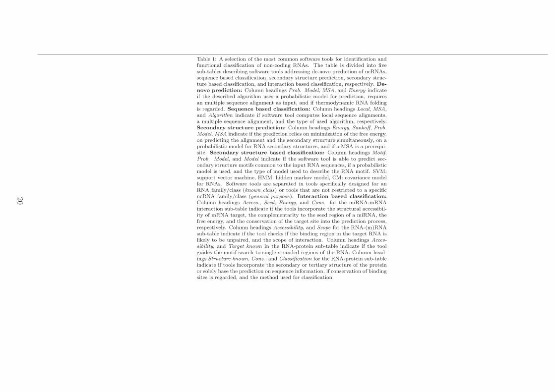
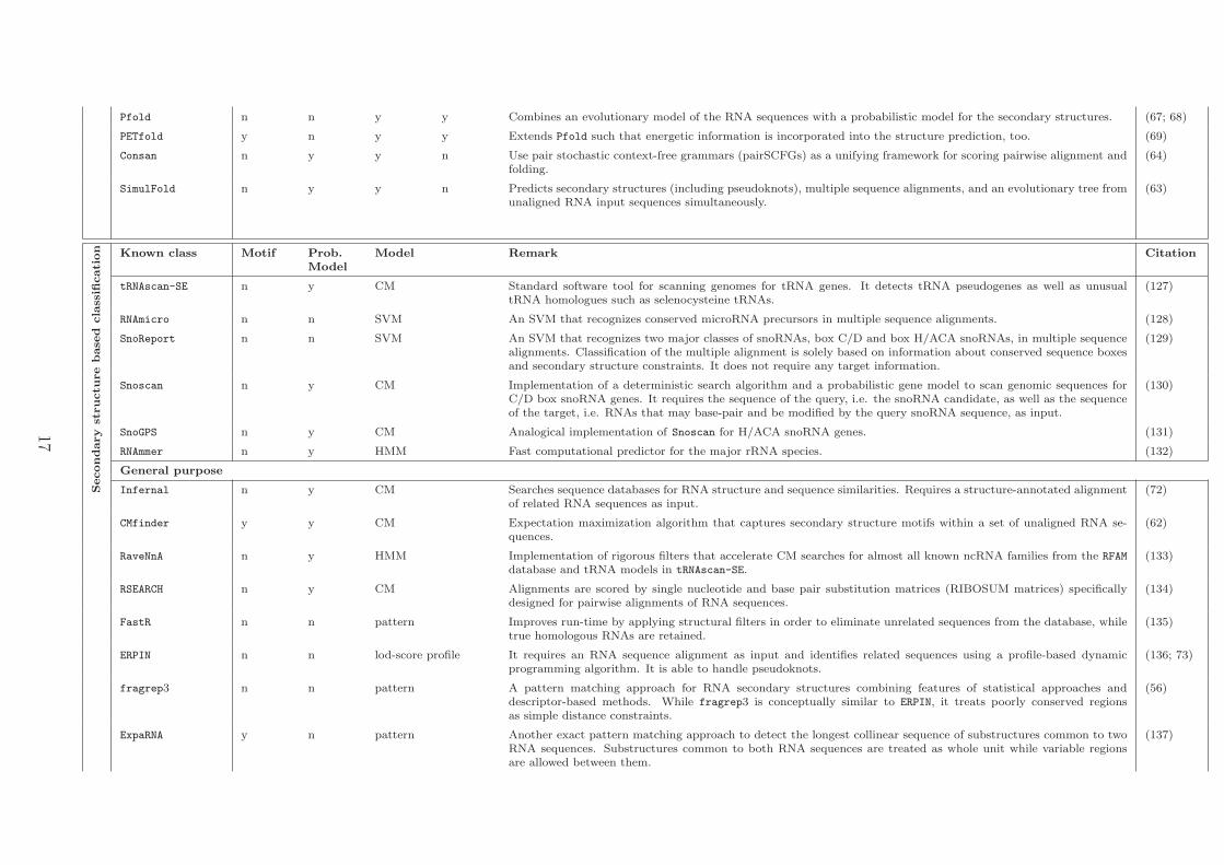
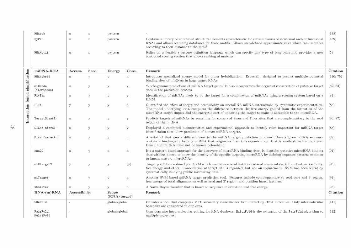
Table 1: A selection of the most common software tools for identification andfunctional classification of non-coding RNAs. The table is divided into fivesub-tables describing software tools addressing de-novo prediction of ncRNAs,sequence based classification, secondary structure prediction, secondary struc-ture based classification, and interaction based classification, respectively. De-novo prediction: Column headings Prob. Model, MSA, and Energy indicateif the described algorithm uses a probabilistic model for prediction, requiresan multiple sequence alignment as input, and if thermodynamic RNA foldingis regarded. Sequence based classification: Column headings Local, MSA,and Algorithm indicate if software tool computes local sequence alignments,a multiple sequence alignment, and the type of used algorithm, respectively.Secondary structure prediction: Column headings Energy, Sankoff, Prob.
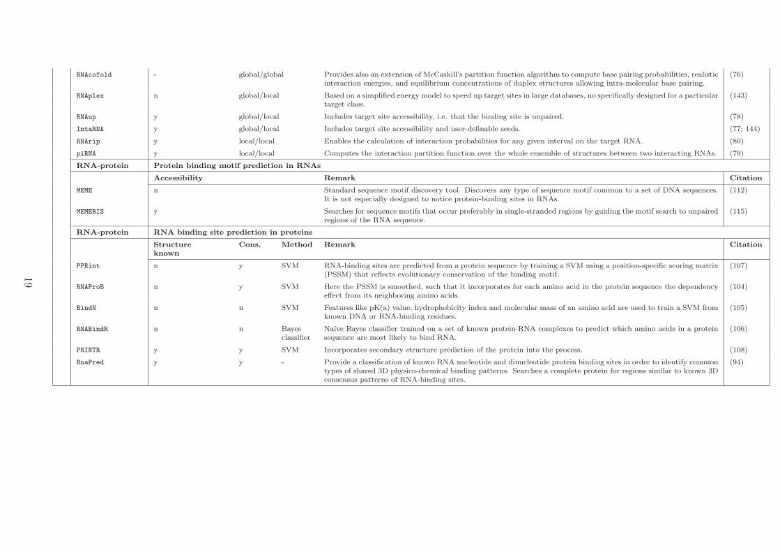
Model, MSA indicate if the prediction relies on minimization of the free energy,on predicting the alignment and the secondary structure simultaneously, on aprobabilistic model for RNA secondary structures, and if a MSA is a prerequi-site. Secondary structure based classification: Column headings Motif,Prob. Model, and Model indicate if the software tool is able to predict sec-ondary structure motifs common to the input RNA sequences, if a probabilisticmodel is used, and the type of model used to describe the RNA motif. SVM:support vector machine, HMM: hidden markov model, CM: covariance modelfor RNAs. Software tools are separated in tools specifically designed for anRNA family/class (known class) or tools that are not restricted to a specificncRNA family/class (general purpose). Interaction based classification:Column headings Access., Seed, Energy, and Cons. for the miRNA-mRNAinteraction sub-table indicate if the tools incorporate the structural accessibil-ity of mRNA target, the complementarity to the seed region of a miRNA, thefree energy, and the conservation of the target site into the prediction process,respectively. Column headings Accessibility, and Scope for the RNA-(m)RNAsub-table indicate if the tool checks if the binding region in the target RNA islikely to be unpaired, and the scope of interaction. Column headings Acces-
sibility, and Target known in the RNA-protein sub-table indicate if the toolguides the motif search to single stranded regions of the RNA. Column head-ings Structure known, Cons., and Classification for the RNA-protein sub-tableindicate if tools incorporate the secondary or tertiary structure of the proteinor solely base the prediction on sequence information, if conservation of bindingsites is regarded, and the method used for classification.
20
4 Notes
4.1 Databases of known regulatory ncRNAs
RFAM: http://rfam.sanger.ac.uk
miRBase: http://www.mirbase.org
NONCODE: http://www.noncode.org
RNAdb: http://research.imb.uq.edu.au/rnadb/
snoRNA base: http://www-snorna.biotoul.fr/
snoRNAdb: http://lowelab.ucsc.edu/snoRNAdb/
4.2 Genome browsers
UCSC: http://genome.ucsc.edu/ UCSC mirror for ncRNAs: http://www.ncrna.org/
glocal/cgi-bin/hgGateway Ensembl: http://www.ensembl.org/index.html
4.3 Useful web services
Vienna RNA Package: http://www.tbi.univie.ac.at/~ivo/RNA/, http://rna.tbi.univie.
ac.at/
LeARN: http://symbiose.toulouse.inra.fr/LeARN/ (145)
Web repository for ncRNA community: http://www.ncrna.org/ SCOR (Database of 3D
structural classification of ncRNAs): http://scor.berkeley.edu/
4.4 Software links in alphabetical order
BindN: http://bioinfo.ggc.org/bindn/
blast: http://blast.ncbi.nlm.nih.gov/Blast.cgi
CMfinder: http://bio.cs.washington.edu/yzizhen/CMfinder/
Consan: http://selab.janelia.org/software/consan/
21
DIANA MicroT: http://www.diana.pcbi.upenn.edu/cgi-bin/micro\_t.cgi
Dynalign: http://rna.urmc.rochester.edu/dynalign.html
ERPIN: http://tagc.univ-mrs.fr/erpin/
evofold: http://users.soe.ucsc.edu/$\sim$jsp/EvoFold/
FOLDALIGN: http://foldalign.ku.dk//software/index.html
FoldalignM: http://foldalign.ku.dk/software/index.html
Gibbs sampler: http://bayesweb.wadsworth.org/gibbs/gibbs.html
Infernal: http://infernal.janelia.org/
IntaRNA: http://www.bioinf.uni-freiburg.de/Software/
LocARNA: http://rna.tbi.univie.ac.at/cgi-bin/LocARNA.cgi
MEME: http://meme.sdsc.edu/meme4\_3\_0/intro.html
MEMERIS: http://www.bioinf.uni-freiburg.de/$\sim$hiller/MEMERIS/
MicroInspector:http://bioinfo.uni-plovdiv.bg/microinspector/
miRanda(Microcosm): http://www.ebi.ac.uk/enright-srv/microcosm/htdocs/targets/
v5/
miRtarget2: http://mirdb.org/miRDB/
miTarget: http://cbit.snu.ac.kr/$\sim$miTarget/
NbmiRTar: http://wotan.wistar.upenn.edu/NBmiRTar/login.php
NcDNAlign: www.bioinf.uni-leipzig.de/Software/NcDNAlign/
qRNA: http://nbc11.biologie.uni-kl.de/framed/left/menu/auto/right/qrna/
PETfold: http://genome.ku.dk/resources/petfold/
Pfold: http://www.daimi.au.dk/$\sim$compbio/pfold/
PicTar: http://pictar.mdc-berlin.de/
PITA: http://genie.weizmann.ac.il/pubs/mir07/mir07\_prediction.html
PMcomp: http://www.tbi.univie.ac.at/RNA/PMcomp/
PPRint: www.imtech.res.in/raghava/pprint/
PRINTR: http://210.42.106.80/printr/
RaveNnA: http://bliss.biology.yale.edu/$\sim$zasha/ravenna/
RNA22: http://cbcsrv.watson.ibm.com/rna22.html
22
RNAalifold: http://rna.tbi.univie.ac.at/cgi-bin/RNAalifold.cgi
RNABindR: http://bindr.gdcb.iastate.edu/RNABindR/
RNAduplex: http://www.tbi.univie.ac.at/RNA/RNAduplex.html
RNAhybrid: http://bibiserv.techfak.uni-bielefeld.de/rnahybrid/
RNAProB:
RNAsnoop: http://www.tbi.univie.ac.at/$\sim$htafer/RNAsnoop/RNAsnoop.html
RNAup: http://www.tbi.univie.ac.at/$\sim$ulim/RNAup/
RNAz: http://rna.tbi.univie.ac.at/cgi-bin/RNAz.cgi, http://www.tbi.univie.ac.
at/~wash/RNAz/
SimulFold: http://people.cs.ubc.ca/~irmtraud/simulfold/
Stemloc: http://biowiki.org/StemLoc
TargetScan/TargetScanS: http://www.targetscan.org/;http://genes.mit.edu/tscan/
targetscanS2005.html
References
[1] Consortium, A. F. B. et al. RNAs everywhere: genome-wide annotation of structured
RNAs. J Exp Zool B Mol Dev Evol 308, 1–25 (2007).
[2] Maeda, N. et al. Transcript annotation in FANTOM3: mouse gene catalog based on
physical cDNAs. PLoS Genet 2, e62 (2006).
[3] Consortium, E. N. C. O. D. E. P. The ENCODE (ENCyclopedia Of DNA Elements)
project. Science 306, 636–640 (2004).
[4] Kapranov, P., Willingham, A. T. & Gingeras, T. R. Genome-wide transcription and
the implications for genomic organization. Nat Rev Genet 8, 413–423 (2007).
[5] Macke, T. J. et al. RNAMotif, an RNA secondary structure definition and search
algorithm. Nucleic Acids Res 29, 4724–4735 (2001).
23
[6] Tinoco, I. & Bustamante, C. How RNA folds. J Mol Biol 293, 271–281 (1999).
[7] Mattick, J. S. The hidden genetic program of complex organisms. Sci Am 291, 60–67
(2004).
[8] Mattick, J. S. & Makunin, I. V. Non-coding RNA. Hum Mol Genet 15 Spec No 1,
R17–R29 (2006).
[9] Prasanth, K. V. & Spector, D. L. Eukaryotic regulatory RNAs: an answer to the
’genome complexity’ conundrum. Genes Dev 21, 11–42 (2007).
[10] Bartel, D. P. MicroRNAs: genomics, biogenesis, mechanism, and function. Cell 116,
281–297 (2004).
[11] Wightman, B., Ha, I. & Ruvkun, G. Posttranscriptional regulation of the heterochronic
gene lin-14 by lin-4 mediates temporal pattern formation in C. elegans. Cell 75, 855–
862 (1993).
[12] Lee, R. C., Feinbaum, R. L. & Ambros, V. The C. elegans heterochronic gene lin-
4 encodes small RNAs with antisense complementarity to lin-14. Cell 75, 843–854
(1993).
[13] Pasquinelli, A. E. et al. Conservation of the sequence and temporal expression of let-7
heterochronic regulatory RNA. Nature 408, 86–89 (2000).
[14] Kato, M. & Slack, F. J. microRNAs: small molecules with big roles - C. elegans to
human cancer. Biol Cell 100, 71–81 (2008).
[15] Seto, A. G., Kingston, R. E. & Lau, N. C. The coming of age for Piwi proteins. Mol
Cell 26, 603–609 (2007).
[16] Senner, C. E. & Brockdorff, N. Xist gene regulation at the onset of X inactivation.
Curr Opin Genet Dev 19, 122–126 (2009).
24
[17] Martianov, I., Ramadass, A., Barros, A. S., Chow, N. & Akoulitchev, A. Repression of
the human dihydrofolate reductase gene by a non-coding interfering transcript. Nature
445, 666–670 (2007).
[18] Beltran, M. et al. A natural antisense transcript regulates Zeb2/Sip1 gene expres-
sion during Snail1-induced epithelial-mesenchymal transition. Genes Dev 22, 756–769
(2008).
[19] Mercer, T. R., Dinger, M. E. & Mattick, J. S. Long non-coding RNAs: insights into
functions. Nat Rev Genet 10, 155–159 (2009).
[20] Le, S. V., Chen, J. H., Currey, K. M. & Maizel, J. V. A program for predicting
significant RNA secondary structures. Comput Appl Biosci 4, 153–159 (1988).
[21] Rivas, E. & Eddy, S. R. Secondary structure alone is generally not statistically signif-
icant for the detection of noncoding RNAs. Bioinformatics 16, 583–605 (2000).
[22] Rivas, E. & Eddy, S. R. Noncoding RNA gene detection using comparative sequence
analysis. BMC Bioinformatics 2, 8 (2001).
[23] Pedersen, J. S. et al. Identification and classification of conserved RNA secondary
structures in the human genome. PLoS Comput Biol 2, e33 (2006).
[24] Washietl, S., Hofacker, I. L. & Stadler, P. F. Fast and reliable prediction of noncoding
RNAs. Proc Natl Acad Sci U S A 102, 2454–2459 (2005).
[25] Washietl, S., Hofacker, I. L., Lukasser, M., Huttenhofer, A. & Stadler, P. F. Mapping
of conserved RNA secondary structures predicts thousands of functional noncoding
RNAs in the human genome. Nat Biotechnol 23, 1383–1390 (2005).
[26] Missal, K., Rose, D. & Stadler, P. F. Non-coding RNAs in ciona intestinalis. Bioin-
formatics 21 Suppl 2, ii77–ii78 (2005).
[27] Missal, K. et al. Prediction of structured non-coding RNAs in the genomes of the
nematodes caenorhabditis elegans and caenorhabditis briggsae. J Exp Zool B Mol Dev
Evol 306, 379–392 (2006).
25

[28] Rose, D. et al. Computational RNomics of drosophilids. BMC Genomics 8, 406 (2007).
[29] Steigele, S., Huber, W., Stocsits, C., Stadler, P. F. & Nieselt, K. Comparative analysis
of structured RNAs in S. cerevisiae indicates a multitude of different functions. BMC
Biol 5, 25 (2007).
[30] Mourier, T. et al. Genome-wide discovery and verification of novel structured RNAs
in plasmodium falciparum. Genome Res 18, 281–292 (2008).
[31] Rose, D. et al. Duplicated RNA genes in teleost fish genomes. J Bioinform Comput
Biol 6, 1157–1175 (2008).
[32] Gesell, T. & Washietl, S. Dinucleotide controlled null models for comparative RNA
gene prediction. BMC Bioinformatics 9, 248 (2008).
[33] Gruber, A., Findeiss, S., Washietl, S., Hofacker, I. & Stadler, P. RNAz 2.0: Improved
noncoding RNA detection. In PSB in press (2010).
[34] Gruber, A. R., Neubck, R., Hofacker, I. L. & Washietl, S. The rnaz web server:
prediction of thermodynamically stable and evolutionarily conserved rna structures.
Nucleic Acids Res 35, W335–W338 (2007).
[35] Washietl, S. Prediction of structural noncoding RNAs with RNAz. Methods Mol Biol
395, 503–526 (2007).
[36] Washietl, S. & Hofacker, I. L. Identifying structural noncoding rnas using rnaz. Curr
Protoc Bioinformatics Chapter 12, Unit 12.7 (2007).
[37] Rose, D., Hertel, J., Reiche, K., Stadler, P. F. & Hackermuller, J. NcDNAlign: plausi-
ble multiple alignments of non-protein-coding genomic sequences. Genomics 92, 65–74
(2008).
[38] Sankoff, D. Simultaneous solution of the RNA folding, alignment, and proto-sequence
problems. SIAM J. Appl. Math. 45, 810–825 (1985).
26
[39] Uzilov, A. V., Keegan, J. M. & Mathews, D. H. Detection of non-coding RNAs
on the basis of predicted secondary structure formation free energy change. BMC
Bioinformatics 7, 173 (2006).
[40] Torarinsson, E., Sawera, M., Havgaard, J. H., Fredholm, M. & Gorodkin, J. Thousands
of corresponding human and mouse genomic regions unalignable in primary sequence
contain common rna structure. Genome Res 16, 885–889 (2006).
[41] Washietl, S. et al. Structured RNAs in the ENCODE selected regions of the human
genome. Genome Res 17, 852–864 (2007).
[42] Hiller, M. et al. Conserved introns reveal novel transcripts in Drosophila melanogaster.
Genome Res 19, 1289–1300 (2009).
[43] Kapranov, P. et al. Large-scale transcriptional activity in chromosomes 21 and 22.
Science 296, 916–919 (2002).
[44] Schadt, E. E. et al. A comprehensive transcript index of the human genome generated
using microarrays and computational approaches. Genome Biol 5, R73 (2004).
[45] Bertone, P. et al. Global identification of human transcribed sequences with genome
tiling arrays. Science 306, 2242–2246 (2004).
[46] Stolc, V. et al. Identification of transcribed sequences in Arabidopsis thaliana by using
high-resolution genome tiling arrays. Proc Natl Acad Sci U S A 102, 4453–4458 (2005).
[47] Kampa, D. et al. Novel RNAs identified from an in-depth analysis of the transcriptome
of human chromosomes 21 and 22. Genome Res 14, 331–342 (2004).
[48] Kapranov, P. et al. RNA maps reveal new RNA classes and a possible function for
pervasive transcription. Science 316, 1484–1488 (2007).
[49] Consortium, E. N. C. O. D. E. P. Identification and analysis of functional elements in
1% of the human genome by the ENCODE pilot project. Nature 447, 799–816 (2007).
27

[50] Berezikov, E. et al. Diversity of microRNAs in human and chimpanzee brain. Nat
Genet 38, 1375–1377 (2006).
[51] Kawai, J. et al. Functional annotation of a full-length mouse cDNA collection. Nature
409, 685–690 (2001).
[52] Okazaki, Y. et al. Analysis of the mouse transcriptome based on functional annotation
of 60,770 full-length cdnas. Nature 420, 563–573 (2002).
[53] Carninci, P. et al. The transcriptional landscape of the mammalian genome. Science
309, 1559–1563 (2005).
[54] Consortium, F. A. N. T. O. M. et al. The transcriptional network that controls growth
arrest and differentiation in a human myeloid leukemia cell line. Nat Genet 41, 553–562
(2009).
[55] Guttman, M. et al. Chromatin signature reveals over a thousand highly conserved
large non-coding rnas in mammals. Nature 458, 223–227 (2009).
[56] Mosig, A., Zhu, L. & Stadler, P. F. Customized strategies for discovering distant
ncRNA homologs. Brief Funct Genomic Proteomic (2009).
[57] Gorodkin, J., Heyer, L. J. & Stormo, G. D. Finding the most significant common
sequence and structure motifs in a set of RNA sequences. Nucleic Acids Res 25,
3724–3732 (1997).
[58] Torarinsson, E., Havgaard, J. H. & Gorodkin, J. Multiple structural alignment and
clustering of RNA sequences. Bioinformatics 23, 926–932 (2007).
[59] Mathews, D. H. & Turner, D. H. Dynalign: an algorithm for finding the secondary
structure common to two RNA sequences. J Mol Biol 317, 191–203 (2002).
[60] Hofacker, I. L., Bernhart, S. H. F. & Stadler, P. F. Alignment of RNA base pairing
probability matrices. Bioinformatics 20, 2222–2227 (2004).
28
[61] Will, S., Reiche, K., Hofacker, I. L., Stadler, P. F. & Backofen, R. Inferring noncoding
RNA families and classes by means of genome-scale structure-based clustering. PLoS
Comput Biol 3, e65 (2007).
[62] Yao, Z., Weinberg, Z. & Ruzzo, W. L. CMfinder–a covariance model based RNA motif
finding algorithm. Bioinformatics 22, 445–452 (2006).
[63] Meyer, I. M. & Miklos, I. SimulFold: simultaneously inferring RNA structures includ-
ing pseudoknots, alignments, and trees using a Bayesian MCMC framework. PLoS
Comput Biol 3, e149 (2007).
[64] Dowell, R. D. & Eddy, S. R. Efficient pairwise RNA structure prediction and alignment
using sequence alignment constraints. BMC Bioinformatics 7, 400 (2006).
[65] Bernhart, S. H., Hofacker, I. L., Will, S., Gruber, A. R. & Stadler, P. F. RNAalifold:
improved consensus structure prediction for RNA alignments. BMC Bioinformatics 9,
474 (2008).
[66] Hofacker, I. L. RNA consensus structure prediction with RNAalifold. Methods Mol
Biol 395, 527–544 (2007).
[67] Knudsen, B. & Hein, J. RNA secondary structure prediction using stochastic context-
free grammars and evolutionary history. Bioinformatics 15, 446–454 (1999).
[68] Knudsen, B. & Hein, J. Pfold: RNA secondary structure prediction using stochastic
context-free grammars. Nucleic Acids Res 31, 3423–3428 (2003).
[69] Seemann, S. E., Gorodkin, J. & Backofen, R. Unifying evolutionary and thermody-
namic information for RNA folding of multiple alignments. Nucleic Acids Res 36,
6355–6362 (2008).
[70] Gardner, P. P., Wilm, A. & Washietl, S. A benchmark of multiple sequence alignment
programs upon structural RNAs. Nucleic Acids Res 33, 2433–2439 (2005).
29
[71] Freyhult, E. K., Bollback, J. P. & Gardner, P. P. Exploring genomic dark matter:
a critical assessment of the performance of homology search methods on noncoding
RNA. Genome Res 17, 117–125 (2007).
[72] Nawrocki, E. P., Kolbe, D. L. & Eddy, S. R. Infernal 1.0: inference of RNA alignments.
Bioinformatics 25, 1335–1337 (2009).
[73] Lambert, A. et al. The ERPIN server: an interface to profile-based RNA motif iden-
tification. Nucleic Acids Res 32, W160–W165 (2004).
[74] Kaczkowski, B. et al. Structural profiles of human miRNA families from pairwise
clustering. Bioinformatics 25, 291–294 (2009).
[75] Rehmsmeier, M., Steffen, P., Hochsmann, M. & Giegerich, R. Fast and effective
prediction of microRNA/target duplexes. RNA 10, 1507–1517 (2004).
[76] Bernhart, S. H. et al. Partition function and base pairing probabilities of RNA het-
erodimers. Algorithms Mol Biol 1, 3 (2006).
[77] Busch, A., Richter, A. S. & Backofen, R. IntaRNA: efficient prediction of bacterial
sRNA targets incorporating target site accessibility and seed regions. Bioinformatics
24, 2849–2856 (2008).
[78] Muckstein, U. et al. Thermodynamics of RNA-RNA binding. Bioinformatics 22,
1177–1182 (2006).
[79] Chitsaz, H., Salari, R., Sahinalp, S. C. & Backofen, R. A partition function algorithm
for interacting nucleic acid strands. Bioinformatics 25, i365–i373 (2009).
[80] Huang, F. W. D., Qin, J., Reidys, C. M. & Stadler, P. F. Partition function and
base pairing probabilities for RNA-RNA interaction prediction. Bioinformatics 25,
2646–2654 (2009).
[81] Alkan, C., Karakoc, E., Nadeau, J. H., Sahinalp, S. C. & Zhang, K. RNA-RNA
interaction prediction and antisense RNA target search. J Comput Biol 13, 267–282
(2006).
30
[82] Enright, A. J. et al. MicroRNA targets in Drosophila. Genome Biol 5, R1 (2003).
[83] John, B. et al. Human MicroRNA targets. PLoS Biol 2, e363 (2004).
[84] Krek, A. et al. Combinatorial microRNA target predictions. Nat Genet 37, 495–500
(2005).
[85] Kertesz, M., Iovino, N., Unnerstall, U., Gaul, U. & Segal, E. The role of site accessi-
bility in microRNA target recognition. Nat Genet 39, 1278–1284 (2007).
[86] Lewis, B. P., hung Shih, I., Jones-Rhoades, M. W., Bartel, D. P. & Burge, C. B.
Prediction of mammalian microRNA targets. Cell 115, 787–798 (2003).
[87] Lewis, B. P., Burge, C. B. & Bartel, D. P. Conserved seed pairing, often flanked by
adenosines, indicates that thousands of human genes are microRNA targets. Cell 120,
15–20 (2005).
[88] Kiriakidou, M. et al. A combined computational-experimental approach predicts hu-
man microRNA targets. Genes Dev 18, 1165–1178 (2004).
[89] Rusinov, V., Baev, V., Minkov, I. N. & Tabler, M. MicroInspector: a web tool for
detection of miRNA binding sites in an RNA sequence. Nucleic Acids Res 33, W696–
W700 (2005).
[90] Wang, X. & Naqa, I. M. E. Prediction of both conserved and nonconserved microRNA
targets in animals. Bioinformatics 24, 325–332 (2008).
[91] Miranda, K. C. et al. A pattern-based method for the identification of MicroRNA
binding sites and their corresponding heteroduplexes. Cell 126, 1203–1217 (2006).
[92] Kim, S.-K., Nam, J.-W., Rhee, J.-K., Lee, W.-J. & Zhang, B.-T. miTarget: microRNA
target gene prediction using a support vector machine. BMC Bioinformatics 7, 411
(2006).
31
[93] Yousef, M., Jung, S., Kossenkov, A. V., Showe, L. C. & Showe, M. K. Naıve Bayes for
microRNA target predictions–machine learning for microRNA targets. Bioinformatics
23, 2987–2992 (2007).
[94] Shulman-Peleg, A., Shatsky, M., Nussinov, R. & Wolfson, H. J. Prediction of interact-
ing single-stranded RNA bases by protein-binding patterns. J Mol Biol 379, 299–316
(2008).
[95] Draper, D. E. Themes in RNA-protein recognition. J Mol Biol 293, 255–270 (1999).
[96] Auweter, S. D., Oberstrass, F. C. & Allain, F. H.-T. Sequence-specific binding of
single-stranded RNA: is there a code for recognition? Nucleic Acids Res 34, 4943–
4959 (2006).
[97] Messias, A. C. & Sattler, M. Structural basis of single-stranded RNA recognition. Acc
Chem Res 37, 279–287 (2004).
[98] Hall, K. B. & Stump, W. T. Interaction of N-terminal domain of U1A protein with an
RNA stem/loop. Nucleic Acids Res 20, 4283–4290 (1992).
[99] Spassov, D. S. & Jurecic, R. The PUF family of RNA-binding proteins: does evo-
lutionarily conserved structure equal conserved function? IUBMB Life 55, 359–366
(2003).
[100] de Moor, C. H., Meijer, H. & Lissenden, S. Mechanisms of translational control by the
3’ UTR in development and differentiation. Semin Cell Dev Biol 16, 49–58 (2005).
[101] Hudson, B. P., Martinez-Yamout, M. A., Dyson, H. J. & Wright, P. E. Recognition of
the mRNA AU-rich element by the zinc finger domain of TIS11d. Nat Struct Mol Biol
11, 257–264 (2004).
[102] Kulinski, T. et al. The apical loop of the HIV-1 TAR RNA hairpin is stabilized by a
cross-loop base pair. J Biol Chem 278, 38892–38901 (2003).
32
[103] Clerte, C. & Hall, K. B. Global and local dynamics of the U1A polyadenylation
inhibition element (PIE) RNA and PIE RNA-U1A complexes. Biochemistry 43, 13404–
13415 (2004).
[104] Cheng, C.-W., Su, E. C.-Y., Hwang, J.-K., Sung, T.-Y. & Hsu, W.-L. Predicting RNA-
binding sites of proteins using support vector machines and evolutionary information.
BMC Bioinformatics 9 Suppl 12, S6 (2008).
[105] Wang, L. & Brown, S. J. BindN: a web-based tool for efficient prediction of DNA
and RNA binding sites in amino acid sequences. Nucleic Acids Res 34, W243–W248
(2006).
[106] Terribilini, M. et al. Prediction of RNA binding sites in proteins from amino acid
sequence. RNA 12, 1450–1462 (2006).
[107] Kumar, M., Gromiha, M. M. & Raghava, G. P. S. Prediction of RNA binding sites in
a protein using SVM and PSSM profile. Proteins 71, 189–194 (2008).
[108] Wang, Y., Xue, Z., Shen, G. & Xu, J. PRINTR: prediction of RNA binding sites in
proteins using SVM and profiles. Amino Acids 35, 295–302 (2008).
[109] Leontis, N. B. & Westhof, E. The annotation of RNA motifs. Comp Funct Genomics
3, 518–524 (2002).
[110] Leontis, N. B. & Westhof, E. Analysis of RNA motifs. Curr Opin Struct Biol 13,
300–308 (2003).
[111] Hermann, T. & Westhof, E. Non-Watson-Crick base pairs in RNA-protein recognition.
Chem Biol 6, R335–R343 (1999).
[112] Bailey, T. L. et al. MEME SUITE: tools for motif discovery and searching. Nucleic
Acids Res 37, W202–W208 (2009).
[113] Thompson, W., McCue, L. A. & Lawrence, C. E. Using the Gibbs motif sampler to
find conserved domains in DNA and protein sequences. Curr Protoc Bioinformatics
Chapter 2, Unit 2.8 (2005).
33
[114] Lawrence, C. E. et al. Detecting subtle sequence signals: a Gibbs sampling strategy
for multiple alignment. Science 262, 208–214 (1993).
[115] Hiller, M., Pudimat, R., Busch, A. & Backofen, R. Using RNA secondary structures
to guide sequence motif finding towards single-stranded regions. Nucleic Acids Res 34,
e117 (2006).
[116] Altschul, S. F., Gish, W., Miller, W., Myers, E. W. & Lipman, D. J. Basic local
alignment search tool. J Mol Biol 215, 403–410 (1990). URL http://dx.doi.org/
10.1006/jmbi.1990.9999.
[117] Smith, T. F. & Waterman, M. S. Identification of common molecular subsequences. J
Mol Biol 147, 195–197 (1981).
[118] Roshan, U. & Livesay, D. R. Probalign: multiple sequence alignment using partition
function posterior probabilities. Bioinformatics 22, 2715–2721 (2006). URL http:
//dx.doi.org/10.1093/bioinformatics/btl472.
[119] Roshan, U., Chikkagoudar, S. & Livesay, D. R. Searching for evolutionary dis-
tant rna homologs within genomic sequences using partition function posterior prob-
abilities. BMC Bioinformatics 9, 61 (2008). URL http://dx.doi.org/10.1186/
1471-2105-9-61.
[120] Hertel, J. et al. Non-coding rna annotation of the genome of trichoplax adhaerens.
Nucleic Acids Res 37, 1602–1615 (2009). URL http://dx.doi.org/10.1093/nar/
gkn1084.
[121] Mosig, A., Sameith, K. & Stadler, P. Fragrep: an efficient search tool for fragmented
patterns in genomic sequences. Genomics Proteomics Bioinformatics 4, 56–60 (2006).
URL http://dx.doi.org/10.1016/S1672-0229(06)60017-X.
[122] Gruber, A. R., Findei, S., Washietl, S., Hofacker, I. L. & Stadler, P. F. Rnaz 2.0:
Improved noncoding rna detection. Pac Symp Biocomput 15, 69–79 (2010).
34
[123] Chenna, R. et al. Multiple sequence alignment with the clustal series of programs.
Nucleic Acids Res 31, 3497–3500 (2003).
[124] Markham, N. R. & Zuker, M. Unafold: software for nucleic acid folding and hy-
bridization. Methods Mol Biol 453, 3–31 (2008). URL http://dx.doi.org/10.1007/
978-1-60327-429-6_1.
[125] Hofacker, I. L. Rna secondary structure analysis using the vienna rna package. Curr
Protoc Bioinformatics Chapter 12, Unit12.2 (2009). URL http://dx.doi.org/10.
1002/0471250953.bi1202s26.
[126] McCaskill, J. S. The equilibrium partition function and base pair binding probabilities
for rna secondary structure. Biopolymers 29, 1105–1119 (1990). URL http://dx.
doi.org/10.1002/bip.360290621.
[127] Lowe, T. M. & Eddy, S. R. trnascan-se: a program for improved detection of transfer
rna genes in genomic sequence. Nucleic Acids Res 25, 955–964 (1997).
[128] Hertel, J. & Stadler, P. F. Hairpins in a haystack: recognizing microrna precursors
in comparative genomics data. Bioinformatics 22, e197–e202 (2006). URL http:
//dx.doi.org/10.1093/bioinformatics/btl257.
[129] Hertel, J., Hofacker, I. L. & Stadler, P. F. Snoreport: computational identification
of snornas with unknown targets. Bioinformatics 24, 158–164 (2008). URL http:
//dx.doi.org/10.1093/bioinformatics/btm464.
[130] Lowe, T. M. & Eddy, S. R. A computational screen for methylation guide snornas in
yeast. Science 283, 1168–1171 (1999).
[131] Schattner, P. et al. Genome-wide searching for pseudouridylation guide snornas: anal-
ysis of the saccharomyces cerevisiae genome. Nucleic Acids Res 32, 4281–4296 (2004).
URL http://dx.doi.org/10.1093/nar/gkh768.
35
[132] Lagesen, K. et al. Rnammer: consistent and rapid annotation of ribosomal rna genes.
Nucleic Acids Res 35, 3100–3108 (2007). URL http://dx.doi.org/10.1093/nar/
gkm160.
[133] Weinberg, Z. & Ruzzo, W. L. Exploiting conserved structure for faster annotation
of non-coding rnas without loss of accuracy. Bioinformatics 20 Suppl 1, i334–i341
(2004). URL http://dx.doi.org/10.1093/bioinformatics/bth925.
[134] Klein, R. J. & Eddy, S. R. Rsearch: finding homologs of single structured rna se-
quences. BMC Bioinformatics 4, 44 (2003). URL http://dx.doi.org/10.1186/
1471-2105-4-44.
[135] Bafna, V. & Zhang, S. Fastr: fast database search tool for non-coding rna. Proc IEEE
Comput Syst Bioinform Conf 52–61 (2004).
[136] Gautheret, D. & Lambert, A. Direct rna motif definition and identification from
multiple sequence alignments using secondary structure profiles. J Mol Biol 313,
1003–1011 (2001). URL http://dx.doi.org/10.1006/jmbi.2001.5102.
[137] Heyne, S., Will, S., Beckstette, M. & Backofen, R. Lightweight comparison of rnas
based on exact sequence-structure matches. Bioinformatics 25, 2095–2102 (2009).
URL http://dx.doi.org/10.1093/bioinformatics/btp065.
[138] R., E. S. RNABOB: a program to search for RNA secondary structure motifs in
sequence databases. unpublished .
[139] Grf, S., Strothmann, D., Kurtz, S. & Steger, G. Hypalib: a database of rnas and rna
structural elements defined by hybrid patterns. Nucleic Acids Res 29, 196–198 (2001).
[140] Krger, J. & Rehmsmeier, M. Rnahybrid: microrna target prediction easy, fast and
flexible. Nucleic Acids Res 34, W451–W454 (2006). URL http://dx.doi.org/10.
1093/nar/gkl243.
36
[141] Dimitrov, R. A. & Zuker, M. Prediction of hybridization and melting for double-
stranded nucleic acids. Biophys J 87, 215–226 (2004). URL http://dx.doi.org/10.
1529/biophysj.103.020743.
[142] Andronescu, M., Zhang, Z. C. & Condon, A. Secondary structure prediction of inter-
acting rna molecules. J Mol Biol 345, 987–1001 (2005). URL http://dx.doi.org/
10.1016/j.jmb.2004.10.082.
[143] Tafer, H. & Hofacker, I. L. RNAplex: a fast tool for RNA-RNA interaction search.
Bioinformatics 24, 2657–2663 (2008).
[144] Richter, A. S., Schleberger, C., Backofen, R. & Steglich, C. Seed-based intarna predic-
tion combined with gfp-reporter system identifies mrna targets of the small rna yfr1.
Bioinformatics (2009). URL http://dx.doi.org/10.1093/bioinformatics/btp609.
[145] Noirot, C., Gaspin, C., Schiex, T. & Gouzy, J. Learn: a platform for detecting,
clustering and annotating non-coding rnas. BMC Bioinformatics 9, 21 (2008). URL
http://dx.doi.org/10.1186/1471-2105-9-21.
37