Embed Size (px)
Citation preview

ICRi
A Progress Report
David Steffen
September 9, 2008

What This Report Covers
• We have had two meetings since our last update
• Since then, we have been working on Use Cases
• To date, we have:
• Evaluated the Translational Use Cases (presented last time)
• Developed 9 New Use Cases
• Possibly inherited a use case from WFWG

Use Case 1: Discovering a Biomarker
• A researcher is trying to identify a new genetic biomarker for HER2/neu negative stage I breast cancer patients.
• Using caTRIP, the researcher queries for HER2/neu negative tissue specimens of Stage I breast cancer patients at LCCC that also have corresponding microarray experiments.
• Analysis of the microarray experiments identify genes that are significantly over-expressed and under-expressed in a number of cases.
• The researcher decides that these results are significant, and related literature suggest a hypothesis that gene A may serve as a biomarker in HER2/neu negative Stage I breast cancer.
• To validate this hypothesis in a significant number of cases the researcher needs a larger data set, so he queries for all the HER2/neu negative specimens of Stage I breast cancer patients with corresponding microarray data and also for appropriate control data from other caTRIP adopting cancer centers.
• After retrieving the microarray experiments the researcher analyzes the data for over-expression of genes A.

Use Case 2: Finding biomaterial to validate a biomarker
• In scenario 1, the researcher has validated a biomarker based on available microarray experiments provided by various caTRIP adopter cancer centers.
• Now, the researcher would like to request biomaterial in the form of formalin-fixed/paraffin embedded tissue specimens from patients with the appropriate clinical outcomes.
• The researcher would like to validate the genetic biomarker in a different series of cases, this time using a different technique such as immunohistochemistry.
• The researcher queries for the presence of appropriate tissue using caTRIP and for the appropriate contact information of the person(s) responsible for the tissue repository.
• The researcher contacts the person(s) to begin the protocol for retrieving biomaterials.

Use Case 3: Extending the use of a biomarker
• The researcher would like to check if genes A could also be used as biomarker for other types of cancer.
• The flow of events will be similar to Scenario 1 with the exception that the specimen query will not be restricted to Stage I breast cancer patients.

Use Case 4: Exploring predictive power of gene expression in breast cancer metastasis
• The researcher would like to explore if gene expression patterns can predict how breast cancer will metastasize.
• He queries all the specimens of breast cancer patients from other caTRIP adopter cancer centers where their metastasis sites are liver, bone and brain.
• The researcher then retrieves the corresponding microarray experiments for these specimens.
• The researcher analyzes the microarray experiments to explore for a correlation between expression profiles and metastasis sites.

Use Case 5: Oncologists in formulating ideas for new clinical studies
The oncologist often wants to first find out the answers to questions such as:
• How many patients have been seen at our institution with disease x"?
• How does that compare with other institutions?
• What is the average survival of patients with disease x?
• How is it different if they are treated with drug x or y?
• How many patients with disease x and TNM stage y at diagnosis?
• How many patients with disease x relapse after treatment y?
This use case, then, is about enabling oncologists to ask these exploratory questions of their clinical databases as well as those at other institutions accessible on caGrid.

Use Case 6: High throughput screen for anti-cancer drugs leads based on robotic microscopy
A basic research scientist has developed a high throughput screen for anti-cancer drugs (leads) based on robotic microscopy. The final output of the process is relatively simple; a two by two matrix with the rows being a few million small molecules and the columns being some biological properties of these compounds, e.g.
• toxicity against several different tumor cell lines,
• toxicity against several different normal cell lines,
• ability of the molecule to enter the cell and its intracellular distribution, and
• impact of the molecule on a number of biological endpoints.
However, the process of generating this output presents a number of challenges.
• The initial output of the robotic microscopy is many thousands of images a day.
• The raw images must be stored so as to be available for future analytical algorithms and should, for similar reasons, be sharable.
• The space required to store these images is tens to hundreds of terabytes per year per instrument.
• Analysis of these images to generate the desired input needs to be automated.
• Many of the best algorithms are proprietary and are embedded in software which is not caBIG compatible and resides within a community not currently engaged with the caBIG community.
• Both the raw images and final results, annotated as to how those results were obtained, needs to be made available to appropriate collaborators both within the academic and commercial sphere so that leads identified in these small molecule libraries can be modified into drug candidates which can then be tested first preclinically and then in clinical trials.
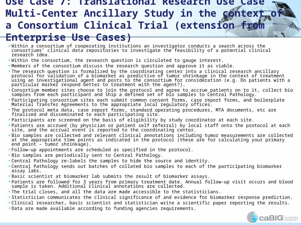
Use Case 7: Translational Research Use Case - Multi-Center Ancillary Study in the context of a Consortium Clinical Trial (extension from Enterprise Use Cases)
• Within a consortium of cooperating institutions an investigator conducts a search across the consortiums’ clinical data repositories to investigate the feasibility of a potential clinical research idea.
• Within the consortium, the research question is circulated to gauge interest.• Members of the consortium discuss the research question and approve it as viable.• The research question is formalized by the coordinating center into a clinical research ancillary protocol for validation of a biomarker as
predictive of tumor shrinkage in the context of treatment using an investigational agent and posts to the consortium for consideration (e.g. Do patients with a particular marker respond better to treatment with the agent?).
• Consortium member sites choose to join the protocol and agree to accrue patients on to it, collect bio samples from each participant and ship a defined set of bio samples to Central Pathology.
• Participating consortium sites each submit common consent forms, case report forms, and boilerplate Material Transfer Agreements to the appropriate local regulatory offices.
• The protocol meta data, case report forms, standard operating procedures, MTA documents, etc are finalized and disseminated to each participating site.
• Participants are screened on the basis of eligibility by study coordinator at each site.• Patients are accrued (by physician or patient self referral) by local staff onto the protocol at each site, and the accrual event is reported to the
coordinating center.• Bio samples are collected and relevant clinical annotations including tumor measurements are collected at the appropriate time points as
indicated in the protocol (these are for calculating your primary end point – tumor shrinkage).• Follow-up appointments are scheduled as specified in the protocol.• Bio samples are periodically sent to Central Pathology.• Central Pathology re-labels the samples to hide the source and identity.• Central Pathology sends out batches of collated bio samples to each of the participating biomarker assay labs.• Basic scientist at biomarker lab submits the result of biomarker assays.• Patients are followed for 3 years from primary treatment date. Annual follow-up visit occurs and blood sample is taken. Additional clinical
annotations are collected.• The trial closes, and all the data are made accessible to the statisticians.• Statistician communicates the clinical significance of and evidence for biomarker response prediction.• Clinical researcher, basic scientist and statistician write a scientific paper reporting the results.• Data are made available according to funding agencies requirements.

Use Case 8: Overlay of protein array data on the regulatory pathways with links to patient and cell culture data.• A clinical research scientist wants to be able to predict the efficacy of tyrosine kinase inhibitors as cancer
chemotherapeutic agents. The fact that many oncogenes are tyrosine kinases would predict that such agents should be effective, but several have been synthesized and tested in clinical trials, and the results have been disappointing in the extreme, with more cases of tumor growth stimulation than inhibition.
• The clinician hypothesizes that these unexpected effects are the result of regulatory feedback loops.
• To test this hypothesis, he requires software tools for modeling regulatory pathways.
• In addition, he needs to determine the state of such pathways in different patients by measuring the state of phosphorylation of the elements (proteins) of these pathways using reverse phase protein arrays.
• Because the consequences of treating the wrong patient with the wrong agent are so severe, the response of the tumor to the inhibitors will be tested in vitro, on cell cultures established from tumor biopsies.
• However, biospecimens and data from those patients who participated in clinical trials of these reagents before their ineffectiveness was appreciated is also available.
• Outputs measured on these cultures and biospecimens will include
• growth rate (determined by flow cytometry or by visually counting of cells at different time points,
• extent of cell death determined similarly,
• photomicrographs, reports of microscopic observations by trained investigators,
• rate of DNA synthesis measured by radioisotope or fluorescent labeled precursor uptake and
• incorporation, and staining with various immune reagents followed by high throughput robotic microscopy and automated image analysis.
• To develop an understanding that will resulting in giving the correct drugs to the correct patients, data from the protein arrays will be overlayed on the regulatory pathways and linked to patient and cell culture data.

Use Case 9: Animal model use case
• Bench scientist chooses candidate glioblastoma genes using human GWAS (eg, TCGA).
• Scientist also utilizes pathway analysis to postulate how multiple "hits" may be involved in tumorigenesis, to direct design of genetically altered mice.
• Utilizes targeted gene transfer to deliver mutated genes to inbred mice.
• Using inbred mice provides uniform genetic background in which researcher can also investigate mutated candidate genes in conjunction with other gene knockouts.
• Scientist finds mutated gene x expressed in gene y knockout mouse results in glioblastoma development that parallels human pathology.
• Scientist validates that model reacts in similar way to current therapeutic treatments.
• Scientist uses mouse model to test new therapeutic treatments, including combinations of drugs chosen to inhibit multiple pathways.
• Clinical scientists utilize mouse model results to design clinical trials to treat glioblastoma, incorporating genomic information on patients.

Use Case Inherited from WFWG
ICRi: What Else?
• Next Meeting (September 19) we will review our progress and charter
• After we complete our Use Cases (October 27) we will develop gap analyses (February 1)

The End