Embed Size (px)
Citation preview
ICOSL Anti-HER2 V-mAbs: Localizing Engineered ICOSL Costimulatory Agonists to HER2+ tumors through trastuzumab Rickel E, Evans L, Swanson R, Levin S, Rixon M, Wolfson M, Bhandari J, MacNeil S, Hoover J, Kornacker M, Capuano I, Peng SL Alpine Immune Sciences, Inc. Seattle, Washington, USA
The presence of tumor infiltrating lymphocytes (TILs) has been associated with improved prognosis in HER2+ breast cancer patients. Antigen specific TCR and costimulatory receptor signaling drive increases in TIL number, effector function, and tumor cytotoxicity. Improving the number and effector phenotype of tumor localized TILs has curative potential by enhancing the adaptive and memory immune response. Targeting HER2 with the monoclonal anti-HER2 antibody trastuzumab has improved survival in HER2+ breast cancer patients and is known to increase peripheral type I immunity, which may be reflected by increased TILs.
The Immunoglobulin Superfamily (IgSF) includes a large, diverse family of immunotherapy targets expressed on immune cells and tumors. Transmembrane IgSF receptors, CD28 and inducible T cell co-stimulator (ICOS), related costimulatory molecules expressed on T cells, interact with CD80/CD86 and ICOS ligand (ICOSL), respectively, and play critical roles in T cell activation and adaptive immunity. Alpine Immune Science’s vIgDTM platform uses directed evolution to derive novel, therapeutically-applicable IgSF extracellular domains with tailored specificity and affinity. The vIgD platform has generated human ICOSL vIgDs capable of binding both ICOS and CD28, activating both pathways. To promote anti-tumor activity of TILs in HER2+ tumors, we developed trastuzumab-ICOSL “V-mAbs” consisting of trastuzumab fused to activating ICOSL vIgDs. These V-mAbs are designed to localize to HER2+ tumors and activate antigen-specific, resident T-cells through costimulatory receptor agonism.
V-mAbs were successfully produced and bound to CD28, ICOS and HER2. In a plate bound costimulation assay, the V-mAbs increased the amount of IFN-γ produced by T-cells stimulated with anti-CD3. When incubated with HER2+ target cells, V-mAbs promoted T-cell proliferation, cytokine secretion, and target cell lysis.
Abstract
The Immunoglobulin Superfamily (IgSF)
Figure 2: vIgDs May Be Used in Multiple Therapeutic Formats Engineering Trastuzumab ICOSL V-mAb Constructs Localized Trastuzumab-ICOSL V-mAbs Enhance Antigen-Specific Responses
Conclusions and Summary
Figure 4. ICOSL and Potential Therapeutic Applications. The ECD domain from an engineered ICOSL domain was attached to the N and/or C-terminus of the heavy and/or light chains of trastuzumab. The resulting trastuzumab-ICOSL V-mAbs were produced in mammalian Expi293 cells, purified with standard Protein A chromatography, and biochemically assessed with analytical size exclusion chromatography (data not shown). In addition, IgV domain variants were also produced (data not shown).
trastuzumab
Figure 1: The vIgD Platform
Trastuzumab
Tumor Suppression V-mAb Dependent Tumor Immunity
ICOSL vIgD
ICOSL V-mAb
Tumor cell
T cell
TCR
MHC
CD28
HER-2
TCR
MHC
HER-2
TCR signal only, no costimulation, no effector function
ICOS
CD28
ICOS
HER-2
HER-2
Tailored High-Affinity Dual ICOS/CD28 Binding of ICOSL vIgD Domains Figure 3. Structure of vIgD Fc fusion proteins with improved binding to counter receptors Yeast outputs were batch cloned into an Fc expression vector, inserts sequenced, and unique clones of interest chosen for transient expression in Expi293 cells. HEK293 cells were transiently transfected with two distinct IgSF receptors. Cells were stained with titrated WT or mutant vIgD hits. Binding was detected with PE-conjugated anti-human Fc(each binding curve). MFI, mean fluorescence intensity.
Table 1. Improved ligand affinity of vIgD-Fc proteins Example dissociation constant (KD) determinations on recombinant ICOSL vIgD-Fc proteins using 2 counter receptors on a ForteBio Octet. FI, fold increase in affinity vs. wild-type (WT). Note: this is an avidity-driven system due to the bivalent nature of both the receptors and the vIgDs.
vIgD Fc: ICOSL ECD IgV and IgC
Domains
Fc
Trastuzumab-ICOSL V-mAbs Retain Engineered Binding to CD28, ICOS, and HER2
10 100 1000 10000 1000000
50000
100000
150000
200000
HER2
Protein [ pM ]
MFI
10 100 1000 10000 1000000
30000
60000
90000
CD28
Protein [ pM ]
MFI
10 100 1000 10000 1000000
30000
60000
90000
120000
ICOS
Protein [ pM ]
MFI
10 100 1000 10000 1000000
20000
40000
60000
MOCK
Protein [ pM ]
MFI
10 100 1000 10000 100000
10
30
50
70
HER2
Protein [ pM ]
% P
ositi
ve
10 100 1000 10000 1000000
20
40
60
80
CD28
Protein [ pM ]
% P
ositi
ve
10 100 1000 10000 1000000
30
60
90
ICOS
Protein [ pM ]
% P
o si t i
v e
10 100 1000 10000 1000000
20
40
60
80
MOCK
Protein [ pM ]
% P
o si t i
v e
Figure 5. Counter Structure Binding: HEK293 cells were transiently transfected with HER2, CD28, CTLA4, or ICOS. Cells were stained with titrated trastuzumab-ICOSL V-mAb or trastuzumab and binding was detected with PE-conjugated anti-human Fc. MFI, mean fluorescence intensity. It should be noted there is a low level of HER2 expression on 293 cells, which is seen in the binding of the proteins to mock transfected cells. Fusion of ICOSL vIgDs to trastuzumab did not disrupt the ability of trastuzumab to bind to HER2. Furthermore, binding to CD28 and ICOS was not disrupted by the presence of trastuzumab. Thus, fusion of our ICOSL vIgD to trastuzumab does not prevent binding to either HER2 or weaken the engineered ability of the ICOSL vIgD to bind CD28 or ICOS.
• The vIgD platform has been harnessed to generate a novel panel of vIgDs with unique binding profiles to CD28 and ICOS. These costimulatory vIgDs were successfully fused to a HER2-specific antibody, creating trastuzumab-ICOSL V-mAbs.
• Trastuzumab-ICOSL V-mAbs are novel ICOS- and CD28-activating immunotherapies for HER2 positive tumors, promoting T-cell proliferation, cytokine secretion, and target cell lysis in a HER2-targeted fashion.
• Trastuzumab-ICOSL V-mAbs may therefore afford new treatment options for HER2+ malignancies, combining multiple anti-cancer mechanisms in a single therapeutic agent.
• The V-mAb approach has broad potential to enable tumor-localized immune modulation via the diverse array of IgSF members.
Trastuzumab-ICOSL V-mAbs Costimulate T-cells
Web: www.alpineimmunesciences.com Twitter: @AlpineImmuneSci
Trastuzumab-ICOSL V-mAbs Enhance T cell Activity in a Target-Dependent Fashion
Figure 6. Costimulation assay Trastuzumab-ICOSL V-mAbs were coated at a final concentration of 40, 10, 2.5 or 0.3 nM in the presence of 10nM anti-CD3 on 96 well plates. 100,000 CFSE labeled T cells/well were added, and 72 hours later, cells and supernatants were harvested for the measurement of proliferation and IFN-γ. V-mAbs enhanced anti-CD3 induced A) IFN-γ production, B) CD4 T-cell proliferation, and C) CD8 T-cell proliferation, showing fusion of the ICOSL vIgD to trastuzumab did not inhibit the ability of the ICOSL vIgD to costimulate T cells.
1 10 1000
10000
20000
30000
40000
50000
Anti-CD3/V-mAb Costim Assay
Protein [ nM ]
IFN
-γ [p
g/m
L]
A
0.1 1 10 1000
30
60
90
CD4+ T-cell Proliferation
Protein [ pM ]
% P
rolif
erat
ion
OKT3 Only
B
0.1 1 10 1000
30
60
90
CD8+ T-cell Proliferation
Protein [ pM ]
% P
r ol i f
e ra t
i on
OKT3 Only
C
1 2 3 40
30000
60000
90000
120000
150000
CD28 Binding
vIgD log[ pM ]
MFI
1 2 3 40
30000
60000
90000
120000
ICOS Binding
vIgD log[ pM ]
MFI
WT ICOSL1st Rnd vIgDs2nd Rnd vIgDs3rd Rnd vigDs
Fc ICOSL IgV+IgC
Combinations with trastuzumab
trastuzumab
Dendritic cell
ICOS
CD28 CD80/86
ICOSL
MHC
MHC
TCR
T cell Tumor cell
TCR
Bead and flow cytometric selection
vIgD
Fc-fusion protein
generation
Counter-structure binding
Functional assays
IgSF ECD Yeast Display Libraries IgSF protein
Therapeutic protein
Random or targeted mutagenesis of ICOSL ECD
Selections: rICOS and rCD28
Binding assays • Flow cytometry • Octet (affinity)
MLR • Proliferation • IFNγ production
Screen yeast outputs for improved binding to rICOS and rCD28
Transient 293 or CHO production followed by Protein A purification
Sequence yeast outputs and identify unique variant hits
Parental ICOSL binds ICOS
• Tailored counter-structures • Improved/high affinity
• Limited counter-structures • Low/modest affinity
Single vIgD domains can be tailored for unique bi- or tri-specific binding and activity profiles. vIgDs can be fused to antibodies or other domains to provide site-directed T cell agonism.
Trastuzumab, a monoclonal antibody to HER2, makes an attractive targeting moiety for our ICOSL vIgD
proteins. While trastuzumab has been a successful treatment for HER2+ breast cancer, and does induce ADCC, it lacks costimulatory functionality, so trastuzumab treatment alone may not be an effective anti-tumor immunity inducing agent able to provide increased chances for long-term remission. By fusing an agonistic vIgD domain to trastuzumab, a molecule would be formed able to provide costimulation in HER2+ tumor settings.
Modern therapies targeting the immune synapse
KD [pM] FI KD [pM] FIWT ICOSL 13880 - 883 -
525 26 332 2.7
783 18 769 1.1
436 32 382 2.3
896 15 1294 0.7447 31 492 1.8401 35 373 2.4390 36 472 1.9293 47 420 2.1368 38 369 2.4
1042 13 337 2.6503 28 543 1.6553 25 362 2.4563 25 477 1.9366 38 477 1.9969 14 340 2.6
1947 7 371 2.4910 15 311 2.8
2nd Rnd ICOSL vIgDs
3rd Rnd ICOSL vIgDs
Sample ID CD28 ICOS
1st Rnd ICOSL vIgDs
Sensor Load
The immunoglobulin superfamily (IgSF) Is the largest protein superfamily Includes cell surface and soluble members/forms involved in cellular recognition, binding, and adhesion Each IgSF member consists of at least one Ig domain of 70-110 aa each
Variable (IgV), constant (IgC1, IgC2), or intermediate (IgI) Has been most well studied in immunology and immuno-oncology,
IgSF and immuno-oncology
T cells at the site of the tumor display IgSF family members ICOS and CD28. Inducible costimulator ligand (ICOSL) provides a positive secondary signal to T cells upon binding to its high affinity receptor, ICOS which results in activation of the T cell and such results as proliferation, IFN-γ release and killing of the tumor cell. Alpine Immune Science’s vIgD platform has generated human ICOSL vIgDs capable of binding both ICOS and CD28, activating both T-cell costimulation pathways.
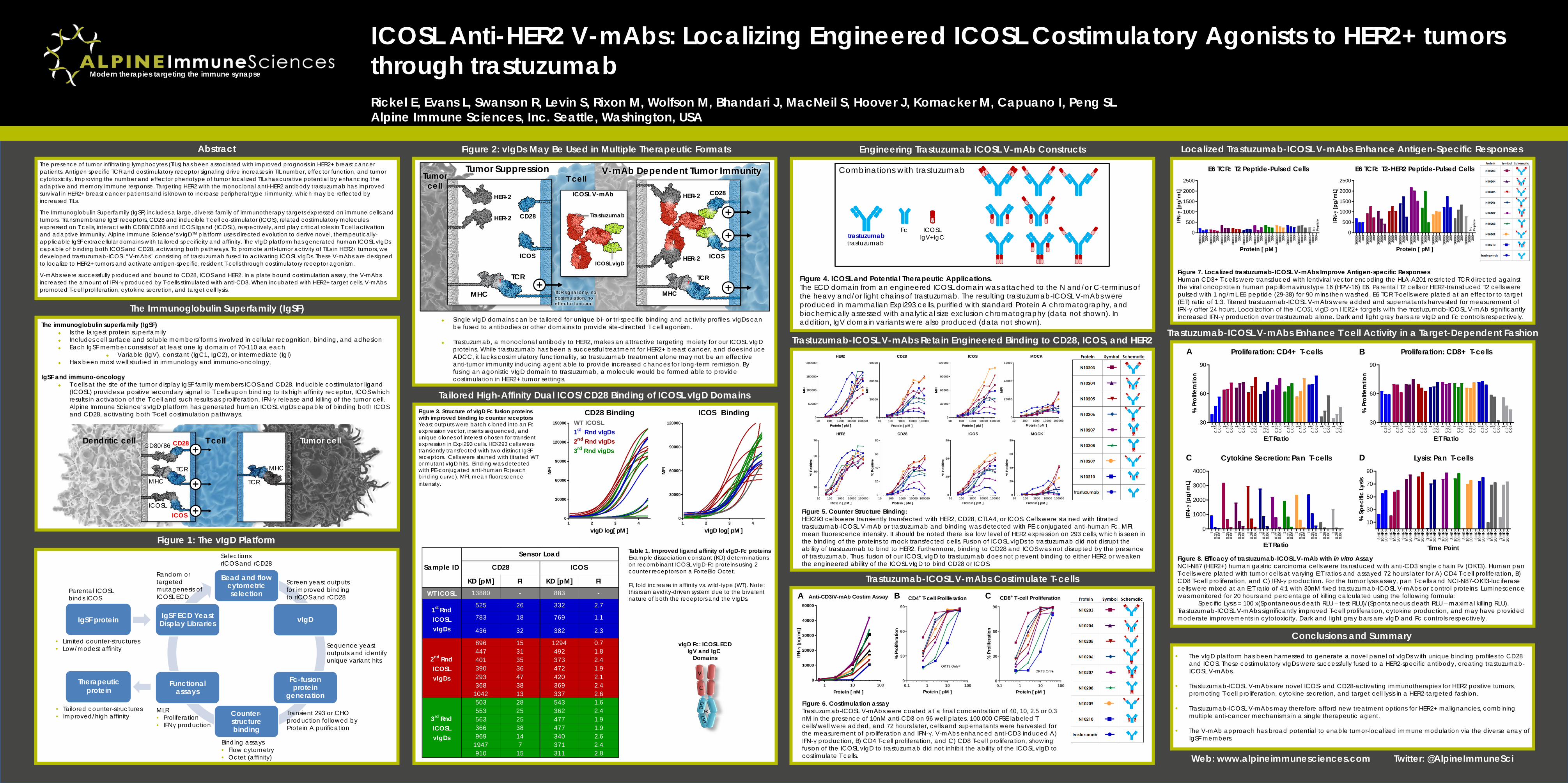
Figure 8. Efficacy of trastuzumab-ICOSL V-mAb with in vitro Assay NCI-N87 (HER2+) human gastric carcinoma cells were transduced with anti-CD3 single chain Fv (OKT3). Human pan T-cells were plated with tumor cells at varying E:T ratios and assayed 72 hours later for A) CD4 T-cell proliferation, B) CD8 T-cell proliferation, and C) IFN-γ production. For the tumor lysis assay, pan T-cells and NCI-N87-OKT3-luciferase cells were mixed at an E:T ratio of 4:1 with 30nM fixed trastuzumab-ICOSL V-mAbs or control proteins. Luminescence was monitored for 20 hours and percentage of killing calculated using the following formula:
Specific Lysis = 100 x(Spontaneous death RLU – test RLU)/(Spontaneous death RLU – maximal killing RLU). Trastuzumab-ICOSL V-mAbs significantly improved T-cell proliferation, cytokine production, and may have provided moderate improvements in cytotoxicity. Dark and light gray bars are vIgD and Fc controls respectively.
A B
C D
3000
030
00 300
3000
030
00 300
3000
030
00 300
3000
030
00 300
3000
030
00 300
3000
030
00 300
3000
030
00 300
3000
030
00 300
3000
030
00 300
3000
030
00 300
3000
030
00 300
0
500
1000
1500
2000
2500
E6 TCR: T2 Peptide-Pulsed Cells
Protein [ pM ]
IFN-
γ [p
g/m
L]
3 00 0
03 0
0 0 3 00
3 00 0
030
00 3 00
3000
03 0
0 0 300
3 00 0
03 0
0 0 3 00
3000
03 0
0 0 300
3 00 0
03 0
0 0 3 00
3000
03 0
0 0 300
3000
03 0
0 0 300
3000
03 0
0 0 3 00
3000
03 0
0 0 300
3000
03 0
0 0 3 00
0
500
1000
1500
2000
2500
E6 TCR: T2-HER2 Peptide-Pulsed Cells
Protein [ pM ]
IFN-
γ [p
g/m
L]
No
Pe
ptid
e
No
Pe
ptid
e
10 .
250.
06 10 .
250.
06 10 .
250.
06 10 .
250.
06 10 .
250.
06 10 .
250.
06 10 .
250.
06 10 .
250.
06 10 .
250.
06 10 .
250.
06 10 .
250.
06
30
60
90
Proliferation: CD4+ T-cells
E:T Ratio
% P
rolif
erat
ion
10 .
250.
06 10 .
250.
06 10 .
250.
06 10 .
250.
06 10 .
250.
06 10 .
250.
06 10 .
250.
06 10 .
250.
06 10 .
250.
06 10 .
250.
06 10 .
250.
06
30
60
90
Proliferation: CD8+ T-cells
E:T Ratio
% P
rolif
erat
ion
10 .
250.
06 10 .
250.
06 10 .
250.
06 10 .
250.
06 10 .
250.
06 10 .
250.
06 10 .
250.
06 10 .
250.
06 10 .
250.
06 10 .
250.
06 10 .
250.
06
0
1000
2000
3000
4000
Cytokine Secretion: Pan T-cells
E:T Ratio
IFN-
γ [p
g/m
L]
3 HR
10 H
R20
HR
3 HR
10 H
R20
HR
3 HR
10 H
R20
HR
3 HR
10 H
R20
HR
3 HR
10 H
R20
HR
3 HR
10 H
R20
HR
3 HR
10 H
R20
HR
3 HR
10 H
R20
HR
3 HR
10 H
R20
HR
3 HR
10 H
R20
HR
3 HR
10 H
R20
HR
10
30
50
70
90
Lysis: Pan T-cells
Time Point
% S
peci
fic L
ysis
Figure 7. Localized trastuzumab-ICOSL V-mAbs Improve Antigen-specific Responses Human CD3+ T-cells were transduced with lentiviral vector encoding the HLA-A201 restricted TCR directed against the viral oncoprotein human papillomavirus type 16 (HPV-16) E6. Parental T2 cells or HER2-transduced T2 cells were pulsed with 1 ng/mL E6 peptide (29-38) for 90 mins then washed. E6 TCR T-cells were plated at an effector to target (E:T) ratio of 1:3. Titered trastuzumab-ICOSL V-mAbs were added and supernatants harvested for measurement of IFN-γ after 24 hours. Localization of the ICOSL vIgD on HER2+ targets with the trastuzumab-ICOSL V-mAb significantly increased IFN-γ production over trastuzumab alone. Dark and light gray bars are vIgD and Fc controls respectively.