Embed Size (px)
Citation preview
Instruction manualInstruction manual
The Instruction Manual for Genomic DNAExtraction from Cultivated animal cells, tissues,Gram negative bacteria and Blood using silicamembrane.
ISO9001 14001
ISO
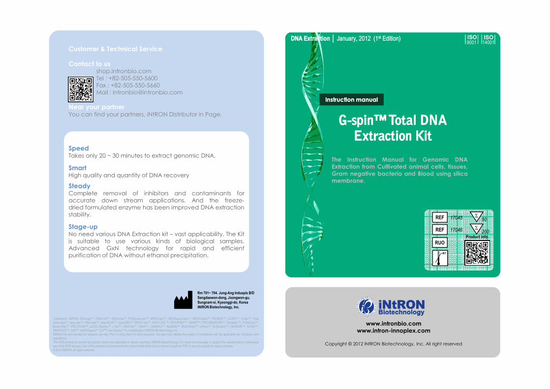
Speed Takes only 20 ~ 30 minutes to extract genomic DNA.
Smart High quality and quantity of DNA recovery
SteadyComplete removal of inhibitors and contaminants foraccurate down stream applications. And the freeze-dried formulated enzyme has been improved DNA extractionstability.
Stage-upNo need various DNA Extraction kit – vast applicability. The Kitis suitable to use various kinds of biological samples.Advanced GxN technology for rapid and efficientpurification of DNA without ethanol precipitation.
Customer & Technical Service
Contact to us shop.intronbio.comTel : +82-505-550-5600Fax : +82-505-550-5660Mail : [email protected]
Near your partner You can find your partners, iNtRON Distributor in Page.
Speed Takes only 20 ~ 30 minutes to extract genomic DNA.
Smart High quality and quantity of DNA recovery
SteadyComplete removal of inhibitors and contaminants foraccurate down stream applications. And the freeze-dried formulated enzyme has been improved DNA extractionstability.
Stage-upNo need various DNA Extraction kit – vast applicability. The Kitis suitable to use various kinds of biological samples.Advanced GxN technology for rapid and efficientpurification of DNA without ethanol precipitation.
Trademarks: iNtRON, DNA-spin™, DNA-midi™, DNA-maxi™, PCRquick-spin™, MEGA-spin™, MEGAquick-spin™, MEGA-bead™, PROBER™, G-DEX™, G-spin™, ViralGene-spin™, easy-spin™, RNA-spin™, easy-BLUE™, easy-RED™, WEST-one™, WEST-ZOL™, PRO-PREP™, SMART™, PRO-MEASURE™, Genelator™, F-Detector™,Broad-Way™, PRO-STAIN™, pLUG, Maxime™, i-Taq™, i-StarTaq™, i-MAX™, i-StarMAX™, RedSafe™, Muta-Direct™, e-Myco™, M-Solution™, CENDORI™, VeTeK™,iNNOPLEX™, GxN™, teleFAXgene™, CLP™ and IQeasy™ is a trademark of iNtRON Biotehcnology, Inc.iNtRON kits are intended for research use only. Prior to using them for other purposes, the user must validate the system in compliance with the applicable law, directives, andregulations.The PCR process is covered by patents issued and applicable in certain countries. iNtRON Biotechnology, Inc. does not encourage or support the unauthorized or unlicenseduse of the PCR process. Use of this product is recommended for persons that either have a license to perform PCR or are not required to obtain a license.© 2012 iNtRON, all rights reserved.
Rm 701~ 704. Jung-Ang Induspia B/DSangdaewon-dong, Joongwon-gu, Sungnam-si, Kyeonggi-do, KoreaiNtRON Biotechnology, Inc.
www.intronbio.comwww.intron-innoplex.com
Copyright © 2012 iNtRON Biotechnology, Inc. All right reserved
REF
RUO
17045 Σ50
REF 17046 Σ200
Product info.
INDEX
Kit Information Description 2Characteristics 2Kit Contents 3Storage 3Consideration Before Use 4Safety Information 4Additional Required Equipment 5Quality Control 5Applications 6
Product Warranty And Satisfaction Guarantee 6
Product Use Limitations 6Technical Assistance 6Protocol List 7
Index1 30
United Kingdom CHEMBIO LTD. Phone : +44 208 123 3116 Fax : +44 800 007 3116URL : http://www.chembio.co.uk
U.S.A. Boca Scientific Phone : +1 561 995 5017Fax : +1 561 995 5018URL : http://www.bocascientific.com
Vietnam VIETLAB Co., Ltd Phone : +844 37821739Fax : +844 37821738Email : [email protected]
◈ Molecular Reagent Kazakhstan BioHim Pribor Phone : +7 727 278 23 16Fax : +7 727 269 2791Email: [email protected]
Spain EUROVET VETERINARIA S.L.Phone : +34 91 8841374Fax : +34 918875465URL : http://www.euroveterinaria.com
◈ Molecular Reagent /Molecular diagnosis
Pakistan HR BIO SCIENCESPhone : +92 42 37247650Fax: +92 42 37247650Email: [email protected]
Philippines Hebborn Analytics INC. Phone : +632 461 7173Fax : +632 418 5877 Email: [email protected]
Romania S.C. Bio Zyme S.R.L. Phone : +40 264 52 32 81Fax : +40 264 52 32 81 URL : http://www.biozyme.ro.
Switzerland LucernaChem AG Phone : +41 (0)41 420 9636Fax : +41 (0)41 420 9656URL : http://www.lucerna-chem.ch
Tunisia RIBO Pharmaceutique & Diagnostique Phone : +216 71981095Fax : +216 71981473Email: [email protected]
U.S.A. Bulldog Bio Inc. Phone : +1 603 570 4248 Fax : +1 603 766 0524 URL : http://www.bulldog-bio.com
Additional Information
Instruction Manual, Jan. 2012, IBT-QMS-GT1704 (R01-2012-01)G-spin™ Total DNA Extraction Kit
Protocol List 7Sample Preparation 7Column Information 8
Protocols Quick Guide – Cell gDNA Extraction 9PROTOCOL A (For Blood, Body Fluids) 10PROTOCOL B (For Tissue, Rodent Tail) 12PROTOCOL C (For Cell, Buffy Coat) 14PROTOCOL D (For Dried Blood Spots) 15PROTOCOL E (For Fixed Tissues) 16PROTOCOL F (For Bacteria) 18PROTOCOL G (For Biological Swabs) 19PROTOCOL H (For Animal Hair) 20
Additional Information Troubleshooting Guide 21Technical Advise 23Experimental Information 25Global Distributors 29
◈ Molecular DiagnosisIran Sina Bio Medical Chemistry Co.Phone : +98 21 2244 2488Fax : +98 21 2244 0888URL: http://www.sinabiomedical.com
Kazakhstan BioHim Pribor Phone : +7 727 278 23 16Fax : +7 727 269 2791Email: [email protected]
Spain EUROVET VETERINARIA S.L.Phone : +34 91 8841374Fax : +34 918875465URL : http://www.euroveterinaria.com
Iran Sina Bio Medical Chemistry Co.Phone : +98 21 2244 2488Fax : +98 21 2244 0888URL: http://www.sinabiomedical. com
AustriaAnopoli Biomedical SystemsPhone : +43 2773 42564Fax : +43 2773 44393URL : http://www.anopoli.com
Jordan / IraqGenetics CompanyPhone : +962 6 5536402 Fax : +962 6 5536398 URL : http://www.genetics-jo.com
Malaysia NHK BIOSCIENCE SOLUTIONS SDNPhone : +60 3 7987 8218Fax : +60 3 7987 8213URL : http://www.nhkbioscience.com
Mongolia SX Biotech Co., Ltd. Phone : +976 5006 0677Fax : +976 7011 1767Email: [email protected]
Pakistan HR BIO SCIENCESPhone : +92 42 37247650Fax: +92 42 37247650Email: [email protected]
Philippines Hebborn Analytics INC. Phone : +632 461 7173Fax : +632 418 5877 Email: [email protected]
Romania S.C. Bio Zyme S.R.L. Phone : +40 264 52 32 81Fax : +40 264 52 32 81 URL : http://www.biozyme.ro.
Switzerland LucernaChem AG Phone : +41 (0)41 420 9636Fax : +41 (0)41 420 9656URL : http://www.lucerna-chem.ch
Tunisia RIBO Pharmaceutique & Diagnostique Phone : +216 71981095Fax : +216 71981473Email: [email protected]
U.S.A. Bulldog Bio Inc. Phone : +1 603 570 4248 Fax : +1 603 766 0524 URL : http://www.bulldog-bio.com
• G-spin™ Total DNA Extraction Mini Kit provides fast and easy method for purificationof total DNA from cultured animal cell, animal tissue, rodent tail, fixed tissue, animalhair, gram negative bacteria, and blood samples for reliable PCR and Southernblotting. Furthermore, we have tested G-spin™ Total DNA Extraction Mini Kit to getmore practical data with a lot numbers of biological samples.
• The simple G-spin™ Total DNA Extraction procedures, which are ideal forsimultaneous processing of multiple samples, yield pure DNA ready for directamplification in just 20 ~ 30 minutes. The G-spin™ Total DNA Extraction Mini Kitprocedure is suitable for use with fresh or frozen whole blood and blood which hasbeen treated with citrate, heparin, or EDTA. Pre-separation of leukocytes is notnecessary.
• G-spin™ Total DNA Extraction Mini Kit requires no phenol/chloroform extraction oralcohol precipitation. DNA is eluted from silica membrane in Buffer CE, TE (10:1),10mM Tris (pH 7.5 ~ 8) or water, ready for direct addition to PCR or other enzymaticreactions. Alternatively, it can be safely stored at –20°C for later use. The purifiedDNA is free of protein, nucleases, and other contaminants or inhibitors. DNA purifiedusing G-spin™ Total DNA Extraction Mini Kit is up to 50 kb in size, with fragments ofapproximately 20–30 kb predominating. DNA of this length denatures completelyduring thermal cycling and can be amplified with high efficiency.
• G-spin™ Total DNA Extraction Mini Kit offers various protocols. You can also extractgenomic DNA from various your biological samples by selecting an appropriateprotocol from Protocol list (see Table 1). If you need some more information inselecting a protocol, please contact our Technical Assistance Team.
DESCRIPTION
Kit Information229
AustraliaScientifix Pty Ltd. Phone : +61 3 85405900 Fax : +61 3 9548 7177URL : http://www.scientifix.com.au
Belgium European Biotech Network Phone : +32 4 3884398Fax : +32 4 3884398URL : http://www.euro-bio-net.com
Canada FroggaBio Phone : +1 416 736 8325Fax : +1 416 736 3399URL : http://www.froggabio.com
China Chinagen Inc. Phone : +86 (0)755 26014525Fax : +86 (0)755 26014527 URL : http://www.chinagen.com.cn
China - Hong Kong Tech Dragon Limited Phone : +852 2646 5368 Fax : +852 2646 5037URL : http://www.techdragon.com.hk
Egypt Biovision Egypt Co.Phone : +20 119007908Fax : +20 223204509Email: [email protected]
FranceEUROMEDEX Phone : +33 3 88 18 07 22 Fax : +33 3 88 18 07 25URL : http://www.euromedex.com
Germany HISS Diagnostics GmbH. Phone : +49 761 389 490
Fax : +49 761 202 0066 URL : http://www.hiss-dx.de
Hungary Bio-Kasztel Kft. Phone : +36 1 381 0694Fax : +36 1 381 0695URL : http://www.kasztel.com
India Biogene Phone: +91 11 42581008/25920048fax: +91 11 42581260URL : http://www.biogene-india.com
Indonesia CV.Kristalindo Biolab Phone : +62 31 5998626Fax : +62 31 5998627Email: [email protected]
IranNANOMEHR CO. Phone : +98 21 4432 3682 Fax : +98 21 4432 3684 URL : http://www.nanomehr.ir
IsraelTalron Biotech Ltd. Phone : +972 8 9472563Fax : +972 8 9471156URL : http://www.talron.co.il
Italy Li StarFISH S.r.l Phone : +39 02 92150794Fax : +39 02 92157285URL : http://www.listarfish.it
Japan Cosmo Bio Co.,LTD. Phone : +81 3 5632 9617Fax : +81 3 5632 9618URL : http://www.cosmobio.co.jp
NetherlandsGoffin Molecular Technologies B.V.Phone : +31 76 508 6000Fax : +31 76 508 6086URL : http://www.goffinmeyv is.com
New Zealand Ngaio Diagnostics Ltd Phone : +64 3 548 4727Fax : +64 3 548 4729URL : http://www.ngaio.co.nz
Spain LABOTAQ, S.C Phone : +34 954 31 7216Fax : +34 954 31 7360URL : http://www.labotaq.com
Taiwan Asian Life Science Co. Ltd. Phone : +886 2 2998 6239Fax : +886 2 8992 0985URL: http://www.asianscicom. com.tw
Taiwan Hong-jing Co., Ltd.Phone : +886 2 3233 8585Fax : +886 2 3233 8686URL : http://www.hongjing.com.twThailand Pacific Science Co. Ltd. Phone : +66 2 433 0068Fax : +66 2 434 2609URL : http://www.Pacificscience.co.th
Turkey BIOCEM Ltd. Co.Phone : +90 212 534 0103Fax : +90 212 631 2061URL : http://www.biocem.com.tr
◈ Molecular Reagent
Additional Information
Instruction Manual, Jan. 2012, IBT-QMS-GT1704 (R01-2012-01)G-spin™ Total DNA Extraction Kit
• G-spin™ Total DNA Extraction Mini Kit provides fast and easy method for purificationof total DNA from cultured animal cell, animal tissue, rodent tail, fixed tissue, animalhair, gram negative bacteria, and blood samples for reliable PCR and Southernblotting. Furthermore, we have tested G-spin™ Total DNA Extraction Mini Kit to getmore practical data with a lot numbers of biological samples.
• The simple G-spin™ Total DNA Extraction procedures, which are ideal forsimultaneous processing of multiple samples, yield pure DNA ready for directamplification in just 20 ~ 30 minutes. The G-spin™ Total DNA Extraction Mini Kitprocedure is suitable for use with fresh or frozen whole blood and blood which hasbeen treated with citrate, heparin, or EDTA. Pre-separation of leukocytes is notnecessary.
• G-spin™ Total DNA Extraction Mini Kit requires no phenol/chloroform extraction oralcohol precipitation. DNA is eluted from silica membrane in Buffer CE, TE (10:1),10mM Tris (pH 7.5 ~ 8) or water, ready for direct addition to PCR or other enzymaticreactions. Alternatively, it can be safely stored at –20°C for later use. The purifiedDNA is free of protein, nucleases, and other contaminants or inhibitors. DNA purifiedusing G-spin™ Total DNA Extraction Mini Kit is up to 50 kb in size, with fragments ofapproximately 20–30 kb predominating. DNA of this length denatures completelyduring thermal cycling and can be amplified with high efficiency.
• G-spin™ Total DNA Extraction Mini Kit offers various protocols. You can also extractgenomic DNA from various your biological samples by selecting an appropriateprotocol from Protocol list (see Table 1). If you need some more information inselecting a protocol, please contact our Technical Assistance Team.
CHARACTERISTICS
• Speed : Takes only 20 ~ 30 minutes to extract genomic DNA.• Smart : High quality and quantity of DNA recovery• Steady : Complete removal of inhibitors and contaminants for accurate down
stream applications. And the freeze-dried formulated enzyme has beenimproved DNA extraction stability.
• Stage-up : No need various DNA Extraction kit – vast applicability. The Kit issuitable to use various kinds of biological samples. Advanced GxNtechnology for rapid and efficient purification of DNA withoutethanol precipitation.
AustraliaScientifix Pty Ltd. Phone : +61 3 85405900 Fax : +61 3 9548 7177URL : http://www.scientifix.com.au
Belgium European Biotech Network Phone : +32 4 3884398Fax : +32 4 3884398URL : http://www.euro-bio-net.com
Canada FroggaBio Phone : +1 416 736 8325Fax : +1 416 736 3399URL : http://www.froggabio.com
China Chinagen Inc. Phone : +86 (0)755 26014525Fax : +86 (0)755 26014527 URL : http://www.chinagen.com.cn
China - Hong Kong Tech Dragon Limited Phone : +852 2646 5368 Fax : +852 2646 5037URL : http://www.techdragon.com.hk
Egypt Biovision Egypt Co.Phone : +20 119007908Fax : +20 223204509Email: [email protected]
FranceEUROMEDEX Phone : +33 3 88 18 07 22 Fax : +33 3 88 18 07 25URL : http://www.euromedex.com
Germany HISS Diagnostics GmbH. Phone : +49 761 389 490
Fax : +49 761 202 0066 URL : http://www.hiss-dx.de
Hungary Bio-Kasztel Kft. Phone : +36 1 381 0694Fax : +36 1 381 0695URL : http://www.kasztel.com
India Biogene Phone: +91 11 42581008/25920048fax: +91 11 42581260URL : http://www.biogene-india.com
Indonesia CV.Kristalindo Biolab Phone : +62 31 5998626Fax : +62 31 5998627Email: [email protected]
IranNANOMEHR CO. Phone : +98 21 4432 3682 Fax : +98 21 4432 3684 URL : http://www.nanomehr.ir
IsraelTalron Biotech Ltd. Phone : +972 8 9472563Fax : +972 8 9471156URL : http://www.talron.co.il
Italy Li StarFISH S.r.l Phone : +39 02 92150794Fax : +39 02 92157285URL : http://www.listarfish.it
Japan Cosmo Bio Co.,LTD. Phone : +81 3 5632 9617Fax : +81 3 5632 9618URL : http://www.cosmobio.co.jp
NetherlandsGoffin Molecular Technologies B.V.Phone : +31 76 508 6000Fax : +31 76 508 6086URL : http://www.goffinmeyv is.com
New Zealand Ngaio Diagnostics Ltd Phone : +64 3 548 4727Fax : +64 3 548 4729URL : http://www.ngaio.co.nz
Spain LABOTAQ, S.C Phone : +34 954 31 7216Fax : +34 954 31 7360URL : http://www.labotaq.com
Taiwan Asian Life Science Co. Ltd. Phone : +886 2 2998 6239Fax : +886 2 8992 0985URL: http://www.asianscicom. com.tw
Taiwan Hong-jing Co., Ltd.Phone : +886 2 3233 8585Fax : +886 2 3233 8686URL : http://www.hongjing.com.twThailand Pacific Science Co. Ltd. Phone : +66 2 433 0068Fax : +66 2 434 2609URL : http://www.Pacificscience.co.th
Turkey BIOCEM Ltd. Co.Phone : +90 212 534 0103Fax : +90 212 631 2061URL : http://www.biocem.com.tr
KIT CONTENTS
Kit Information3
Label Contents50 Columns
Contents200 Columns
Buffer CL 25 ml 90 ml
Buffer BL1 25 ml 90 ml
Buffer WA1 40 ml 160 ml
Buffer WB2 10 ml 40 ml
Buffer CE3 20 ml 40 ml
Spin Column4 / Collection Tube5 50 ea 200 ea
RNase A (Lyophilized powder)6 3 mg x 1 vial 3 mg x 4 vials
Proteinase K (Lyophilized powder)6 22 mg x 1 vial 22 mg x 4 vials
28
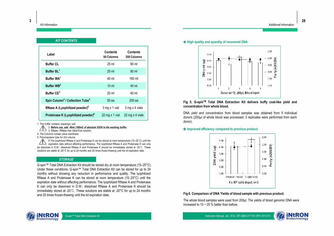
Fig 5. G-spinTM Total DNA Extraction Kit delivers buffy coat-like yield andconcentration from whole blood.
DNA yield and concentration from blood samples was obtained from 5 individualdonors (200µl of whole blood was processed; 3 replicates were performed from eachdonor).
◈ High quality and quantity of recovered DNA
Additional Information
Instruction Manual, Jan. 2012, IBT-QMS-GT1704 (R01-2012-01)G-spin™ Total DNA Extraction Kit
1. This buffer contains chaotropic salt.2. Before use, add 40ml (160ml) of absolute EtOH to the washing buffer.3. DNase / RNase free Ultra-Pure solution.
4. The Columns contain silica membrane5. Polypropylene tube for 2ml volume
6 The lyophilized RNase A and Proteinase K can be stored at room temperature (15–25°C) until theexpiration date without affecting performance. The lyophilized RNase A and Proteinase K can only
be dissolved in D.W.; dissolved RNase A and Proteinase K should be immediately stored at -20℃. Thesesolutions are stable at -20°C for up to 24 months and 20 times frozen-thawing until the kit expiration date.
G-spin™ Total DNA Extraction Kit should be stored dry at room temperature (15–25°C).Under these conditions, G-spin™ Total DNA Extraction Kit can be stored for up to 24months without showing any reduction in performance and quality. The lyophilizedRNase A and Proteinase K can be stored at room temperature (15–25°C) until theexpiration date without affecting performance. The lyophilized RNase A and ProteinaseK can only be dissolved in D.W.; dissolved RNase A and Proteinase K should beimmediately stored at -20℃. These solutions are stable at -20°C for up to 24 monthsand 20 times frozen-thawing until the kit expiration date.
STORAGE
Fig 5. G-spinTM Total DNA Extraction Kit delivers buffy coat-like yield andconcentration from whole blood.
DNA yield and concentration from blood samples was obtained from 5 individualdonors (200µl of whole blood was processed; 3 replicates were performed from eachdonor).
Fig 6. Comparison of DNA Yields of blood sample with previous product.
The whole blood samples were used from 200μl. The yields of blood genomic DNA wereincreased to 15 ~ 25 % better than before.
◈ Improved efficiency compared to previous product
Kit Information4
CONSIDERATION BEFORE USE
§ Lyophilized RNase A : Dissolve the RNase A in 0.3 ml of pure D.W. to eachvial. The lyophilized RNase A can be stored at room temperature (15–25°C)
until the expiration date without affecting performance. The lyophilized RNase A canonly be dissolved in D.W.; dissolved RNase A should be immediately stored at -20℃.The RNase A solution is stable at -20°C for up to 24 months and 20 times frozen-thawing until the kit expiration date
§ Lyophilized Proteinase K : Dissolve the Proteinase K in 1.1 ml of pure D.W.to each vial. The lyophilized Proteinase K can be stored at room temperature
(15–25°C) until the expiration date without affecting performance. The lyophilizedProteinase K can only be dissolved in D.W.; dissolved Proteinase K should beimmediately stored at -20℃. The Proteinase K solution is stable at -20°C for up to 24months and 20 times frozen-thawing until the kit expiration date
§ Buffer WBBuffer WB is supplied as concentrate. Before using for the first time, be sure to add40 ml (160 ml)of absolute ethanol (96 - 100%) to obtain a working solution.
§ Preheat a water bath or heating block§ Equilibrate samples to room temperature (15-25°C).§ Equilibrate Buffer IE or distilled water for elution to room temperature.§ If Buffer CL or Buffer BL contains precipitates, dissolve by heating to 70°C with
gentle agitation.§ Centrifugation
All centrifugation steps are carried out at RT (15 - 25°C)
27Additional Information
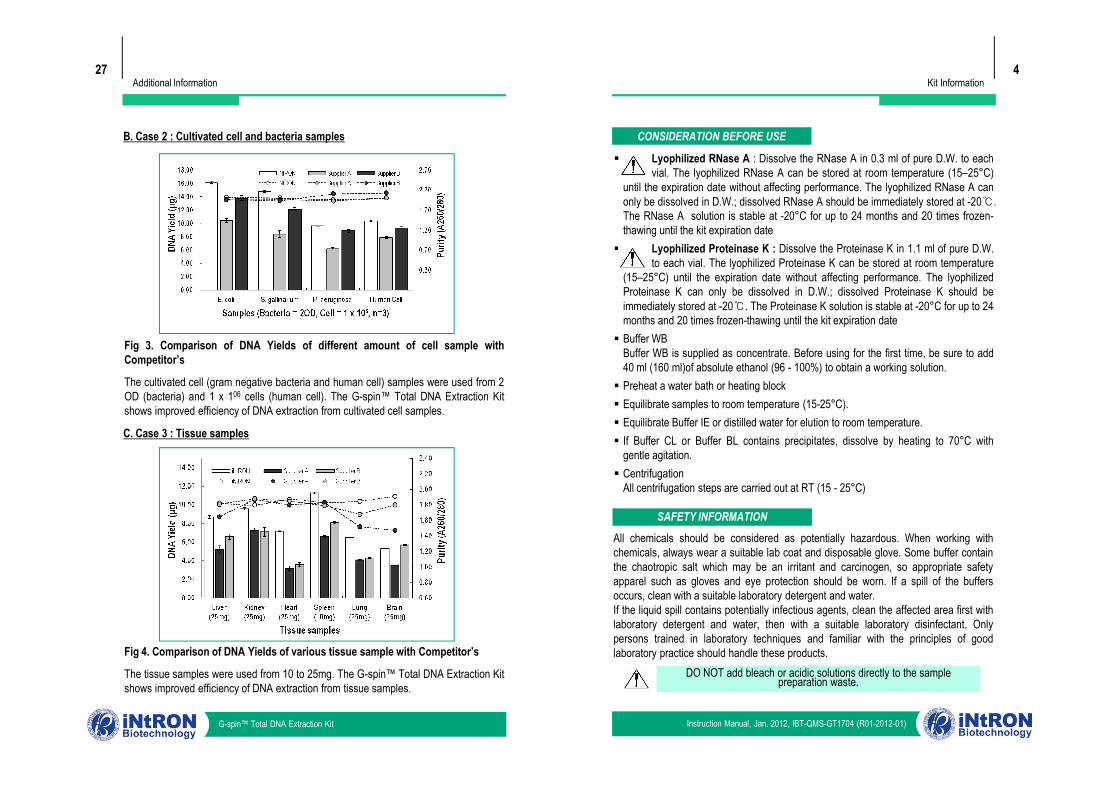
B. Case 2 : Cultivated cell and bacteria samples
Fig 3. Comparison of DNA Yields of different amount of cell sample withCompetitor’s
The cultivated cell (gram negative bacteria and human cell) samples were used from 2OD (bacteria) and 1 x 10⁶ cells (human cell). The G-spin™ Total DNA Extraction Kitshows improved efficiency of DNA extraction from cultivated cell samples.
Instruction Manual, Jan. 2012, IBT-QMS-GT1704 (R01-2012-01)G-spin™ Total DNA Extraction Kit
SAFETY INFORMATION
All chemicals should be considered as potentially hazardous. When working withchemicals, always wear a suitable lab coat and disposable glove. Some buffer containthe chaotropic salt which may be an irritant and carcinogen, so appropriate safetyapparel such as gloves and eye protection should be worn. If a spill of the buffersoccurs, clean with a suitable laboratory detergent and water.If the liquid spill contains potentially infectious agents, clean the affected area first withlaboratory detergent and water, then with a suitable laboratory disinfectant. Onlypersons trained in laboratory techniques and familiar with the principles of goodlaboratory practice should handle these products.
DO NOT add bleach or acidic solutions directly to the sample preparation waste.
§ Lyophilized RNase A : Dissolve the RNase A in 0.3 ml of pure D.W. to eachvial. The lyophilized RNase A can be stored at room temperature (15–25°C)
until the expiration date without affecting performance. The lyophilized RNase A canonly be dissolved in D.W.; dissolved RNase A should be immediately stored at -20℃.The RNase A solution is stable at -20°C for up to 24 months and 20 times frozen-thawing until the kit expiration date
§ Lyophilized Proteinase K : Dissolve the Proteinase K in 1.1 ml of pure D.W.to each vial. The lyophilized Proteinase K can be stored at room temperature
(15–25°C) until the expiration date without affecting performance. The lyophilizedProteinase K can only be dissolved in D.W.; dissolved Proteinase K should beimmediately stored at -20℃. The Proteinase K solution is stable at -20°C for up to 24months and 20 times frozen-thawing until the kit expiration date
§ Buffer WBBuffer WB is supplied as concentrate. Before using for the first time, be sure to add40 ml (160 ml)of absolute ethanol (96 - 100%) to obtain a working solution.
§ Preheat a water bath or heating block§ Equilibrate samples to room temperature (15-25°C).§ Equilibrate Buffer IE or distilled water for elution to room temperature.§ If Buffer CL or Buffer BL contains precipitates, dissolve by heating to 70°C with
gentle agitation.§ Centrifugation
All centrifugation steps are carried out at RT (15 - 25°C)
Fig 3. Comparison of DNA Yields of different amount of cell sample withCompetitor’s
The cultivated cell (gram negative bacteria and human cell) samples were used from 2OD (bacteria) and 1 x 10⁶ cells (human cell). The G-spin™ Total DNA Extraction Kitshows improved efficiency of DNA extraction from cultivated cell samples.
C. Case 3 : Tissue samples
Fig 4. Comparison of DNA Yields of various tissue sample with Competitor’s
The tissue samples were used from 10 to 25mg. The G-spin™ Total DNA Extraction Kitshows improved efficiency of DNA extraction from tissue samples.
Kit Information5
ADDITIONAL REQUIRED EQUIPMENTG-spin™ Total DNA Extraction Kit provides almost all reagents for extracting DNA,including RNase A and Proteinase K. However, you should prepare some equipmentsand reagents as follows for a fast and easy extraction. When working with chemicals,always wear a suitable lab coat, disposable gloves and protective goggles.Common equipment and reagents• Equipment for disruption and homogenization, mechanical tissue grinder like pestle• Pipettes and pipette tips • Water bath or heating block• Vortex mixer • Microcentrifuge with rotor for 2.0 ml tubes• Microcentrifuge tubes (1.5 ml) • Liquid nitrogen• Absolute ethanol (EtOH, 96~100%) • 80% ethanol• Ice • Other general lab equipments• 1X PBS Buffer • Xylene Solution (for paraffin block)
QUALITY CONTROL§ In accordance with iNtRON’s ISO-certified Total Quality Management System, each
lot of G-spin™ Total DNA Extraction Kit is tested against predeterminedspecifications to ensure consistent product quality. The quality of the isolatedgenomic DNA was checked by restriction analysis, agarose gel electrophoresis, andspectrophotometric determination.
§ G-spin™ column control : The DNA binding capacity was tested by determining therecovery with 10 ~ 15 μg of genomic DNA from 1 x 10⁶ cultivated cells.
§ RNase A / Proteinase K : In case of RNase A, the activity was determined 20K ~25K unit per mg of protein using tolura yeast RNA hydration test. Also, in case ofProteinase K, the activity was determined from cleavage of the substrate releasing p-nitroaniline which can be measured spectrophotometrically at 410nm.
§ Buffer control : Conductivity and pH of buffers were tested and found to be within thepre-determinated ranges descrbed below.
26
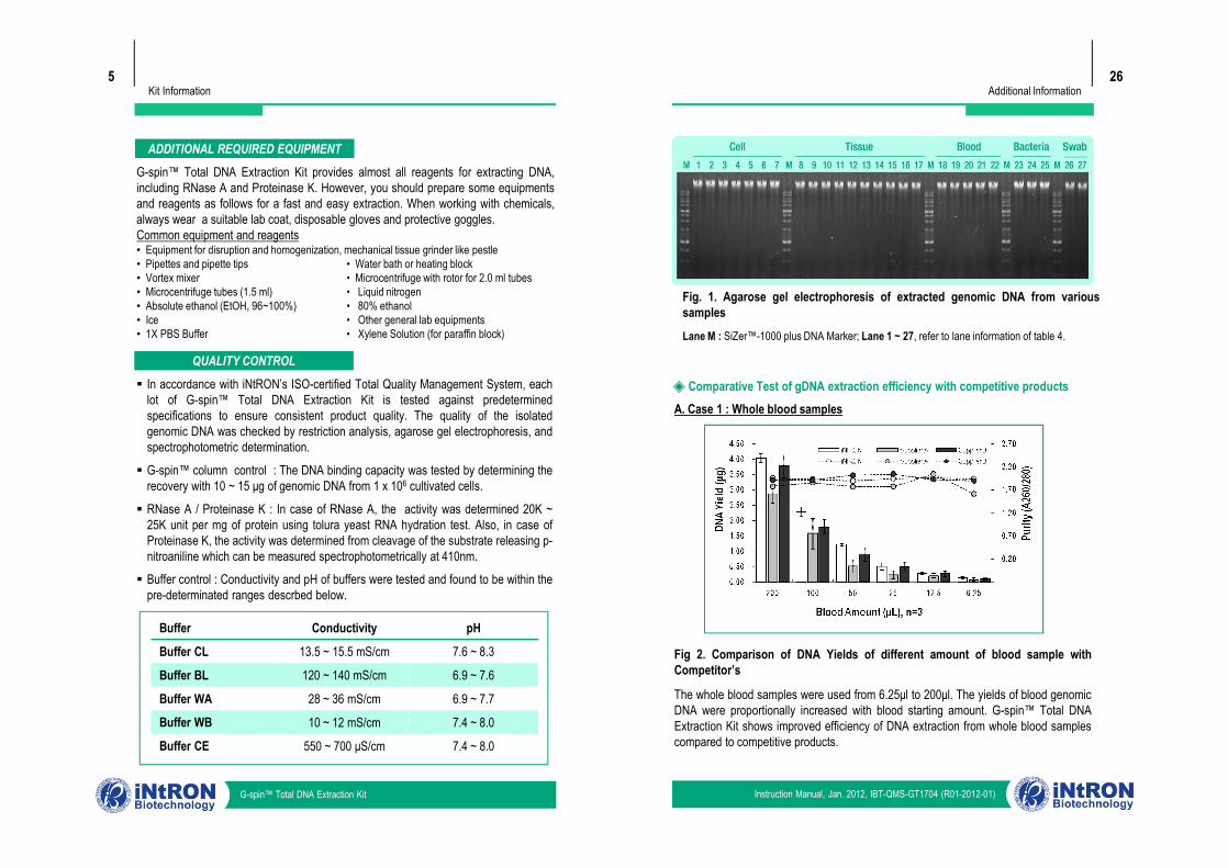
A. Case 1 : Whole blood samples◈ Comparative Test of gDNA extraction efficiency with competitive products
Additional Information
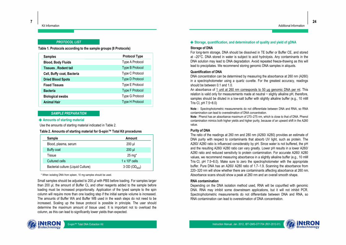
Fig. 1. Agarose gel electrophoresis of extracted genomic DNA from varioussamplesLane M : SiZer™-1000 plus DNA Marker; Lane 1 ~ 27, refer to lane information of table 4.
Instruction Manual, Jan. 2012, IBT-QMS-GT1704 (R01-2012-01)G-spin™ Total DNA Extraction Kit
§ In accordance with iNtRON’s ISO-certified Total Quality Management System, eachlot of G-spin™ Total DNA Extraction Kit is tested against predeterminedspecifications to ensure consistent product quality. The quality of the isolatedgenomic DNA was checked by restriction analysis, agarose gel electrophoresis, andspectrophotometric determination.
§ G-spin™ column control : The DNA binding capacity was tested by determining therecovery with 10 ~ 15 μg of genomic DNA from 1 x 10⁶ cultivated cells.
§ RNase A / Proteinase K : In case of RNase A, the activity was determined 20K ~25K unit per mg of protein using tolura yeast RNA hydration test. Also, in case ofProteinase K, the activity was determined from cleavage of the substrate releasing p-nitroaniline which can be measured spectrophotometrically at 410nm.
§ Buffer control : Conductivity and pH of buffers were tested and found to be within thepre-determinated ranges descrbed below.
Buffer Conductivity pH
Buffer CL 13.5 ~ 15.5 mS/cm 7.6 ~ 8.3
Buffer BL 120 ~ 140 mS/cm 6.9 ~ 7.6
Buffer WA 28 ~ 36 mS/cm 6.9 ~ 7.7
Buffer WB 10 ~ 12 mS/cm 7.4 ~ 8.0
Buffer CE 550 ~ 700 μS/cm 7.4 ~ 8.0
Fig 2. Comparison of DNA Yields of different amount of blood sample withCompetitor’s
The whole blood samples were used from 6.25μl to 200μl. The yields of blood genomicDNA were proportionally increased with blood starting amount. G-spin™ Total DNAExtraction Kit shows improved efficiency of DNA extraction from whole blood samplescompared to competitive products.
Kit Information6
APPLICATIONS§ Cancer research ▪ Gram negative bacterial research§ Human genetic research ▪ Viral DNA Research§ Detection Assay : PCR, real time PCR§ DNA hybridization : Southern blotting, Microarray
PRODUCT WARRANTY AND SATISFACTION GUARANTEEAll products undergo extensive quality control test and are warranted to perform asdescribed when used correctly. Immediately any problems should be reported. Satisfactionguarantee is conditional upon the customer providing full details of the problem to iNtRONwithin 60 days, and returning the product to iNtRON for examination.
PRODUCT USE LIMITATIONS
The G-spin™ Total DNA Extraction Kit is intended for research use only. Prior to using itfor other purposes, the user must validate the system in compliance with the applicable law,directives, and regulations.
G-spin™ Total DNA Extraction Kit is developed, designed, and sold for research purposeonly. They are not to be used for human or animal diagnosis of diseases. Do not useinternally or externally in humans or animals. Be careful in the handling of the products.
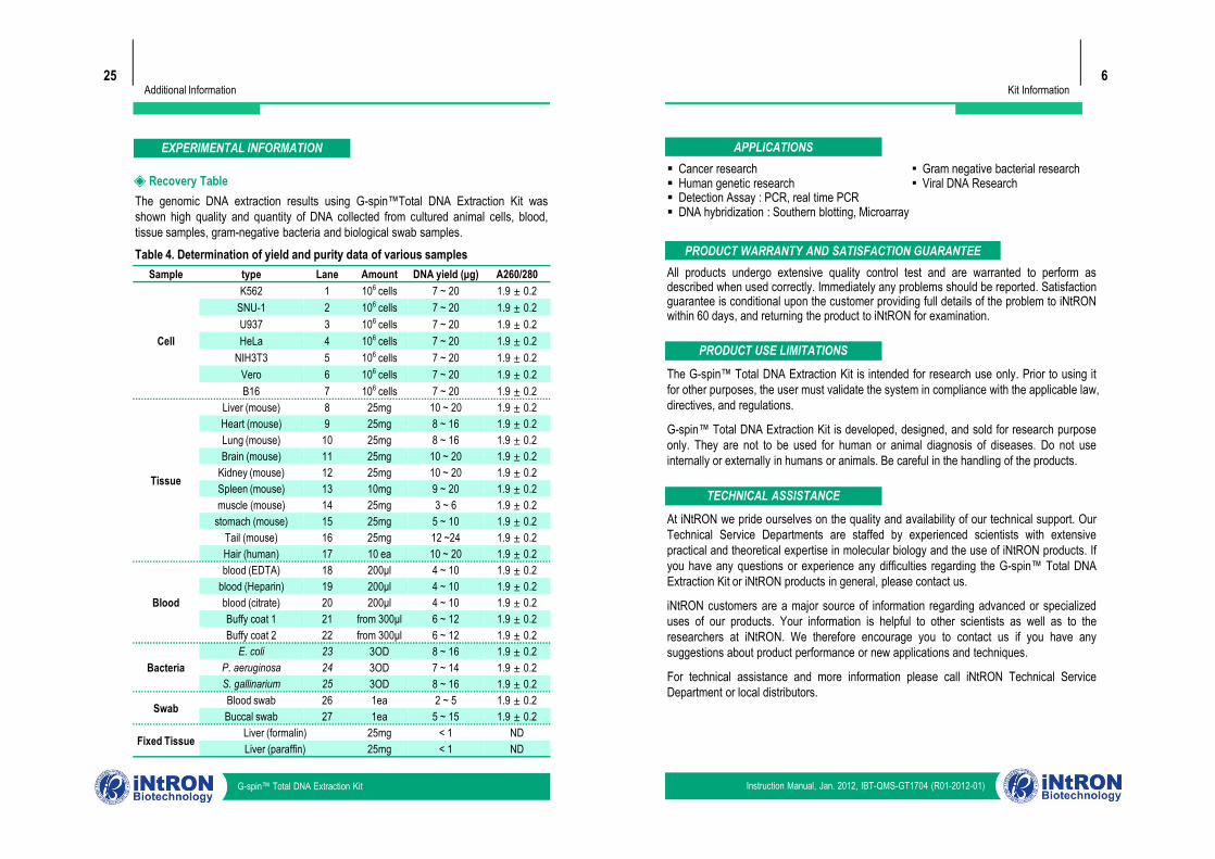
25Additional Information
The genomic DNA extraction results using G-spin™Total DNA Extraction Kit wasshown high quality and quantity of DNA collected from cultured animal cells, blood,tissue samples, gram-negative bacteria and biological swab samples.
◈ Recovery Table
Table 4. Determination of yield and purity data of various samplesSample type Lane Amount DNA yield (μg) A260/280
Cell
K562 1 106 cells 7 ~ 20 1.9 ± 0.2 SNU-1 2 106 cells 7 ~ 20 1.9 ± 0.2 U937 3 106 cells 7 ~ 20 1.9 ± 0.2 HeLa 4 106 cells 7 ~ 20 1.9 ± 0.2
NIH3T3 5 106 cells 7 ~ 20 1.9 ± 0.2 Vero 6 106 cells 7 ~ 20 1.9 ± 0.2 B16 7 106 cells 7 ~ 20 1.9 ± 0.2
Liver (mouse) 8 25mg 10 ~ 20 1.9 ± 0.2 Heart (mouse) 9 25mg 8 ~ 16 1.9 ± 0.2
EXPERIMENTAL INFORMATION
Instruction Manual, Jan. 2012, IBT-QMS-GT1704 (R01-2012-01)G-spin™ Total DNA Extraction Kit
The G-spin™ Total DNA Extraction Kit is intended for research use only. Prior to using itfor other purposes, the user must validate the system in compliance with the applicable law,directives, and regulations.
G-spin™ Total DNA Extraction Kit is developed, designed, and sold for research purposeonly. They are not to be used for human or animal diagnosis of diseases. Do not useinternally or externally in humans or animals. Be careful in the handling of the products.
TECHNICAL ASSISTANCE
At iNtRON we pride ourselves on the quality and availability of our technical support. OurTechnical Service Departments are staffed by experienced scientists with extensivepractical and theoretical expertise in molecular biology and the use of iNtRON products. Ifyou have any questions or experience any difficulties regarding the G-spin™ Total DNAExtraction Kit or iNtRON products in general, please contact us.
iNtRON customers are a major source of information regarding advanced or specializeduses of our products. Your information is helpful to other scientists as well as to theresearchers at iNtRON. We therefore encourage you to contact us if you have anysuggestions about product performance or new applications and techniques.
For technical assistance and more information please call iNtRON Technical ServiceDepartment or local distributors.
Tissue
Heart (mouse) 9 25mg 8 ~ 16 1.9 ± 0.2 Lung (mouse) 10 25mg 8 ~ 16 1.9 ± 0.2 Brain (mouse) 11 25mg 10 ~ 20 1.9 ± 0.2
Kidney (mouse) 12 25mg 10 ~ 20 1.9 ± 0.2 Spleen (mouse) 13 10mg 9 ~ 20 1.9 ± 0.2 muscle (mouse) 14 25mg 3 ~ 6 1.9 ± 0.2
stomach (mouse) 15 25mg 5 ~ 10 1.9 ± 0.2 Tail (mouse) 16 25mg 12 ~24 1.9 ± 0.2 Hair (human) 17 10 ea 10 ~ 20 1.9 ± 0.2
Blood
blood (EDTA) 18 200μl 4 ~ 10 1.9 ± 0.2 blood (Heparin) 19 200μl 4 ~ 10 1.9 ± 0.2 blood (citrate) 20 200μl 4 ~ 10 1.9 ± 0.2 Buffy coat 1 21 from 300μl 6 ~ 12 1.9 ± 0.2 Buffy coat 2 22 from 300μl 6 ~ 12 1.9 ± 0.2
BacteriaE. coli 23 3OD 8 ~ 16 1.9 ± 0.2
P. aeruginosa 24 3OD 7 ~ 14 1.9 ± 0.2 S. gallinarium 25 3OD 8 ~ 16 1.9 ± 0.2
Swab Blood swab 26 1ea 2 ~ 5 1.9 ± 0.2 Buccal swab 27 1ea 5 ~ 15 1.9 ± 0.2
Fixed Tissue Liver (formalin) 25mg < 1 NDLiver (paraffin) 25mg < 1 ND
Kit Information7
PROTOCOL LISTTable 1. Protocols according to the sample groups (8 Protocols)
SamplesBlood, Body FluidsTissues , Rodent tailCell, Buffy coat, BacteriaDried Blood SpotsFixed TissuesBacteriaBiological swabsAnimal Hair
Protocol TypeType A ProtocolType B ProtocolType C ProtocolType D ProtocolType E ProtocolType F ProtocolType G ProtocolType H Protocol
SAMPLE PREPARATION◈ Amounts of starting materialUse the amounts of starting material indicated in Table 2.Table 2. Amounts of starting material for G-spin™ Total Kit procedures
24Additional Information
Storage of DNAFor long-term storage, DNA should be dissolved in TE buffer or Buffer CE, and storedat –20°C. DNA stored in water is subject to acid hydrolysis. Any contaminants in theDNA solution may lead to DNA degradation. Avoid repeated freeze-thawing as this willlead to precipitates. We recommend storing genomic DNA samples in aliquots.Quantification of DNADNA concentration can be determined by measuring the absorbance at 260 nm (A260)in a spectrophotometer using a quartz cuvette. For the greatest accuracy, readingsshould be between 0.1 and 1.0.An absorbance of 1 unit at 260 nm corresponds to 50 μg genomic DNA per ml. Thisrelation is valid only for measurements made at neutral ~ slighly alkaline pH, therefore,samples should be diluted in a low-salt buffer with slightly alkaline buffer (e.g., 10 mMTris·Cl, pH 7.5~8.0)Note : Spectrophotometric measurements do not differentiate between DNA and RNA, so RNAcontamination can lead to overestimation of DNA concentration.Note : Phenol has an absorbance maximum of 270–275 nm, which is close to that of DNA. Phenolcontamination mimics both higher yields and higher purity, because of an upward shift in the A260value.
Purity of DNAThe ratio of the readings at 260 nm and 280 nm (A260/ A280) provides an estimate ofDNA purity with respect to contaminants that absorb UV light, such as protein. TheA260/ A280 ratio is influenced considerably by pH. Since water is not buffered, the pHand the resulting A260/ A280 ratio can vary greatly. Lower pH results in a lower A260/A280 ratio and reduced sensitivity to protein contamination. For accurate A260/ A280values, we recommend measuring absorbance in a slightly alkaline buffer (e.g., 10 mMTris·Cl, pH 7.5~8.0). Make sure to zero the spectrophotometer with the appropriatebuffer. Pure DNA has an A260/ A280 ratio of 1.7–1.9. Scanning the absorbance from220–320 nm will show whether there are contaminants affecting absorbance at 260 nm.Absorbance scans should show a peak at 260 nm and an overall smooth shape.RNA contaminationDepending on the DNA isolation method used, RNA will be copurified with genomicDNA. RNA may inhibit some downstream applications, but it will not inhibit PCR.Spectrophotometric measurements do not differentiate between DNA and RNA, soRNA contamination can lead to overestimation of DNA concentration.
◈ Storage, quantification, and determination of quality and yield of gDNA
Instruction Manual, Jan. 2012, IBT-QMS-GT1704 (R01-2012-01)G-spin™ Total DNA Extraction Kit
Use the amounts of starting material indicated in Table 2.Table 2. Amounts of starting material for G-spin™ Total Kit procedures
Sample AmountBlood, plasma, serum 200 μlBuffy coat 200 μlTissue 25 mg*Cultured cells 1 x 10⁶ cellsBacterial culture (Liquid Culture) 3 OD (OD600)
* When isolating DNA from spleen, 10 mg samples should be used.
Small samples should be adjusted to 200 μl with PBS before loading. For samples largerthan 200 μl, the amount of Buffer CL and other reagents added to the sample beforeloading must be increased proportionally. Application of the lysed sample to the spincolumn will require more than one loading step if the initial sample volume is increased.The amounts of Buffer WA and Buffer WB used in the wash steps do not need to beincreased. Scaling up the tissue protocol is possible in principle. The user shoulddetermine the maximum amount of tissue used. It is important not to overload thecolumn, as this can lead to significantly lower yields than expected.
Storage of DNAFor long-term storage, DNA should be dissolved in TE buffer or Buffer CE, and storedat –20°C. DNA stored in water is subject to acid hydrolysis. Any contaminants in theDNA solution may lead to DNA degradation. Avoid repeated freeze-thawing as this willlead to precipitates. We recommend storing genomic DNA samples in aliquots.Quantification of DNADNA concentration can be determined by measuring the absorbance at 260 nm (A260)in a spectrophotometer using a quartz cuvette. For the greatest accuracy, readingsshould be between 0.1 and 1.0.An absorbance of 1 unit at 260 nm corresponds to 50 μg genomic DNA per ml. Thisrelation is valid only for measurements made at neutral ~ slighly alkaline pH, therefore,samples should be diluted in a low-salt buffer with slightly alkaline buffer (e.g., 10 mMTris·Cl, pH 7.5~8.0)Note : Spectrophotometric measurements do not differentiate between DNA and RNA, so RNAcontamination can lead to overestimation of DNA concentration.Note : Phenol has an absorbance maximum of 270–275 nm, which is close to that of DNA. Phenolcontamination mimics both higher yields and higher purity, because of an upward shift in the A260value.
Purity of DNAThe ratio of the readings at 260 nm and 280 nm (A260/ A280) provides an estimate ofDNA purity with respect to contaminants that absorb UV light, such as protein. TheA260/ A280 ratio is influenced considerably by pH. Since water is not buffered, the pHand the resulting A260/ A280 ratio can vary greatly. Lower pH results in a lower A260/A280 ratio and reduced sensitivity to protein contamination. For accurate A260/ A280values, we recommend measuring absorbance in a slightly alkaline buffer (e.g., 10 mMTris·Cl, pH 7.5~8.0). Make sure to zero the spectrophotometer with the appropriatebuffer. Pure DNA has an A260/ A280 ratio of 1.7–1.9. Scanning the absorbance from220–320 nm will show whether there are contaminants affecting absorbance at 260 nm.Absorbance scans should show a peak at 260 nm and an overall smooth shape.RNA contaminationDepending on the DNA isolation method used, RNA will be copurified with genomicDNA. RNA may inhibit some downstream applications, but it will not inhibit PCR.Spectrophotometric measurements do not differentiate between DNA and RNA, soRNA contamination can lead to overestimation of DNA concentration.
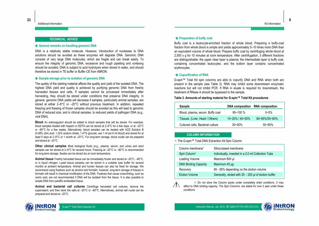
Kit Information8
◈ Preparation of buffy coatBuffy coat is a leukocyte-enriched fraction of whole blood. Preparing a buffy-coatfraction from whole blood is simple and yields approximately 5–10 times more DNA thanan equivalent volume of whole blood. Prepare buffy coat by centrifuging whole blood at2,500 x g for 10 minutes at room temperature. After centrifugation, 3 different fractionsare distinguishable: the upper clear layer is plasma; the intermediate layer is buffy coat,containing concentrated leukocytes; and the bottom layer contains concentratederythrocytes.
◈ Copurification of RNAG-spin™ Total Kit spin columns are able to copurify DNA and RNA when both arepresent in the sample (see Table 3). RNA may inhibit some downstream enzymaticreactions but will not inhibit PCR. If RNA in eluate is required for downstream, thetreatment of RNase A should be bypassed to the sample.Table 3. Amounts of starting material for G-spin™ Total Kit procedures
Sample DNA composition RNA composition
Blood, plasma, serum, Buffy coat 95~100 % 0~5%
Tissues (Liver, Heart / Others) 15~20% / 40~50% 80~85%/50~60%
Cultured cells, Bacterial culture 30~50% 50~80%
23Additional Information
TECHNICAL ADVICE
DNA is a relatively stable molecule. However, introduction of nucleases to DNAsolutions should be avoided as these enzymes will degrade DNA. Genomic DNAconsists of very large DNA molecules, which are fragile and can break easily. Toensure the integrity of genomic DNA, excessive and rough pipetting and vortexingshould be avoided. DNA is subject to acid hydrolysis when stored in water, and shouldtherefore be stored in TE buffer or Buffer CE from iNtRON.
◈ General remarks on handling genomic DNA
The quality of the starting material affects the quality and yield of the isolated DNA. Thehighest DNA yield and quality is achieved by purifying genomic DNA from freshlyharvested tissues and cells. If samples cannot be processed immediately afterharvesting, they should be stored under conditions that preserve DNA integrity. Ingeneral, genomic DNA yields will decrease if samples, particularly animal samples, arestored at either 2–8°C or –20°C without previous treatment. In addition, repeatedfreezing and thawing of frozen samples should be avoided as this will lead to genomicDNA of reduced size, and in clinical samples, to reduced yields of pathogen DNA (e.g.,viral DNA).Blood An anticoagulant should be added to blood samples that will be stored. For example,blood samples treated with heparin or EDTA can be stored at 2–8°C for a few days, or at –20°Cor –80°C for a few weeks. Alternatively, blood samples can be treated with ACD Solution B(0.48% citric acid, 1.32% sodium citrate, 1.47% glucose; use 1 ml per 6 ml blood) and stored for atleast 5 days at 2–8°C or 1 month at –20°C. For long-term storage, blood nuclei can be preparedand stored at –20°C.
Other clinical samples Most biological fluids (e.g., plasma, serum, and urine) and stoolsamples can be stored at 2–8°C for several hours. Freezing at –20°C or –80°C is recommendedfor long-term storage. Swabs can be stored dry at room temperature.
Animal tissue Freshly harvested tissue can be immediately frozen and stored at –20°C, –80°C,or in liquid nitrogen. Lysed tissue samples can be stored in a suitable lysis buffer for severalmonths at ambient temperature. Animal and human tissues can also be fixed for storage. Werecommend using fixatives such as alcohol and formalin; however, long-term storage of tissues informalin will result in chemical modification of the DNA. Fixatives that cause cross-linking, such asosmic acid, are not recommended if DNA will be isolated from the tissue. It is also possible toisolate DNA from paraffin-embedded tissue.
Animal and bacterial cell cultures Centrifuge harvested cell cultures, remove thesupernatant, and then store the cells at –20°C or –80°C. Alternatively, animal cell nuclei can beprepared and stored at –20°C.
◈ Sample storage prior to isolation of genomic DNA
Instruction Manual, Jan. 2012, IBT-QMS-GT1704 (R01-2012-01)G-spin™ Total DNA Extraction Kit
• The G-spin™ Total DNA Extraction Kit Spin Column
1. Do not store the Column packs under completely dried conditions. It mayaffect to DNA binding capacity. The Spin Columns are stable for over 2 year under theseconditions
COLUMN INFORMATION
Column membrane1
Spin Column1
Loading VolumeDNA Binding CapacityRecoveryElution Volume
Silica-based membraneIndividually, inserted in a 2.0 ml Collection TubeMaximum 800 μlMaximum 45 μg85 - 95% depending on the elution volumeGenerally, eluted with 30 – 200 μl of elution buffer
Sample DNA composition RNA composition
Blood, plasma, serum, Buffy coat 95~100 % 0~5%
Tissues (Liver, Heart / Others) 15~20% / 40~50% 80~85%/50~60%
Cultured cells, Bacterial culture 30~50% 50~80%
The quality of the starting material affects the quality and yield of the isolated DNA. Thehighest DNA yield and quality is achieved by purifying genomic DNA from freshlyharvested tissues and cells. If samples cannot be processed immediately afterharvesting, they should be stored under conditions that preserve DNA integrity. Ingeneral, genomic DNA yields will decrease if samples, particularly animal samples, arestored at either 2–8°C or –20°C without previous treatment. In addition, repeatedfreezing and thawing of frozen samples should be avoided as this will lead to genomicDNA of reduced size, and in clinical samples, to reduced yields of pathogen DNA (e.g.,viral DNA).Blood An anticoagulant should be added to blood samples that will be stored. For example,blood samples treated with heparin or EDTA can be stored at 2–8°C for a few days, or at –20°Cor –80°C for a few weeks. Alternatively, blood samples can be treated with ACD Solution B(0.48% citric acid, 1.32% sodium citrate, 1.47% glucose; use 1 ml per 6 ml blood) and stored for atleast 5 days at 2–8°C or 1 month at –20°C. For long-term storage, blood nuclei can be preparedand stored at –20°C.
Other clinical samples Most biological fluids (e.g., plasma, serum, and urine) and stoolsamples can be stored at 2–8°C for several hours. Freezing at –20°C or –80°C is recommendedfor long-term storage. Swabs can be stored dry at room temperature.
Animal tissue Freshly harvested tissue can be immediately frozen and stored at –20°C, –80°C,or in liquid nitrogen. Lysed tissue samples can be stored in a suitable lysis buffer for severalmonths at ambient temperature. Animal and human tissues can also be fixed for storage. Werecommend using fixatives such as alcohol and formalin; however, long-term storage of tissues informalin will result in chemical modification of the DNA. Fixatives that cause cross-linking, such asosmic acid, are not recommended if DNA will be isolated from the tissue. It is also possible toisolate DNA from paraffin-embedded tissue.
Animal and bacterial cell cultures Centrifuge harvested cell cultures, remove thesupernatant, and then store the cells at –20°C or –80°C. Alternatively, animal cell nuclei can beprepared and stored at –20°C.
Protocols9
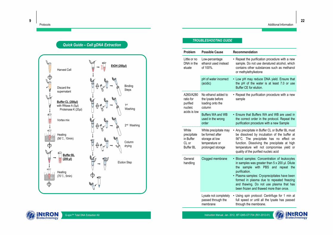
Quick Guide – Cell gDNA Extraction
Harvest Cell
Discard the supernatant
Buffer CL (200μl)with RNase A (5μl)
Proteinase K (20μl)
Vortex mix
EtOH (200μl)
BindingSteps
1st
Washing
22
TROUBLESHOOTING GUIDE
Additional Information
Problem Possible Cause Recommendation
Little or no DNA in the eluate
Low-percentage ethanol used instead of 100%.
• Repeat the purification procedure with a newsample. Do not use denatured alcohol, whichcontains other substances such as methanolor methylethylketone
pH of water incorrect (acidic)
• Low pH may reduce DNA yield. Ensure thatthe pH of the water is at least 7.0 or useBuffer CE for elution.
A260/A280 ratio for purified nucleic acids is low
No ethanol added to the lysate before loading onto the column
• Repeat the purification procedure with a newsample
Buffers WA and WB used in the wrong order
• Ensure that Buffers WA and WB are used inthe correct order in the protocol. Repeat thepurification procedure with a new Sample
Instruction Manual, Jan. 2012, IBT-QMS-GT1704 (R01-2012-01)G-spin™ Total DNA Extraction Kit
Vortex mix
Buffer BL (200 μl)
Heating(56℃, 10min)
Heating(70℃, 5min)
2nd Washing
Columndrying
Elution Step
Buffers WA and WB used in the wrong order
• Ensure that Buffers WA and WB are used inthe correct order in the protocol. Repeat thepurification procedure with a new Sample
White precipitate in Buffer CL or Buffer BL
White precipitate may be formed after storage at low temperature or prolonged storage
• Any precipitate in Buffer CL or Buffer BL mustbe dissolved by incubation of the buffer at56°C. The precipitate has no effect onfunction. Dissolving the precipitate at hightemperature will not compromise yield orquality of the purified nucleic acid
General handling
Clogged membrane • Blood samples: Concentration of leukocytesin samples was greater than 5 x 200 μl. Dilutethe sample with PBS and repeat thepurification.• Plasma samples: Cryoprecipitates have been
formed in plasma due to repeated freezingand thawing. Do not use plasma that hasbeen frozen and thawed more than once.
Lysate not completely passed through the membrane
• Using spin protocol: Centrifuge for 1 min atfull speed or until all the lysate has passedthrough the membrane.
Protocols10
PROTOCOL A (for Blood, body fluids)
1.Pipet 200 μl of whole blood or body fluids into a 1.5 ml microcentrifuge tube (notprovided).Note : If the volume of sample is less than 200 μl, use Buffer CL or PBS Buffer to make up thevolume to 200ml
2.Add 20 μl of Proteinase K and 5 μl of RNase A Solution into sample tube andgently mix.
Note : It is possible to add Proteinase K to blood sample that have already beenmeasured into 1.5 ml tube. It is important to assure that it is properly mixed after adding
the Proteinase K and RNase A solution.
3. Add 200 μl of Buffer BL into upper sample tube and mix thoroughly. Forcomplete lysis, mix 3 or 4 times during incubation by inverting tube.
Note : Avoid any vigorous vortexing because doing so may induce genomic DNAbreakage. In order to assure efficient lysis, it is important that the blood sample andBuffer BL are mixed thoroughly to yield a lysis solution.
4. Incubate the lysate at 56°C for 10 min.Note : For complete lysis, mix 3 or 4 times during incubation by inverting tube. If it lysis perfectly,the red color of lysate becomes the dark green.
5.Briefly centrifuge the 1.5 ml tube to remove drops from the inside of the rid.
6.Add 200 μl of absolute ethanol into the lysate, and mix well by gently inverting 5- 6 times or by pipetting. DO NOT vortex. After mixing, briefly centrifuge the 1.5ml tube to remove drops from inside of the lid.Note : This step is an equilibration step for binding genomic DNA to column membrane. It isimportant to assure proper mixing after adding the ethanol, until phase separation could not beobserved.
7.Carefully apply the mixture from step 7 to the Spin Column (in a 2 ml CollectionTube) without wetting the rim, close the cap, and centrifuge at 13,000 rpm for 1min. Discard the filtrate and place the Spin Column in a new 2 ml CollectionTube (additionally supplied).Note : Close each Spin Column in order to avoid aerosol formation during centrifugation. Do nottransfer any solid materials.
8.Add 700 μl of Buffer WA to the Spin Column without wetting the rim, andcentrifuge for 1 min at 13,000 rpm. Discard the flow-through and reuse theCollection Tube.
21
TROUBLESHOOTING GUIDE
Additional Information
Problem Possible Cause Recommendation
Colored residues remain on the spin column after washing
Inefficient cell lysisdue to insufficient mixing of the sample with Buffer BL
• Repeat the DNA purification procedure with anew sample. Be sure to mix the sample andBuffer BL immediately and thoroughly bypulse-vortexing.
Inefficient cell lysisdue to decreased protease activity
• Repeat the DNA purification procedure witha new sample and with freshly preparedProteinase K stock solution. Be sure to storethe stock solution at 2–8°C immediately afteruse. Ensure that Proteinase K is not addeddirectly to Buffer BL
No ethanol added to the lysate before loading onto the column
• Repeat the purification procedure with a newsample.
Instruction Manual, Jan. 2012, IBT-QMS-GT1704 (R01-2012-01)G-spin™ Total DNA Extraction Kit
1.Pipet 200 μl of whole blood or body fluids into a 1.5 ml microcentrifuge tube (notprovided).Note : If the volume of sample is less than 200 μl, use Buffer CL or PBS Buffer to make up thevolume to 200ml
2.Add 20 μl of Proteinase K and 5 μl of RNase A Solution into sample tube andgently mix.
Note : It is possible to add Proteinase K to blood sample that have already beenmeasured into 1.5 ml tube. It is important to assure that it is properly mixed after adding
the Proteinase K and RNase A solution.
3. Add 200 μl of Buffer BL into upper sample tube and mix thoroughly. Forcomplete lysis, mix 3 or 4 times during incubation by inverting tube.
Note : Avoid any vigorous vortexing because doing so may induce genomic DNAbreakage. In order to assure efficient lysis, it is important that the blood sample andBuffer BL are mixed thoroughly to yield a lysis solution.
4. Incubate the lysate at 56°C for 10 min.Note : For complete lysis, mix 3 or 4 times during incubation by inverting tube. If it lysis perfectly,the red color of lysate becomes the dark green.
5.Briefly centrifuge the 1.5 ml tube to remove drops from the inside of the rid.
6.Add 200 μl of absolute ethanol into the lysate, and mix well by gently inverting 5- 6 times or by pipetting. DO NOT vortex. After mixing, briefly centrifuge the 1.5ml tube to remove drops from inside of the lid.Note : This step is an equilibration step for binding genomic DNA to column membrane. It isimportant to assure proper mixing after adding the ethanol, until phase separation could not beobserved.
7.Carefully apply the mixture from step 7 to the Spin Column (in a 2 ml CollectionTube) without wetting the rim, close the cap, and centrifuge at 13,000 rpm for 1min. Discard the filtrate and place the Spin Column in a new 2 ml CollectionTube (additionally supplied).Note : Close each Spin Column in order to avoid aerosol formation during centrifugation. Do nottransfer any solid materials.
8.Add 700 μl of Buffer WA to the Spin Column without wetting the rim, andcentrifuge for 1 min at 13,000 rpm. Discard the flow-through and reuse theCollection Tube.
No ethanol added to the lysate before loading onto the column
Little or no DNA in the eluate
Low concentration of cells or viruses in the sample
• Concentrate a larger volume of a new cell-free sample to 200 μl using a Centricon®-100(Amicon, USA). Repeat the DNA purificationprocedure, adding 5–10 μg of carrier to eachlysate if the sample has a low DNA content. Ifwhole blood was used, prepare buffy coat
Inefficient cell lysisdue to insufficient mixing with Buffer BL
• Repeat the DNA purification procedure with anew sample. Be sure to mix the sample andBuffer BL immediately and thoroughly bypulse-vortexing.
Inefficient cell lysis orprotein degradation inBuffer CL or BufferBL
• Repeat the procedure with a new sample.Ensure that the tissue sample is cut into smallpieces and extend the incubation time.Ensure that no residual particulates arevisible
Protocols11
9. Add 700 μl of Buffer WB to the Spin Column without wetting the rim, andcentrifuge for 1 min at 13,000 rpm. Discard the flow-through and place theColumn into a new 2.0 ml Collection Tube (additionally supplied), Then againcentrifuge for additionally 1 min to dry the membrane. Discard the flow-throughand Collection Tube altogether.Note : It is very important to dry the membrane of the Spin Column since residual ethanol mayinhibit subsequent reactions. Following the centrifugation, remove carefully the Spin Columnfrom the Collection Tube without contacting with the flow-through, since this will result incarryover of ethanol.Note : Ensure that 40 (160) ml of absolute ethanol has been added to Buffer WB.
10. Place the Spin Column into a new 1.5 ml tube (not supplied), and 30 - 100 μl ofBuffer CE directly onto the membrane. Incubate for 1 min at room temperatureand then centrifuge for 1 min at 13,000 rpm to elute.Note : Elution with 30 μl (instead of 50 μl) increases the final DNA concentration, but reducesoverall DNA yield.Note : A new 1.5 ml tube can be used for the second elution step to prevent dilution of the firsteluate. Alternatively, the tube can be reused for the second elution step to combine the eluates.
Protocols20
PROTOCOL H (for Animal hair)
1.Prepare 10 pieces of hair.Note : In the case of hair sample, the purification result is changed greatly according with orwithout hair root in hair sample. If it is possible, it is desirable to use sample which contains hairroot. In case of no hair root sample, it is available to detect the result only PCR amplification
2.Cut the hair sample from hair root 1 cm length, then carefully transfer thesample into a new 1.5 ml tube.Note : Carefully handle to sample without loss of hair root because the hair root tends toadhesive on surface of solid material.
3.Crash the hair sample 10 ~ 20 times using micro-pestle with 50 μl of Buffer CL.Note : The hair is not solubilized in Buffer CL, But hair root part is soluble in Buffer CG. It isdifficult too small to estimate in the unaided whether the sample is homogenized well or not.Thus, crash the sample 10 ~ 20 times using micro-pestle.
4.Add 150 μl Buffer CL (include that use in homogenizing step), 20 μl Proteinase Kand 5 μl RNase A Solution into sample tube and mix by vortexing vigorously.
Note : Be sure that Proteinase K and RNase A solutions are always kept under freezer(below -10℃). In case of transcriptionally active tissues, such as liver and kidney, large
amount of RNA be copurified with genomic DNA. RNA may inhibit downstream enzymaticreaction, but will not affect PCR.
5. Incubate the lysate at 56℃ (preheated heat block or water bath) for 10 ~ 30 min.Note : To help lysis tissue sample, mix the tube by inverting every 2 min during the incubation.Lysis time varies depending on the type of sample.
6.After lysis completely, add 200 μl of Buffer BL into upper sample tube and mixthoroughly. Then incubate the mixture at 70℃ for 5min.Note : Avoid any vigorous vortexing because doing so many induces genomic DNA breakage. Inorder to assure efficient lysis, it is important that the lysate sample and Buffer BL are mixedthoroughly.
7.Centrifuge the sample tube at 13,000 rpm for 5 min to remove un-lysed tissueparticles. Then carefully transfer 350 ~ 400 μl of the supernatant into a new 1.5ml tube (not provided).Note : If insoluble tissue clumps remains in homogenated mixture, it might cause spin columnclogging, sometimes.
8.Follow the Protocol A (for Blood, Body fluids) from Step 6.
Instruction Manual, Jan. 2012, IBT-QMS-GT1704 (R01-2012-01)G-spin™ Total DNA Extraction Kit
1.Prepare 10 pieces of hair.Note : In the case of hair sample, the purification result is changed greatly according with orwithout hair root in hair sample. If it is possible, it is desirable to use sample which contains hairroot. In case of no hair root sample, it is available to detect the result only PCR amplification
2.Cut the hair sample from hair root 1 cm length, then carefully transfer thesample into a new 1.5 ml tube.Note : Carefully handle to sample without loss of hair root because the hair root tends toadhesive on surface of solid material.
3.Crash the hair sample 10 ~ 20 times using micro-pestle with 50 μl of Buffer CL.Note : The hair is not solubilized in Buffer CL, But hair root part is soluble in Buffer CG. It isdifficult too small to estimate in the unaided whether the sample is homogenized well or not.Thus, crash the sample 10 ~ 20 times using micro-pestle.
4.Add 150 μl Buffer CL (include that use in homogenizing step), 20 μl Proteinase Kand 5 μl RNase A Solution into sample tube and mix by vortexing vigorously.
Note : Be sure that Proteinase K and RNase A solutions are always kept under freezer(below -10℃). In case of transcriptionally active tissues, such as liver and kidney, large
amount of RNA be copurified with genomic DNA. RNA may inhibit downstream enzymaticreaction, but will not affect PCR.
5. Incubate the lysate at 56℃ (preheated heat block or water bath) for 10 ~ 30 min.Note : To help lysis tissue sample, mix the tube by inverting every 2 min during the incubation.Lysis time varies depending on the type of sample.
6.After lysis completely, add 200 μl of Buffer BL into upper sample tube and mixthoroughly. Then incubate the mixture at 70℃ for 5min.Note : Avoid any vigorous vortexing because doing so many induces genomic DNA breakage. Inorder to assure efficient lysis, it is important that the lysate sample and Buffer BL are mixedthoroughly.
7.Centrifuge the sample tube at 13,000 rpm for 5 min to remove un-lysed tissueparticles. Then carefully transfer 350 ~ 400 μl of the supernatant into a new 1.5ml tube (not provided).Note : If insoluble tissue clumps remains in homogenated mixture, it might cause spin columnclogging, sometimes.
8.Follow the Protocol A (for Blood, Body fluids) from Step 6.
Protocols12
PROTOCOL B (for Tissue, Rodent tail)
1.Take out the target organ from laboratory animal.Note : The fresh animal tissue can be used directly to isolation of genomic DNA. But if thetissues are not used immediately, those should be stored with liquid nitrogen (below -196℃) ordeep freezer (below - 80℃) for long-term.
2.Slice off the prepared sample to suitable size by the scalpel or scissor.Note : To reduce disruption and homogenization time, we recommend to slice it off. Incase of enzymatic sample lysis, cut the sample 0.6 ~ 1.2 cm (mouse) or 0.3 ~ 0.6 cm(Rat) length, then slice the sample into pieces as small as possible.
3.Place the sliced sample material into a grinding jar (mortar). Add liquid nitrogento the mortar and freeze. Keep the sample submerged in liquid nitrogen, anddisrupt carefully until the sample is homogenized completely. Allow the liquidnitrogen to evaporate, and proceed immediately to step 4.
Note : Disruption and homogenization time depends on the tissue samples. Werecommend the samples to be disrupted completely until no tissue clumps are not visible.
Clumps of issue sample will be difficult to lyse properly and will result in a lower yield of DNA. It’svery important to keep the sample frozen in liquid nitrogen during disruption and homogenizationstep to inhibit low DNA yields and degraded DNA. Be careful to handle liquid nitrogen.
4. Measure 25 mg of ground tissue sample, and then transfer into 1.5 ml tubeusing a spatula.Note : In order to prevent from thawing the frozen sample during transfer it, use pre-chilled thespatula and 1.5ml tube (When pre-chill the tube, the lid of tube MUST always be OPEN) withliquid nitrogen. The freeze-thaw repetition of frozen sample will result in the DNA degradation.And more, exceeding the recommended optimal amount of starting material will result ininefficient lysis, resulting in low DNA yield and purity. Ensure that the amount of starting materialis used, if the genomic DNA is prepared from spleen and thymus tissue, no more 10 mg shouldbe used.
5.Add 200 μl Buffer CL, 20 μl Proteinase K and 5 μl RNase A Solution into sampletube and mix by vortexing vigorously.
Note : Be sure that Proteinase K and RNase A solutions are always kept under freezer(below -10℃).
Protocols19
1.Prepare sample.Note : To collect a sample, scrape the swab firmly against the surface of each samplemore than 6 times. Air-dry the swab for at least 2 hr after collection. After samplecollection, samples can be kept at room temperature when processed immediately. If
storage is necessary, freeze swab sample at - 20 °C.
2.Place single swab into a 1.5 ml micro-centrifuge tube.Note : Cotton or DACRON swabs are cut from the stick by scissors.
3.Add 400 μl of Buffer CL, 20 μl of Proteinase K Solution and 5 μl of RNase A intosample tube and mix vortexing vigorously. Then Incubate the lysate at 56°C for30 min.Note : Be sure that Proteinase K solutions are always kept under freezer (below -10°C).
4.Briefly centrifuge the 1.5 ml tube to remove drops from the inside of the rid.
5.Add 400 μl of Buffer BL into the lysate, and mix well by gently inverting 5 - 6times. After mixing, incubate the lysate at 70°C for 5 min.
6.Briefly centrifuge the 1.5 ml tube to remove drops from the inside of the rid.
7.Add 400 μl of absolute ethanol into the lysate, and mix well by gently inverting 5- 6 times or by pipetting. DO NOT vortex. After mixing, briefly centrifuge the 1.5ml tube to remove drops from inside of the lid.Note : This step is an equilibration step for binding genomic DNA to column membrane. It isimportant to assure proper mixing after adding the ethanol, until not showing 2-phase which isnot mixed. Also, this step conduces to pass efficiently cell lysate through a column.
8.Carefully apply 800 μl of the mixture from step 7 to the Spin Column (in a 2 mlCollection Tube) without wetting the rim. Close the cap and centrifuge at 13,000rpm for 1 min. Discard the filtrate and place the Spin Column in a 2 ml CollectionTube (reuse).Note : Close each Spin Column in order to avoid aerosol formation during centrifugation. Do nottransfer any solid materials.
9.Repeat step 8 by applying up to 500 - 600 μl of the remaining mixture from step7 to the Spin Column. Discard the filtrate and place the Spin Column in a new 2ml Collection Tube (additionally supplied).
10. Follow the Protocol A (for Blood, Body fluids) from Step 8
PROTOCOL G (for Biological swabs)
Instruction Manual, Jan. 2012, IBT-QMS-GT1704 (R01-2012-01)G-spin™ Total DNA Extraction Kit
1.Take out the target organ from laboratory animal.Note : The fresh animal tissue can be used directly to isolation of genomic DNA. But if thetissues are not used immediately, those should be stored with liquid nitrogen (below -196℃) ordeep freezer (below - 80℃) for long-term.
2.Slice off the prepared sample to suitable size by the scalpel or scissor.Note : To reduce disruption and homogenization time, we recommend to slice it off. Incase of enzymatic sample lysis, cut the sample 0.6 ~ 1.2 cm (mouse) or 0.3 ~ 0.6 cm(Rat) length, then slice the sample into pieces as small as possible.
3.Place the sliced sample material into a grinding jar (mortar). Add liquid nitrogento the mortar and freeze. Keep the sample submerged in liquid nitrogen, anddisrupt carefully until the sample is homogenized completely. Allow the liquidnitrogen to evaporate, and proceed immediately to step 4.
Note : Disruption and homogenization time depends on the tissue samples. Werecommend the samples to be disrupted completely until no tissue clumps are not visible.
Clumps of issue sample will be difficult to lyse properly and will result in a lower yield of DNA. It’svery important to keep the sample frozen in liquid nitrogen during disruption and homogenizationstep to inhibit low DNA yields and degraded DNA. Be careful to handle liquid nitrogen.
4. Measure 25 mg of ground tissue sample, and then transfer into 1.5 ml tubeusing a spatula.Note : In order to prevent from thawing the frozen sample during transfer it, use pre-chilled thespatula and 1.5ml tube (When pre-chill the tube, the lid of tube MUST always be OPEN) withliquid nitrogen. The freeze-thaw repetition of frozen sample will result in the DNA degradation.And more, exceeding the recommended optimal amount of starting material will result ininefficient lysis, resulting in low DNA yield and purity. Ensure that the amount of starting materialis used, if the genomic DNA is prepared from spleen and thymus tissue, no more 10 mg shouldbe used.
5.Add 200 μl Buffer CL, 20 μl Proteinase K and 5 μl RNase A Solution into sampletube and mix by vortexing vigorously.
Note : Be sure that Proteinase K and RNase A solutions are always kept under freezer(below -10℃).
1.Prepare sample.Note : To collect a sample, scrape the swab firmly against the surface of each samplemore than 6 times. Air-dry the swab for at least 2 hr after collection. After samplecollection, samples can be kept at room temperature when processed immediately. If
storage is necessary, freeze swab sample at - 20 °C.
2.Place single swab into a 1.5 ml micro-centrifuge tube.Note : Cotton or DACRON swabs are cut from the stick by scissors.
3.Add 400 μl of Buffer CL, 20 μl of Proteinase K Solution and 5 μl of RNase A intosample tube and mix vortexing vigorously. Then Incubate the lysate at 56°C for30 min.Note : Be sure that Proteinase K solutions are always kept under freezer (below -10°C).
4.Briefly centrifuge the 1.5 ml tube to remove drops from the inside of the rid.
5.Add 400 μl of Buffer BL into the lysate, and mix well by gently inverting 5 - 6times. After mixing, incubate the lysate at 70°C for 5 min.
6.Briefly centrifuge the 1.5 ml tube to remove drops from the inside of the rid.
7.Add 400 μl of absolute ethanol into the lysate, and mix well by gently inverting 5- 6 times or by pipetting. DO NOT vortex. After mixing, briefly centrifuge the 1.5ml tube to remove drops from inside of the lid.Note : This step is an equilibration step for binding genomic DNA to column membrane. It isimportant to assure proper mixing after adding the ethanol, until not showing 2-phase which isnot mixed. Also, this step conduces to pass efficiently cell lysate through a column.
8.Carefully apply 800 μl of the mixture from step 7 to the Spin Column (in a 2 mlCollection Tube) without wetting the rim. Close the cap and centrifuge at 13,000rpm for 1 min. Discard the filtrate and place the Spin Column in a 2 ml CollectionTube (reuse).Note : Close each Spin Column in order to avoid aerosol formation during centrifugation. Do nottransfer any solid materials.
9.Repeat step 8 by applying up to 500 - 600 μl of the remaining mixture from step7 to the Spin Column. Discard the filtrate and place the Spin Column in a new 2ml Collection Tube (additionally supplied).
10. Follow the Protocol A (for Blood, Body fluids) from Step 8
Protocols13
6.Incubate the lysate at 56℃ (preheated heat block or water bath) for 10 ~ 30 min.Note : To help lysis tissue sample, mix the tube by inverting every 2 min during the incubation.Lysis time varies depending on the type of sample. However G-spin Total DNA Extraction Kitprovides strong lysis mechanism against tissue sample. In case of cultured cell, it is enough tolysis completely for 10 ~ 15 mins, respectively. After incubation the lysate may appear viscous,but should not be gelatinous as it may clog the spin column.
7.After lysis completely, add 200 μl of Buffer BL into upper sample tube and mixthoroughly. Then incubate the mixture at 70℃ for 5min.Note : Avoid any vigorous vortexing because doing so many induce genomic DNA breakage. Inorder to assure efficient lysis, it is important that the lysate sample and Buffer BL are mixedthoroughly.
8.Centrifuge the sample tube at 13,000 rpm for 5 min to remove un-lysed tissueparticles. Then carefully transfer 350 ~ 400 μl of the supernatant into a new 1.5ml tube (not provided).Note : If insoluble tissue clumps remains in homogenated mixture, it might cause spin columnclogging.
9.Follow the Protocol A (for Blood, body fluids) from Step 6.
Protocols18
PROTOCOL F (for Bacteria)
1.Prepare Gram negative bacteria sample.Note : Streak or spread cell on solid media plate (ex. LB, SOB plate etc.) . Incubate for 14 ~ 16hrat 37℃. Pick up the single colony from media plate. Inoculate single colony to 5 ml liquid culturemedia (ex. LB, SOB etc), then incubate for overnight at 37℃ until OD600 value of 0.8 ~ 1.0 ona spectrophotometer. OD600 values depend on the length of the light path and therefore differbetween spectrophotometers.
2.Transfer 1 ~ 2 ml cultured bacteria cell into 2 ml tube.Note : If an excess of starting amount is applied more than the recommended optimal amount ofstarting material, it will result in inefficient lysis, resulting in low yield and purity.
3. Pellet bacteria by centrifugation for 1 min at 13,000 rpm, and discardsupernatant. Resuspend completely the cell pellet with remnant supernatant bytapping or vigorously vortexing.
Note : It is essential that the pellet and remnant supernatant are mixed thoroughly toyield a homogeneous solution.
4.Add 200 μl Buffer CL, 20 μl Proteinase K and 5 μl RNase A Solution into sampletube and mix vortexing vigorously.
Note : Be sure that Proteinase K and RNase A solutions are always kept under freezer(below -10℃).
5. Incubate lysate at 56℃ (preheated heat block or water bath) for 10 ~ 30 min.Note : To help lysis sample, mix the tube by inverting every 2 min during the incubation. In caseof Gram negative bacteria sample, it is enough to lysis for 10 ~ 20 mins.
6.After lysis completely, add 200 μl of Buffer BL into upper sample tube and mixthoroughly. Then incubate the mixture at 70℃ for 5min.Note : Avoid any vigorous vortexing because doing so many induce genomic DNA breakage. Inorder to assure efficient lysis, it is important that the lysate sample and Buffer BL are mixedthoroughly.
7.Centrifuge the sample tube at 13,000 rpm for 5 min to remove un-lysed tissueparticles. Then carefully transfer 350 ~ 400 μl of the supernatant into a new 1.5ml tube (not provided).Note : If insoluble tissue clumps remains in homogenated mixture, it might cause spin columnclogging.
8.Follow the Protocol A (for Blood, Body fluids) from Step 6.
Instruction Manual, Jan. 2012, IBT-QMS-GT1704 (R01-2012-01)G-spin™ Total DNA Extraction Kit
1.Prepare Gram negative bacteria sample.Note : Streak or spread cell on solid media plate (ex. LB, SOB plate etc.) . Incubate for 14 ~ 16hrat 37℃. Pick up the single colony from media plate. Inoculate single colony to 5 ml liquid culturemedia (ex. LB, SOB etc), then incubate for overnight at 37℃ until OD600 value of 0.8 ~ 1.0 ona spectrophotometer. OD600 values depend on the length of the light path and therefore differbetween spectrophotometers.
2.Transfer 1 ~ 2 ml cultured bacteria cell into 2 ml tube.Note : If an excess of starting amount is applied more than the recommended optimal amount ofstarting material, it will result in inefficient lysis, resulting in low yield and purity.
3. Pellet bacteria by centrifugation for 1 min at 13,000 rpm, and discardsupernatant. Resuspend completely the cell pellet with remnant supernatant bytapping or vigorously vortexing.
Note : It is essential that the pellet and remnant supernatant are mixed thoroughly toyield a homogeneous solution.
4.Add 200 μl Buffer CL, 20 μl Proteinase K and 5 μl RNase A Solution into sampletube and mix vortexing vigorously.
Note : Be sure that Proteinase K and RNase A solutions are always kept under freezer(below -10℃).
5. Incubate lysate at 56℃ (preheated heat block or water bath) for 10 ~ 30 min.Note : To help lysis sample, mix the tube by inverting every 2 min during the incubation. In caseof Gram negative bacteria sample, it is enough to lysis for 10 ~ 20 mins.
6.After lysis completely, add 200 μl of Buffer BL into upper sample tube and mixthoroughly. Then incubate the mixture at 70℃ for 5min.Note : Avoid any vigorous vortexing because doing so many induce genomic DNA breakage. Inorder to assure efficient lysis, it is important that the lysate sample and Buffer BL are mixedthoroughly.
7.Centrifuge the sample tube at 13,000 rpm for 5 min to remove un-lysed tissueparticles. Then carefully transfer 350 ~ 400 μl of the supernatant into a new 1.5ml tube (not provided).Note : If insoluble tissue clumps remains in homogenated mixture, it might cause spin columnclogging.
8.Follow the Protocol A (for Blood, Body fluids) from Step 6.
Protocols14
PROTOCOL C (for Cell, Buffy coat)
1.Prepare the sample according to 1a or 1b.1a. Cells grown in suspension : Transfer the culture fluid into 15 ml or 50 ml ofcentrifuge tube and pellet the cell by centrifugation for 5 min at 3,000 rpm. Remove thesupernatant completely and wash the pellet with PBS or fresh media. Then resuspend thewashed cell pellet in appropriate volume of PBS or fresh media.1b. Cells grown in monolayer ; Cells grown in monolayer can be detached from cultureflask (or plate) by either 1) Trypsinization or 2) Using a cell scraper.
1) To Trypsinize cells : Remove the medium and wash the cells with preheated (at 37℃)PBS. Then aspirate the PBS and add trypsin solution. After cells have become detachedfrom culture flask (or dish), collect and wash the cells with PBS, then resuspend thewashed cell pellet in appropriate volume of PBS or fresh media.2) Using a cell scrape, detach cells from culture flask or dish. Collect and wash the cellswith PBS, then resuspend the washed cell pellet in appropriate volume of PBS or freshmedia.
2.Determinate the cell number using cell counter (eg. hemocytometer) andtransfer the appropriated number of cells (1 ~ 3 x 10⁶ cells) to a new 1.5 mlmicrocentrifuge tube.
3.Pellet the cell by centrifugation for 1 min at 13,000 rpm and discard thesupernatant. Resuspend completely the cell pellet with remnant supernatant bytapping or vigorously vortexing.Note : In order to ensure efficient lysis, it is essential that the cell pellet and remnant supernatantare mixed thoroughly to yield a homogeneous solution.
4.Add 200 μl Buffer CL, 20 μl Proteinase K and 5 μl RNase A Solution into sampletube and mix by vortexing vigorously .
Note : Be sure that Proteinase K and RNase A solutions are always kept under freezer(below -10℃). In case of transcriptionally active cultured cell, large amount of RNAmight be copurified with genomic DNA. RNA may inhibit downstream enzymatic reaction
but, will not affect PCR.
5.Incubate the lysate at 56℃ (preheated heat block or water bath) for 10 ~ 30 min.Note : To help lysis of tissue sample, mix the tube by inverting every 2 min during the incubation.Lysis time varies depending on the type of sample. In case of cultured cell, it is enough to lysecompletely for 10 ~ 15 min.
6.After lysis completely, add 200 μl of Buffer BL into upper sample tube and mixthroughly. Then incubate the mixture at 70℃ for 5min.Note : Avoid any vigorous vortexing, a possible cause of genomic DNA breakage. In order toassure efficient lysis, it is important that the lysate sample and Buffer BL are mixed thoroughly.
7.Follow the Protocol A (for Blood, body fluids) from Step 6.
Protocols17
12.Add 150 μl Buffer CL, 20 μl Proteinase K and 5 μl RNase A Solution into sampletube and mix by vortexing vigorously.Note : Be sure that Proteinase K and RNase A solutions are always kept under freezer (below -10℃). In case of transcriptionally active tissues, such as liver and kidney, contain large amountof RNA might be copurified with genomic DNA. RNA may inhibit downstream enzymatic reaction,but will not affect PCR.
13.Incubate the lysate at 56℃ (preheated heat block or water bath) for 10 ~ 30 min.Note : To help lysis tissue sample, mix the tube by inverting every 2 min during the incubation.Lysis time varies depending on the type of sample. However, G-spin™ Total DNA Extraction MiniKit provides strong lysis mechanism against various tissue sample. In case of cultured cell, it isenough to lysis completely for 15 ~ 20 min, respectively. After incubation the lysate may appearviscous, but should not be gelatinous as it may clog the spin column.
14.After lysis is completed, centrifuge the sample tube at 13,000 rpm for 5 min toremove un-lysed tissue particles. Then carefully transfer 180 ~ 200 μl of thesupernatant into a new 1.5 ml tube (not provided).Note : If insoluble tissue clumps remains in homogenated mixture, it might cause spin columnclogging.
15.Add 200 μl of Buffer BL into upper sample tube and mix thoroughly. Thenincubate the mixture at 70℃ for 5min.Note : Avoid any vigorous vortexing, a possible cause of genomic DNA breakage. In order toassure efficient lysis, it is important that the lysate sample and Buffer BL are mixed thoroughly.
16.Centrifuge the sample tube at 13,000 rpm for 5 min to remove un-lysed tissueparticles. Then carefully transfer 350 ~ 400 μl of the supernatant into a new 1.5ml tube (not provided).
17.Follow the Protocol A (for Blood, body fluids) from Step 6.
Instruction Manual, Jan. 2012, IBT-QMS-GT1704 (R01-2012-01)G-spin™ Total DNA Extraction Kit
1.Prepare the sample according to 1a or 1b.1a. Cells grown in suspension : Transfer the culture fluid into 15 ml or 50 ml ofcentrifuge tube and pellet the cell by centrifugation for 5 min at 3,000 rpm. Remove thesupernatant completely and wash the pellet with PBS or fresh media. Then resuspend thewashed cell pellet in appropriate volume of PBS or fresh media.1b. Cells grown in monolayer ; Cells grown in monolayer can be detached from cultureflask (or plate) by either 1) Trypsinization or 2) Using a cell scraper.
1) To Trypsinize cells : Remove the medium and wash the cells with preheated (at 37℃)PBS. Then aspirate the PBS and add trypsin solution. After cells have become detachedfrom culture flask (or dish), collect and wash the cells with PBS, then resuspend thewashed cell pellet in appropriate volume of PBS or fresh media.2) Using a cell scrape, detach cells from culture flask or dish. Collect and wash the cellswith PBS, then resuspend the washed cell pellet in appropriate volume of PBS or freshmedia.
2.Determinate the cell number using cell counter (eg. hemocytometer) andtransfer the appropriated number of cells (1 ~ 3 x 10⁶ cells) to a new 1.5 mlmicrocentrifuge tube.
3.Pellet the cell by centrifugation for 1 min at 13,000 rpm and discard thesupernatant. Resuspend completely the cell pellet with remnant supernatant bytapping or vigorously vortexing.Note : In order to ensure efficient lysis, it is essential that the cell pellet and remnant supernatantare mixed thoroughly to yield a homogeneous solution.
4.Add 200 μl Buffer CL, 20 μl Proteinase K and 5 μl RNase A Solution into sampletube and mix by vortexing vigorously .
Note : Be sure that Proteinase K and RNase A solutions are always kept under freezer(below -10℃). In case of transcriptionally active cultured cell, large amount of RNAmight be copurified with genomic DNA. RNA may inhibit downstream enzymatic reaction
but, will not affect PCR.
5.Incubate the lysate at 56℃ (preheated heat block or water bath) for 10 ~ 30 min.Note : To help lysis of tissue sample, mix the tube by inverting every 2 min during the incubation.Lysis time varies depending on the type of sample. In case of cultured cell, it is enough to lysecompletely for 10 ~ 15 min.
6.After lysis completely, add 200 μl of Buffer BL into upper sample tube and mixthroughly. Then incubate the mixture at 70℃ for 5min.Note : Avoid any vigorous vortexing, a possible cause of genomic DNA breakage. In order toassure efficient lysis, it is important that the lysate sample and Buffer BL are mixed thoroughly.
7.Follow the Protocol A (for Blood, body fluids) from Step 6.
12.Add 150 μl Buffer CL, 20 μl Proteinase K and 5 μl RNase A Solution into sampletube and mix by vortexing vigorously.Note : Be sure that Proteinase K and RNase A solutions are always kept under freezer (below -10℃). In case of transcriptionally active tissues, such as liver and kidney, contain large amountof RNA might be copurified with genomic DNA. RNA may inhibit downstream enzymatic reaction,but will not affect PCR.
13.Incubate the lysate at 56℃ (preheated heat block or water bath) for 10 ~ 30 min.Note : To help lysis tissue sample, mix the tube by inverting every 2 min during the incubation.Lysis time varies depending on the type of sample. However, G-spin™ Total DNA Extraction MiniKit provides strong lysis mechanism against various tissue sample. In case of cultured cell, it isenough to lysis completely for 15 ~ 20 min, respectively. After incubation the lysate may appearviscous, but should not be gelatinous as it may clog the spin column.
14.After lysis is completed, centrifuge the sample tube at 13,000 rpm for 5 min toremove un-lysed tissue particles. Then carefully transfer 180 ~ 200 μl of thesupernatant into a new 1.5 ml tube (not provided).Note : If insoluble tissue clumps remains in homogenated mixture, it might cause spin columnclogging.
15.Add 200 μl of Buffer BL into upper sample tube and mix thoroughly. Thenincubate the mixture at 70℃ for 5min.Note : Avoid any vigorous vortexing, a possible cause of genomic DNA breakage. In order toassure efficient lysis, it is important that the lysate sample and Buffer BL are mixed thoroughly.
16.Centrifuge the sample tube at 13,000 rpm for 5 min to remove un-lysed tissueparticles. Then carefully transfer 350 ~ 400 μl of the supernatant into a new 1.5ml tube (not provided).
17.Follow the Protocol A (for Blood, body fluids) from Step 6.
ProtocolsProtocols15 16
PROTOCOL E (for Fixed tissues)
1. Slice of the paraffin block into thin pieces.
2.Place a small section (not more than 25 mg) of paraffin fixed tissue in a 2.0 mltube.Note : Ensure that correct amount of starting material is used. If the genomic DNA is preparedfrom spleen or thymus tissue, no more than 10 mg should be used. To maximize the purificationyield, remove the paraffin only part as much as possible.
3.Add 1.2 ml xylene and mix by vortexing vigorously. Then centrifuge at full speedfor 5 min at room temperature.Note : This step describes the removal of paraffin by extraction with xylene.
4.Remove supernatant by pipetting. Do not remove any of the pellet.
5.Repeat the step 3 ~ 4 once.
6.Add 1.2 ml absolute ethanol and mix by vortexing vigorously. Then centrifuge atfull speed for 5 min at room temperature.
7.Remove supernatant by pipetting. Do not remove any of the pellet.
8.Repeat the step 6 ~ 7 once.
9. Incubate the open tube at 65°C for 10–15 min until the ethanol has evaporatedcompletely.
Note : It is the most important key point of DNA extraction from paraffin embedded tissueto remove the remnant ethanol.
10.Transfer the dried tissue sample into a new 1.5ml tube.
11.Crash the tissue 10 ~ 20 times using pestle without buffer adding. Afterpreliminary crashing, add 50 μl of Buffer CL to the sample tube. Keep thesample submerged in Buffer CL, and disrupt carefully until the sample ishomogenized completely.Note : Disruption and homogenization time vary depending on the tissue samples. Werecommend to be disrupted completely until tissue clumps are not visible. Clumps of tissuesample will not be lysed properly and will therefore result in a lower yield of DNA.
PROTOCOL D (for Dried Blood Spots)
1. Prepare Dried Blood Spot sample.Note : Dried blood spot samples can be used for performing a number of different tests. Bloodsample are usually prepared from simple capillary blood samples obtained using lancets.Sometimes venous blood may be collected by needle and syringe for other purposes, in thiscase drawn blood can be simply spotted directly onto the collection paper. The blood is allowedto thoroughly saturate the paper and is air dried for a minimum of 3 hours. The blood collectionpaper is specially manufactured for blood spot. It is important that special paper be used and notsubstituted with regular filter paper.There are 2 main grades of blood collection paper, 903 and 2292 (Schleicher and Schuell). It isimportant that all samples be collected on the same grade of paper that is used for the bloodspot standard in the laboratory. These paper are manufactured from 100% pure cotton linterswith no wet-strength additives. Single drops typically absorbed in less than ten seconds. Bloodconstituents easily eluted : Simply utilize water or solvent methods, depending on analyte ofinterest.
2. Place 3 punched-out circles from dried blood spot into 1.5ml tube.Note : Cut 5~7 mm diameter punches from a dried blood spot with a single-hole paper puncher.
3.Add 200 μl PBS and vortex vigorously. Incubate at 85℃ for 10 min. Brieflycentrifuge to remove drops from inside the lid.Note : This step is pre-treating step. During the incubation time, the treated PBS solutionbecomes red blood color.
4. Follow the Protocol A (for Blood) from Step 3.
• Paraffin embedded block : follow the protocol from step 1• Formalin fixed tissue : follow the protocol from step 6
Instruction Manual, Jan. 2012, IBT-QMS-GT1704 (R01-2012-01)G-spin™ Total DNA Extraction Kit
1. Slice of the paraffin block into thin pieces.
2.Place a small section (not more than 25 mg) of paraffin fixed tissue in a 2.0 mltube.Note : Ensure that correct amount of starting material is used. If the genomic DNA is preparedfrom spleen or thymus tissue, no more than 10 mg should be used. To maximize the purificationyield, remove the paraffin only part as much as possible.
3.Add 1.2 ml xylene and mix by vortexing vigorously. Then centrifuge at full speedfor 5 min at room temperature.Note : This step describes the removal of paraffin by extraction with xylene.
4.Remove supernatant by pipetting. Do not remove any of the pellet.
5.Repeat the step 3 ~ 4 once.
6.Add 1.2 ml absolute ethanol and mix by vortexing vigorously. Then centrifuge atfull speed for 5 min at room temperature.
7.Remove supernatant by pipetting. Do not remove any of the pellet.
8.Repeat the step 6 ~ 7 once.
9. Incubate the open tube at 65°C for 10–15 min until the ethanol has evaporatedcompletely.
Note : It is the most important key point of DNA extraction from paraffin embedded tissueto remove the remnant ethanol.
10.Transfer the dried tissue sample into a new 1.5ml tube.
11.Crash the tissue 10 ~ 20 times using pestle without buffer adding. Afterpreliminary crashing, add 50 μl of Buffer CL to the sample tube. Keep thesample submerged in Buffer CL, and disrupt carefully until the sample ishomogenized completely.Note : Disruption and homogenization time vary depending on the tissue samples. Werecommend to be disrupted completely until tissue clumps are not visible. Clumps of tissuesample will not be lysed properly and will therefore result in a lower yield of DNA.
1. Prepare Dried Blood Spot sample.Note : Dried blood spot samples can be used for performing a number of different tests. Bloodsample are usually prepared from simple capillary blood samples obtained using lancets.Sometimes venous blood may be collected by needle and syringe for other purposes, in thiscase drawn blood can be simply spotted directly onto the collection paper. The blood is allowedto thoroughly saturate the paper and is air dried for a minimum of 3 hours. The blood collectionpaper is specially manufactured for blood spot. It is important that special paper be used and notsubstituted with regular filter paper.There are 2 main grades of blood collection paper, 903 and 2292 (Schleicher and Schuell). It isimportant that all samples be collected on the same grade of paper that is used for the bloodspot standard in the laboratory. These paper are manufactured from 100% pure cotton linterswith no wet-strength additives. Single drops typically absorbed in less than ten seconds. Bloodconstituents easily eluted : Simply utilize water or solvent methods, depending on analyte ofinterest.
2. Place 3 punched-out circles from dried blood spot into 1.5ml tube.Note : Cut 5~7 mm diameter punches from a dried blood spot with a single-hole paper puncher.
3.Add 200 μl PBS and vortex vigorously. Incubate at 85℃ for 10 min. Brieflycentrifuge to remove drops from inside the lid.Note : This step is pre-treating step. During the incubation time, the treated PBS solutionbecomes red blood color.
4. Follow the Protocol A (for Blood) from Step 3.