Embed Size (px)
Citation preview
Dysplastic Nevi An Update
Ibrahim Khalifeh, M.D.
Outline•
History of Dysplastic Nevi (DN)
•
Epidemiology of DN•
DN and Melanoma risk
•
Current definitions of DN•
Histology of DN
•
Molecular classification•
Treatment of DN
Introduction•
Controversy and confusion–
Nomenclature
•
B-K mole•
Atypical nevus
•
Clark’s nevus•
Dysplastic nevus
•
Nevus with architectural disorder (NAD)–
Diagnosis
•
Clinical and histological–
Relation to malignant melanoma
–
Treatment
History of Dysplastic Nevi (DN)
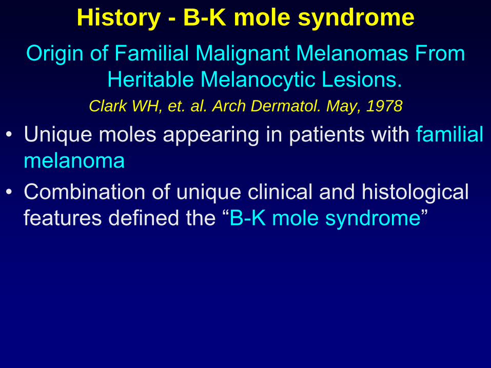
History - B-K mole syndromeOrigin of Familial Malignant Melanomas From
Heritable Melanocytic Lesions.Clark WH, et. al. Arch Dermatol. May, 1978
•
Unique moles appearing in patients with familial melanoma
•
Combination of unique clinical and histological features defined the “B-K mole syndrome”
History - B-K mole syndrome
•
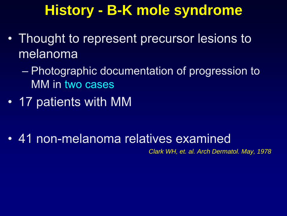
Thought to represent precursor lesions to melanoma–
Photographic documentation of progression to MM in two cases
•
17 patients with MM
•
41 non-melanoma relatives examinedClark WH, et. al. Arch Dermatol. May, 1978
History - B-K mole syndrome
•
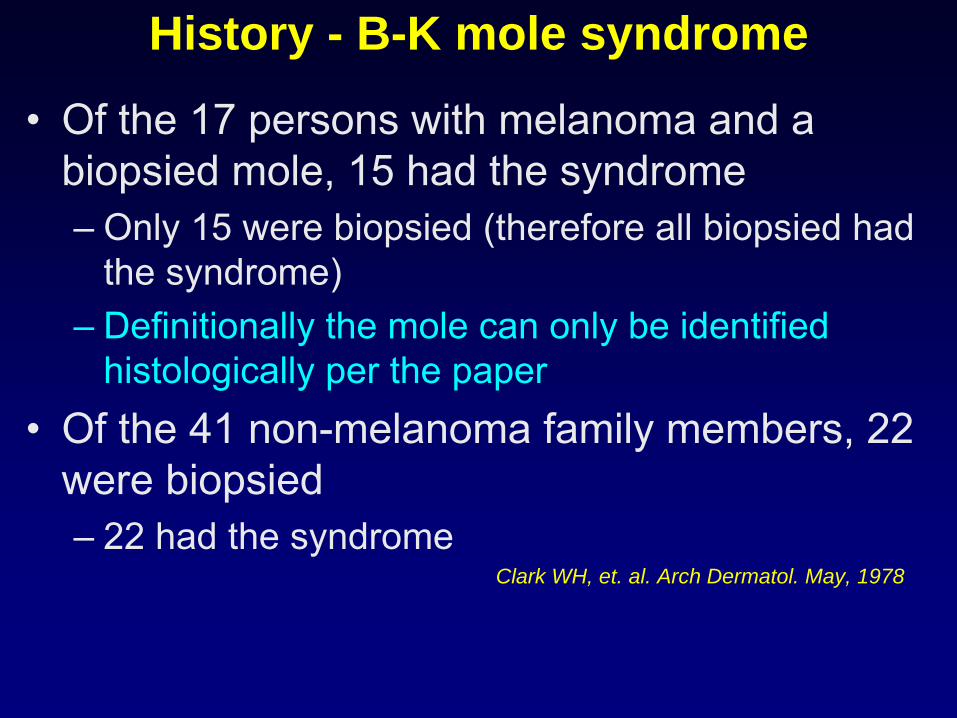
Of the 17 persons with melanoma and a biopsied mole, 15 had the syndrome–
Only 15 were biopsied (therefore all biopsied had the syndrome)
–
Definitionally the mole can only be identified histologically per the paper
•
Of the 41 non-melanoma family members, 22 were biopsied–
22 had the syndrome
Clark WH, et. al. Arch Dermatol. May, 1978
History - B-K mole syndrome
•
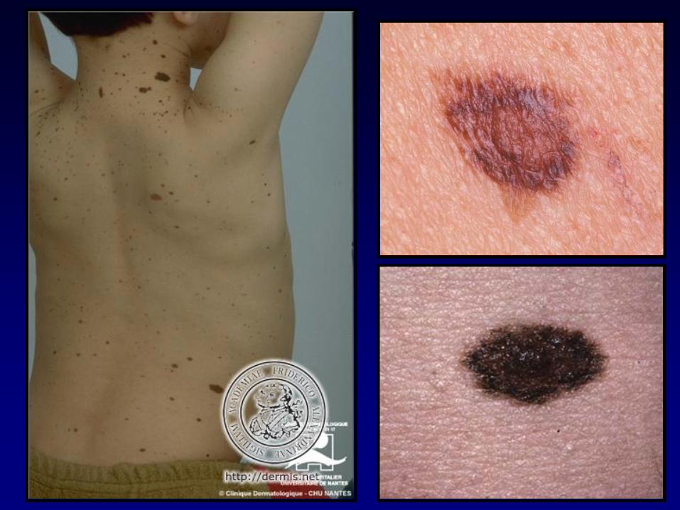
Affected patient may have <10 or >100 moles–
Every cutaneous surface, scalp to feet
–
Most prominent on trunk –
Prototypic B-K mole is ~10mm
in diameter,
irregular in outline, haphazard mixture of tan, brown, black, and pink
–
Striking variability
from one mole to the next•
Patients with B-K mole syndrome thought to be at “extremely high risk for development of MM”
Clark WH, et. al. Arch Dermatol. May, 1978
History - B-K mole syndrome •
Histological definition–
Compound melanocytic nevus
–
Atypical melanocytic hyperplasia•
Melanocytic dysplasia – “individual melanocytes or small clusters of melanocytes that have some of the structural features of malignant melanocytes”
•
Atypical melanocytes tend to be isolated in the basilar epidermal area or disposed in nests
•
Atypical melanocytes may also be seen in the upper part of the papillary dermis
Clark WH, et. al. Arch Dermatol. May, 1978
History - B-K mole syndrome
•
Histological definition–
Atypical melanocytic hyperplasia
•
Individual melanocytes : large and pale, spindled or epithelioid
•
Cytoplasm: abundant and filled with fine, “dusty” melanin granules
•
Mitotic figures may be identified
–
Mesenchymal changes•
Papillary dermis is widened due to fibroplasia and new blood vessel formation
–
Lymphocytic infiltrate
Clark WH, et. al. Arch Dermatol. May, 1978
History – Dysplastic nevus syndrome
Dysplastic Nevus Syndrome: A Phenotypic Association of Sporadic Cutaneous
MelanomaElder DE, et. al. Cancer. Oct, 1980
–
First description of dysplastic nevi in patients with non-familial MM
(79 patients)
–
“…behave as formal histogenetic precursors of melanoma.”
–
B-K mole syndrome becomes DNS sporadic and familial types
–
Syndrome patients may display as few as 1 DN

History – Dysplastic nevus syndrome
The Dysplastic Nevus Syndrome: Our definition Elder DE, et al. Am J Dermatopathol. Oct, 1982
•
DN are not “obligatory”
precursors of MM-
400 members
of 14 melanoma prone families
-
111 had DN & 67 had melanoma
-
Intermediate
between common nevi and MM (clinically and histopathologically)
-
22-36%
of sporadic MM arise in DN
-
Presence of DN in familial melanoma families defines those kindred at increased risk of developing MM
Epidemiology of DN
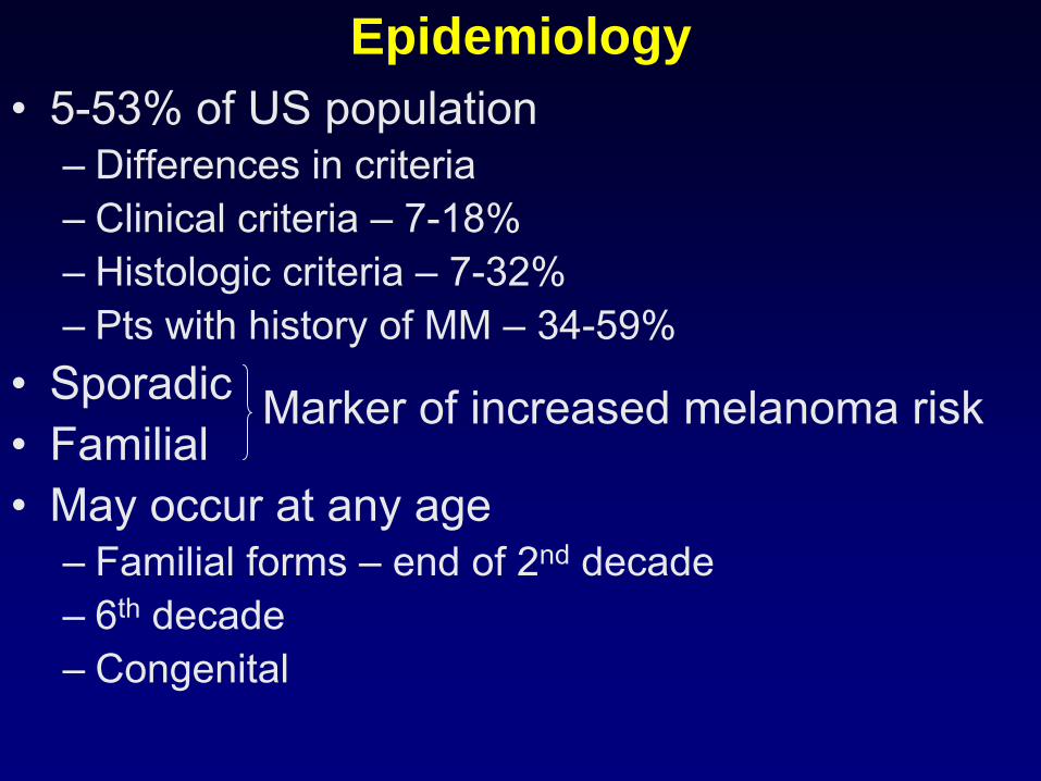
Epidemiology•
5-53% of US population–
Differences in criteria
–
Clinical criteria –
7-18%–
Histologic criteria –
7-32%
–
Pts with history of MM –
34-59%•
Sporadic
•
Familial•
May occur at any age–
Familial forms –
end of 2nd
decade
–
6th
decade
–
Congenital
Marker of increased melanoma risk
Dysplastic Nevi and Melanoma Risk
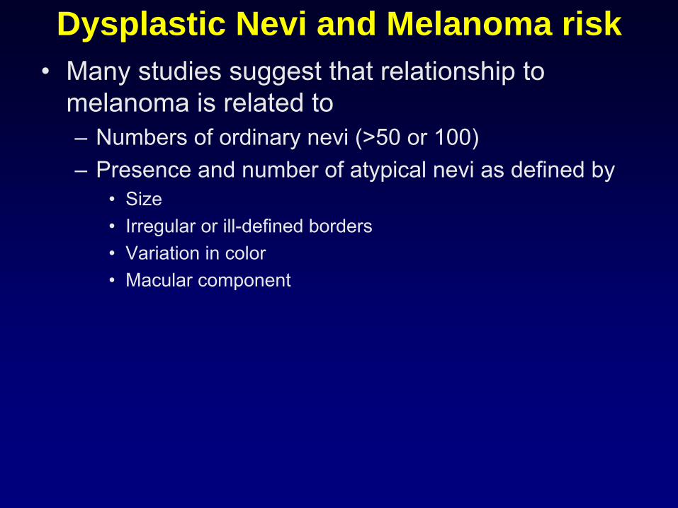
Dysplastic Nevi and Melanoma risk•
Many studies suggest that relationship to melanoma is related to –
Numbers of ordinary nevi (>50 or 100)
–
Presence and number of atypical nevi as defined by•
Size•
Irregular or ill-defined borders•
Variation in color•
Macular component
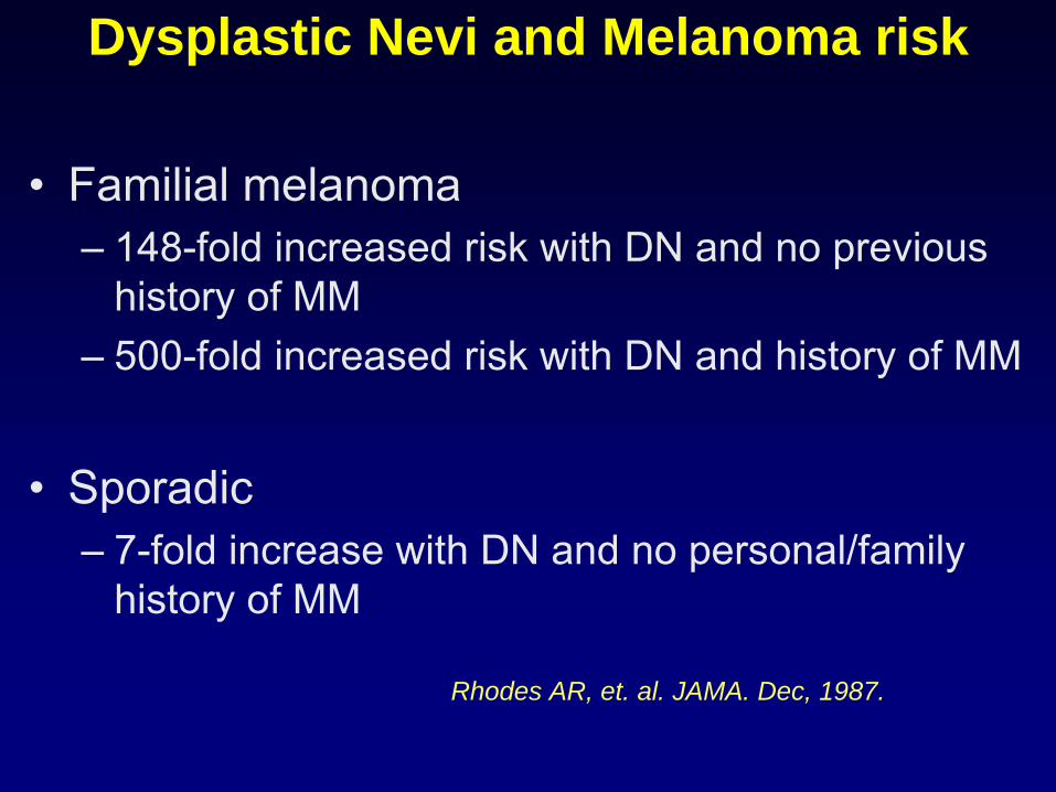
Dysplastic Nevi and Melanoma risk
•
Familial melanoma–
148-fold increased risk with DN and no previous history of MM
–
500-fold increased risk with DN and history of MM
•
Sporadic–
7-fold increase with DN and no personal/family history of MM
Rhodes AR, et. al. JAMA. Dec, 1987.
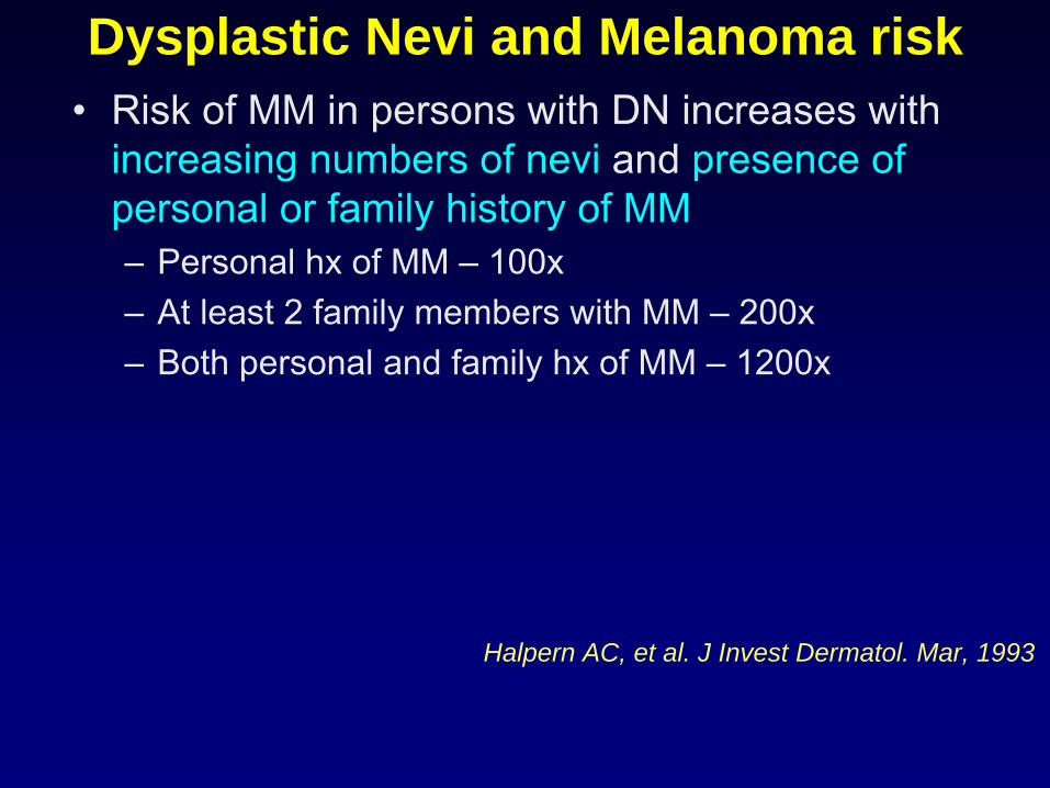
Dysplastic Nevi and Melanoma risk•
Risk of MM in persons with DN increases with increasing numbers of nevi
and presence of
personal or family history of MM–
Personal hx
of MM –
100x
–
At least 2 family members with MM –
200x–
Both personal and family hx
of MM –
1200x
Halpern AC, et al. J Invest Dermatol. Mar, 1993
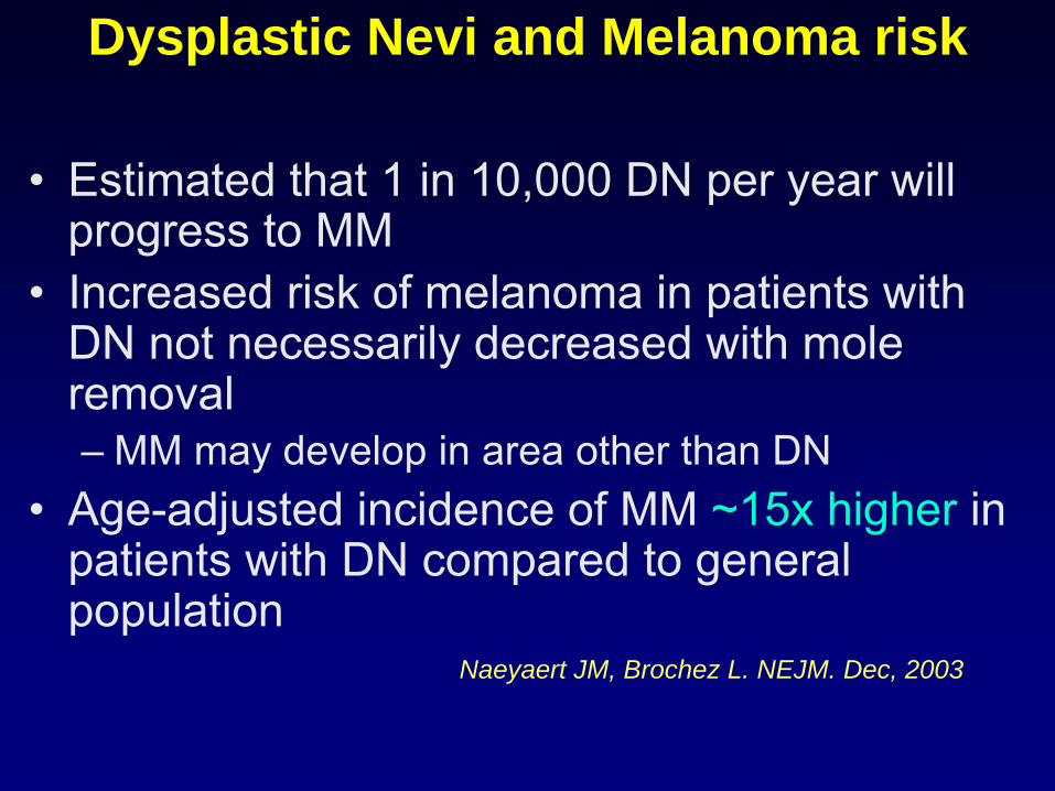
Dysplastic Nevi and Melanoma risk
•
Estimated that 1 in 10,000 DN per year will progress to MM
•
Increased risk of melanoma in patients with DN not necessarily decreased with mole removal–
MM may develop in area other than DN
•
Age-adjusted incidence of MM ~15x higher
in patients with DN compared to general population
Naeyaert JM, Brochez L. NEJM. Dec, 2003
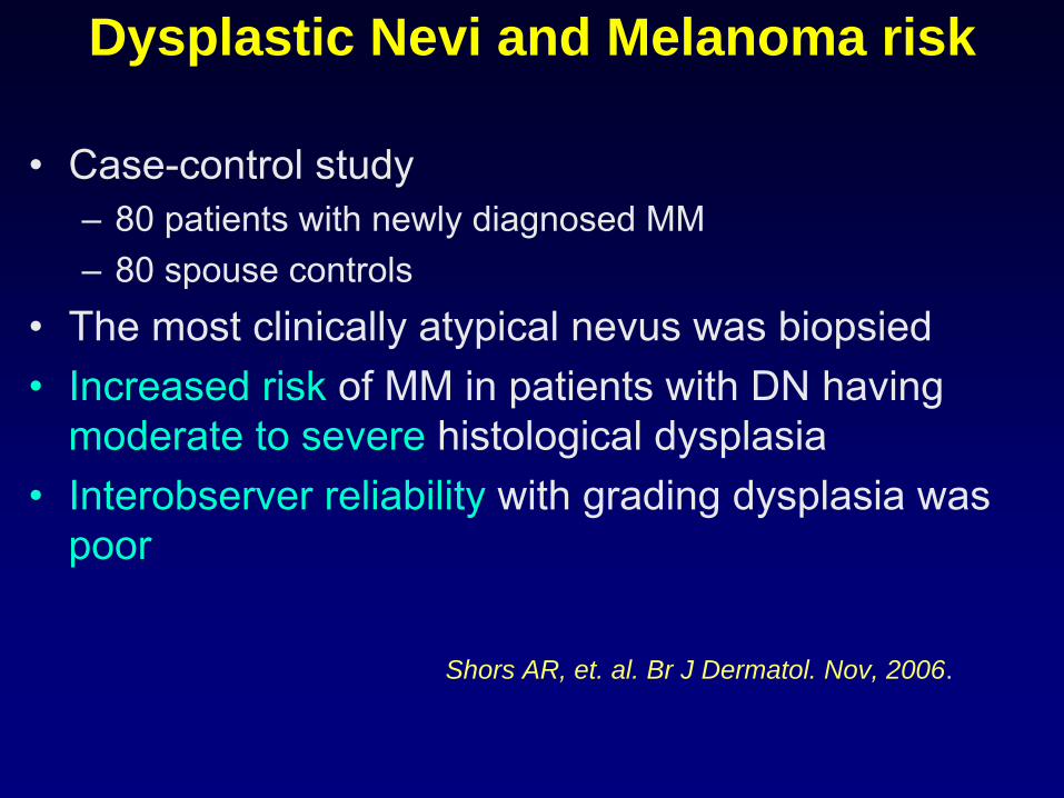
Dysplastic Nevi and Melanoma risk
•
Case-control study–
80 patients with newly diagnosed MM
–
80 spouse controls•
The most clinically atypical nevus was biopsied
•
Increased risk
of MM in patients with DN having moderate to severe
histological dysplasia
•
Interobserver reliability
with grading dysplasia was poor
Shors AR, et. al. Br J Dermatol. Nov, 2006.
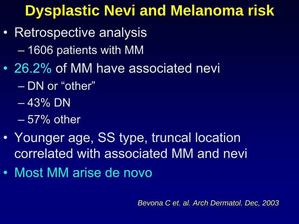
Dysplastic Nevi and Melanoma risk•
Retrospective analysis–
1606 patients with MM
•
26.2%
of MM have associated nevi–
DN or “other”
–
43% DN–
57% other
•
Younger age, SS type, truncal location correlated with associated MM and nevi
•
Most MM arise de novo
Bevona C et. al. Arch Dermatol. Dec, 2003
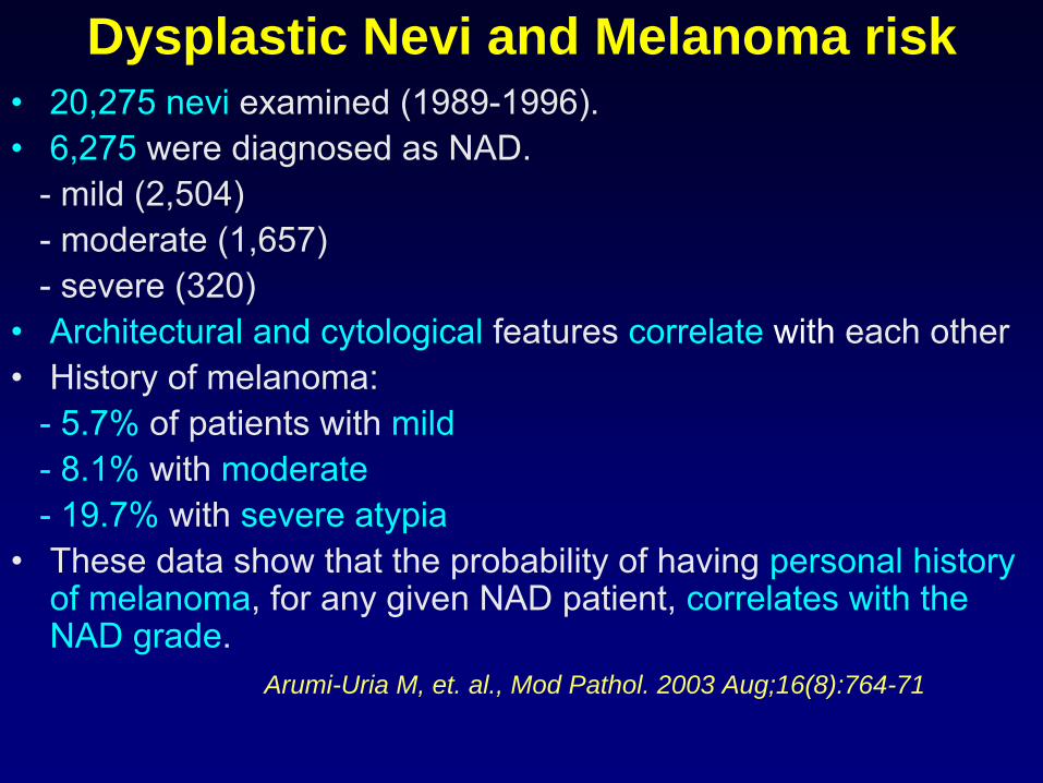
Dysplastic Nevi and Melanoma risk•
20,275 nevi
examined (1989-1996).
•
6,275
were diagnosed as NAD.-
mild (2,504)
-
moderate (1,657)-
severe (320)
•
Architectural and cytological
features correlate with
each other •
History of melanoma:
- 5.7% of patients with mild- 8.1% with moderate- 19.7% with severe atypia
•
These data show that the probability of having personal history of melanoma, for any given NAD patient, correlates with the NAD grade.
Arumi-Uria M, et. al., Mod Pathol. 2003 Aug;16(8):764-71
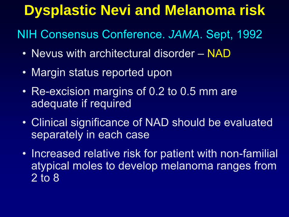
Dysplastic Nevi and Melanoma riskNIH Consensus Conference. JAMA. Sept, 1992•
Nevus with architectural disorder –
NAD
•
Margin status reported upon
•
Re-excision margins of 0.2 to 0.5 mm are adequate if required
•
Clinical significance of NAD should be evaluated separately in each case
•
Increased relative risk for patient with non-familial atypical moles to develop melanoma ranges from 2 to 8
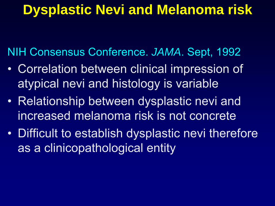
Dysplastic Nevi and Melanoma risk
NIH Consensus Conference. JAMA. Sept, 1992•
Correlation between clinical impression of atypical nevi and histology is variable
•
Relationship between dysplastic nevi and increased melanoma risk is not concrete
•
Difficult to establish dysplastic nevi therefore as a clinicopathological entity
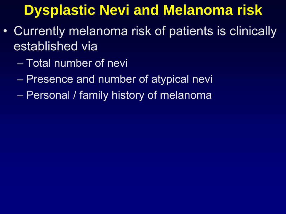
Dysplastic Nevi and Melanoma risk•
Currently melanoma risk of patients is clinically established via–
Total number of nevi
–
Presence and number of atypical nevi–
Personal / family history of melanoma
Current definitions – Dysplastic nevus
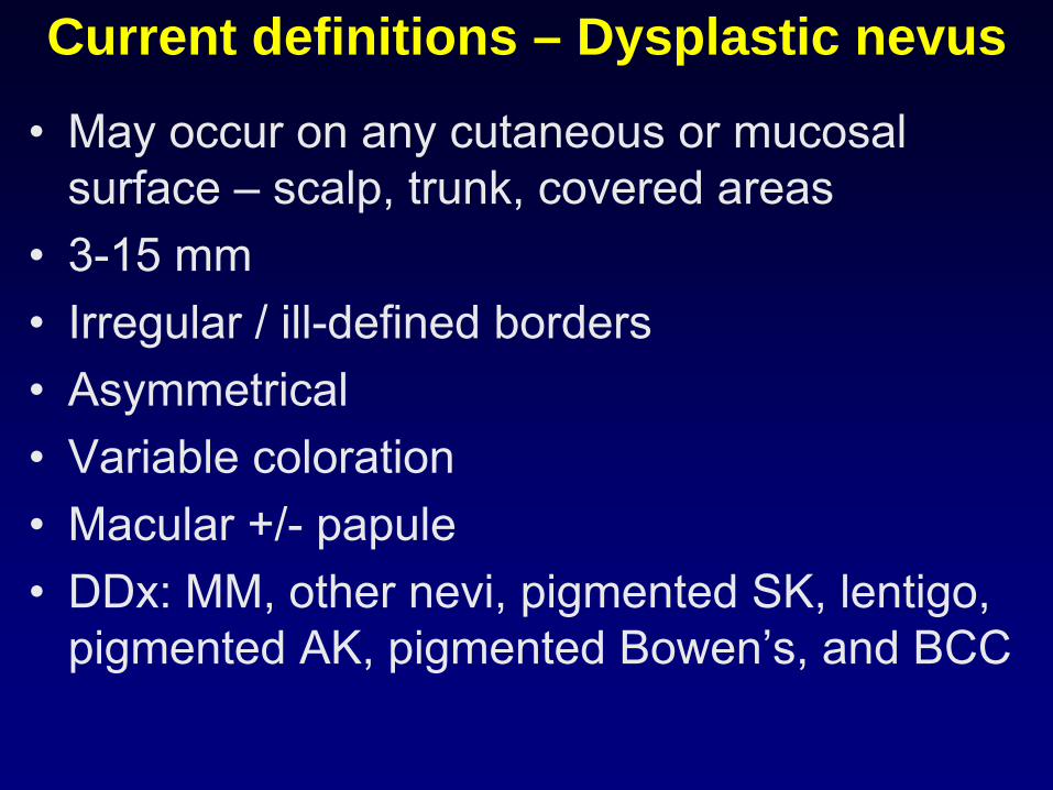
Current definitions – Dysplastic nevus
•
May occur on any cutaneous or mucosal surface –
scalp, trunk, covered areas
•
3-15 mm•
Irregular / ill-defined borders
•
Asymmetrical•
Variable coloration
•
Macular +/-
papule•
DDx: MM, other nevi, pigmented SK, lentigo, pigmented AK, pigmented Bowen’s, and BCC
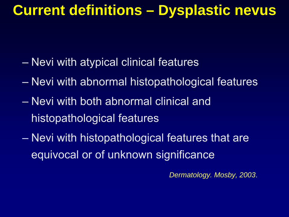
Current definitions – Dysplastic nevus
–
Nevi with atypical clinical features
–
Nevi with abnormal histopathological features
–
Nevi with both abnormal clinical and histopathological features
–
Nevi with histopathological features that are equivocal or of unknown significance
Dermatology. Mosby, 2003.
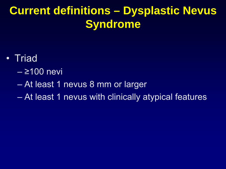
Current definitions – Dysplastic Nevus Syndrome
•
Triad–
≥100 nevi
–
At least 1 nevus 8 mm or larger–
At least 1 nevus with clinically atypical features
Histology of Dysplastic nevus
Histology
•
Scanning (20x) magnification
–
Shoulder
–
Stromal response with fibrosis and inflammation
Shea CR. Hum Pathol. 1999 May;30(5):500-5
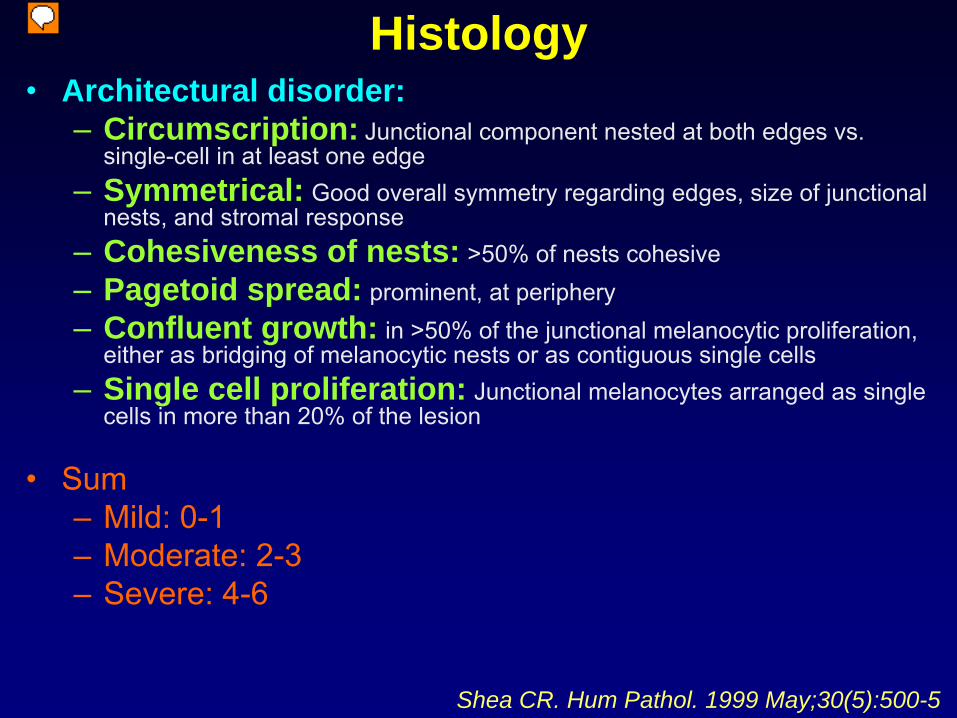
Histology•
Architectural disorder:–
Circumscription: Junctional component nested at both edges vs. single-cell in at least one edge
–
Symmetrical: Good overall symmetry regarding edges, size of junctional nests, and stromal response
–
Cohesiveness of nests: >50% of nests cohesive–
Pagetoid spread: prominent, at periphery
–
Confluent growth: in >50% of the junctional melanocytic proliferation, either as bridging of melanocytic nests or as contiguous single cells
–
Single cell proliferation: Junctional melanocytes arranged as single cells in more than 20% of the lesion
•
Sum–
Mild: 0-1
–
Moderate: 2-3–
Severe: 4-6
Shea CR. Hum Pathol. 1999 May;30(5):500-5
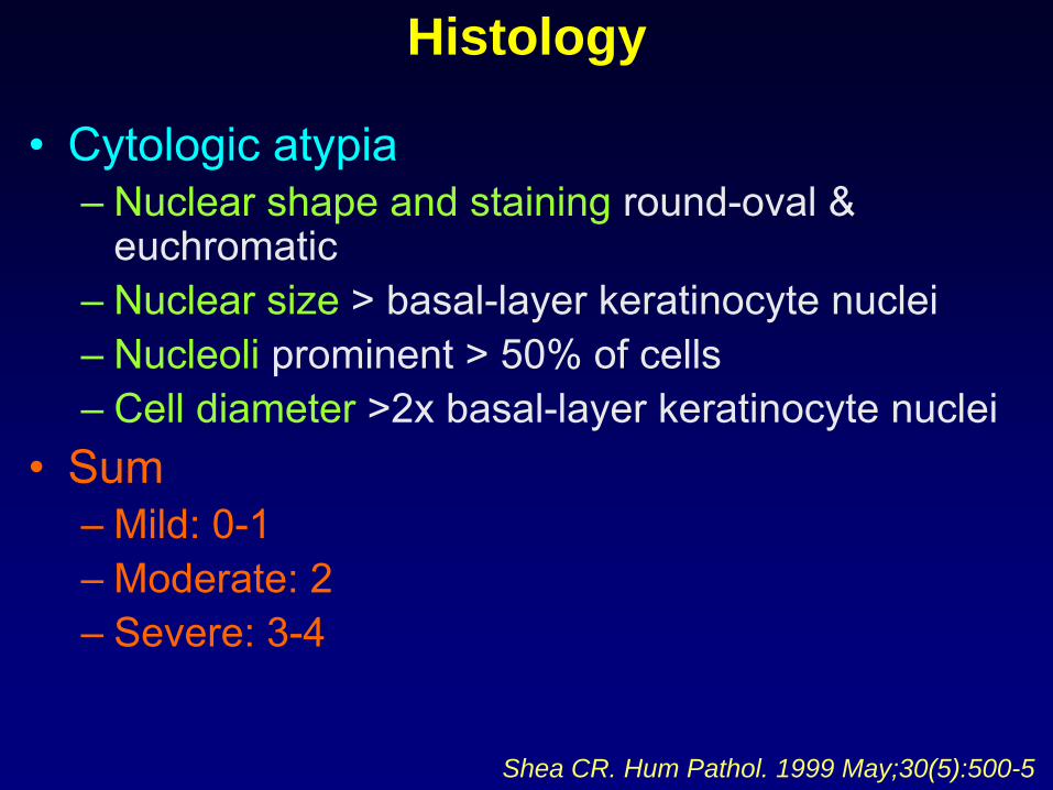
Histology
•
Cytologic atypia–
Nuclear shape and staining
round-oval &
euchromatic–
Nuclear size
> basal-layer keratinocyte nuclei
–
Nucleoli
prominent > 50% of cells–
Cell diameter
>2x basal-layer keratinocyte nuclei
•
Sum–
Mild: 0-1
–
Moderate: 2–
Severe: 3-4
Shea CR. Hum Pathol. 1999 May;30(5):500-5
Molecular Classification
Molecular Classification•
Comparative genomic hybridization (CGH)
•
DNA microarray-derived gene expression
Treatment of Dysplastic nevus
Treatment•
Assessment in conjunction with clinical history
•
Observation –
serial photographs•
Removal–
Shave
–
Punch, ellipse•
Margins–
2 mm
–
5 mm•
Re-excision–
Severely atypical
–
Lack of consensus
Treatment•
Prophylactic removal –
“de-moling”
•
Self-examination•
Ocular examination
•
Sun protection