Embed Size (px)
Citation preview
IBD: Ulcerative ColitisMonday, February 10, 2014The Fairmont Royal YorkToronto, ON
Accreditation• This event has been approved as an accredited (Section1)
group learning activity as defined by the Maintenance of Certification program of the RCPSC. It has been produced under RCPSC guidelines for the development of co-developed educational activities between CAG and Takeda Canada Inc.
Learning ObjectivesAt the end of this session, participants will be able to:• Review data on new biologic treatment options, their
advantages/disadvantages and treatment algorithm• Summarize the data on the need for dysplasia surveillance
including which patients to surveil and how best to monitor such patients
• Understand the correlation between symptoms and mucosal disease activity, and describe what is meant by mucosal healing
• Describe the markers used to assess mucosal healing, and the patients in whom mucosal healing should be the treatment target
FacultyCo-ChairsBrian Bressler, MD, MSc, FRCPCDirector, Advanced IBD Training ProgramClinical Assistant Professor of Medicine, Division of GastroenterologyUniversity of British Columbia
John Marshall, MD, MSc, FRCPC, AGAFProfessor of Medicine (Division of Gastroenterology)McMaster UniversityChief of Clinical Gastroenterology ServiceHamilton Health Sciences

FacultySpeakersBrian Feagan, MD, FRCPCDirector, Robarts Clinical TrialsProfessor of Medicine, Epidemiology & BiostatisticsUniversity of Western OntarioLondon Health Sciences CentreRobarts Research Institute
David Rubin, MD, FACG, AGAF, FACP Professor of MedicineCo-Director, Inflammatory Bowel Disease CenterInterim Chief, Section of Gastroenterology, Hepatology and NutritionUniversity of Chicago Medicine
Mark Silverberg, MD, PhD, FRCPCAssociate Professor of Medicine, University of TorontoStaff Gastroenterologist, Mount Sinai Hospital IBD GroupSenior Investigator, Lunenfeld-Tanenbaum Research InstZane Cohen Centre for Digestive Diseases

Dysplasia Surveillance: Do we need it?David Rubin, MD, FACG, AGAF, FACP
Learning objectives:
This presentation will address the following:
1. Why new data suggests that in 2014 we may no longer need to perform dysplasia surveillance in 2014,
2. In which patients with UC we should consider dysplasia surveillance, and
3. How best to survey those patients with UC that require such monitoring.
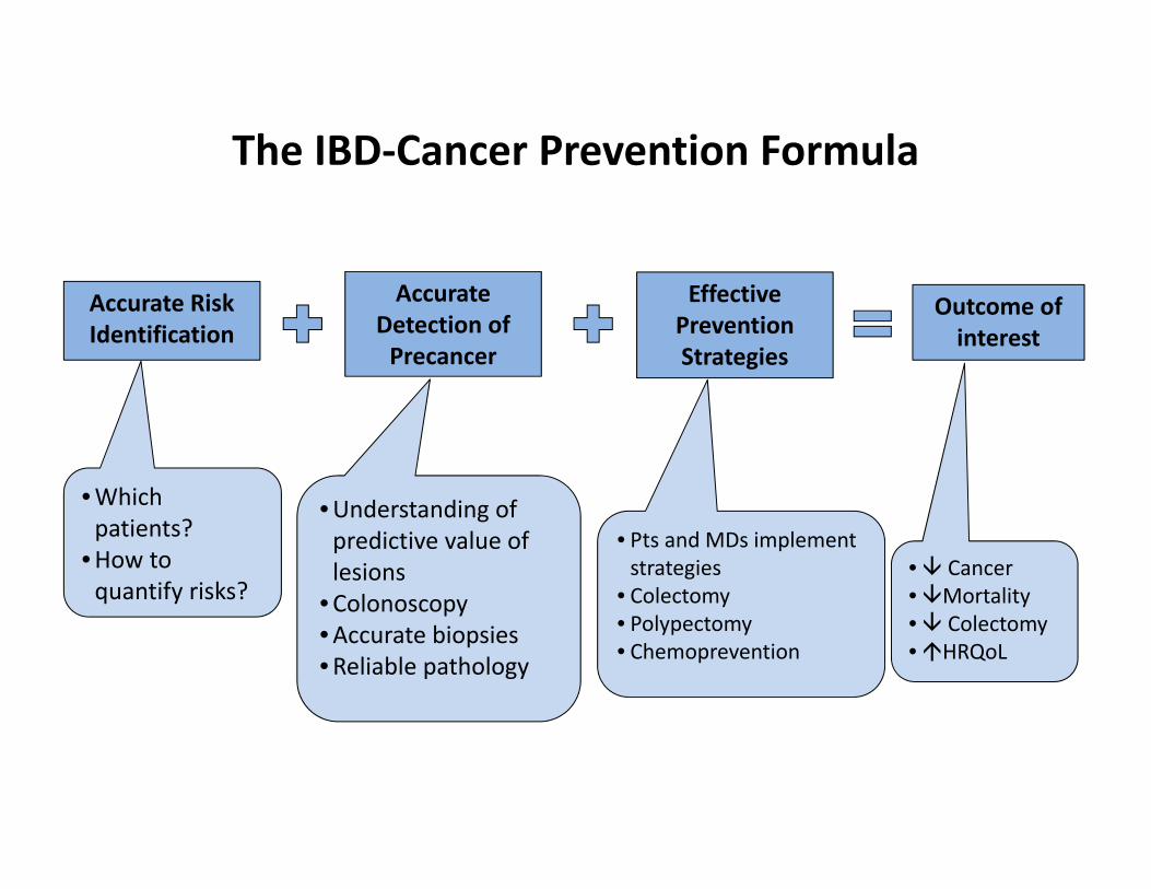
The IBD‐Cancer Prevention Formula
Accurate Risk Identification
Effective Prevention Strategies
Accurate Detection of Precancer
•Which patients?
•How to quantify risks?
• Pts and MDs implement strategies
• Colectomy• Polypectomy• Chemoprevention
Outcome of interest
• Cancer•Mortality• Colectomy•HRQoL
•Understanding of predictive value of lesions
•Colonoscopy•Accurate biopsies•Reliable pathology
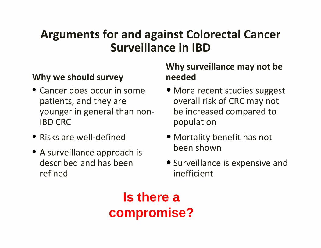
Arguments for and against Colorectal Cancer Surveillance in IBD
Why we should survey• Cancer does occur in some patients, and they are younger in general than non‐IBD CRC
• Risks are well‐defined• A surveillance approach is described and has been refined
Why surveillance may not be needed•More recent studies suggest overall risk of CRC may not be increased compared to population
•Mortality benefit has not been shown
• Surveillance is expensive and inefficient
Is there a compromise?
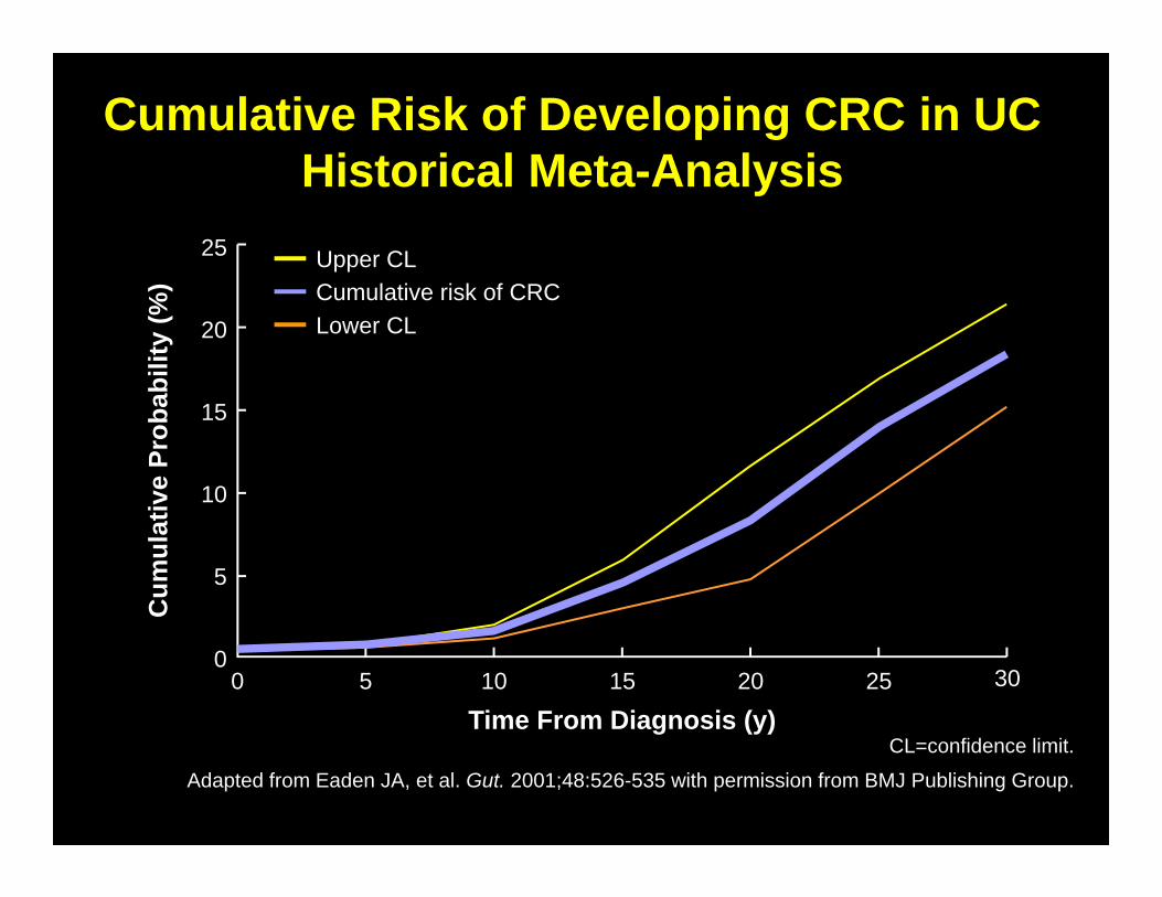
CL=confidence limit.
Adapted from Eaden JA, et al. Gut. 2001;48:526-535 with permission from BMJ Publishing Group.
25
20
15
10
Cum
ulat
ive
Prob
abili
ty (%
)
030
Time From Diagnosis (y)5 15
5
10 200 25
Upper CLCumulative risk of CRCLower CL
Cumulative Risk of Developing CRC in UCHistorical Meta-Analysis
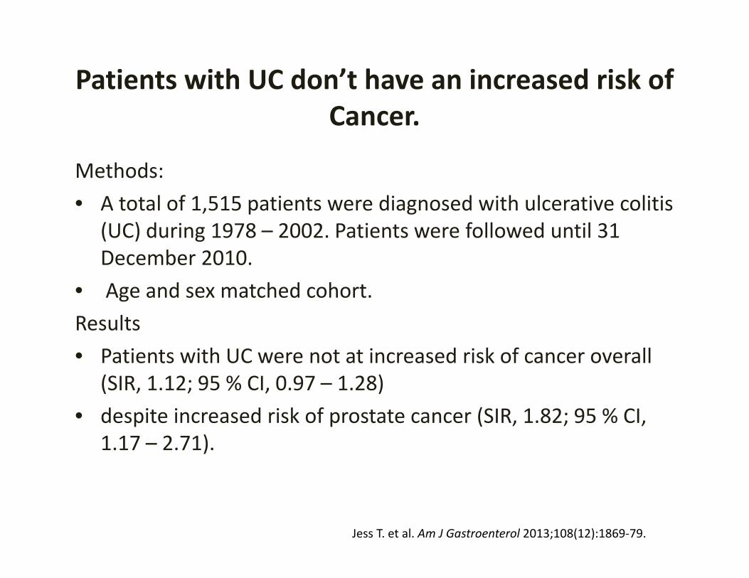
Patients with UC don’t have an increased risk of Cancer.
Methods:• A total of 1,515 patients were diagnosed with ulcerative colitis
(UC) during 1978 – 2002. Patients were followed until 31 December 2010.
• Age and sex matched cohort.Results• Patients with UC were not at increased risk of cancer overall
(SIR, 1.12; 95 % CI, 0.97 – 1.28) • despite increased risk of prostate cancer (SIR, 1.82; 95 % CI,
1.17 – 2.71).
Jess T. et al. Am J Gastroenterol 2013;108(12):1869‐79.
0
10
20
30
40
0 10 20 30 40
Cum
ulat
ive
inci
denc
e of
co
lore
ctal
can
cer (
%)
Cum
ulat
ive
inci
denc
e of
co
lore
ctal
can
cer (
%)
Years from diagnosisYears from diagnosisNo. at risk
376 220 109 46 10No. at risk
376 220 109 46 10
ObservedExpectedObservedExpected
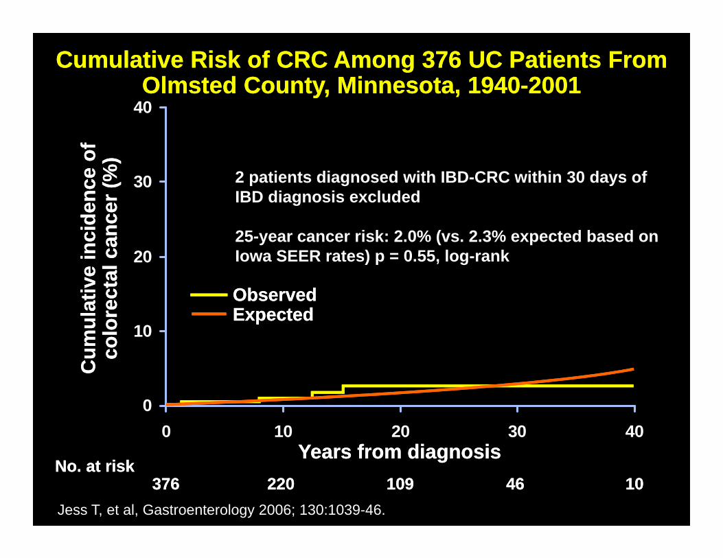
Cumulative Risk of CRC Among 376 UC Patients From Olmsted County, Minnesota, 1940-2001
Cumulative Risk of CRC Among 376 UC Patients From Olmsted County, Minnesota, 1940-2001
Jess T, et al, Gastroenterology 2006; 130:1039-46.
2 patients diagnosed with IBD-CRC within 30 days of IBD diagnosis excluded
25-year cancer risk: 2.0% (vs. 2.3% expected based onIowa SEER rates) p = 0.55, log-rank
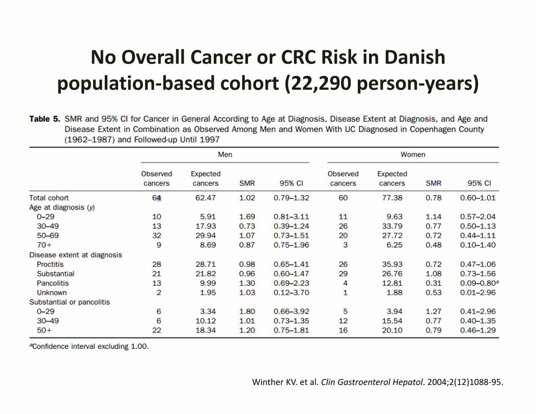
No Overall Cancer or CRC Risk in Danish population‐based cohort (22,290 person‐years)
Winther KV. et al. Clin Gastroenterol Hepatol. 2004;2(12)1088‐95.
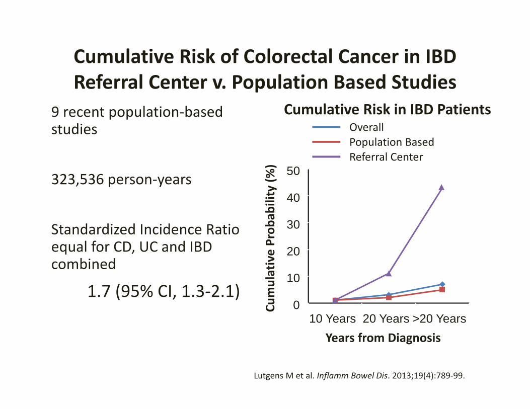
Cumulative Risk of Colorectal Cancer in IBDReferral Center v. Population Based Studies
Years from Diagnosis
Cumulative Prob
ability (%
)
Overall
Referral Center
Cumulative Risk in IBD Patients
0
10
20
30
40
50
10 Years 20 Years >20 Years
Population Based
• 9 recent population‐based studies
• 323,536 person‐years
• Standardized Incidence Ratio equal for CD, UC and IBD combined
1.7 (95% CI, 1.3‐2.1)
Lutgens M et al. Inflamm Bowel Dis. 2013;19(4):789‐99.
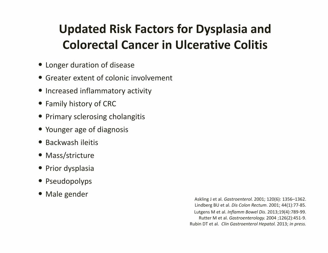
Updated Risk Factors for Dysplasia and Colorectal Cancer in Ulcerative Colitis
• Longer duration of disease• Greater extent of colonic involvement • Increased inflammatory activity• Family history of CRC• Primary sclerosing cholangitis• Younger age of diagnosis• Backwash ileitis• Mass/stricture• Prior dysplasia • Pseudopolyps• Male gender
Askling J et al. Gastroenterol. 2001; 120(6): 1356–1362.Lindberg BU et al. Dis Colon Rectum. 2001; 44(1):77‐85.Lutgens M et al. Inflamm Bowel Dis. 2013;19(4):789‐99.Rutter M et al. Gastroenterology. 2004 ;126(2):451‐9.
Rubin DT et al. Clin Gastroenterol Hepatol. 2013; in press.
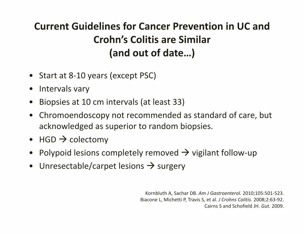
Current Guidelines for Cancer Prevention in UC and Crohn’s Colitis are Similar
(and out of date…)
• Start at 8‐10 years (except PSC)• Intervals vary• Biopsies at 10 cm intervals (at least 33)• Chromoendoscopy not recommended as standard of care, but
acknowledged as superior to random biopsies.• HGD colectomy• Polypoid lesions completely removed vigilant follow‐up• Unresectable/carpet lesions surgery
Kornbluth A, Sachar DB. Am J Gastroenterol. 2010;105:501‐523. Biacone L, Michetti P, Travis S, et al. J Crohns Colitis. 2008;2:63‐92.
Cairns S and Schofield JH. Gut. 2009.
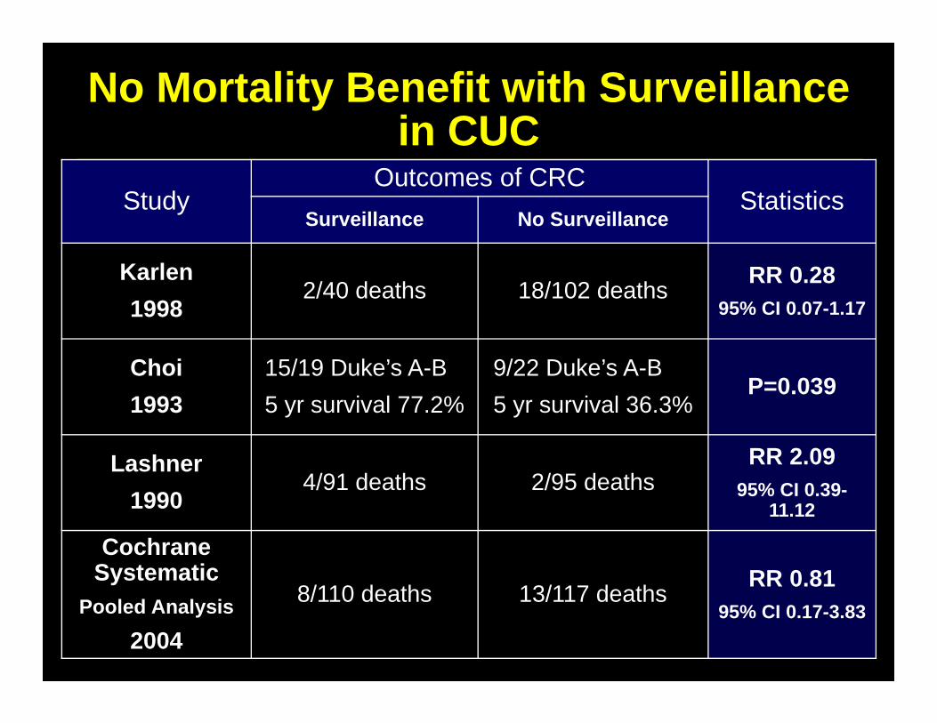
No Mortality Benefit with Surveillance in CUC
StudyOutcomes of CRC
StatisticsSurveillance No Surveillance
Karlen1998
2/40 deaths 18/102 deaths RR 0.2895% CI 0.07-1.17
Choi1993
15/19 Duke’s A-B5 yr survival 77.2%
9/22 Duke’s A-B5 yr survival 36.3%
P=0.039
Lashner1990
4/91 deaths 2/95 deathsRR 2.09
95% CI 0.39-11.12
Cochrane Systematic
Pooled Analysis
2004
8/110 deaths 13/117 deaths RR 0.8195% CI 0.17-3.83
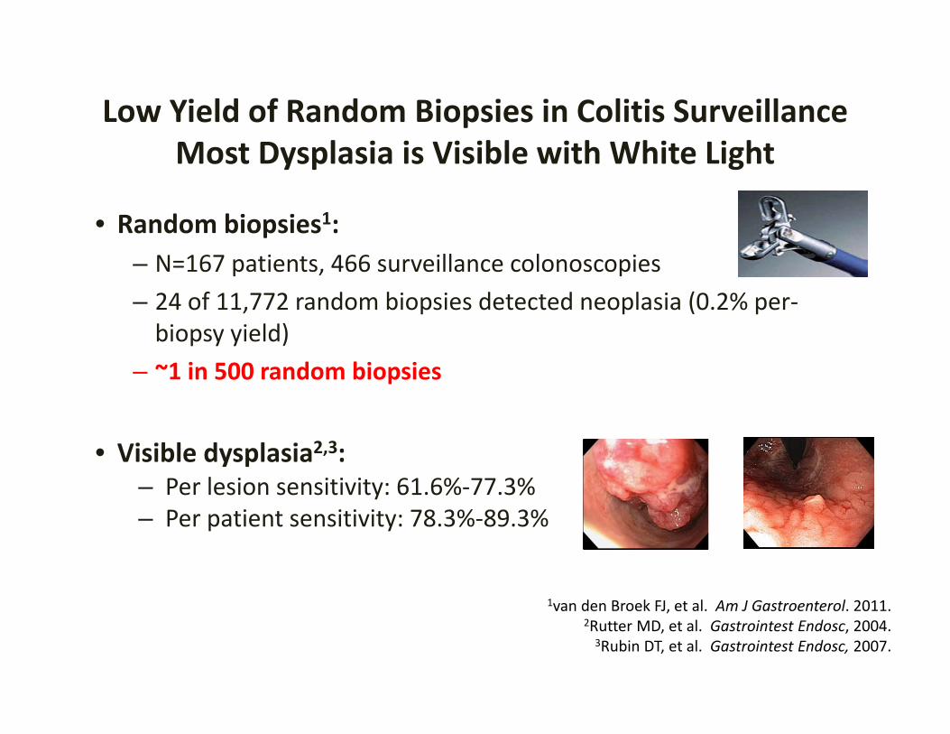
Low Yield of Random Biopsies in Colitis Surveillance Most Dysplasia is Visible with White Light
• Random biopsies1: – N=167 patients, 466 surveillance colonoscopies– 24 of 11,772 random biopsies detected neoplasia (0.2% per‐biopsy yield)
– ~1 in 500 random biopsies
• Visible dysplasia2,3:– Per lesion sensitivity: 61.6%‐77.3%– Per patient sensitivity: 78.3%‐89.3%
1van den Broek FJ, et al. Am J Gastroenterol. 2011.2Rutter MD, et al. Gastrointest Endosc, 2004.3Rubin DT, et al. Gastrointest Endosc, 2007.
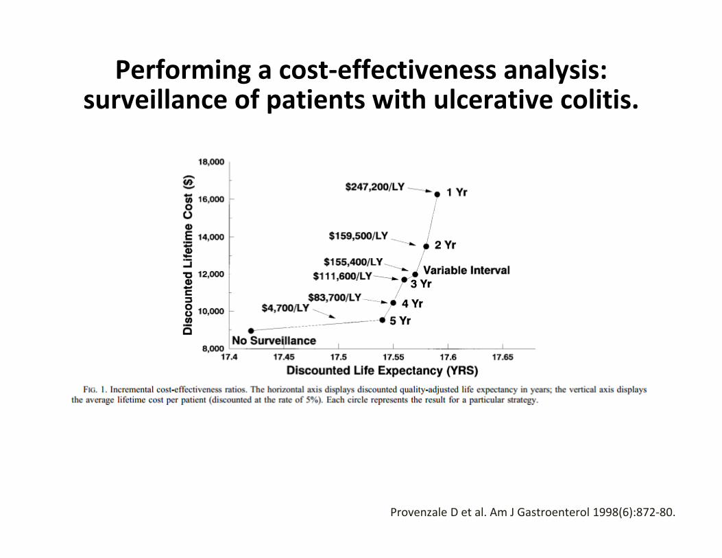
Performing a cost‐effectiveness analysis: surveillance of patients with ulcerative colitis.
Provenzale D et al. Am J Gastroenterol 1998(6):872‐80.
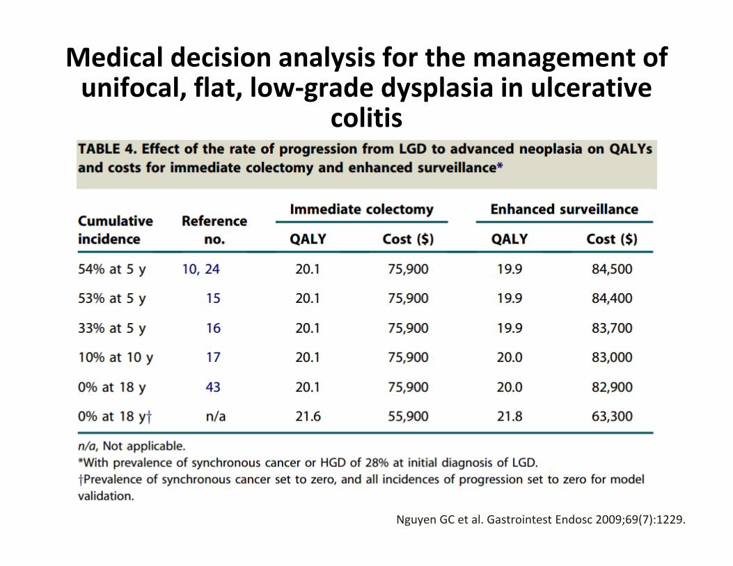
Medical decision analysis for the management of unifocal, flat, low‐grade dysplasia in ulcerative
colitis
Nguyen GC et al. Gastrointest Endosc 2009;69(7):1229.
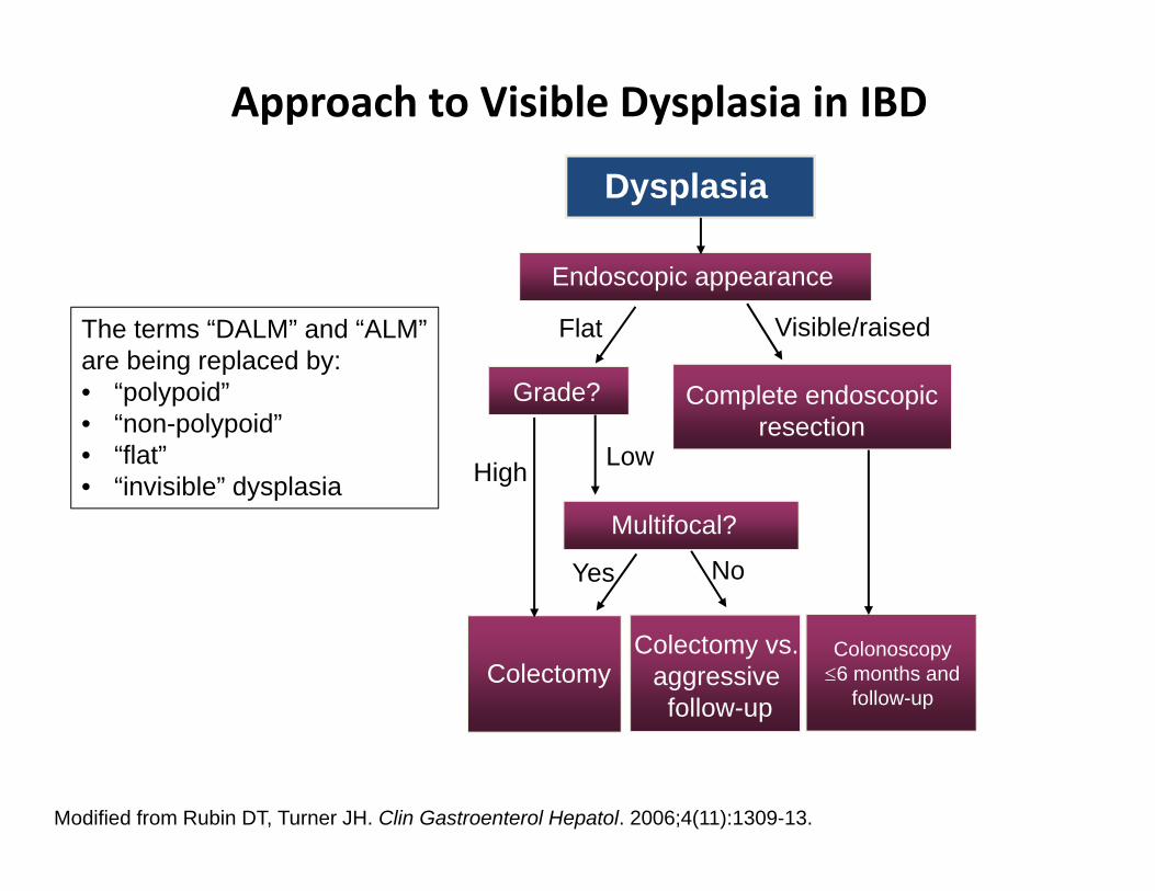
Approach to Visible Dysplasia in IBD
Modified from Rubin DT, Turner JH. Clin Gastroenterol Hepatol. 2006;4(11):1309-13.
Dysplasia
Multifocal?
ColectomyColectomy vs.
aggressive follow-up
Grade?
Flat
High Low
Yes No
Endoscopic appearance
Complete endoscopic resection
Colonoscopy 6 months and
follow-up
Visible/raisedThe terms “DALM” and “ALM” are being replaced by:• “polypoid” • “non-polypoid” • “flat” • “invisible” dysplasia
We Should Update our Surveillance Approach
Selective PatientsBetter Techniques
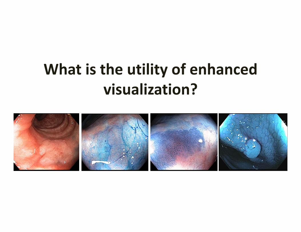
What is the utility of enhanced visualization?
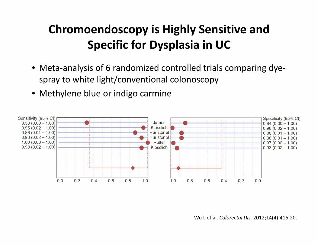
Chromoendoscopy is Highly Sensitive and Specific for Dysplasia in UC
• Meta‐analysis of 6 randomized controlled trials comparing dye‐spray to white light/conventional colonoscopy
• Methylene blue or indigo carmine
Wu L et al. Colorectal Dis. 2012;14(4):416‐20.
What Happens to Dysplasia Found on Chromoendoscopy?
• Are we missing occult cancers?• Dysplasia in the current age has a different predictive value
than dysplasia found with earlier technology• Current therapies prevent progression of dysplasia• Chromoendoscopy studies:
• Follow‐up in only one study• Marion (NYC)
– Follow‐up with colectomy specimens– 5 of original 102 had colectomy due to unresectable LGD – No CRC
Marion J, et al. Am J Gastroenterol, 2008;103:2342..
Challenges to Chromoendoscopy in IBD
• Perception of time consuming and expensive (time plus supplies)
• Unclear if it changes outcomes (cancer or mortality)• Many patients don’t “qualify” for it due to poor prep
or too much inflammation• No consensus on its use in our field • No defined training pathway or competency
requirement• Comparison to newer high definition scopes not
completed
My Approach to Chromoendoscopy
• WHO: – Pancolonic: High risk (PSC, previous confirmed dysplasia) – Segmental: Lesions found and require clarification
• PREP: needs to be CLEAN and in remission• TYPE: Methylene blue diluted (my preference)• HOW:
– Standard scope. Power wash. Segmental exams. Raised lesions AND abnormal microscopic/pit patterns.
• FOLLOW‐UP: Depends…
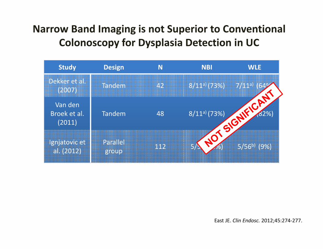
Narrow Band Imaging is not Superior to Conventional Colonoscopy for Dysplasia Detection in UC
Study Design N NBI WLE
Dekker et al.(2007) Tandem 42 8/11a) (73%) 7/11a) (64%)
Van den Broek et al. (2011)
Tandem 48 8/11a) (73%) 9/11a) (82%)
Ignjatovic et al. (2012)
Parallelgroup 112 5/56b) (9%) 5/56b) (9%)
NBI Narrow band imaging; WLE; White light endoscopy.a)Proportion of total dysplastic lesions detected overall; b)Proportion of patients with at least one dysplastic lesion.
East JE. Clin Endosc. 2012;45:274‐277.
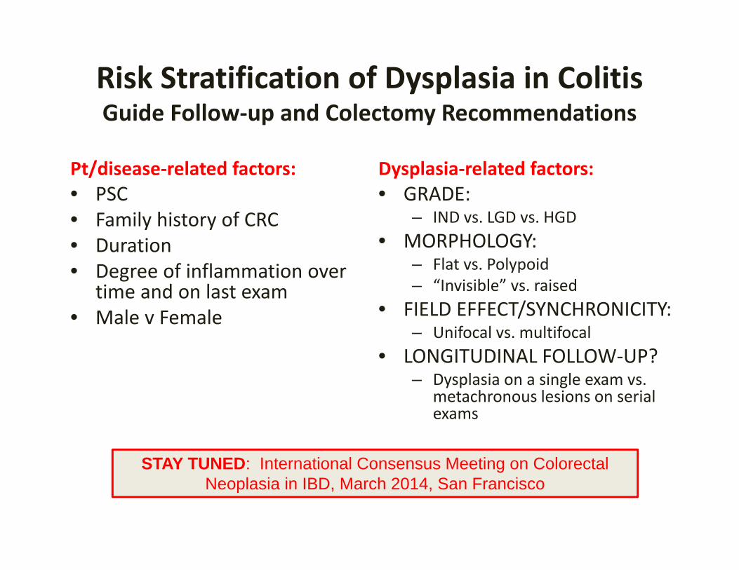
Risk Stratification of Dysplasia in ColitisGuide Follow‐up and Colectomy Recommendations
Pt/disease‐related factors:• PSC• Family history of CRC• Duration• Degree of inflammation over
time and on last exam• Male v Female
Dysplasia‐related factors:• GRADE:
– IND vs. LGD vs. HGD• MORPHOLOGY:
– Flat vs. Polypoid– “Invisible” vs. raised
• FIELD EFFECT/SYNCHRONICITY: – Unifocal vs. multifocal
• LONGITUDINAL FOLLOW‐UP?– Dysplasia on a single exam vs.
metachronous lesions on serial exams
STAY TUNED: International Consensus Meeting on Colorectal Neoplasia in IBD, March 2014, San Francisco
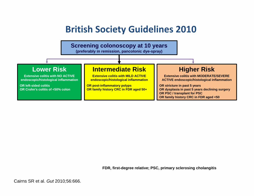
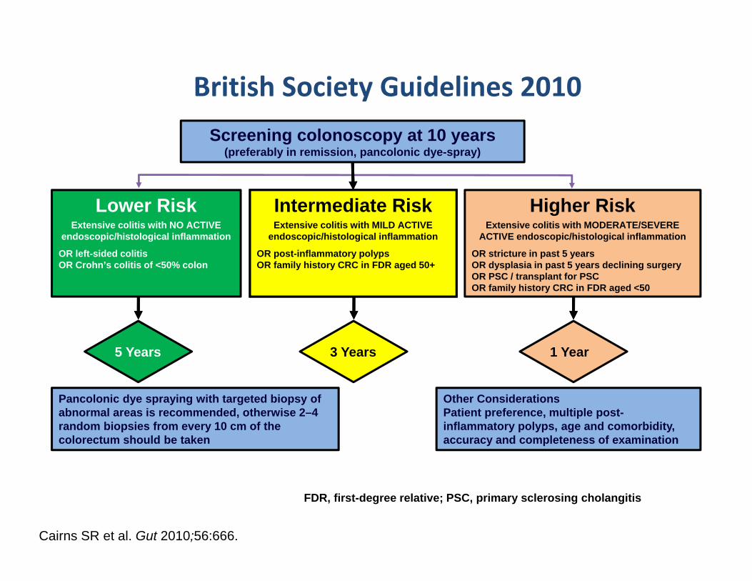
Cairns SR et al. Gut 2010;56:666.
Screening colonoscopy at 10 years(preferably in remission, pancolonic dye-spray)
Lower RiskExtensive colitis with NO ACTIVE
endoscopic/histological inflammation
OR left-sided colitisOR Crohn’s colitis of <50% colon
Intermediate RiskExtensive colitis with MILD ACTIVE
endoscopic/histological inflammation
OR post-inflammatory polypsOR family history CRC in FDR aged 50+
Higher RiskExtensive colitis with MODERATE/SEVERE
ACTIVE endoscopic/histological inflammation
OR stricture in past 5 yearsOR dysplasia in past 5 years declining surgeryOR PSC / transplant for PSCOR family history CRC in FDR aged <50
FDR, first-degree relative; PSC, primary sclerosing cholangitis
British Society Guidelines 2010
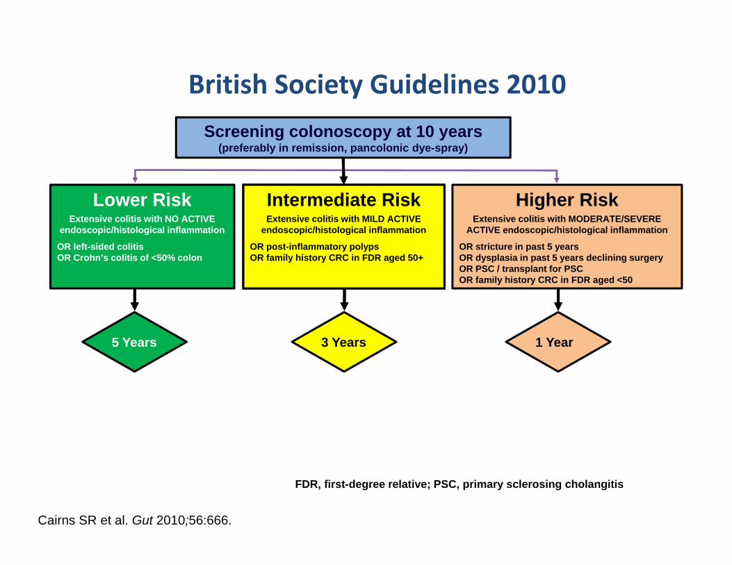
Cairns SR et al. Gut 2010;56:666.
Lower RiskExtensive colitis with NO ACTIVE
endoscopic/histological inflammation
OR left-sided colitisOR Crohn’s colitis of <50% colon
Intermediate RiskExtensive colitis with MILD ACTIVE
endoscopic/histological inflammation
OR post-inflammatory polypsOR family history CRC in FDR aged 50+
Higher RiskExtensive colitis with MODERATE/SEVERE
ACTIVE endoscopic/histological inflammation
OR stricture in past 5 yearsOR dysplasia in past 5 years declining surgeryOR PSC / transplant for PSCOR family history CRC in FDR aged <50
5 Years 3 Years 1 Year
British Society Guidelines 2010Screening colonoscopy at 10 years
(preferably in remission, pancolonic dye-spray)
FDR, first-degree relative; PSC, primary sclerosing cholangitis
Cairns SR et al. Gut 2010;56:666.
Lower RiskExtensive colitis with NO ACTIVE
endoscopic/histological inflammation
OR left-sided colitisOR Crohn’s colitis of <50% colon
Intermediate RiskExtensive colitis with MILD ACTIVE
endoscopic/histological inflammation
OR post-inflammatory polypsOR family history CRC in FDR aged 50+
Higher RiskExtensive colitis with MODERATE/SEVERE
ACTIVE endoscopic/histological inflammation
OR stricture in past 5 yearsOR dysplasia in past 5 years declining surgeryOR PSC / transplant for PSCOR family history CRC in FDR aged <50
Pancolonic dye spraying with targeted biopsy of abnormal areas is recommended, otherwise 2–4 random biopsies from every 10 cm of the colorectum should be taken
Other ConsiderationsPatient preference, multiple post-inflammatory polyps, age and comorbidity, accuracy and completeness of examination
British Society Guidelines 2010Screening colonoscopy at 10 years
(preferably in remission, pancolonic dye-spray)
FDR, first-degree relative; PSC, primary sclerosing cholangitis
5 Years 3 Years 1 Year
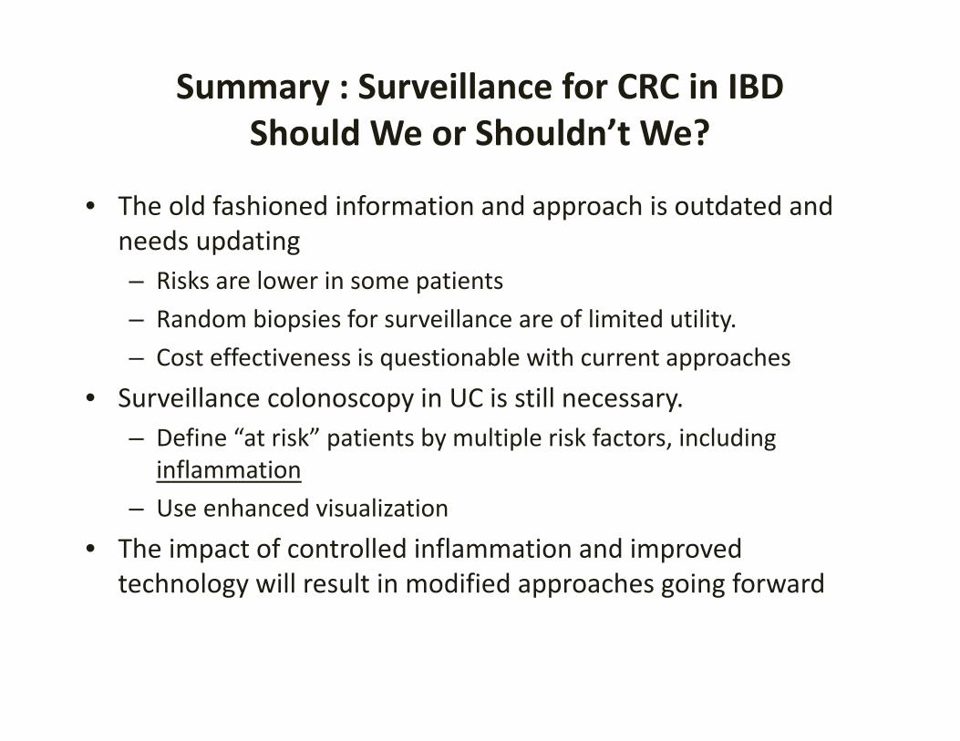
Summary : Surveillance for CRC in IBDShould We or Shouldn’t We?
• The old fashioned information and approach is outdated and needs updating– Risks are lower in some patients– Random biopsies for surveillance are of limited utility.– Cost effectiveness is questionable with current approaches
• Surveillance colonoscopy in UC is still necessary.– Define “at risk” patients by multiple risk factors, including
inflammation– Use enhanced visualization
• The impact of controlled inflammation and improved technology will result in modified approaches going forward