Embed Size (px)
Citation preview
Ib-202-18
Immune system continued(Chapt 43 all)
Antigen Recognition by Lymphocytes
• The vertebrate body is populated by two main types of lymphocytes– B lymphocytes (B cells) and T lymphocytes (T cells) which
circulate through the blood. In microscope can’t tell difference.
• The plasma membranes of both B cells and T cells--– have about 100,000 embedded antigen receptors that all
recognize the same epitope (specific). Other B cells recognize other epitopes. There are enough different B cells with different specificities to recognize most all the epitopes present in the microbial world.
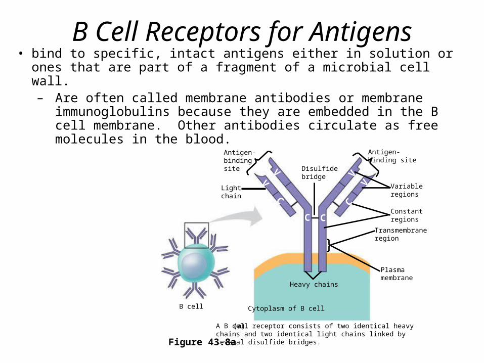
B Cell Receptors for Antigens• bind to specific, intact antigens either in solution or ones that are part
of a fragment of a microbial cell wall.– Are often called membrane antibodies or membrane
immunoglobulins because they are embedded in the B cell membrane. Other antibodies circulate as free molecules in the blood.
Figure 43.8a
Antigen-bindingsite
Antigen-binding site
Disulfidebridge
Lightchain
Heavy chains
Cytoplasm of B cell
V
A B cell receptor consists of two identical heavy chains and two identical light chains linked by several disulfide bridges.
(a)
Variableregions
Constantregions
Transmembraneregion
Plasmamembrane
B cell
V
V
CC C
C
V

Antigen-Binding site
chain
Disulfide bridge
chain
T cell
A T cell receptor consists of one chain and one chain linked by a disulfide bridge. (TCR)
(b)
Variableregions
Constantregions
Transmembraneregion
Plasmamembrane
Cytoplasm of T cell
T Cell Receptors for Antigens and the Role of the MHC
• Each T cell receptor consists of two different polypeptide chains embedded in the T cell membrane
Figure 43.8b
V V
C C
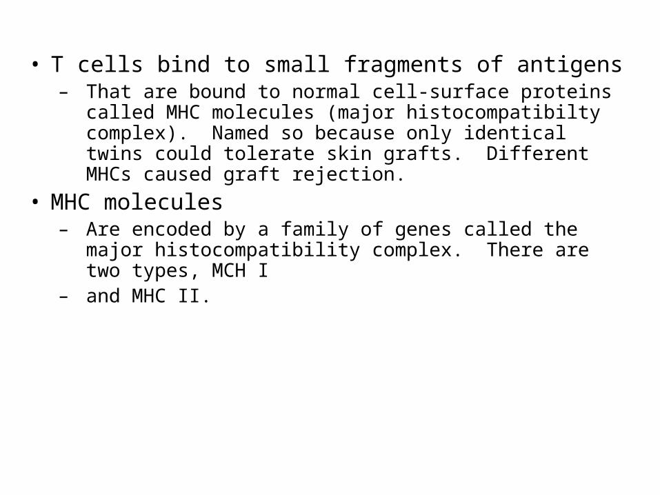
• T cells bind to small fragments of antigens– That are bound to normal cell-surface proteins called MHC
molecules (major histocompatibilty complex). Named so because only identical twins could tolerate skin grafts. Different MHCs caused graft rejection.
• MHC molecules– Are encoded by a family of genes called the major
histocompatibility complex. There are two types, MCH I– and MHC II.

• Virus infected cells produce MHC molecules (MHC I)– Which bind to antigen fragments from degradation
of the antigen proteins and then are transported to the cell surface in a process called antigen presentation. The antigens are peptides of about 9-11 amino acids derived from the foreign protein.
• A nearby cytotoxic T cell– Can then recognize the antigen fragment displayed
on the cell’s surface by means of its specific T cell receptor.

MHC I
Figure 43.9a
Infected cell
Antigenfragment
Class I MHCmolecule
T cellreceptor
(a) Cytotoxic T cell
A fragment offoreign protein(antigen) inside thecell associates withan MHC moleculeand is transportedto the cell surface.
1
The combination ofMHC molecule andantigen is recognizedby a T cell, alerting itto the infection.
2
1
2
• Class I MHC molecules, found on almost all nucleated cells of the body– Display peptide antigens to cytotoxic T cells
Cytotoxic T cell then destroys the infected cell!
Antigen and MHC

Cytotoxic T cell
Perforin
Granzymes
CD8TCR
Class I MHCmolecule
Targetcell Peptide
antigen
Pore
ReleasedcytotoxicT cell
Apoptotictarget cell
Cancercell
CytotoxicT cell
A specific cytotoxic T cell binds to a class I MHC–antigen complex on a target cell via its TCR with the aid of CD8. This interaction, along with cytokines from helper T cells, leads to the activation of the cytotoxic cell.
1 The activated T cell releases perforin molecules, which form pores in the target cell membrane, and proteolytic enzymes (granzymes), which enter the target cell by endocytosis.
2 The granzymes initiate apoptosis within the target cells, leading to fragmentation of thenucleus, release of small apoptotic bodies, and eventual cell death. The released cytotoxic T cell can attack other target cells.
3
1
2
3
Figure 43.16
• The activated cytotoxic T cell– Secretes proteins that destroy the infected target
cell
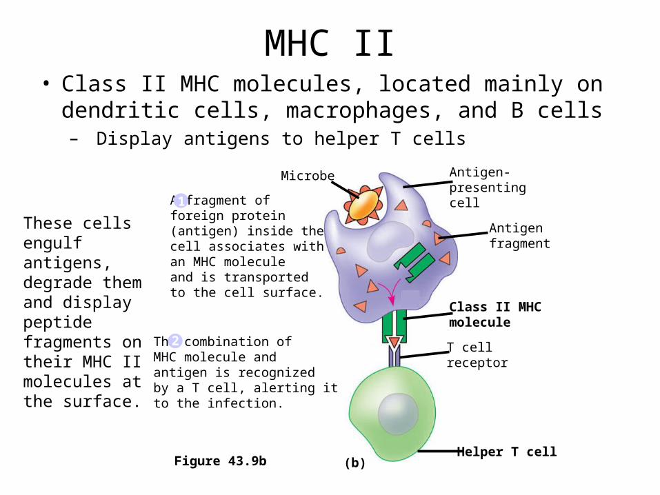
MHC II• Class II MHC molecules, located mainly on dendritic
cells, macrophages, and B cells– Display antigens to helper T cells
1
2
Figure 43.9b
Microbe Antigen-presentingcell
Antigenfragment
Class II MHCmolecule
T cellreceptor
Helper T cell
A fragment offoreign protein(antigen) inside thecell associates withan MHC moleculeand is transportedto the cell surface.
1
The combination ofMHC molecule andantigen is recognizedby a T cell, alerting itto the infection.
2
(b)
These cells engulf antigens, degrade them and display peptide fragments on their MHC II molecules at the surface.
21
3
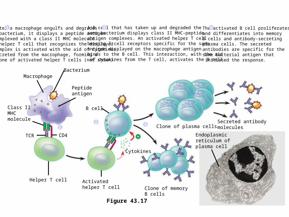
B cell
Bacterium
Peptide antigen
Class II MHCmolecule
TCR
Helper T cell
CD4
Activated helper T cell Clone of memory
B cells
Cytokines
Clone of plasma cellsSecreted antibodymolecules
Endoplasmicreticulum of plasma cell
Macrophage
After a macrophage engulfs and degradesa bacterium, it displays a peptide antigencomplexed with a class II MHC molecule.A helper T cell that recognizes the displayed complex is activated with the aid of cytokines secreted from the macrophage, forming a clone of activated helper T cells (not shown).
1 A B cell that has taken up and degraded the same bacterium displays class II MHC–peptide antigen complexes. An activated helper T cellbearing T cell receptors specific for the same antigen displayed on the macrophage antigen binds to the B cell. This interaction, with the aid of cytokines from the T cell, activates the B cell.
2 The activated B cell proliferatesand differentiates into memoryB cells and antibody-secreting plasma cells. The secreted antibodies are specific for the same bacterial antigen that initiated the response.
3
Figure 43.17
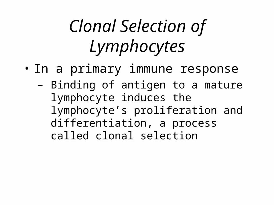
Clonal Selection of Lymphocytes
• In a primary immune response– Binding of antigen to a mature lymphocyte
induces the lymphocyte’s proliferation and differentiation, a process called clonal selection
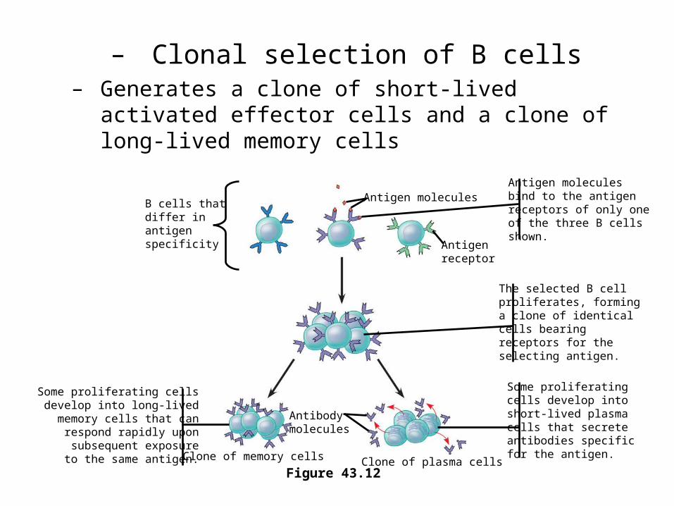
– Clonal selection of B cells– Generates a clone of short-lived activated effector
cells and a clone of long-lived memory cells
Figure 43.12
Antigen molecules
Antigenreceptor
B cells thatdiffer inantigenspecificity
Antibodymolecules
Clone of memory cells Clone of plasma cells
Antigen moleculesbind to the antigenreceptors of only oneof the three B cellsshown.
The selected B cellproliferates, forminga clone of identicalcells bearingreceptors for theselecting antigen.
Some proliferatingcells develop intoshort-lived plasmacells that secreteantibodies specificfor the antigen.
Some proliferating cellsdevelop into long-livedmemory cells that canrespond rapidly uponsubsequent exposureto the same antigen.

• The role of helper T cells in acquired immunity
Figure 43.15
After a dendritic cell engulfs and degrades a bacterium, it displays bacterial antigen fragments (peptides) complexed with a class II MHC molecule on the cell surface. A specific helper T cell binds to the displayed complex via its TCR with the aid of CD4. This interaction promotes secretion of cytokines by the dendritic cell.
Proliferation of the T cell, stimulatedby cytokines from both the dendritic cell and the T cell itself, gives rise toa clone of activated helper T cells(not shown), all with receptors for thesame MHC–antigen complex.
The cells in this clonesecrete other cytokines that help activate B cellsand cytotoxic T cells that are specific for the sameAntigen.
Cell-mediatedimmunity(attack on
infected cells)
Humoralimmunity
(secretion ofantibodies byplasma cells)
Dendriticcell
Dendriticcell
Bacterium
Peptide antigen
Class II MHCmolecule
TCR
CD4
Helper T cell
Cytokines
Cytotoxic T cell
B cell
1
2 3
1
2 3
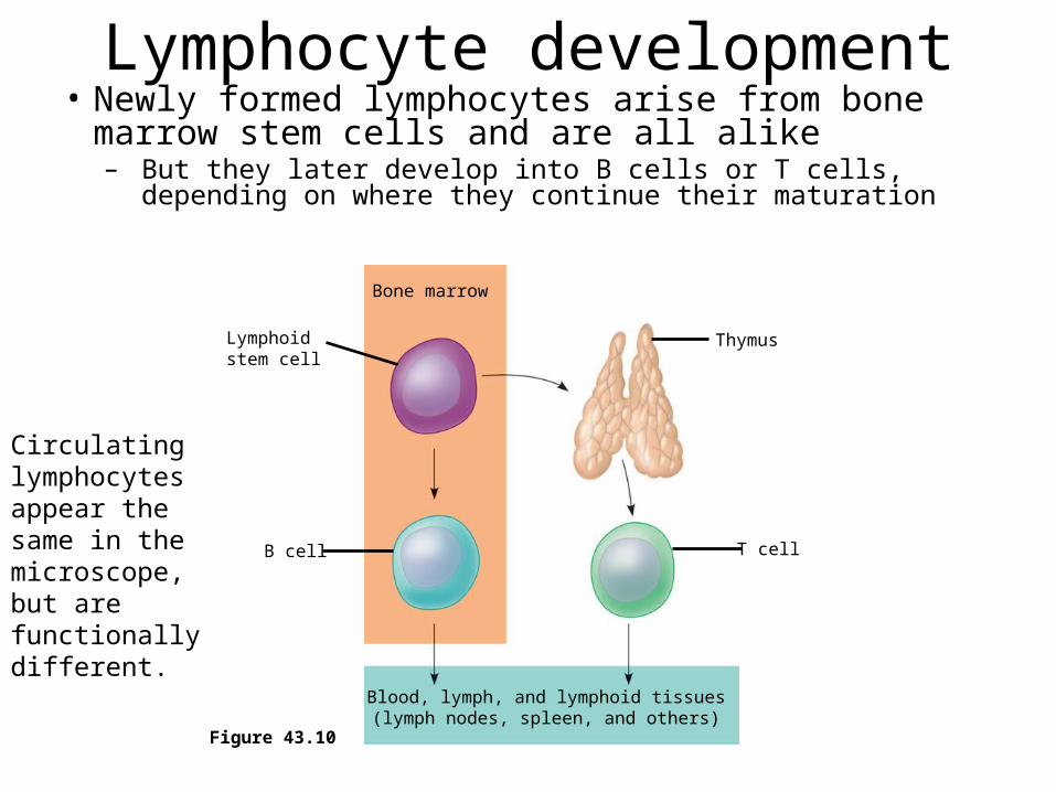
Lymphocyte development• Newly formed lymphocytes arise from bone
marrow stem cells and are all alike– But they later develop into B cells or T cells, depending
on where they continue their maturation
Figure 43.10
Bone marrow
Lymphoidstem cell
B cell
Blood, lymph, and lymphoid tissues(lymph nodes, spleen, and others)
T cell
Thymus
Circulating lymphocytes appear the same in the microscope, but are functionally different.

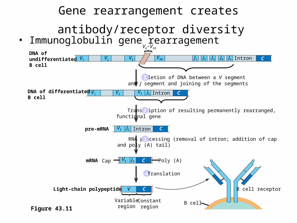
Generation of Lymphocyte Diversity by Gene Rearrangement
Lymphocytes arise from pluripotent stem cells in the bone marrow. Early in development, random, permanent gene rearrangement takes place.– Forms functional genes encoding the B or T
cell antigen receptor chains
DNA ofundifferentiatedB cell
DNA of differentiatedB cell
pre-mRNA
mRNA Cap
B cell
B cell receptorLight-chain polypeptide
Intron
Intron
Intron
Variableregion
Constantregion
V1V2 V3
V4–V39
V40 J1 J2 J3 J4 J5
V1 V2V3 J5
V3 J5
V3 J5
V C
C
C
C
C
Poly (A)
Figure 43.11
Deletion of DNA between a V segmentand J segment and joining of the segments1
Gene rearrangement creates antibody/receptor diversity • Immunoglobulin gene rearragement
Transcription of resulting permanently rearranged,functional gene2
RNA processing (removal of intron; addition of capand poly (A) tail)3
4 Translation
Testing and Removal of Self-Reactive Lymphocytes
• As B and T cells are maturing in the bone and thymus their antigen receptors are tested for possible self-reactivity
• Lymphocytes bearing receptors for antigens (self proteins or fragments of proteins) already present in the body are destroyed by apoptosis or rendered nonfunctional.
• MHC I and II cells are both expressed at high levels in the thymus and bone marrow.
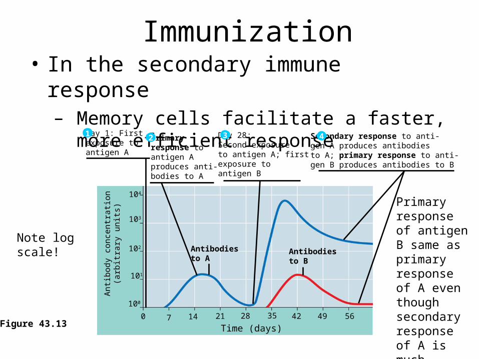
Immunization• In the secondary immune response
– Memory cells facilitate a faster, more efficient responseA
ntib
od
y co
nce
ntr
atio
n(a
rbitr
ary
un
its)
104
103
102
101
100
0 7 14 21 28 35 42 49 56
Time (days)Figure 43.13
Antibodiesto A
Antibodiesto B
Primaryresponse toantigen Aproduces anti-bodies to A
2Day 1: First exposure toantigen A
1 Day 28: Second exposureto antigen A; firstexposure to antigen B
3 Secondary response to anti-gen A produces antibodiesto A; primary response to anti-gen B produces antibodies to B
4
Primary response of antigen B same as primary response of A even though secondary response of A is much greater.
Note log scale!

• Concept 43.3: Humoral and cell-mediated immunity defend against different types of threats
• Acquired immunity includes two branches– The humoral immune response involves the
activation and clonal selection of B cells, resulting in the production of secreted antibodies
– The cell-mediated immune response involves the activation and clonal selection of cytotoxic T cells

• The roles of the major participants in the acquired immune response
Figure 43.14
Humoral immune response Cell-mediated immune response
First exposure to antigen
Intact antigensAntigens engulfed and
displayed by dendritic cellsAntigens displayed
by infected cells
Activate Activate Activate
Gives rise to Gives rise to Gives rise to
B cellHelperT cell
CytotoxicT cell
Plasmacells
MemoryB cells
Active and memory helperT cells
Memory cytotoxic
T cells
Active cytotoxic
T cells
Secrete antibodies that defend againstpathogens and toxins in extracellular fluid
Defend against infected cells, cancer cells, and transplanted tissues
Secretedcytokinesactivate
Humoral = blood!
B cell and helper T cell recognize same antigen so latter stimulates the former!
Antibody Classes
• The five major classes of antibodies, or immunoglobulins– Differ in their distributions and functions
within the body

• The five classes of immunoglobulins
Figure 43.18
First Ig class produced after initial exposure to antigen; then its concentration in the blood declines
Most abundant Ig class in blood; also present in tissue fluids
Only Ig class that crosses placenta, thus conferring passive immunity on fetus
Promotes opsonization, neutralization, and agglutination of antigens; less effective in complement activation than IgM (see Figure 43.19)
Present in secretions such as tears, saliva, mucus, and breast milk
Triggers release from mast cells and basophils of histamine and other chemicals that cause allergic reactions (see Figure 43.20)
Present primarily on surface of naive B cells that havenot been exposed to antigens
IgM(pentamer)
IgG(monomer)
IgA(dimer)
IgE(monomer)
J chain
Secretorycomponent
J chain
Transmembraneregion
IgD(monomer)
Promotes neutralization and agglutination of antigens; very effective in complement activation (see Figure 43.19)
Provides localized defense of mucous membranes byagglutination and neutralization of antigens (seeFigure 43.19)
Presence in breast milk confers passive immunity onnursing infant
Acts as antigen receptor in antigen-stimulated proliferation and differentiation of B cells (clonal selection)
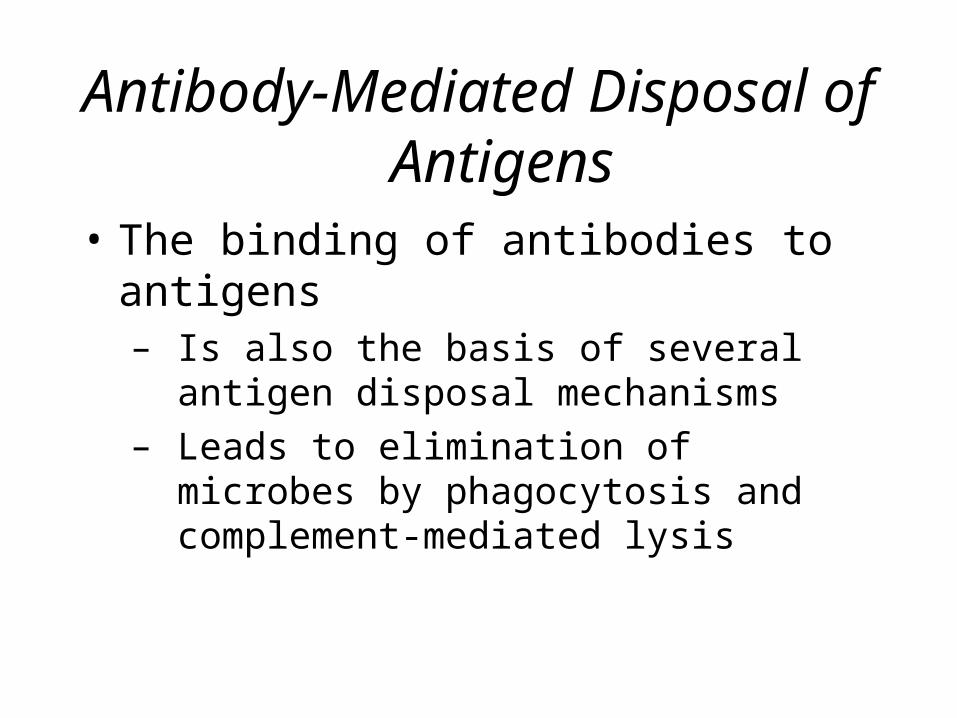
Antibody-Mediated Disposal of Antigens
• The binding of antibodies to antigens– Is also the basis of several antigen disposal
mechanisms– Leads to elimination of microbes by
phagocytosis and complement-mediated lysis
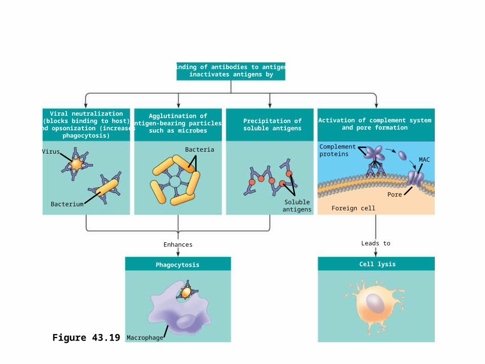
• Antibody-mediated mechanisms of antigen disposal
Binding of antibodies to antigensinactivates antigens by
Viral neutralization(blocks binding to host)
and opsonization (increasesphagocytosis)
Agglutination ofantigen-bearing particles,
such as microbes
Precipitation ofsoluble antigens
Activation of complement systemand pore formation
Bacterium
Virus Bacteria
Solubleantigens Foreign cell
Complementproteins
MAC
Pore
Enhances
Phagocytosis
Leads to
Cell lysis
MacrophageFigure 43.19

Active and Passive Immunization• Active immunity
– Develops naturally in response to an infection– Can also develop following immunization, also called
vaccination
• Passive immunity, which provides immediate, short-term protection– Is conferred naturally when IgG crosses the placenta
from mother to fetus or when IgA passes from mother to infant in breast milk
– Can be conferred artificially by injecting antibodies into a non-immune person. Immunoglobulins for snake bites (anti rattle snake serum) to treat victims of snake bites. Need to know what kind of snake injected the venom.
• In immunization– A nonpathogenic form of a microbe or part of
a microbe elicits an immune response to an immunological memory for that microbe
• Concept 43.4: The immune system’s ability to distinguish self from nonself limits tissue transplantation
• The immune system– Can wage war against cells from other individuals
• Transplanted tissues– Are usually destroyed by the recipient’s immune
system

Blood Groups and Transfusions
• Certain antigens on red blood cells – Determine whether a person has type A, B,
AB, or O blood
• Antibodies to non-self blood types– Already exist in the body probably from
exposure to saccharides on bacterial walls.
• Transfusion with incompatible blood– Leads to destruction of the transfused cells
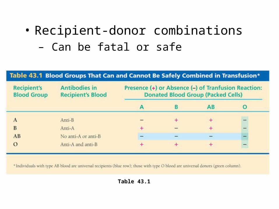
• Recipient-donor combinations– Can be fatal or safe
Table 43.1
Tissue and Organ Transplants• MHC molecules
– Are responsible for stimulating the rejection of tissue grafts and organ transplants
• The chances of successful transplantation are increased– If the donor and recipient MHC tissue types are well
matched– If the recipient is given immunosuppressive drugs
• Lymphocytes in bone marrow transplants– May cause a graft versus host reaction in recipients
• Concept 43.5: Exaggerated, self-directed, or diminished immune responses can cause disease
• If the delicate balance of the immune system is disrupted– The effects on the individual can range from minor
to often fatal consequences
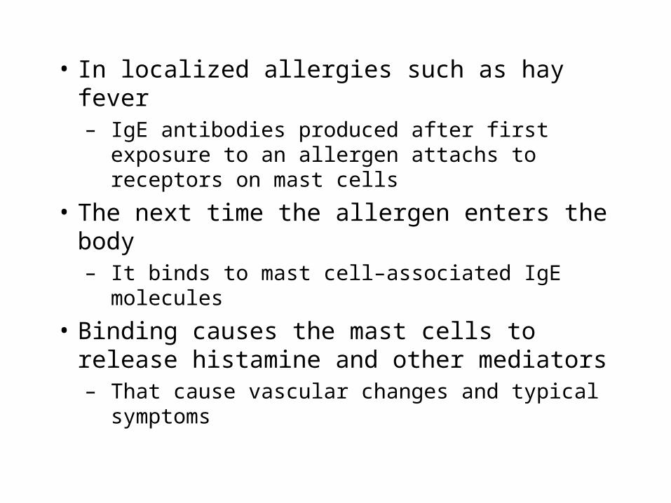
• In localized allergies such as hay fever– IgE antibodies produced after first exposure to an
allergen attach to receptors on mast cells
• In localized allergies such as hay fever– IgE antibodies produced after first exposure to
an allergen attachs to receptors on mast cells
• The next time the allergen enters the body– It binds to mast cell–associated IgE molecules
• Binding causes the mast cells to release histamine and other mediators– That cause vascular changes and typical
symptoms
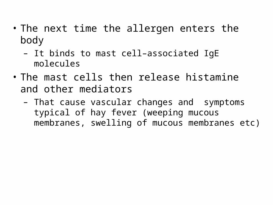
• The next time the allergen enters the body– It binds to mast cell–associated IgE molecules
• The mast cells then release histamine and other mediators– That cause vascular changes and symptoms typical
of hay fever (weeping mucous membranes, swelling of mucous membranes etc)
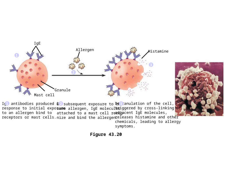
• The allergic response
Figure 43.20
IgE antibodies produced in response to initial exposure to an allergen bind to receptors or mast cells.
1 On subsequent exposure to the same allergen, IgE molecules attached to a mast cell recog-nize and bind the allergen.
2 Degranulation of the cell,triggered by cross-linking of adjacent IgE molecules, releases histamine and other chemicals, leading to allergysymptoms.
3
1
2
3
Allergen
IgE
Histamine
GranuleMast cell
• An acute allergic response sometimes leads to anaphylactic shock because of massive release of histimine. Dilates vasculature. Blood pools in capillary beds leading to blood pressure drops low enough so that the heart has no blood to pump. – Can occur within seconds of exposure to an allergen.
Treat with epinephrin to counteract the effect of histamine.
– Allergies to bees stings, peanuts and certain shell fish protein. Some people have such an allergy to penicillium.
Autoimmune Diseases
• In individuals with autoimmune diseases– The immune system loses tolerance for self
and turns against certain molecules of the body
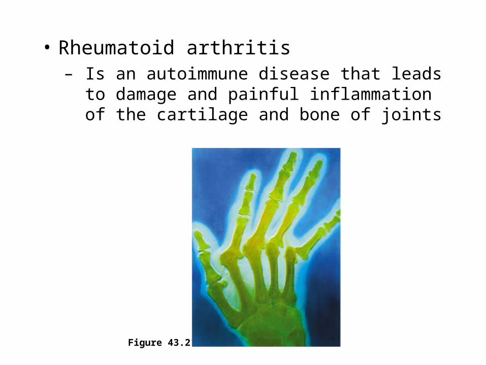
• Rheumatoid arthritis– Is an autoimmune disease that leads to damage
and painful inflammation of the cartilage and bone of joints
Figure 43.21
• Other examples of autoimmune diseases include– Systemic lupus erythematosus (reaction against
histones and DNA leads to fever, arthritis and kidney dysfunction)
– Multiple sclerosis (reaction against myelin sheath of the nervous system leads to loss of motor control of muscles).
– Insulin-dependent diabetes (destruction of Beta cells in pancreas leads to child onset diabetes. Can’t control blood sugar level at 100 mg/100ml blood.

Immunodeficiency Diseases• An inborn or primary immunodeficiency
– Results from hereditary or congenital defects that prevent proper functioning of innate, humoral, and/or cell-mediated defenses
• In severe combined immunodeficiency (SCID)– Both the humoral and cell-mediated branches of
acquired immunity fail to function
– Sometimes can solve problem by bone marrow transplant but hope other persons cells will not attack recipients cells
• Acquired Immunodeficiency Syndrome (AIDS)
• People with AIDS– Are highly susceptible to opportunistic
infections and cancers that take advantage of an immune system in collapse. Fungus invade the lungs skin cancers etc.
• Acquired Immunodeficiency Syndrome (AIDS)• People with AIDS
– Are highly susceptible to opportunistic infections and cancers that take advantage of an immune system in collapse. Fungus invade the lungs causing pneumonia ,skin cancers etc.
• Because AIDS arises from the loss of helper T cells– Both humoral and cell-mediated immune responses
are impaired– Virus uses CD-4 receptor on helper T cell to bind to
cell. Some Africans and a few others lack CD4 and are positive for virus antibody, but do not develop AIDs. So some day may be a large population of AIDs resistant people
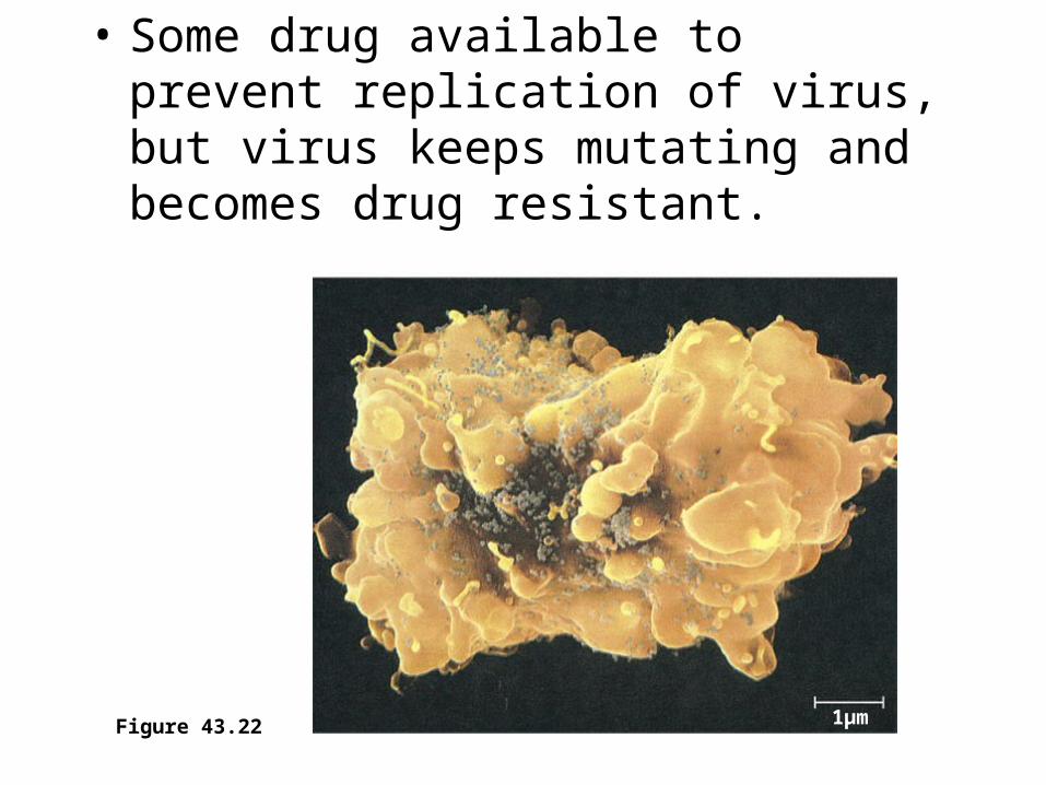
• Some drug available to prevent replication of virus, but virus keeps mutating and becomes drug resistant.
1µmFigure 43.22