Embed Size (px)
Citation preview

Hindawi Publishing CorporationCase Reports in CardiologyVolume 2012, Article ID 652086, 9 pagesdoi:10.1155/2012/652086
Case Report
Iatrogenic Aortocoronary Arteriovenous Fistulafollowing Coronary Artery Bypass Surgery: A Case Reportand Complete Review of the Literature
Jonathan D. Gardner,1 William R. Maddox,2 and Joe B. Calkins Jr.2, 3
1 Internal Medicine Department, Georgia Health Sciences University, Augusta, GA 30912, USA2 Cardiology Department, Georgia Health Sciences University, Augusta, GA 30912, USA3 Cardiology Department, Charlie Norwood VA Medical Center, Augusta, GA 30904, USA
Correspondence should be addressed to Jonathan D. Gardner, [email protected]
Received 2 October 2012; Accepted 18 October 2012
Academic Editors: G. Devlin and T. Kasai
Copyright © 2012 Jonathan D. Gardner et al. This is an open access article distributed under the Creative Commons AttributionLicense, which permits unrestricted use, distribution, and reproduction in any medium, provided the original work is properlycited.
The case of a patient who presented with angina following a coronary artery bypass (CABG) operation during which the leftinternal mammary artery was inadvertently anastomosed to a cardiac vein is presented. The literature concerning previouslyreported cases of aortocoronary arteriovenous fistulas (ACAVF) due to inadvertent grafting of a coronary vein is reviewed andthe significance of this complication is discussed. ACAVF due to inadvertent grafting of a coronary vein is a rare complication ofCABG and may be a more common cause of graft failure than has previously been recognized. Distortion of cardiac anatomy,the presence of epicardial fat, and an intramyocardial course of the artery intended for grafting are predisposing factors. Somepatients present with angina pectoris and heart failure whereas others have no symptoms. The diagnostic test of choice is coronaryangiography. Cardiac MRI and CT have a limited role due to the smaller size and the more clearly defined course of these fistulas.Asymptomatic patients are simply observed since spontaneous closure of these fistulas is reported. Symptomatic patients can betreated with combined medical management and percutaneous methods.
1. Introduction
Iatrogenic aortocoronary arteriovenous fistula (ACAVF)resulting from placement of an arterial graft to a cardiac veinis a rare complication of CABG. We present a case involvinggrafting of the left internal mammary artery (LIMA) to a leftcoronary vein and a review of the literature.
2. Patient Presentation
The patient is a 69-year-old male with hypertension, hyper-lipidemia, and type 2 diabetes mellitus who presented withexertional chest pain, dyspnea, decreased functional capacityand occasional palpitations. A myocardial perfusion studyone month earlier had demonstrated inferolateral ischemiaand preserved left ventricular systolic function (EF 69%).
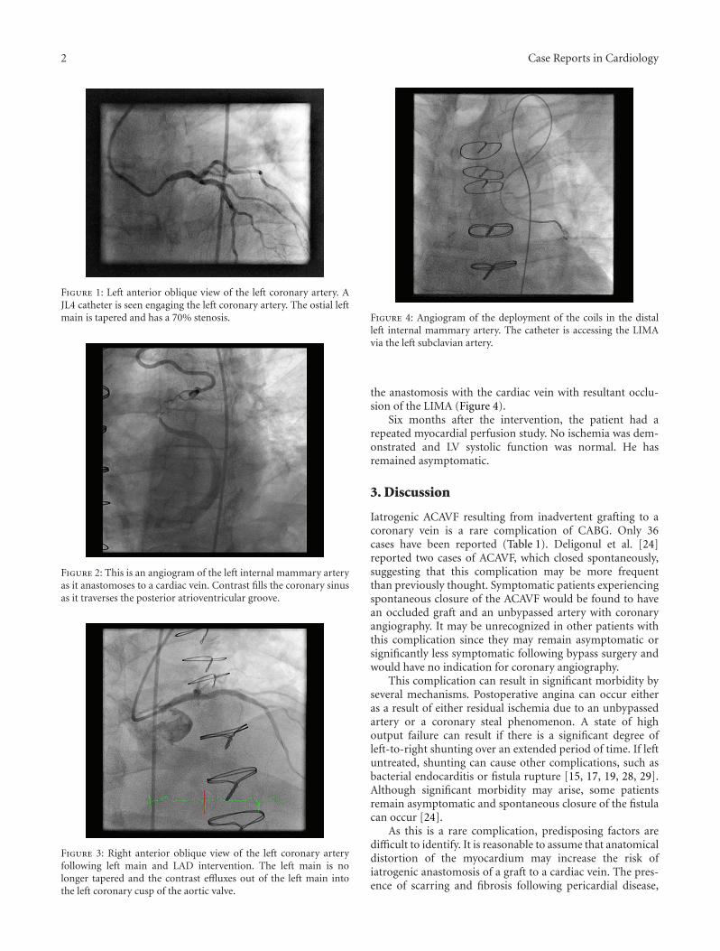
Subsequent left heart catheterization (LHC) showed70% ostial left main stenosis (Figure 1), 60% left anterior
descending artery (LAD) stenosis, and complete occlusion ofthe mid circumflex artery with filling via right to left collater-als. The right coronary artery (RCA) had 70% stenosis in itsmidportion and left ventricular systolic function was normal.He underwent CABG with the following grafts: LIMA to theLAD, SVG to OM, and SVG to the PDA. Postoperative coursewas uncomplicated.
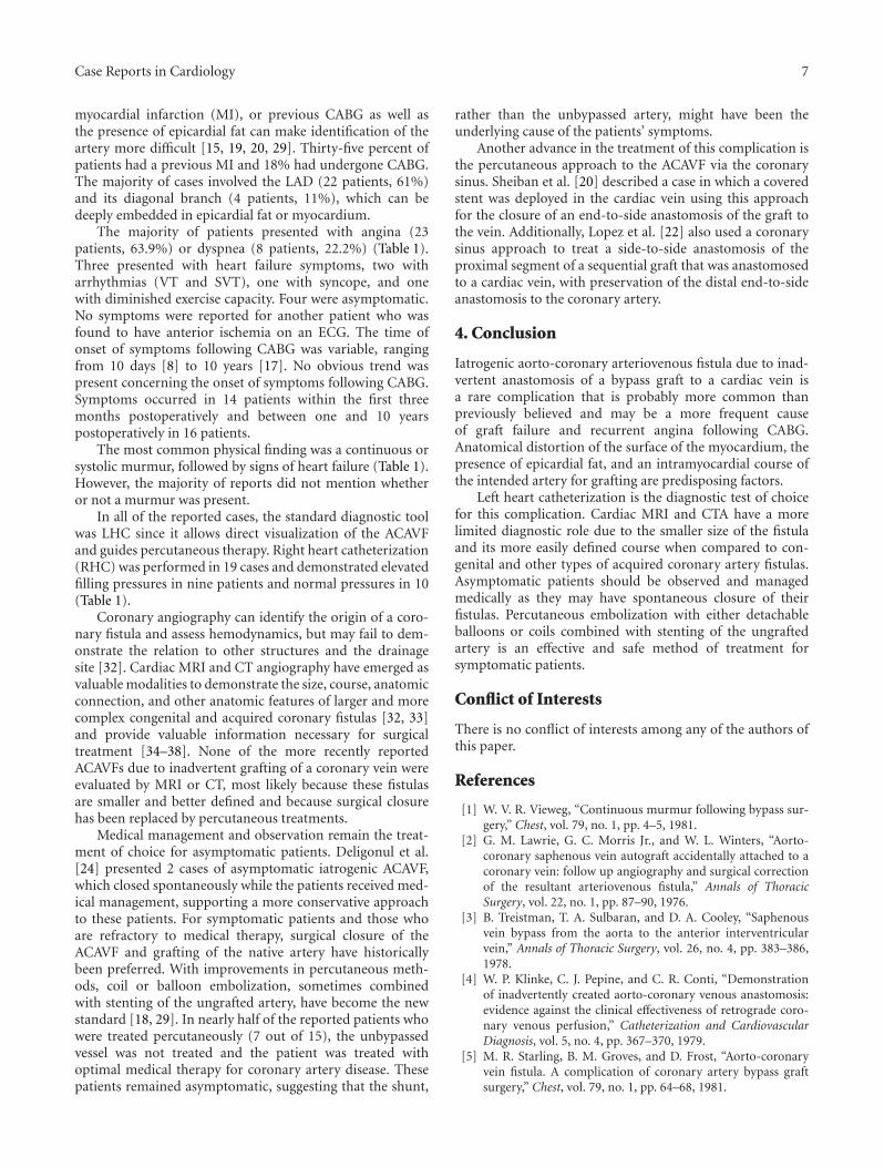
Three months later, the patient presented with exertionalchest pain similar to his pain prior to surgery. Repeated LHCshowed no change in the native coronary arteries and patentSVG to OM and SVG to PDA with good flow. Angiography ofthe LIMA demonstrated that it was anastomosed to a cardiacvein with resultant flow into the coronary sinus (Figure 2).
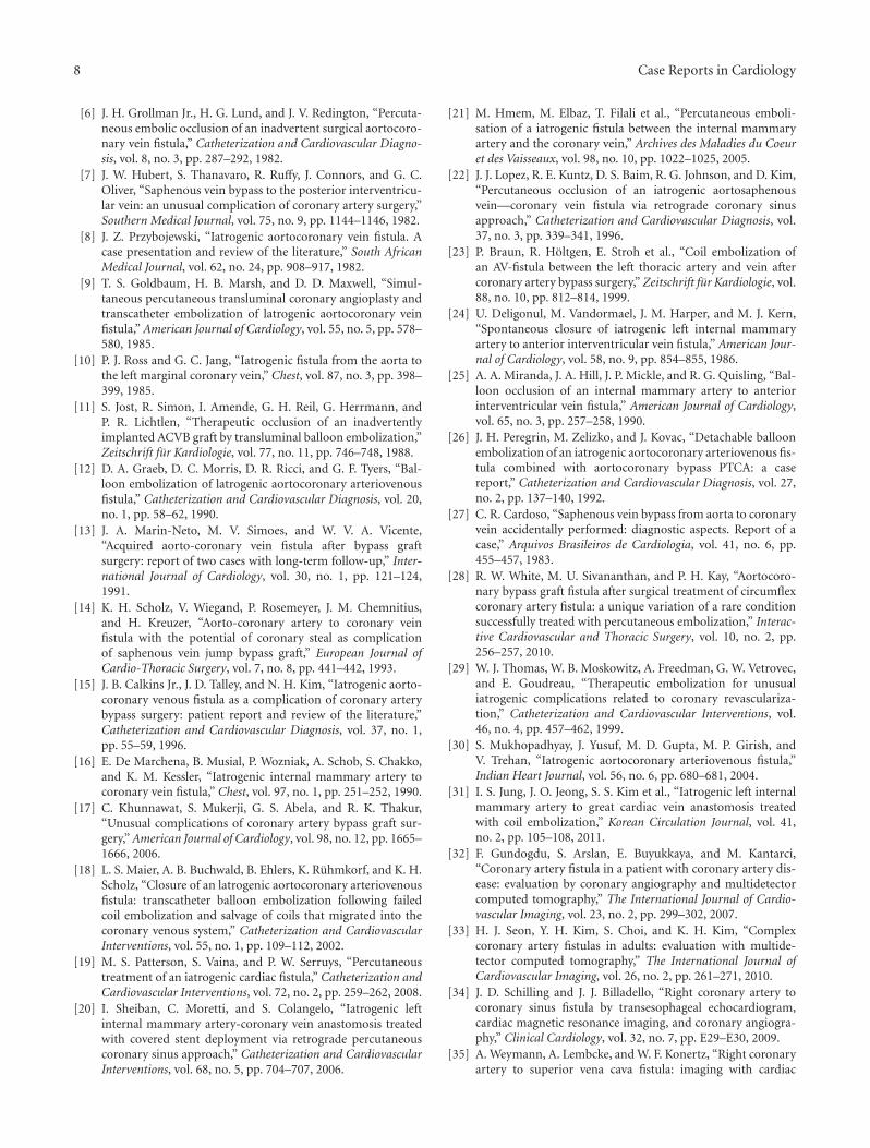
Percutaneous coronary intervention (PCI) was per-formed with placement of three drug eluting stents in theLM and ostial/proximal LAD. There was no residual stenosis(Figure 3). Subsequently, eight 3 mm stainless steel coils weredeployed in the distal portion of the LIMA just proximal to
2 Case Reports in Cardiology
Figure 1: Left anterior oblique view of the left coronary artery. AJL4 catheter is seen engaging the left coronary artery. The ostial leftmain is tapered and has a 70% stenosis.
Figure 2: This is an angiogram of the left internal mammary arteryas it anastomoses to a cardiac vein. Contrast fills the coronary sinusas it traverses the posterior atrioventricular groove.
Figure 3: Right anterior oblique view of the left coronary arteryfollowing left main and LAD intervention. The left main is nolonger tapered and the contrast effluxes out of the left main intothe left coronary cusp of the aortic valve.
Figure 4: Angiogram of the deployment of the coils in the distalleft internal mammary artery. The catheter is accessing the LIMAvia the left subclavian artery.
the anastomosis with the cardiac vein with resultant occlu-sion of the LIMA (Figure 4).
Six months after the intervention, the patient had arepeated myocardial perfusion study. No ischemia was dem-onstrated and LV systolic function was normal. He hasremained asymptomatic.
3. Discussion
Iatrogenic ACAVF resulting from inadvertent grafting to acoronary vein is a rare complication of CABG. Only 36cases have been reported (Table 1). Deligonul et al. [24]reported two cases of ACAVF, which closed spontaneously,suggesting that this complication may be more frequentthan previously thought. Symptomatic patients experiencingspontaneous closure of the ACAVF would be found to havean occluded graft and an unbypassed artery with coronaryangiography. It may be unrecognized in other patients withthis complication since they may remain asymptomatic orsignificantly less symptomatic following bypass surgery andwould have no indication for coronary angiography.
This complication can result in significant morbidity byseveral mechanisms. Postoperative angina can occur eitheras a result of either residual ischemia due to an unbypassedartery or a coronary steal phenomenon. A state of highoutput failure can result if there is a significant degree ofleft-to-right shunting over an extended period of time. If leftuntreated, shunting can cause other complications, such asbacterial endocarditis or fistula rupture [15, 17, 19, 28, 29].Although significant morbidity may arise, some patientsremain asymptomatic and spontaneous closure of the fistulacan occur [24].
As this is a rare complication, predisposing factors aredifficult to identify. It is reasonable to assume that anatomicaldistortion of the myocardium may increase the risk ofiatrogenic anastomosis of a graft to a cardiac vein. The pres-ence of scarring and fibrosis following pericardial disease,

Case Reports in Cardiology 3
Ta
ble
1:R
epor
tsof
inad
vert
ent
atta
chm
ent
ofa
bypa
ssgr
aft
toa
card
iac
vein
.
Au
thor
Pati
ent
Sym
ptom
s/on
set
afte
rC
AB
GM
urm
ur
Gra
ft/I
nte
nde
dA
rter
y/A
ctu
alA
nas
tam
osis
Shu
nt
Hem
odyn
amic
sC
AB
GP
revi
ous
MI
Trea
tmen
t
Vie
weg
[1]
53M
CH
F/6
wee
ksC
onti
nu
ous
2nd
LIC
SSV
G/L
AD
/an
teri
orca
rdia
cve
in
Shu
nt
byhy
drog
enin
hal
atio
n;n
orm
alpu
lse
ox
Mild
elev
atio
nof
righ
th
eart
pres
sure
sFi
rst
No
Gra
ftre
mov
al;
regr
afti
ng
ofSV
Gto
LAD
Law
rie
etal
.[2]
44M
An
gin
ape
ctor
is;
<3
mon
ths
Syst
olic
;bas
eto
nec
kSV
G/L
AD
/LA
Dve
inN
otm
enti
oned
Not
men
tion
edT
hir
dN
oG
raft
ligat
ion
;SV
Gto
LAD
Trei
stm
anet
al.[
3]55
MSV
T,pa
lpit
atio
ns,
syn
cop
e;3.
5ye
ars
Con
tin
uou
s3r
dL
ICS
SVG
/LA
D/a
nte
rior
inte
rven
tric
ula
rve
inN
orm
alox
imet
ryM
ildel
evat
ion
ofri
ght
hea
rtpr
essu
res
Firs
tA
nte
rola
tera
lM
IN
one
Klin
keet
al.[
4]40
MA
ngi
na
pect
oris
;5m
onth
sN
otm
enti
oned
SVG
/LA
D/a
nte
rior
inte
rven
tric
ula
rve
inC
oron
ary
sin
us
02sa
tura
tion
90%
Nor
mal
Firs
tN
oC
AB
G
Star
ling
etal
.[5]
47M
Non
ere
port
ed.
An
teri
oris
chem
ia;
3w
eeks
Con
tin
uou
s2n
dL
ICS
toap
exSV
G/L
AD
/an
teri
orca
rdia
cve
inN
one
Nor
mal
Firs
tPo
ster
olat
eral
MI
Fist
ula
ligat
ion
;SV
Gto
diag
onal
Star
ling
etal
.[5]
66M
Asy
mpt
omat
icC
onti
nu
ous
2nd
and
3rd
LIC
Sto
the
apex
SVG
/pro
xim
alLA
Dto
dist
alLA
D/a
nte
rior
card
iac
vein
dist
alLA
DN
one
Nor
mal
Firs
tA
nte
rose
ptal
MI
Obs
erva
tion
Gro
llman
Jr.e
tal
.[6]
52M
Fati
gue,
dysp
nea
;1ye
arN
one
SVG
/an
tero
late
ralb
ran
chof
Cx/
ante
rior
inte
rven
tric
ula
rve
in1.
1:1
RV
ED
P8
mm
Hg
LVE
DP
25m
mH
gFi
rst
An
tero
apic
alM
I
Perc
uta
neo
us
occl
usi
onof
SVG
wit
h2
coils
Hu
bert
etal
.[7]
55M
CH
F,V
T;1
mon
thC
onti
nu
ous
ULS
B
SVG
/LV
bran
chof
RC
AP
Lof
Cx/
LVbr
anch
post
erio
rin
terv
entr
icu
lar
vein
Nor
mal
oxim
etry
RA
20m
mH
gFi
rst
Infe
rior
MI
Lig
atio
nof
fist
ula
Prz
yboj
ewsk
i[8]
43M
An
gin
ape
ctor
is;1
0da
ysC
onti
nu
ous
2nd
and
3rd
LIC
SSV
G/L
AD
/LA
Dve
inN
otm
enti
oned
Not
men
tion
edFi
rst
An
tero
late
ral
MI
Lig
atio
n;
rep
eat
CA
BG
Gol
dbau
met
al.[
9]53
MA
ngi
na
pect
oris
,ex
erti
onal
dysp
nea
;4
year
sN
one
SVG
/LA
D/a
nte
rior
inte
rven
tric
ula
rve
inSm
all;
not
quan
tifi
edPA
42/1
9LV
ED
P19
Firs
tA
nte
rior
MI
PT
CA
ofLA
D;
perc
uta
neo
us
occl
usi
onof
SVG
wit
hco
ils
Ros
san
dJa
ng
[10]
44M
An
gin
alp
ecto
ris;
onse
tn
otm
enti
oned
Syst
olic
;ULS
BSV
G/i
nte
rmed
iate
orC
x/le
ftm
argi
nal
vein
1.4
:1N
orm
alSe
con
dIn
feri
orM
IN
one
rep
orte
d
Jost
etal
.[11
]57
MA
ngi
na
pect
oris
;2ye
ars
Not
men
tion
edSV
G/L
AD
/an
teri
orca
rdia
cve
in18
%of
pulm
onar
yfl
owN
otm
enti
oned
Firs
tN
oE
mbo
lizat
ion
wit
hsi
licon
eba
lloon

4 Case Reports in Cardiology
Ta
ble
1:C
onti
nu
ed.
Au
thor
Pati
ent
Sym
ptom
s/on
set
afte
rC
AB
GM
urm
ur
Gra
ft/I
nte
nde
dA
rter
y/A
ctu
alA
nas
tam
osis
Shu
nt
Hem
odyn
amic
sC
AB
GP
revi
ous
MI
Trea
tmen
t
Gra
ebet
al.[
12]
56F
An
gin
ape
ctor
is;1
year
Not
men
tion
edSV
G/P
DA
/PD
VSm
all
Nor
mal
Firs
tN
o
Bal
loon
embo
lizat
ion
ofP
DA
(un
succ
essf
ul)
Mar
in-N
eto
etal
.[13
]57
MD
yspn
ea,c
hes
tpa
in;1
mon
th(n
ois
chem
iade
tect
ed)
Syst
olic
;pu
lmon
icar
eaSV
G/fi
rst
diag
onal
/an
teri
orca
rdia
cve
in23
%of
pulm
onar
yfl
owN
orm
alFi
rst
Infe
rior
MI
Non
e
Mar
in-N
eto
etal
.[13
]84
MA
ngi
na
pect
oris
;14
mon
ths
Not
men
tion
edSV
G/fi
rst
diag
onal
/an
tero
late
ral
coro
nar
yve
in
12%
ofpu
lmon
ary
flow
Not
men
tion
edFi
rst
No
PT
CA
ofn
ewR
CA
lesi
on,
no
trea
tmen
tof
fist
ula
Sch
olz
etal
.[14
]49
MA
ngi
na
pect
oris
15m
onth
sSy
stol
icU
LSB
SVG
/OM
1,O
M2/
OM
1,co
ron
ary
vein
Smal
lN
orm
alFi
rst
No
Obs
erva
tion
Cal
kin
sJr
.et
al.[
15]
51F
An
gin
ape
ctor
is;
2-3
mon
ths
Non
eSV
G/O
M1,
OM
2/O
M1
coro
nar
yve
inN
one
RV
55/1
5PA
55/1
7Se
con
dN
oC
oil
embo
lizat
ion
De
Mar
chen
aet
al.[
16]
73M
Dim
inis
hed
exer
cise
capa
city
,dy
spn
ea;2
mon
ths
Non
eLI
MA
/LA
D/g
reat
card
iac
vein
Smal
lR
V68
/12
RA
12m
mH
gFi
rst
No
Obs
erva
tion
Khu
nn
awat
etal
.[17
]75
FD
yspn
ea;1
0ye
ars
S3,n
om
urm
ur
SVG
/RC
A/c
ardi
acve
inN
otm
enti
oned
Not
men
tion
edFi
rst
LBB
Bon
EK
G
Not
men
tion
ed
Khu
nn
awat
etal
.[17
]57
MD
yspn
ea;6
year
sN
one
LIM
A/L
AD
/LA
Dca
rdia
cve
inN
otm
enti
oned
Not
men
tion
edFi
rst
Not
men
tion
ed
Not
men
tion
ed
Mai
eret
al.[
18]
50M
Dys
pnea
and
An
gin
ape
ctor
is;2
year
s
Syst
olic
mu
rmu
rat
ULS
BSV
G/D
1/co
ron
ary
vein
Larg
ele
ftto
righ
t
Pu
lmon
ary
C.O
.6.
6L
/mm
,sys
tem
icC
O4.
8L
/mm
,n
orm
alpu
lmon
ary
pres
sure
s
Firs
tN
otm
enti
oned
PT
CA
;fai
led
perc
uta
neo
us
coil
embo
lizat
ion
led
tope
rcu
tan
eou
str
ansc
ath
eter
deta
chab
leba
lloon
ofSV
Gto
D1
Patt
erso
net
al.[
19]
67M
angi
na
Pect
oris
;7m
onth
sN
otm
enti
oned
LIM
A-R
IMA
/PL/
PL
vein
Pre
sen
tN
otm
enti
oned
Firs
tN
otm
enti
oned
PC
Ito
reva
scu
lari
zeC
x;th
enco
ilem
boliz
atio
nof
RIM
A

Case Reports in Cardiology 5
Ta
ble
1:C
onti
nu
ed.
Au
thor
Pati
ent
Sym
ptom
s/on
set
afte
rC
AB
GM
urm
ur
Gra
ft/I
nte
nde
dA
rter
y/A
ctu
alA
nas
tam
osis
Shu
nt
Hem
odyn
amic
sC
AB
GP
revi
ous
MI
Trea
tmen
t
Shei
ban
etal
.[20
]73
MPo
siti
vest
ress
test
and
angi
na
wit
hex
erti
on;2
mon
ths
Not
men
tion
edLI
MA
/LA
D/G
CV
Pre
sen
t“a
rter
iove
nou
sst
eal”
Mod
erat
eL-
RSh
un
t
Not
men
tion
edFi
rst
No
PC
Iw
ith
DE
Sof
LAD
and
PC
Iw
ith
cove
red
sten
tof
GC
Vth
rou
ghco
ron
ary
sin
us
Hm
emet
al.[
21]
76M
Dys
pnea
,LE
edem
a,C
HF/
RH
F;2
mon
ths
Non
eL
IMA
/LA
D/L
IMA
/car
diac
vein
Not
men
tion
edN
otm
enti
oned
Firs
tA
SQ
wav
es
Coi
lem
boliz
atio
nto
prox
imal
LIM
A
Lop
ezet
al.[
22]
74M
Res
tan
gin
a;3
mon
ths
Not
men
tion
edSV
G-O
M2-
OM
3/SV
G-L
mar
gin
alve
inof
OM
3N
osi
gnifi
can
tle
ftto
righ
tsh
un
tN
otm
enti
oned
Seco
nd
Not
men
tion
ed
PC
I,em
boliz
atio
nof
mar
gin
alve
in
Bra
un
etal
.[23
]58
MA
ngi
na;
6m
onth
sN
otm
enti
oned
LIM
A/L
AD
/LIM
A/c
ardi
acve
inN
otm
enti
oned
Not
men
tion
edSe
con
dN
otm
enti
oned
RC
A-P
TC
A;
coil
embo
lizat
ion
Del
igon
ule
tal
.[24
]66
MA
sym
ptom
atic
Non
eLI
MA
/LA
D/a
nte
rior
inte
rven
tric
ula
rve
inSm
all
Non
eFi
rst
No
Spon
tan
eou
scl
osu
re
Del
igon
ule
tal
.[24
]57
MA
sym
ptom
atic
Non
eLI
MA
/LA
D/a
nte
rior
inte
rven
tric
ula
rve
inSm
all
Non
eFi
rst
No
Spon
tan
eou
scl
osu
re
Mir
anda
etal
.[25
]66
MA
ngi
na;
2w
eeks
Not
men
tion
edLI
MA
/LA
D/a
nte
rior
inte
rven
tric
ula
rve
inSm
all
Pu
lmon
ary
arte
rypu
lse
ox66
%Se
con
dN
o
PT
CA
todi
agon
albr
anch
graf
tfo
llow
edby
ballo
onoc
clu
sion
offi
stu
la

6 Case Reports in Cardiology
Ta
ble
1:C
onti
nu
ed.
Au
thor
Pati
ent
Sym
ptom
s/on
set
afte
rC
AB
GM
urm
ur
Gra
ft/I
nte
nde
dA
rter
y/A
ctu
alA
nas
tam
osis
Shu
nt
Hem
odyn
amic
sC
AB
GP
revi
ous
MI
Trea
tmen
t
Pere
grin
etal
.[26
]54
MU
nst
able
angi
na;
1ye
arSy
stol
ic/d
iast
olic
left
para
ster
nal
Gra
ft/d
iago
nal
bran
ch/g
raft
ven
aco
rdis
mag
na
Not
men
tion
edN
otm
enti
oned
Firs
tIn
feri
orM
I
PT
CA
ofR
CA
and
ballo
onoc
clu
sion
ofAV
fist
ula
Car
doso
[27]
55F
An
gin
a
Con
tin
uou
sse
con
dle
ftin
terc
osta
lssp
ace
SVG
/LA
D/L
AD
vein
Aor
tic
oxyg
ensa
tura
tion
92%
,co
ron
ary
sin
us
satu
rati
on80
%,
SVC
satu
rati
on73
%
LV17
1/19
,RV
43/1
2,m
ean
PC
WP
21Fi
rst
No
Not
men
tion
ed
Wh
ite
etal
.[28
]56
MA
ngi
na
and
trop
onin
elev
atio
n;
5ye
ars
Not
men
tion
edLC
X-v
ein
graf
t-m
argi
nal
/LC
X-c
oron
ary
sin
us
Non
eN
one
Firs
tN
oM
RI;
coil
embo
lizat
ion
Th
omas
etal
.[29
]76
MA
ngi
na
wit
hex
erti
on;3
mon
ths
Not
men
tion
edLI
MA
/LA
D/A
IVN
otm
enti
oned
Not
men
tion
edFi
rst
Not
men
tion
ed
PC
Iw
ith
sten
tin
gan
dP
CI
wit
hem
boliz
atio
n
Mu
khop
adhy
ayet
al.[
30]
60M
Exe
rtio
nal
angi
na
Not
men
tion
edLI
MA
/LA
D/c
ardi
acve
inLe
ftto
righ
tsh
un
t
RH
and
PApr
essu
res
nor
mal
,02
sat
was
84%
onst
epu
pin
coro
nar
ysi
nu
san
d72
%in
the
RA
to80
%in
PA
Firs
tN
otm
enti
oned
PC
Iw
ith
faile
dco
ilem
boliz
atio
n,
defe
rred
tosu
rgic
alco
rrec
tion
Jun
get
al.[
31]
50M
Asy
mpt
omat
icN
otm
enti
oned
LIM
A/L
AD
/GC
VN
otm
enti
oned
EC
HO
show
edLV
hypo
kin
esis
and
LVsy
stol
icdy
sfu
nct
ion
.Fi
rst
Not
men
tion
ed
CT
angi
ogra
m;
PC
I/D
ES
and
Coi
lem
boliz
atio
n
Cu
rren
tCas
e69
M
An
gin
aw
ith
exer
tion
left
arm
radi
atio
n;3
mon
ths
Non
eL
IMA
/LA
D/L
IMA
/car
diac
vein
Non
e
Aor
ta11
9/54
,mea
n77
,LV
syst
olic
:130
,LV
ED
P:1
4,LV
-an
giog
raph
yE
F=
55%
Firs
tN
o
PC
I/D
ES
and
coil
embo
lizat
ion
toL
IMA
Case Reports in Cardiology 7
myocardial infarction (MI), or previous CABG as well asthe presence of epicardial fat can make identification of theartery more difficult [15, 19, 20, 29]. Thirty-five percent ofpatients had a previous MI and 18% had undergone CABG.The majority of cases involved the LAD (22 patients, 61%)and its diagonal branch (4 patients, 11%), which can bedeeply embedded in epicardial fat or myocardium.
The majority of patients presented with angina (23patients, 63.9%) or dyspnea (8 patients, 22.2%) (Table 1).Three presented with heart failure symptoms, two witharrhythmias (VT and SVT), one with syncope, and onewith diminished exercise capacity. Four were asymptomatic.No symptoms were reported for another patient who wasfound to have anterior ischemia on an ECG. The time ofonset of symptoms following CABG was variable, rangingfrom 10 days [8] to 10 years [17]. No obvious trend waspresent concerning the onset of symptoms following CABG.Symptoms occurred in 14 patients within the first threemonths postoperatively and between one and 10 yearspostoperatively in 16 patients.
The most common physical finding was a continuous orsystolic murmur, followed by signs of heart failure (Table 1).However, the majority of reports did not mention whetheror not a murmur was present.
In all of the reported cases, the standard diagnostic toolwas LHC since it allows direct visualization of the ACAVFand guides percutaneous therapy. Right heart catheterization(RHC) was performed in 19 cases and demonstrated elevatedfilling pressures in nine patients and normal pressures in 10(Table 1).
Coronary angiography can identify the origin of a coro-nary fistula and assess hemodynamics, but may fail to dem-onstrate the relation to other structures and the drainagesite [32]. Cardiac MRI and CT angiography have emerged asvaluable modalities to demonstrate the size, course, anatomicconnection, and other anatomic features of larger and morecomplex congenital and acquired coronary fistulas [32, 33]and provide valuable information necessary for surgicaltreatment [34–38]. None of the more recently reportedACAVFs due to inadvertent grafting of a coronary vein wereevaluated by MRI or CT, most likely because these fistulasare smaller and better defined and because surgical closurehas been replaced by percutaneous treatments.
Medical management and observation remain the treat-ment of choice for asymptomatic patients. Deligonul et al.[24] presented 2 cases of asymptomatic iatrogenic ACAVF,which closed spontaneously while the patients received med-ical management, supporting a more conservative approachto these patients. For symptomatic patients and those whoare refractory to medical therapy, surgical closure of theACAVF and grafting of the native artery have historicallybeen preferred. With improvements in percutaneous meth-ods, coil or balloon embolization, sometimes combinedwith stenting of the ungrafted artery, have become the newstandard [18, 29]. In nearly half of the reported patients whowere treated percutaneously (7 out of 15), the unbypassedvessel was not treated and the patient was treated withoptimal medical therapy for coronary artery disease. Thesepatients remained asymptomatic, suggesting that the shunt,
rather than the unbypassed artery, might have been theunderlying cause of the patients’ symptoms.
Another advance in the treatment of this complication isthe percutaneous approach to the ACAVF via the coronarysinus. Sheiban et al. [20] described a case in which a coveredstent was deployed in the cardiac vein using this approachfor the closure of an end-to-side anastomosis of the graft tothe vein. Additionally, Lopez et al. [22] also used a coronarysinus approach to treat a side-to-side anastomosis of theproximal segment of a sequential graft that was anastomosedto a cardiac vein, with preservation of the distal end-to-sideanastomosis to the coronary artery.
4. Conclusion
Iatrogenic aorto-coronary arteriovenous fistula due to inad-vertent anastomosis of a bypass graft to a cardiac vein isa rare complication that is probably more common thanpreviously believed and may be a more frequent causeof graft failure and recurrent angina following CABG.Anatomical distortion of the surface of the myocardium, thepresence of epicardial fat, and an intramyocardial course ofthe intended artery for grafting are predisposing factors.
Left heart catheterization is the diagnostic test of choicefor this complication. Cardiac MRI and CTA have a morelimited diagnostic role due to the smaller size of the fistulaand its more easily defined course when compared to con-genital and other types of acquired coronary artery fistulas.Asymptomatic patients should be observed and managedmedically as they may have spontaneous closure of theirfistulas. Percutaneous embolization with either detachableballoons or coils combined with stenting of the ungraftedartery is an effective and safe method of treatment forsymptomatic patients.
Conflict of Interests
There is no conflict of interests among any of the authors ofthis paper.
References
[1] W. V. R. Vieweg, “Continuous murmur following bypass sur-gery,” Chest, vol. 79, no. 1, pp. 4–5, 1981.
[2] G. M. Lawrie, G. C. Morris Jr., and W. L. Winters, “Aorto-coronary saphenous vein autograft accidentally attached to acoronary vein: follow up angiography and surgical correctionof the resultant arteriovenous fistula,” Annals of ThoracicSurgery, vol. 22, no. 1, pp. 87–90, 1976.
[3] B. Treistman, T. A. Sulbaran, and D. A. Cooley, “Saphenousvein bypass from the aorta to the anterior interventricularvein,” Annals of Thoracic Surgery, vol. 26, no. 4, pp. 383–386,1978.
[4] W. P. Klinke, C. J. Pepine, and C. R. Conti, “Demonstrationof inadvertently created aorto-coronary venous anastomosis:evidence against the clinical effectiveness of retrograde coro-nary venous perfusion,” Catheterization and CardiovascularDiagnosis, vol. 5, no. 4, pp. 367–370, 1979.
[5] M. R. Starling, B. M. Groves, and D. Frost, “Aorto-coronaryvein fistula. A complication of coronary artery bypass graftsurgery,” Chest, vol. 79, no. 1, pp. 64–68, 1981.
8 Case Reports in Cardiology
[6] J. H. Grollman Jr., H. G. Lund, and J. V. Redington, “Percuta-neous embolic occlusion of an inadvertent surgical aortocoro-nary vein fistula,” Catheterization and Cardiovascular Diagno-sis, vol. 8, no. 3, pp. 287–292, 1982.
[7] J. W. Hubert, S. Thanavaro, R. Ruffy, J. Connors, and G. C.Oliver, “Saphenous vein bypass to the posterior interventricu-lar vein: an unusual complication of coronary artery surgery,”Southern Medical Journal, vol. 75, no. 9, pp. 1144–1146, 1982.
[8] J. Z. Przybojewski, “Iatrogenic aortocoronary vein fistula. Acase presentation and review of the literature,” South AfricanMedical Journal, vol. 62, no. 24, pp. 908–917, 1982.
[9] T. S. Goldbaum, H. B. Marsh, and D. D. Maxwell, “Simul-taneous percutaneous transluminal coronary angioplasty andtranscatheter embolization of latrogenic aortocoronary veinfistula,” American Journal of Cardiology, vol. 55, no. 5, pp. 578–580, 1985.
[10] P. J. Ross and G. C. Jang, “Iatrogenic fistula from the aorta tothe left marginal coronary vein,” Chest, vol. 87, no. 3, pp. 398–399, 1985.
[11] S. Jost, R. Simon, I. Amende, G. H. Reil, G. Herrmann, andP. R. Lichtlen, “Therapeutic occlusion of an inadvertentlyimplanted ACVB graft by transluminal balloon embolization,”Zeitschrift fur Kardiologie, vol. 77, no. 11, pp. 746–748, 1988.
[12] D. A. Graeb, D. C. Morris, D. R. Ricci, and G. F. Tyers, “Bal-loon embolization of latrogenic aortocoronary arteriovenousfistula,” Catheterization and Cardiovascular Diagnosis, vol. 20,no. 1, pp. 58–62, 1990.
[13] J. A. Marin-Neto, M. V. Simoes, and W. V. A. Vicente,“Acquired aorto-coronary vein fistula after bypass graftsurgery: report of two cases with long-term follow-up,” Inter-national Journal of Cardiology, vol. 30, no. 1, pp. 121–124,1991.
[14] K. H. Scholz, V. Wiegand, P. Rosemeyer, J. M. Chemnitius,and H. Kreuzer, “Aorto-coronary artery to coronary veinfistula with the potential of coronary steal as complicationof saphenous vein jump bypass graft,” European Journal ofCardio-Thoracic Surgery, vol. 7, no. 8, pp. 441–442, 1993.
[15] J. B. Calkins Jr., J. D. Talley, and N. H. Kim, “Iatrogenic aorto-coronary venous fistula as a complication of coronary arterybypass surgery: patient report and review of the literature,”Catheterization and Cardiovascular Diagnosis, vol. 37, no. 1,pp. 55–59, 1996.
[16] E. De Marchena, B. Musial, P. Wozniak, A. Schob, S. Chakko,and K. M. Kessler, “Iatrogenic internal mammary artery tocoronary vein fistula,” Chest, vol. 97, no. 1, pp. 251–252, 1990.
[17] C. Khunnawat, S. Mukerji, G. S. Abela, and R. K. Thakur,“Unusual complications of coronary artery bypass graft sur-gery,” American Journal of Cardiology, vol. 98, no. 12, pp. 1665–1666, 2006.
[18] L. S. Maier, A. B. Buchwald, B. Ehlers, K. Ruhmkorf, and K. H.Scholz, “Closure of an latrogenic aortocoronary arteriovenousfistula: transcatheter balloon embolization following failedcoil embolization and salvage of coils that migrated into thecoronary venous system,” Catheterization and CardiovascularInterventions, vol. 55, no. 1, pp. 109–112, 2002.
[19] M. S. Patterson, S. Vaina, and P. W. Serruys, “Percutaneoustreatment of an iatrogenic cardiac fistula,” Catheterization andCardiovascular Interventions, vol. 72, no. 2, pp. 259–262, 2008.
[20] I. Sheiban, C. Moretti, and S. Colangelo, “Iatrogenic leftinternal mammary artery-coronary vein anastomosis treatedwith covered stent deployment via retrograde percutaneouscoronary sinus approach,” Catheterization and CardiovascularInterventions, vol. 68, no. 5, pp. 704–707, 2006.
[21] M. Hmem, M. Elbaz, T. Filali et al., “Percutaneous emboli-sation of a iatrogenic fistula between the internal mammaryartery and the coronary vein,” Archives des Maladies du Coeuret des Vaisseaux, vol. 98, no. 10, pp. 1022–1025, 2005.
[22] J. J. Lopez, R. E. Kuntz, D. S. Baim, R. G. Johnson, and D. Kim,“Percutaneous occlusion of an iatrogenic aortosaphenousvein—coronary vein fistula via retrograde coronary sinusapproach,” Catheterization and Cardiovascular Diagnosis, vol.37, no. 3, pp. 339–341, 1996.
[23] P. Braun, R. Holtgen, E. Stroh et al., “Coil embolization ofan AV-fistula between the left thoracic artery and vein aftercoronary artery bypass surgery,” Zeitschrift fur Kardiologie, vol.88, no. 10, pp. 812–814, 1999.
[24] U. Deligonul, M. Vandormael, J. M. Harper, and M. J. Kern,“Spontaneous closure of iatrogenic left internal mammaryartery to anterior interventricular vein fistula,” American Jour-nal of Cardiology, vol. 58, no. 9, pp. 854–855, 1986.
[25] A. A. Miranda, J. A. Hill, J. P. Mickle, and R. G. Quisling, “Bal-loon occlusion of an internal mammary artery to anteriorinterventricular vein fistula,” American Journal of Cardiology,vol. 65, no. 3, pp. 257–258, 1990.
[26] J. H. Peregrin, M. Zelizko, and J. Kovac, “Detachable balloonembolization of an iatrogenic aortocoronary arteriovenous fis-tula combined with aortocoronary bypass PTCA: a casereport,” Catheterization and Cardiovascular Diagnosis, vol. 27,no. 2, pp. 137–140, 1992.
[27] C. R. Cardoso, “Saphenous vein bypass from aorta to coronaryvein accidentally performed: diagnostic aspects. Report of acase,” Arquivos Brasileiros de Cardiologia, vol. 41, no. 6, pp.455–457, 1983.
[28] R. W. White, M. U. Sivananthan, and P. H. Kay, “Aortocoro-nary bypass graft fistula after surgical treatment of circumflexcoronary artery fistula: a unique variation of a rare conditionsuccessfully treated with percutaneous embolization,” Interac-tive Cardiovascular and Thoracic Surgery, vol. 10, no. 2, pp.256–257, 2010.
[29] W. J. Thomas, W. B. Moskowitz, A. Freedman, G. W. Vetrovec,and E. Goudreau, “Therapeutic embolization for unusualiatrogenic complications related to coronary revasculariza-tion,” Catheterization and Cardiovascular Interventions, vol.46, no. 4, pp. 457–462, 1999.
[30] S. Mukhopadhyay, J. Yusuf, M. D. Gupta, M. P. Girish, andV. Trehan, “Iatrogenic aortocoronary arteriovenous fistula,”Indian Heart Journal, vol. 56, no. 6, pp. 680–681, 2004.
[31] I. S. Jung, J. O. Jeong, S. S. Kim et al., “Iatrogenic left internalmammary artery to great cardiac vein anastomosis treatedwith coil embolization,” Korean Circulation Journal, vol. 41,no. 2, pp. 105–108, 2011.
[32] F. Gundogdu, S. Arslan, E. Buyukkaya, and M. Kantarci,“Coronary artery fistula in a patient with coronary artery dis-ease: evaluation by coronary angiography and multidetectorcomputed tomography,” The International Journal of Cardio-vascular Imaging, vol. 23, no. 2, pp. 299–302, 2007.
[33] H. J. Seon, Y. H. Kim, S. Choi, and K. H. Kim, “Complexcoronary artery fistulas in adults: evaluation with multide-tector computed tomography,” The International Journal ofCardiovascular Imaging, vol. 26, no. 2, pp. 261–271, 2010.
[34] J. D. Schilling and J. J. Billadello, “Right coronary artery tocoronary sinus fistula by transesophageal echocardiogram,cardiac magnetic resonance imaging, and coronary angiogra-phy,” Clinical Cardiology, vol. 32, no. 7, pp. E29–E30, 2009.
[35] A. Weymann, A. Lembcke, and W. F. Konertz, “Right coronaryartery to superior vena cava fistula: imaging with cardiac

Case Reports in Cardiology 9
catheterization, 320-detector row computed tomography,magnetic resonance imaging, and transoesophageal echocar-diography,” European Heart Journal, vol. 30, no. 17, article2146, 2009.
[36] A. Maleszka, G. Kleikamp, K. Minami, A. Peterschroder, andR. Korfer, “Giant coronary arteriovenous fistula: a case reportand review of the literature,” Zeitschrift fur Kardiologie, vol. 94,no. 1, pp. 38–43, 2005.
[37] U. Aydogan, E. Onursal, T. Cantez, C. Barlas, B. Tanman, andL. Gurgan, “Giant congenital coronary artery fistula to leftsuperior vena cava and right atrium with compression of leftpulmonary vein simulating cor triatriatum—diagnostic valueof magnetic resonance imaging,” European Journal of Cardio-Thoracic Surgery, vol. 8, no. 2, pp. 97–99, 1994.
[38] L. Gruberg, L. F. Satler, A. J. Pfister, L. H. Monsein, and M.B. Leon, “A large coronary artery saphenous vein bypass graftaneurysm with a fistula: case report and review of the liter-ature,” Catheterization and Cardiovascular Interventions, vol.48, no. 2, pp. 214–216, 1999.

Submit your manuscripts athttp://www.hindawi.com
Stem CellsInternational
Hindawi Publishing Corporationhttp://www.hindawi.com Volume 2014
Hindawi Publishing Corporationhttp://www.hindawi.com Volume 2014
MEDIATORSINFLAMMATION
of
Hindawi Publishing Corporationhttp://www.hindawi.com Volume 2014
Behavioural Neurology
EndocrinologyInternational Journal of
Hindawi Publishing Corporationhttp://www.hindawi.com Volume 2014
Hindawi Publishing Corporationhttp://www.hindawi.com Volume 2014
Disease Markers
Hindawi Publishing Corporationhttp://www.hindawi.com Volume 2014
BioMed Research International
OncologyJournal of
Hindawi Publishing Corporationhttp://www.hindawi.com Volume 2014
Hindawi Publishing Corporationhttp://www.hindawi.com Volume 2014
Oxidative Medicine and Cellular Longevity
Hindawi Publishing Corporationhttp://www.hindawi.com Volume 2014
PPAR Research
The Scientific World JournalHindawi Publishing Corporation http://www.hindawi.com Volume 2014
Immunology ResearchHindawi Publishing Corporationhttp://www.hindawi.com Volume 2014
Journal of
ObesityJournal of
Hindawi Publishing Corporationhttp://www.hindawi.com Volume 2014
Hindawi Publishing Corporationhttp://www.hindawi.com Volume 2014
Computational and Mathematical Methods in Medicine
OphthalmologyJournal of
Hindawi Publishing Corporationhttp://www.hindawi.com Volume 2014
Diabetes ResearchJournal of
Hindawi Publishing Corporationhttp://www.hindawi.com Volume 2014
Hindawi Publishing Corporationhttp://www.hindawi.com Volume 2014
Research and TreatmentAIDS
Hindawi Publishing Corporationhttp://www.hindawi.com Volume 2014
Gastroenterology Research and Practice
Hindawi Publishing Corporationhttp://www.hindawi.com Volume 2014
Parkinson’s Disease
Evidence-Based Complementary and Alternative Medicine
Volume 2014Hindawi Publishing Corporationhttp://www.hindawi.com

![[PPT]Case Study: CABG - Weeblyheidiaschultz.weebly.com/uploads/2/8/9/2/2892613/nutn... · Web viewThe pt was tx to our hospital with unstable angina and need for CABG surgery. The](https://img.pdfslide.us/doc/110x75/5ae068987f8b9af05b8d98df/pptcase-study-cabg-viewthe-pt-was-tx-to-our-hospital-with-unstable-angina-and.jpg)