Embed Size (px)
Citation preview
IAEA HumAn HEAltH SErIESno. 9
Appropriate Use of FDG-PET for the Management
of Cancer Patients
INTERNATIONAL ATOMIC ENERGY AGENCYVIENNA
ISBN 978–92–0–101610–2ISSN 2075–3772
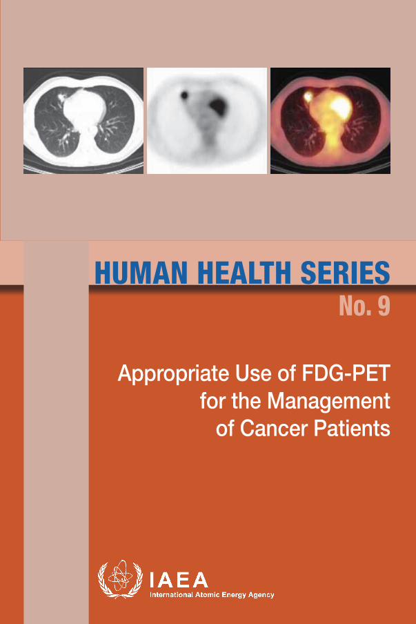
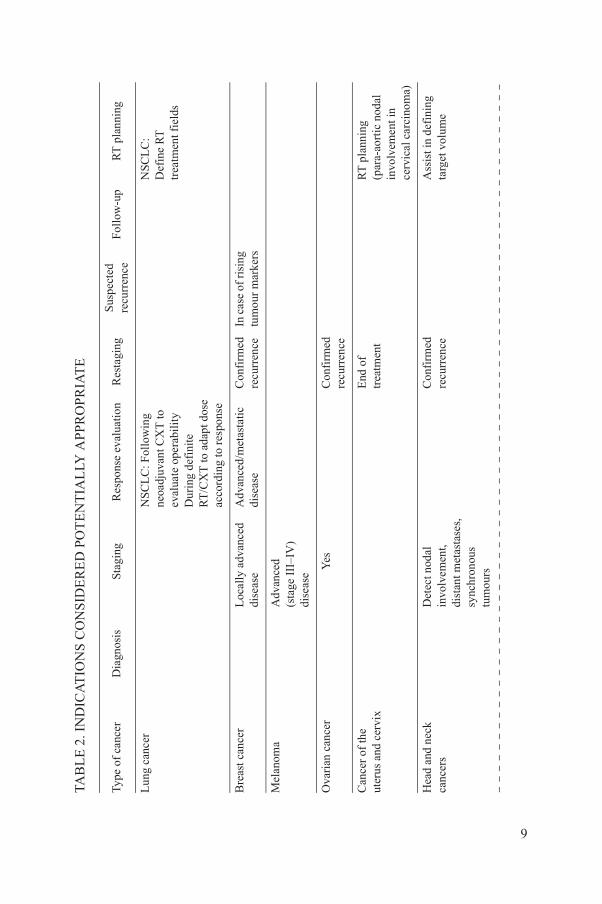
This book provides guidance on the value and appropriateness of the use of positron emission tomography (PET), either alone or in combination with computed tomography (CT) scanners using 2-fluoro-2-deoxy-D-glucose (FDG) labelled with fluorine-18, in the management of patients affected by cancer. The concept of appropriateness provides a tool for determining which diagnostic investigations and therapies should be implemented, with the overall aim of optimizing health resource allocation, recognizing not only the cost of the intervention but also the consequences of failure to implement innovations of proven effectiveness. The book includes clinical scenarios for FDG-PET/CT indications; in all, 21 different types of cancer are considered, with seven different possible indications each.
IAEA HumAn HEAltH SErIES
Appropriate U
se of FDG
-PET for the M
anagement of C
ancer Patients
IAEA HumAn HEAltH SErIES no. 9
10-06881_P1438_cover.indd 1 2010-04-22 11:25:23
IAEA HUMAN HEALTH SERIES PUBLICATIONS
The mandate of the IAEA human health programme originates from Article II of its Statute, which states that the “Agency shall seek to accelerate and enlarge the contribution of atomic energy to peace, health and prosperity throughout the world”. The main objective of the human health programme is to enhance the capabilities of IAEA Member States in addressing issues related to the prevention, diagnosis and treatment of health problems through the development and application of nuclear techniques, within a framework of quality assurance.
Publications in the IAEA Human Health Series provide information in the areas of: radiation medicine, including diagnostic radiology, diagnostic and therapeutic nuclear medicine, and radiation therapy; dosimetry and medical radiation physics; and stable isotope techniques and other nuclear applications in nutrition. The publications have a broad readership and are aimed at medical practitioners, researchers and other professionals. International experts assist the IAEA Secretariat in drafting and reviewing these publications. Some of the publications in this series may also be endorsed or co-sponsored by international organizations and professional societies active in the relevant fields. There are two categories of publications in this series:
IAEA HUMAN HEALTH SERIESPublications in this category present analyses or provide information of an
advisory nature, for example guidelines, codes and standards of practice, and quality assurance manuals. Monographs and high level educational material, such as graduate texts, are also published in this series.
IAEA HUMAN HEALTH REPORTSHuman Health Reports complement information published in the IAEA Human
Health Series in areas of radiation medicine, dosimetry and medical radiation physics, and nutrition. These publications include reports of technical meetings, the results of IAEA coordinated research projects, interim reports on IAEA projects, and educational material compiled for IAEA training courses dealing with human health related subjects. In some cases, these reports may provide supporting material relating to publications issued in the IAEA Human Health Series.
All of these publications can be downloaded cost free from the IAEA web site:http://www.iaea.org/Publications/index.html
Further information is available from:Marketing and Sales UnitInternational Atomic Energy AgencyVienna International CentrePO Box 1001400 Vienna, Austria
Readers are invited to provide their impressions on these publications. Information may be provided via the IAEA web site, by mail at the address given above, or by email to:
RELATED PUBLICATIONS
www.iaea.org/books
A Guide to clinicAl pet in oncoloGy: improvinG clinicAl mAnAGement of cAncer pAtientsiAeA tecdoc series no. 1605IAEA-TECDOC-1605 (58 pp; 2008)ISBN 978–92–0–110608–7 Price: €15.00
the role of pet/ct in rAdiAtion treAtment plAnninG for cAncer pAtient treAtmentiAeA tecdoc series no. 1603IAEA-TECDOC-1603 (40 pp.; 2008)ISBN 978–92–0–110408–3 Price: €15.00
QuAlity mAnAGement Audits in nucleAr medicine prActicesSTI/PUB/1371 (57 pp.; 2008)ISBN 978–92–0–112108–0 Price: €25.00
strAteGies for clinicAl implementAtion And QuAlity mAnAGement of pet trAcersSTI/PUB/1344 (197 pp.; 2009)ISBN 978–92–0–107008–1 Price: €44.00
operAtionAl GuidAnce on hospitAl rAdiophArmAcy: A sAfe And effective ApproAchSTI/PUB/1342 (75 pp.; 2008)ISBN 978–92–0–106708–1 Price: €32.00
QuAlity AssurAnce for pet And pet/ct systemsiAeA human health series no. 1STI/PUB/1393 (145 pp.; 2009)ISBN 978–92–0–103609–4 Price: €32.00
rAdiAtion protection in newer medicAl imAGinG techniQues: pet/ctsafety reports series no. 58STI/PUB/1343 (54 pp.; 2008)ISBN 978–92–0–106808–8 Price: €28.00
cyclotron produced rAdionuclides: Guidelines for settinG up A fAcilitytechnical reports series no. 471STI/DOC/010/471 (213 pp.; 2009)ISBN 978–92–0–103109–9 Price: €45.00
plAnninG A clinicAl pet centreiAeA human health series no. 11STI/PUB/1457 (140 pp; 2010)ISBN 978–92–0–104610–9 Price: €42.00
10-06881_P1438_cover.indd 2 2010-04-22 11:25:23

APPROPRIATE USE OF FDG-PETFOR THE MANAGEMENT OF
CANCER PATIENTS

The following States are Members of the International Atomic Energy Agency:
AFGHANISTANALBANIAALGERIAANGOLAARGENTINAARMENIAAUSTRALIAAUSTRIAAZERBAIJANBAHRAINBANGLADESHBELARUSBELGIUMBELIZEBENINBOLIVIABOSNIA AND HERZEGOVINABOTSWANABRAZILBULGARIABURKINA FASOBURUNDICAMBODIACAMEROONCANADACENTRAL AFRICAN
REPUBLICCHADCHILECHINACOLOMBIACONGOCOSTA RICACÔTE D’IVOIRECROATIACUBACYPRUSCZECH REPUBLICDEMOCRATIC REPUBLIC
OF THE CONGODENMARKDOMINICAN REPUBLICECUADOREGYPTEL SALVADOR
GHANAGREECEGUATEMALAHAITIHOLY SEEHONDURASHUNGARYICELANDINDIAINDONESIAIRAN, ISLAMIC REPUBLIC OF IRAQIRELANDISRAELITALYJAMAICAJAPANJORDANKAZAKHSTANKENYAKOREA, REPUBLIC OFKUWAITKYRGYZSTANLATVIALEBANONLESOTHOLIBERIALIBYAN ARAB JAMAHIRIYALIECHTENSTEINLITHUANIALUXEMBOURGMADAGASCARMALAWIMALAYSIAMALIMALTAMARSHALL ISLANDSMAURITANIAMAURITIUSMEXICOMONACOMONGOLIAMONTENEGROMOROCCOMOZAMBIQUE
NORWAYOMANPAKISTANPALAUPANAMAPARAGUAYPERUPHILIPPINESPOLANDPORTUGALQATARREPUBLIC OF MOLDOVAROMANIARUSSIAN FEDERATIONSAUDI ARABIASENEGALSERBIASEYCHELLESSIERRA LEONESINGAPORESLOVAKIASLOVENIASOUTH AFRICASPAINSRI LANKASUDANSWEDENSWITZERLANDSYRIAN ARAB REPUBLICTAJIKISTANTHAILANDTHE FORMER YUGOSLAV
REPUBLIC OF MACEDONIATUNISIATURKEYUGANDAUKRAINEUNITED ARAB EMIRATESUNITED KINGDOM OF
GREAT BRITAIN AND NORTHERN IRELAND
UNITED REPUBLIC OF TANZANIA
The Agency’s Statute was approved on 23 October 1956 by the Conference on the Statute of theIAEA held at United Nations Headquarters, New York; it entered into force on 29 July 1957. TheHeadquarters of the Agency are situated in Vienna. Its principal objective is “to accelerate and enlarge thecontribution of atomic energy to peace, health and prosperity throughout the world’’.
ERITREAESTONIAETHIOPIAFINLANDFRANCEGABONGEORGIAGERMANY
MYANMARNAMIBIANEPAL NETHERLANDSNEW ZEALANDNICARAGUANIGERNIGERIA
UNITED STATES OF AMERICAURUGUAYUZBEKISTANVENEZUELAVIETNAMYEMENZAMBIAZIMBABWE
IAEA HUMAN HEALTH SERIES No. 9
APPROPRIATE USE OF FDG-PETFOR THE MANAGEMENT OF
CANCER PATIENTS
INTERNATIONAL ATOMIC ENERGY AGENCYVIENNA, 2010

IAEA Library Cataloguing in Publication Data
Appropriate use of FDG-PET for the management of cancer patients. — Vienna :International Atomic Energy Agency, 2010.
p. ; 24 cm. — (IAEA human health series, ISSN 2075–3772 ; no. 9)STI/PUB/1438
COPYRIGHT NOTICE
All IAEA scientific and technical publications are protected by the terms of the Universal Copyright Convention as adopted in 1952 (Berne) and as revised in 1972 (Paris). The copyright has since been extended by the World Intellectual Property Organization (Geneva) to include electronic and virtual intellectual property. Permission to use whole or parts of texts contained in IAEA publications in printed or electronic form must be obtained and is usually subject to royalty agreements. Proposals for non-commercial reproductions and translations are welcomed and considered on a case-by-case basis. Enquiries should be addressed to the IAEA Publishing Section at:
Marketing and Sales Unit, Publishing SectionInternational Atomic Energy AgencyVienna International CentrePO Box 1001400 Vienna, Austriafax: +43 1 2600 29302tel.: +43 1 2600 22417email: [email protected] http://www.iaea.org/books
© IAEA, 2010
Printed by the IAEA in AustriaApril 2010
STI/PUB/1438
ISBN 978–92–0–101610–2Includes bibliographical references.
1. Cancer — Patients — Treatment. 2. Tomography, Emission — Methodology. 3. Positrons — Emission. I. International Atomic Energy Agency. II. Series.
IAEAL 10–00631
FOREWORD
The global incidence of cancer is increasing in both developed and developing countries and will become a heavy health burden in the coming decade. This increase in the cancer rate will bring with it challenges for health care systems, clinicians, and patients and their families. Technologies that improve the decision making process and optimize treatment have the potential to benefit society as a whole.
The purpose of this publication is to develop a consensus based on evidence from existing systematic reviews, to make health care providers aware of the value and the appropriateness of the introduction of positron emission tomography (PET), either alone or in combination with computed tomography (PET/CT) using 2-fluoro-2-deoxy-D-glucose (FDG) labelled with 18F, in the management of patients affected by cancer.
Although the concept of appropriateness has been defined in terms of clinical utility, it may also be used to assist in the allocation of limited resources in an environment of shrinking health budgets. There is, however, the danger that new interventions will be underutilized, because they are viewed by health care administrators as inappropriate. This could be due to a narrow interpretation of appropriateness that is based solely on the cost of the intervention, isolated from the potential cost savings derived from its use. In reality, therefore, there might be a series of interventions, services and health services of proven effectiveness whose necessary implementation requires an increase in costs, at least in the short and medium terms.
Thus, if decision makers are to rely only on appropriateness criteria in decisions to fund health services, they must accept that the main aim of appropriateness is the optimization of resource allocation and not simply the reduction of costs. Therefore they must also focus on the inappropriateness of failing to introduce innovations of proven effectiveness.
While the use of PET is well established and integrated into oncological practice in many developed countries, it is limited or absent in many developing countries. Based on these considerations, the IAEA recognizes the need to make reliable information widely available to support Member States in the use of PET scanning. Within the Asia–Pacific region, the IAEA has initiated technical cooperation projects addressing the technical aspects and quality assurance of PET scanning, and aimed at identifying the indications for PET scanning most
likely to provide the greatest benefit to both individual patients and the health system.The regional project on Strengthening Clinical Applications of PET in RCA Member States (RAS/6/049), under the Regional Co-operative Agreement for Research, Development and Training Related to Nuclear Science and Technology
(RCA) programme, was formulated to address this need in the Asia–Pacific region. As an integral component of this project, the IAEA convened an expert consultant group to consider the available systematic reviews and to draft a list of indications for PET scanning. The expert consultant group was also requested to consider specific issues that may affect the utility of PET scanning in the Asia–Pacific region.
The recommendations included here have been written and approved by the IAEA to promote the optimal use of FDG-PET imaging procedures. These broad recommendations cannot be rigidly applied to all patients in all clinical settings. This publication represents the state of knowledge at the time of writing regarding the utility of FDG-PET in the treatment of cancers that are common in the Asia–Pacific region. Since FDG-PET is a rapidly evolving technology, this report will require periodic updating, and readers are advised to seek the most recent reports pertinent to this particular area.
The IAEA officers responsible for this publication were M. Dondi of the Division of Human Health and M.P. Dias of the Division for Asia and the Pacific.
EDITORIAL NOTE
Although great care has been taken to maintain the accuracy of information contained in this publication, neither the IAEA nor its Member States assume any responsibility for consequences which may arise from its use.
The use of particular designations of countries or territories does not imply any judgement by the publisher, the IAEA, as to the legal status of such countries or territories, of
their authorities and institutions or of the delimitation of their boundaries.The mention of names of specific companies or products (whether or not indicated as registered) does not imply any intention to infringe proprietary rights, nor should it be construed as an endorsement or recommendation on the part of the IAEA.
The IAEA has no responsibility for the persistence or accuracy of URLs for external or third party Internet web sites referred to in this book and does not guarantee that any content on such web sites is, or will remain, accurate or appropriate.
CONTENTS
1. INTRODUCTION . . . . . . . . . . . . . . . . . . . . . . . . . . . . . . . . . . . . . . . . 1
1.1. Background . . . . . . . . . . . . . . . . . . . . . . . . . . . . . . . . . . . . . . . . 11.2. Objective . . . . . . . . . . . . . . . . . . . . . . . . . . . . . . . . . . . . . . . . . . 11.3. Search strategy . . . . . . . . . . . . . . . . . . . . . . . . . . . . . . . . . . . . . . 21.4. Definitions of the appropriateness criteria for the use of PET . . . 21.5. Definitions of indications for PET scanning . . . . . . . . . . . . . . . 31.6. Structure . . . . . . . . . . . . . . . . . . . . . . . . . . . . . . . . . . . . . . . . . . . 4
2. CLINICAL SCENARIOS FOR FDG-PET/CT INDICATIONS . . . . 5
2.1. Summary of results . . . . . . . . . . . . . . . . . . . . . . . . . . . . . . . . . . 5
3. NON-SMALL CELL LUNG CANCER (NSCLC) . . . . . . . . . . . . . . 16
3.1. Diagnosis . . . . . . . . . . . . . . . . . . . . . . . . . . . . . . . . . . . . . . . . . . 163.2. Staging . . . . . . . . . . . . . . . . . . . . . . . . . . . . . . . . . . . . . . . . . . . . 163.3. Response evaluation . . . . . . . . . . . . . . . . . . . . . . . . . . . . . . . . . 173.4. Restaging . . . . . . . . . . . . . . . . . . . . . . . . . . . . . . . . . . . . . . . . . . 183.5. Suspected recurrence . . . . . . . . . . . . . . . . . . . . . . . . . . . . . . . . 183.6. Follow-up . . . . . . . . . . . . . . . . . . . . . . . . . . . . . . . . . . . . . . . . . 183.7. RT planning . . . . . . . . . . . . . . . . . . . . . . . . . . . . . . . . . . . . . . . . 18
4. SMALL CELL LUNG CANCER (SCLC) . . . . . . . . . . . . . . . . . . . . . 20
4.1. Diagnosis . . . . . . . . . . . . . . . . . . . . . . . . . . . . . . . . . . . . . . . . . . 204.2. Staging . . . . . . . . . . . . . . . . . . . . . . . . . . . . . . . . . . . . . . . . . . . 204.3. Response evaluation . . . . . . . . . . . . . . . . . . . . . . . . . . . . . . . . . . 204.4. Restaging . . . . . . . . . . . . . . . . . . . . . . . . . . . . . . . . . . . . . . . . . . 204.5. Suspected recurrence . . . . . . . . . . . . . . . . . . . . . . . . . . . . . . . . 214.6. Follow-up . . . . . . . . . . . . . . . . . . . . . . . . . . . . . . . . . . . . . . . . . 214.7. RT planning . . . . . . . . . . . . . . . . . . . . . . . . . . . . . . . . . . . . . . . . 21
5. LYMPHOMA . . . . . . . . . . . . . . . . . . . . . . . . . . . . . . . . . . . . . . . . . . . 22
5.1. Diagnosis . . . . . . . . . . . . . . . . . . . . . . . . . . . . . . . . . . . . . . . . . . 225.2. Staging . . . . . . . . . . . . . . . . . . . . . . . . . . . . . . . . . . . . . . . . . . . . 225.3. Response evaluation . . . . . . . . . . . . . . . . . . . . . . . . . . . . . . . . . . 22
5.4. Restaging . . . . . . . . . . . . . . . . . . . . . . . . . . . . . . . . . . . . . . . . . . 235.5. Suspected recurrence . . . . . . . . . . . . . . . . . . . . . . . . . . . . . . . . . 235.6. Follow-up . . . . . . . . . . . . . . . . . . . . . . . . . . . . . . . . . . . . . . . . . . 235.7. RT planning . . . . . . . . . . . . . . . . . . . . . . . . . . . . . . . . . . . . . . . . 23
6. BREAST CANCER . . . . . . . . . . . . . . . . . . . . . . . . . . . . . . . . . . . . . . 25
6.1. Diagnosis . . . . . . . . . . . . . . . . . . . . . . . . . . . . . . . . . . . . . . . . . . 256.2. Staging . . . . . . . . . . . . . . . . . . . . . . . . . . . . . . . . . . . . . . . . . . . . 256.3. Response evaluation . . . . . . . . . . . . . . . . . . . . . . . . . . . . . . . . . . 266.4. Restaging . . . . . . . . . . . . . . . . . . . . . . . . . . . . . . . . . . . . . . . . . . 266.5. Suspected recurrence . . . . . . . . . . . . . . . . . . . . . . . . . . . . . . . . . 266.6. Follow-up . . . . . . . . . . . . . . . . . . . . . . . . . . . . . . . . . . . . . . . . . . 276.7. RT planning . . . . . . . . . . . . . . . . . . . . . . . . . . . . . . . . . . . . . . . . 27
7. MELANOMA . . . . . . . . . . . . . . . . . . . . . . . . . . . . . . . . . . . . . . . . . . . 28
7.1. Diagnosis . . . . . . . . . . . . . . . . . . . . . . . . . . . . . . . . . . . . . . . . . . 287.2. Staging . . . . . . . . . . . . . . . . . . . . . . . . . . . . . . . . . . . . . . . . . . . . 287.3. Response evaluation . . . . . . . . . . . . . . . . . . . . . . . . . . . . . . . . . . 297.4. Restaging . . . . . . . . . . . . . . . . . . . . . . . . . . . . . . . . . . . . . . . . . . 297.5. Suspected recurrence . . . . . . . . . . . . . . . . . . . . . . . . . . . . . . . . . 297.6. Follow-up . . . . . . . . . . . . . . . . . . . . . . . . . . . . . . . . . . . . . . . . . . 307.7. RT planning . . . . . . . . . . . . . . . . . . . . . . . . . . . . . . . . . . . . . . . . 30
8. OVARIAN CANCER . . . . . . . . . . . . . . . . . . . . . . . . . . . . . . . . . . . . 31
8.1. Diagnosis . . . . . . . . . . . . . . . . . . . . . . . . . . . . . . . . . . . . . . . . . . 318.2. Staging . . . . . . . . . . . . . . . . . . . . . . . . . . . . . . . . . . . . . . . . . . . . 318.3. Response evaluation . . . . . . . . . . . . . . . . . . . . . . . . . . . . . . . . . . 318.4. Restaging . . . . . . . . . . . . . . . . . . . . . . . . . . . . . . . . . . . . . . . . . . 318.5. Suspected recurrence . . . . . . . . . . . . . . . . . . . . . . . . . . . . . . . . . 328.6. Follow-up . . . . . . . . . . . . . . . . . . . . . . . . . . . . . . . . . . . . . . . . . . 328.7. RT planning . . . . . . . . . . . . . . . . . . . . . . . . . . . . . . . . . . . . . . . . 32
9. CANCER OF THE UTERUS AND CERVIX . . . . . . . . . . . . . . . . . 34
9.1. Diagnosis . . . . . . . . . . . . . . . . . . . . . . . . . . . . . . . . . . . . . . . . . . 349.2. Staging . . . . . . . . . . . . . . . . . . . . . . . . . . . . . . . . . . . . . . . . . . . . 349.3. Response evaluation . . . . . . . . . . . . . . . . . . . . . . . . . . . . . . . . . . 349.4. Restaging . . . . . . . . . . . . . . . . . . . . . . . . . . . . . . . . . . . . . . . . . . 34
9.5. Suspected recurrence . . . . . . . . . . . . . . . . . . . . . . . . . . . . . . . . . 359.6. Follow-up . . . . . . . . . . . . . . . . . . . . . . . . . . . . . . . . . . . . . . . . . . 359.7. RT planning . . . . . . . . . . . . . . . . . . . . . . . . . . . . . . . . . . . . . . . . 35
10. HEAD AND NECK CANCERS . . . . . . . . . . . . . . . . . . . . . . . . . . . . . 37
10.1. Diagnosis . . . . . . . . . . . . . . . . . . . . . . . . . . . . . . . . . . . . . . . . . . 3710.2. Staging . . . . . . . . . . . . . . . . . . . . . . . . . . . . . . . . . . . . . . . . . . . . 3710.3. Response evaluation . . . . . . . . . . . . . . . . . . . . . . . . . . . . . . . . . . 3810.4. Restaging . . . . . . . . . . . . . . . . . . . . . . . . . . . . . . . . . . . . . . . . . . 3810.5. Suspected recurrence . . . . . . . . . . . . . . . . . . . . . . . . . . . . . . . . 3810.6. Follow-up . . . . . . . . . . . . . . . . . . . . . . . . . . . . . . . . . . . . . . . . . . 3910.7. RT planning . . . . . . . . . . . . . . . . . . . . . . . . . . . . . . . . . . . . . . . . 39
11. KIDNEY CANCER . . . . . . . . . . . . . . . . . . . . . . . . . . . . . . . . . . . . . . 40
11.1. Diagnosis . . . . . . . . . . . . . . . . . . . . . . . . . . . . . . . . . . . . . . . . . . 4011.2. Staging . . . . . . . . . . . . . . . . . . . . . . . . . . . . . . . . . . . . . . . . . . . . 4011.3. Response evaluation . . . . . . . . . . . . . . . . . . . . . . . . . . . . . . . . . . 4011.4. Restaging . . . . . . . . . . . . . . . . . . . . . . . . . . . . . . . . . . . . . . . . . . 4011.5. Suspected recurrence . . . . . . . . . . . . . . . . . . . . . . . . . . . . . . . . . 4111.6. Follow-up . . . . . . . . . . . . . . . . . . . . . . . . . . . . . . . . . . . . . . . . . . 4111.7. RT planning . . . . . . . . . . . . . . . . . . . . . . . . . . . . . . . . . . . . . . . . 41
12. GERMINAL TUMOURS . . . . . . . . . . . . . . . . . . . . . . . . . . . . . . . . . . 43
12.1. Diagnosis . . . . . . . . . . . . . . . . . . . . . . . . . . . . . . . . . . . . . . . . . . 4312.2. Staging . . . . . . . . . . . . . . . . . . . . . . . . . . . . . . . . . . . . . . . . . . . . 4312.3. Response evaluation . . . . . . . . . . . . . . . . . . . . . . . . . . . . . . . . . . 4312.4. Restaging . . . . . . . . . . . . . . . . . . . . . . . . . . . . . . . . . . . . . . . . . . 4312.5. Suspected recurrence . . . . . . . . . . . . . . . . . . . . . . . . . . . . . . . . . 4412.6. Follow-up . . . . . . . . . . . . . . . . . . . . . . . . . . . . . . . . . . . . . . . . . . 4412.7. RT planning . . . . . . . . . . . . . . . . . . . . . . . . . . . . . . . . . . . . . . . . 44
13. CANCER OF UNKNOWN PRIMARY (CUP) . . . . . . . . . . . . . . . . . 45
13.1. Diagnosis . . . . . . . . . . . . . . . . . . . . . . . . . . . . . . . . . . . . . . . . . . 4513.2. Staging . . . . . . . . . . . . . . . . . . . . . . . . . . . . . . . . . . . . . . . . . . . . 4513.3. Response evaluation . . . . . . . . . . . . . . . . . . . . . . . . . . . . . . . . . . 4513.4. Restaging . . . . . . . . . . . . . . . . . . . . . . . . . . . . . . . . . . . . . . . . . . 4513.5. Suspected recurrence . . . . . . . . . . . . . . . . . . . . . . . . . . . . . . . . . 46
13.6. Follow-up . . . . . . . . . . . . . . . . . . . . . . . . . . . . . . . . . . . . . . . . . . 4613.7. RT planning . . . . . . . . . . . . . . . . . . . . . . . . . . . . . . . . . . . . . . . . 46
14. COLORECTAL CANCER . . . . . . . . . . . . . . . . . . . . . . . . . . . . . . . . . 47
14.1. Diagnosis . . . . . . . . . . . . . . . . . . . . . . . . . . . . . . . . . . . . . . . . . . 4714.2. Staging . . . . . . . . . . . . . . . . . . . . . . . . . . . . . . . . . . . . . . . . . . . . 4714.3. Response evaluation . . . . . . . . . . . . . . . . . . . . . . . . . . . . . . . . . . 4714.4. Restaging . . . . . . . . . . . . . . . . . . . . . . . . . . . . . . . . . . . . . . . . . . 4714.5. Suspected recurrence . . . . . . . . . . . . . . . . . . . . . . . . . . . . . . . . . 4814.6. Follow-up . . . . . . . . . . . . . . . . . . . . . . . . . . . . . . . . . . . . . . . . . . 4814.7. RT planning . . . . . . . . . . . . . . . . . . . . . . . . . . . . . . . . . . . . . . . . 48
15. GASTRIC CARCINOMA . . . . . . . . . . . . . . . . . . . . . . . . . . . . . . . . . 50
15.1. Diagnosis . . . . . . . . . . . . . . . . . . . . . . . . . . . . . . . . . . . . . . . . . . 5015.2. Staging . . . . . . . . . . . . . . . . . . . . . . . . . . . . . . . . . . . . . . . . . . . . 5015.3. Response evaluation . . . . . . . . . . . . . . . . . . . . . . . . . . . . . . . . . . 5015.4. Restaging . . . . . . . . . . . . . . . . . . . . . . . . . . . . . . . . . . . . . . . . . . 5115.5. Suspected recurrence . . . . . . . . . . . . . . . . . . . . . . . . . . . . . . . . . 5115.6. Follow-up . . . . . . . . . . . . . . . . . . . . . . . . . . . . . . . . . . . . . . . . . . 5115.7. RT planning . . . . . . . . . . . . . . . . . . . . . . . . . . . . . . . . . . . . . . . . 51
16. SARCOMAS (SOFT TISSUE AND BONE) . . . . . . . . . . . . . . . . . . . 53
16.1. Diagnosis . . . . . . . . . . . . . . . . . . . . . . . . . . . . . . . . . . . . . . . . . . 5316.2. Staging . . . . . . . . . . . . . . . . . . . . . . . . . . . . . . . . . . . . . . . . . . . . 5316.3. Response evaluation . . . . . . . . . . . . . . . . . . . . . . . . . . . . . . . . . . 5316.4. Restaging . . . . . . . . . . . . . . . . . . . . . . . . . . . . . . . . . . . . . . . . . . 5416.5. Suspected recurrence . . . . . . . . . . . . . . . . . . . . . . . . . . . . . . . . . 5416.6. Follow-up . . . . . . . . . . . . . . . . . . . . . . . . . . . . . . . . . . . . . . . . . . 5416.7. RT planning . . . . . . . . . . . . . . . . . . . . . . . . . . . . . . . . . . . . . . . . 54
17. PRIMARY TUMOURS OF THE CENTRAL NERVOUSSYSTEM . . . . . . . . . . . . . . . . . . . . . . . . . . . . . . . . . . . . . . . . . . . . . . . 56
17.1. Diagnosis . . . . . . . . . . . . . . . . . . . . . . . . . . . . . . . . . . . . . . . . . . 5617.2. Staging . . . . . . . . . . . . . . . . . . . . . . . . . . . . . . . . . . . . . . . . . . . . 5617.3. Response evaluation . . . . . . . . . . . . . . . . . . . . . . . . . . . . . . . . . . 5617.4. Restaging . . . . . . . . . . . . . . . . . . . . . . . . . . . . . . . . . . . . . . . . . . 5717.5. Suspected recurrence . . . . . . . . . . . . . . . . . . . . . . . . . . . . . . . . . 57
17.6. Follow-up . . . . . . . . . . . . . . . . . . . . . . . . . . . . . . . . . . . . . . . . . . 5717.7. RT planning . . . . . . . . . . . . . . . . . . . . . . . . . . . . . . . . . . . . . . . . 58
18. NASOPHARYNGEAL CARCINOMAS . . . . . . . . . . . . . . . . . . . . . . 59
18.1. Diagnosis . . . . . . . . . . . . . . . . . . . . . . . . . . . . . . . . . . . . . . . . . . 5918.2. Staging . . . . . . . . . . . . . . . . . . . . . . . . . . . . . . . . . . . . . . . . . . . . 5918.3. Response evaluation . . . . . . . . . . . . . . . . . . . . . . . . . . . . . . . . . . 5918.4. Restaging . . . . . . . . . . . . . . . . . . . . . . . . . . . . . . . . . . . . . . . . . . 5918.5. Suspected recurrence . . . . . . . . . . . . . . . . . . . . . . . . . . . . . . . . . 6018.6. Follow-up . . . . . . . . . . . . . . . . . . . . . . . . . . . . . . . . . . . . . . . . . . 6018.7. RT planning . . . . . . . . . . . . . . . . . . . . . . . . . . . . . . . . . . . . . . . . 60
19. GASTROINTESTINAL STROMAL TUMOURS (GISTS) . . . . . . . 62
19.1. Diagnosis . . . . . . . . . . . . . . . . . . . . . . . . . . . . . . . . . . . . . . . . . . 6219.2. Staging . . . . . . . . . . . . . . . . . . . . . . . . . . . . . . . . . . . . . . . . . . . . 6219.3. Response evaluation . . . . . . . . . . . . . . . . . . . . . . . . . . . . . . . . . . 6219.4. Restaging . . . . . . . . . . . . . . . . . . . . . . . . . . . . . . . . . . . . . . . . . . 6219.5. Suspected recurrence . . . . . . . . . . . . . . . . . . . . . . . . . . . . . . . . 6319.6. Follow-up . . . . . . . . . . . . . . . . . . . . . . . . . . . . . . . . . . . . . . . . . . 6319.7. RT planning . . . . . . . . . . . . . . . . . . . . . . . . . . . . . . . . . . . . . . . . 63
20. PANCREATIC ADENOCARCINOMA . . . . . . . . . . . . . . . . . . . . . . . 64
20.1. Diagnosis . . . . . . . . . . . . . . . . . . . . . . . . . . . . . . . . . . . . . . . . . . 6420.2. Staging . . . . . . . . . . . . . . . . . . . . . . . . . . . . . . . . . . . . . . . . . . . . 6420.3. Response evaluation . . . . . . . . . . . . . . . . . . . . . . . . . . . . . . . . . . 6420.4. Restaging . . . . . . . . . . . . . . . . . . . . . . . . . . . . . . . . . . . . . . . . . . 6420.5. Suspected recurrence . . . . . . . . . . . . . . . . . . . . . . . . . . . . . . . . . 6520.6. Follow-up . . . . . . . . . . . . . . . . . . . . . . . . . . . . . . . . . . . . . . . . . . 6520.7. RT planning . . . . . . . . . . . . . . . . . . . . . . . . . . . . . . . . . . . . . . . . 65
21. CHOLANGIO- AND GALLBLADDER CARCINOMAS . . . . . . . . 67
21.1. Diagnosis . . . . . . . . . . . . . . . . . . . . . . . . . . . . . . . . . . . . . . . . . . 67
21.2. Staging . . . . . . . . . . . . . . . . . . . . . . . . . . . . . . . . . . . . . . . . . . . . 6721.3. Response evaluation . . . . . . . . . . . . . . . . . . . . . . . . . . . . . . . . . . 6721.4. Restaging . . . . . . . . . . . . . . . . . . . . . . . . . . . . . . . . . . . . . . . . . . 6721.5. Suspected recurrence . . . . . . . . . . . . . . . . . . . . . . . . . . . . . . . . . 68
21.6. Follow-up . . . . . . . . . . . . . . . . . . . . . . . . . . . . . . . . . . . . . . . . . . 6821.7. RT planning . . . . . . . . . . . . . . . . . . . . . . . . . . . . . . . . . . . . . . . . 68
22. OESOPHAGEAL CANCER . . . . . . . . . . . . . . . . . . . . . . . . . . . . . . . 70
22.1. Diagnosis . . . . . . . . . . . . . . . . . . . . . . . . . . . . . . . . . . . . . . . . . . 7022.2. Staging . . . . . . . . . . . . . . . . . . . . . . . . . . . . . . . . . . . . . . . . . . . . 7022.3. Response evaluation . . . . . . . . . . . . . . . . . . . . . . . . . . . . . . . . . . 7022.4. Restaging . . . . . . . . . . . . . . . . . . . . . . . . . . . . . . . . . . . . . . . . . . 7122.5. Suspected recurrence . . . . . . . . . . . . . . . . . . . . . . . . . . . . . . . . . 7122.6. Follow-up . . . . . . . . . . . . . . . . . . . . . . . . . . . . . . . . . . . . . . . . . . 7122.7. RT planning . . . . . . . . . . . . . . . . . . . . . . . . . . . . . . . . . . . . . . . . 71
23. THYROID CANCER . . . . . . . . . . . . . . . . . . . . . . . . . . . . . . . . . . . . . 73
23.1. Diagnosis . . . . . . . . . . . . . . . . . . . . . . . . . . . . . . . . . . . . . . . . . . 7323.2. Staging . . . . . . . . . . . . . . . . . . . . . . . . . . . . . . . . . . . . . . . . . . . . 7323.3. Response evaluation . . . . . . . . . . . . . . . . . . . . . . . . . . . . . . . . . . 7323.4. Restaging and suspected recurrence . . . . . . . . . . . . . . . . . . . . . 7323.5. Follow-up . . . . . . . . . . . . . . . . . . . . . . . . . . . . . . . . . . . . . . . . . . 7423.6. RT planning . . . . . . . . . . . . . . . . . . . . . . . . . . . . . . . . . . . . . . . . 74
CONTRIBUTORS TO DRAFTING AND REVIEW . . . . . . . . . . . . . . . . . 75
1. INTRODUCTION
1.1. BACKGROUND
In the past decade, appropriateness has become a guiding principle to justify the introduction of new health care interventions, from the use of new drugs or new treatment modalities to the implementation of new diagnostic procedures. The concept of appropriateness, with a decision aid for its assessment, provides clinicians and funders with a tool to determine which diagnostic investigations and therapies should be implemented. In the context of diagnostic investigations, new investigations are deemed appropriate when the difference between the expected incremental information and the expected or possible adverse effects is sufficiently large that the investigation is warranted for the indication concerned. The decision tool for rating appropriateness includes a literature review and synthesis of the evidence according to designated indications.
Although the concept of appropriateness has been defined in terms of clinical utility, it may also be used to assist in the allocation of limited resources in an environment of shrinking health budgets. There is, however, the danger that new interventions will be underutilized, because they are viewed by health care administrators as inappropriate. This could be due to a narrow interpretation of appropriateness that is based solely on the cost of the intervention, isolated from the potential cost savings derived from its use. In reality, therefore, there might be a series of interventions, services and health services of proven effectiveness that are widely underutilized, whose necessary implementation requires, at least in the short and medium terms, an increase in costs.
Funding decision makers must accept that the main aim of appropriateness is not cost reduction, but rather optimization of health resource allocation, recognizing the consequences of failure to implement innovations of proven effectiveness. It is only through acceptance of this perspective that innovations of proven effectiveness will be introduced for the benefit of both individuals and society.
1.2. OBJECTIVE
1
The purpose of this publication is to develop a consensus based on evidence from existing systematic reviews, to make health care providers aware of the value and the appropriateness of the introduction of positron emission tomography (PET) or PET combined with computed tomography (PET/CT) using 2-fluoro-2-deoxy-D-glucose (FDG) labelled with 18F in the management of patients affected by cancer.

1.3. SEARCH STRATEGY
The search of the available scientific publications was initially confined to systematic reviews of PET scanning in oncology using full ring PET and/or PET/CT that were published prior to 2009. However, owing to the rapid recent improvements in PET technology, for indications not deemed ‘appropriate’ (see definition below) in the systematic reviews, a literature review of publications more recent than the current systematic review was undertaken, to determine whether more recent information changed the classification of appropriateness, as defined below.
1.4. DEFINITIONS OF THE APPROPRIATENESS CRITERIAFOR THE USE OF PET
The use of PET for clinical indications can be considered appropriate, potentially appropriate, possibly appropriate or inappropriate. The appropriateness criteria for the usefulness of PET are defined as follows:
Appropriate (all the conditions below must be met)
— There is evidence of improved diagnostic performance (higher sensitivity and specificity) compared with other current techniques.
— The information derived from the PET scan influences clinical practice.— The information derived from the PET scan has a plausible impact on the
patient’s outcome, either through adoption of more effective therapeutic strategies or through non-adoption of ineffective or harmful practices.
Potentially appropriate (potentially useful)
There is evidence of improved diagnostic performance (greater sensitivity and specificity) compared with other current techniques, but evidence of an impact on treatment and outcome is lacking.
Possibly appropriate (appropriateness not yet documented)
2
There is insufficient evidence for assessment, although there is a strong rationale for clinical benefit from PET.
Inappropriate
Improved accuracy of tumour staging will not alter management, or the performance of PET is poorer than that of other current techniques.
1.5. DEFINITIONS OF INDICATIONS FOR PET SCANNING
Seven different indications for PET scanning are considered here: diagnosis, staging, response evaluation, restaging, suspected recurrence, follow-up and radiotherapy (RT) planning. They are defined as follows:
Diagnosis
— Characterization of mass lesion: indication of whether a mass lesion is benign or malignant;
— PET guided biopsy: assistance in guiding biopsy to the region of a tumour with the highest metabolic activity, identified on the PET scan by the area(s) of highest FDG uptake;
— Detection of occult primary cancer (cancer of unknown primary site); — Raised tumour markers: determination of the presence of cancer;— Metastasis: determination of the primary site when metastases have been
detected.
Staging
Assessment of the extent of disease prior to initiation of treatment.
Response evaluation
Assessment of treatment response during or after therapy.
Restaging
Assessment of the extent of disease following initial therapy or when
3
recurrence has been confirmed.
Suspected recurrence
Assessment of the presence of cancer following clinical and/or biochemical suspicion of recurrence.

Follow-up
Surveillance in the absence of clinical evidence of recurrence.
RT planning
Aid in the placement of radiation fields (this assumes that there has been a decision to use RT).
1.6. STRUCTURE
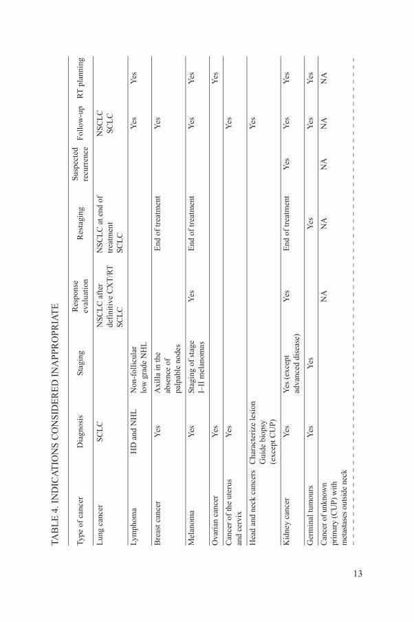
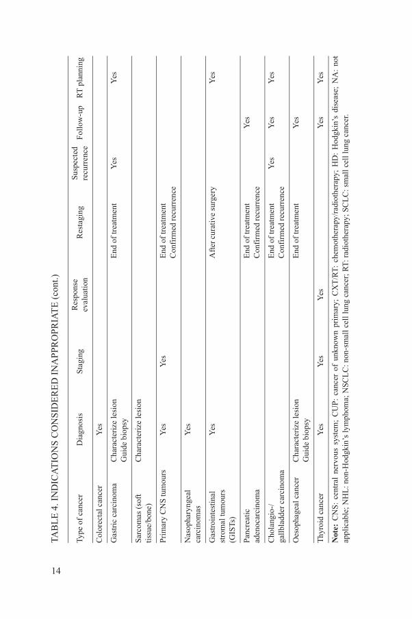
Indications for the use of FDG-PET/CT in the management of 21 types of cancer are outlined in Section 2 and presented in more detail in Sections 3–23. Seven different possible indications are considered for each type of cancer, with recommendations given as to the appropriateness of FDG-PET/CT for each indication.
4
2. CLINICAL SCENARIOSFOR FDG-PET/CT INDICATIONS
Overall, 21 different types of cancer are considered here, with seven different possible indications for each. It should be noted that the recommendations refer to ‘average individuals’. Specific clinical conditions may require the referring physician to take decisions that may differ from the evaluations included in this publication.
2.1. SUMMARY OF RESULTS
The following cancers have been considered:
(1) Non-small cell lung cancer (NSCLC)(2) Small cell lung cancer (SCLC)(3) Lymphoma (4) Breast cancer(5) Melanoma(6) Ovarian cancer(7) Cancer of the uterus and cervix(8) Head and neck cancers(9) Kidney cancer(10) Germinal tumours(11) Cancer of unknown primary (CUP)(12) Colorectal cancer(13) Gastric carcinoma(14) Sarcomas (soft tissue and bone)(15) Primary tumours of the central nervous system(16) Nasopharyngeal carcinomas(17) Gastrointestinal stromal tumours (GISTs)(18) Pancreatic adenocarcinoma(19) Cholangio- and gallbladder carcinomas(20) Oesophageal cancer
5
(21) Thyroid cancer.
Cancers for which FDG-PET has no established role, such as prostate and hepatocellular carcinoma, are not discussed in this publication. Also, as most gastro-entero-pancreatic tumours (GEPTs) and mucinous adenocarcinomas are not FDG avid, FDG-PET is usually inappropriate for them.
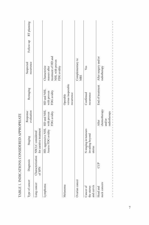
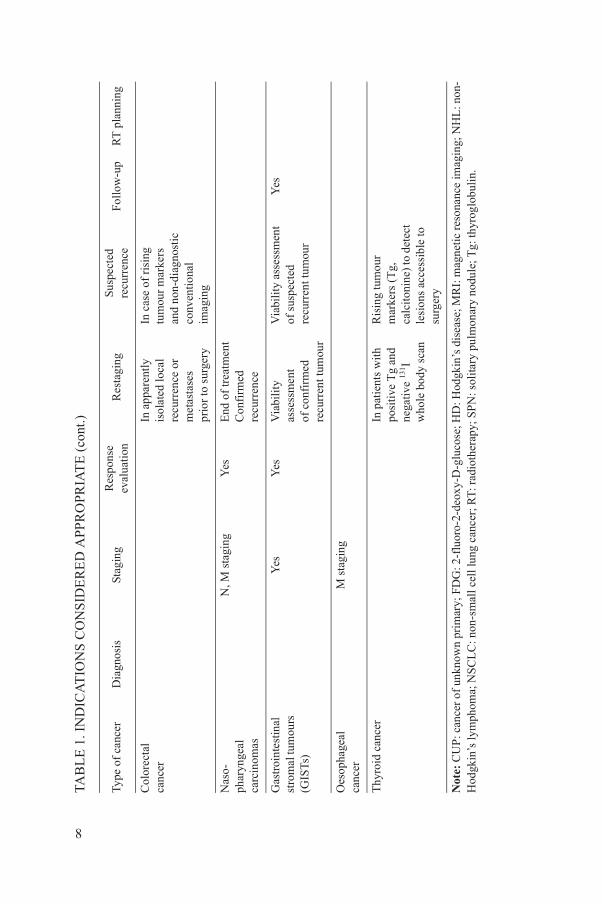
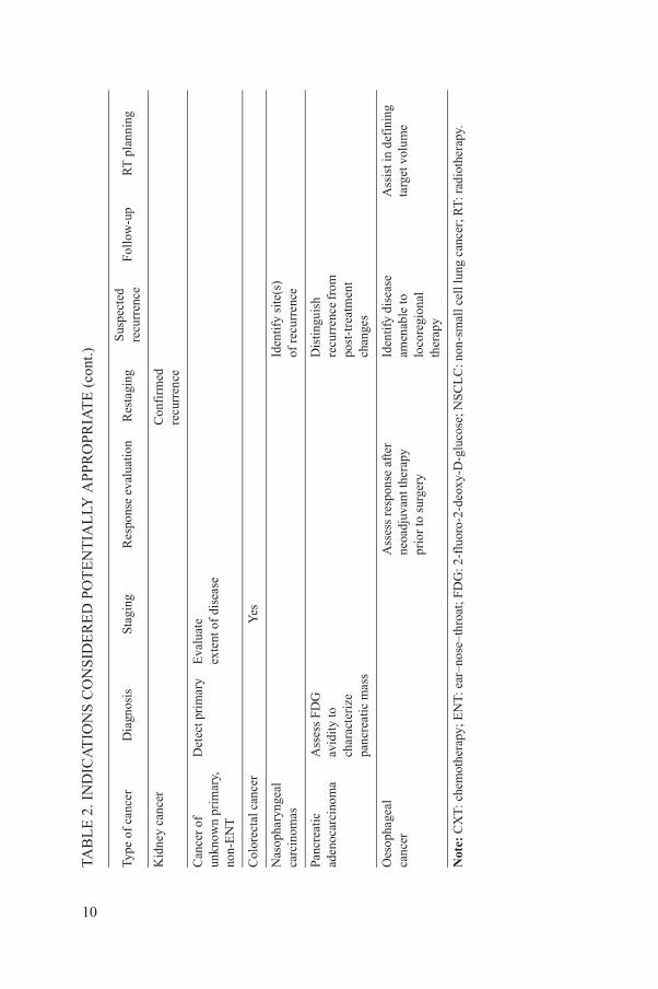
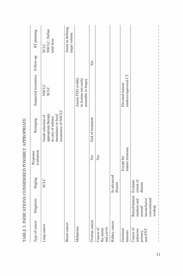
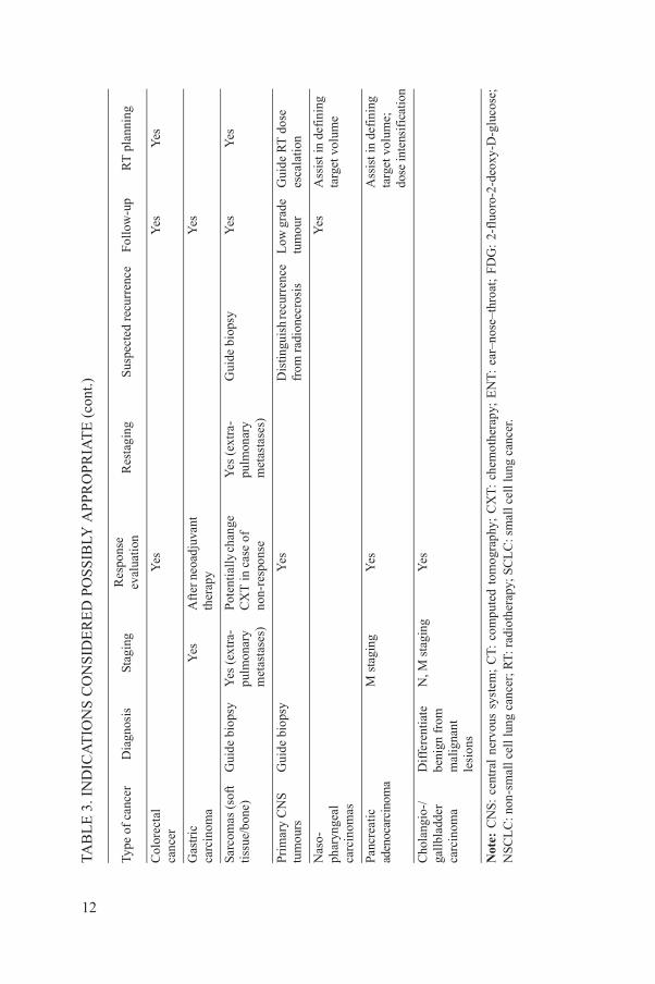
Tables 1–4 summarize clinical indications for which the use of FDG-PET is recognized as appropriate, potentially appropriate, possibly appropriate and inappropriate, respectively.
6
Text continues on p. 15.
AT
ION
S C
ON
SID
ER
ED
APP
RO
PRIA
TE
Dia
gnos
isSt
agin
gR
espo
nse
eval
uati
on
Res
tagi
ngSu
spec
ted
recu
rren
ceFo
llow
-up
RT
pla
nnin
g
arac
teri
zati
on
SP
NN
SCL
C c
onsi
dere
d fo
r cu
rativ
e tr
eatm
ent
HD
, agg
ress
ive
NH
LA
sses
s FD
G a
vidi
tyH
D a
nd N
HL
w
ith
prov
en
FD
G a
vidi
ty
HD
and
NH
L
wit
h pr
oven
FD
G a
vidi
ty
Cha
ract
eriz
em
asse
s af
ter
trea
tmen
t of
HD
and
N
HL
with
pro
ven
FD
G a
vidi
ty
Ope
rabl
eve
rsus
inop
erab
le
recu
rren
ce
Com
plem
enta
ry to
M
RI
N s
tagi
ng in
tum
ours
in
vadi
ng b
eyon
d ut
erus
Con
firm
ed
recu
rren
ceY
es
CU
PA
fter
ch
emot
hera
py
and/
or
radi
othe
rapy
End
of
trea
tmen
tA
fter
sur
gery
and
/or
radi
othe
rapy
7
TAB
LE
1. I
ND
IC
Type
of
canc
er
Lun
g ca
ncer
Ch
of
Lym
phom
a
Mel
anom
a
Ova
rian
can
cer
Can
cer
of
the
uter
us
and
cerv
ix
Hea
d an
dne
ck c
ance
rs
In a
ppar
entl
y is
olat
ed lo
cal
recu
rren
ce o
r m
etas
tase
spr
ior
to s
urge
ry
In c
ase
of r
isin
g tu
mou
r m
arke
rs
and
non-
diag
nost
ic
conv
entio
nal
imag
ing
N, M
sta
ging
Yes
End
of
trea
tmen
tC
onfi
rmed
re
curr
ence
Yes
Yes
Via
bili
ty
asse
ssm
ent
of c
onfi
rmed
re
curr
ent t
umou
r
Via
bili
ty a
sses
smen
t of
sus
pect
ed
recu
rren
t tum
our
Yes
M s
tagi
ng
In p
atie
nts
wit
h po
siti
ve T
g an
d ne
gativ
e 13
1 I w
hole
bod
y sc
an
Ris
ing
tum
our
mar
kers
(T
g,
calc
iton
ine)
to d
etec
t le
sion
s ac
cess
ible
to
surg
ery
f un
know
n pr
imar
y; F
DG
: 2-f
luor
o-2-
deox
y-D
-glu
cose
; HD
: Hod
gkin
’s d
isea
se; M
RI:
mag
neti
c re
sona
nce
imag
ing;
NH
L: n
on-
a; N
SC
LC
: non
-sm
all c
ell l
ung
canc
er; R
T: r
adio
ther
apy;
SP
N: s
olita
ry p
ulm
onar
y no
dule
; Tg:
thyr
oglo
buli
n.
AT
ION
S C
ON
SID
ER
ED
APP
RO
PRIA
TE
(co
nt.)
Dia
gnos
isSt
agin
gR
espo
nse
eval
uati
on
Res
tagi
ngSu
spec
ted
recu
rren
ceFo
llow
-up
RT
pla
nnin
g
8
Col
orec
tal
canc
er
Nas
o-ph
aryn
geal
ca
rcin
omas
Gas
troi
ntes
tinal
st
rom
al tu
mou
rs
(GIS
Ts)
Oes
opha
geal
ca
ncer
Thy
roid
can
cer
Not
e: C
UP
: can
cer
oH
odgk
in’s
lym
phom
TAB
LE
1. I
ND
IC
Type
of
canc
er
AT
ION
S C
ON
SID
ER
ED
PO
TE
NT
IAL
LY A
PPR
OP
RIA
TE
Dia
gnos
isSt
agin
gR
espo
nse
eval
uatio
nR
esta
ging
Sus
pect
ed
recu
rren
ceFo
llow
-up
RT
pla
nnin
g
NS
CL
C: F
ollo
win
g ne
oadj
uvan
t CX
T to
ev
alua
te o
pera
bili
tyD
urin
g de
fini
te
RT
/CX
T to
ada
pt d
ose
acco
rdin
g to
res
pons
e
NSC
LC
:D
efin
e R
T
trea
tmen
t fie
lds
Loc
ally
adv
ance
d di
seas
eA
dvan
ced/
met
asta
tic
dise
ase
Con
firm
ed
recu
rren
ceIn
cas
e of
risi
ng
tum
our
mar
kers
Adv
ance
d(s
tage
III
–IV
) di
seas
e
Yes
Con
firm
ed
recu
rren
ce
End
of
trea
tmen
tR
T p
lann
ing
(par
a-ao
rtic
nod
al
invo
lvem
ent i
n ce
rvic
al c
arci
nom
a)
Det
ect n
odal
in
volv
emen
t, di
stan
t met
asta
ses,
sy
nchr
onou
s tu
mou
rs
Con
firm
ed
recu
rren
ceA
ssis
t in
defi
ning
ta
rget
vol
ume
9
TAB
LE
2. I
ND
IC
Type
of
canc
er
Lun
g ca
ncer
Bre
ast c
ance
r
Mel
anom
a
Ova
rian
can
cer
Can
cer
of th
e ut
erus
and
cer
vix
Hea
d an
d ne
ck
canc
ers
Con
firm
ed
recu
rren
ce
Det
ect p
rim
ary
Eva
luat
eex
tent
of
dise
ase
Yes
Iden
tify
site
(s)
of r
ecur
renc
e
Ass
ess
FD
G
avid
ity to
ch
arac
teri
ze
panc
reat
ic m
ass
Dis
tingu
ish
recu
rren
ce fr
om
post
-tre
atm
ent
chan
ges
Ass
ess
resp
onse
aft
er
neoa
djuv
ant t
hera
py
prio
r to
sur
gery
Iden
tify
dise
ase
amen
able
to
loco
regi
onal
th
erap
y
Ass
ist i
n de
fini
ng
targ
et v
olum
e
erap
y; E
NT
: ear
–nos
e–th
roat
; FD
G: 2
-flu
oro-
2-de
oxy-
D-g
luco
se; N
SC
LC
: non
-sm
all c
ell l
ung
canc
er; R
T: r
adio
ther
apy.
AT
ION
S C
ON
SID
ER
ED
PO
TE
NT
IAL
LY A
PPR
OP
RIA
TE
(co
nt.)
Dia
gnos
isSt
agin
gR
espo
nse
eval
uatio
nR
esta
ging
Sus
pect
ed
recu
rren
ceFo
llow
-up
RT
pla
nnin
g
10
Kid
ney
canc
er
Can
cer
of
unkn
own
prim
ary,
no
n-E
NT
Col
orec
tal c
ance
r
Nas
opha
ryng
eal
carc
inom
as
Panc
reat
ic
aden
ocar
cino
ma
Oes
opha
geal
ca
ncer
Not
e: C
XT
: che
mot
h
TAB
LE
2. I
ND
IC
Type
of
canc
er
AT
ION
S C
ON
SID
ER
ED
PO
SS
IBLY
AP
PRO
PR
IAT
E
iagn
osis
Stag
ing
Res
pons
e ev
alua
tion
R
esta
ging
Sus
pect
ed r
ecur
renc
eFo
llow
-up
RT
pla
nnin
g
SC
LC
Gui
de s
elec
tion
of
appr
opri
ate
ther
apy
in c
ase
of s
olita
ry
met
asta
ses
or lo
cal
recu
rren
ce o
f N
SC
LC
NSC
LC
SC
LC
SC
LC
NS
CL
C: D
efin
e to
tal d
ose
Ass
ist i
n de
fini
ng
targ
et v
olum
e
Ass
ess
FD
G a
vidi
ty
in le
sion
s no
t eas
ily
amen
able
to b
iops
y
Yes
End
of
trea
tmen
tY
es
Yes
In a
dvan
ced
dise
ase
Exc
ept f
or
mat
ure
tera
tom
aE
leva
ted
tum
our
mar
kers
/equ
ivoc
al C
T
ised
tum
our
rker
s an
d m
al/
oncl
usiv
e ve
ntio
nal
rkup
Eva
luat
e ex
tent
of
dise
ase
11
TAB
LE
3. I
ND
IC
Type
of
canc
erD
Lun
g ca
ncer
Bre
ast c
ance
r
Mel
anom
a
Ova
rian
can
cer
Can
cer
of
the
uter
us
and
cerv
ix
Kid
ney
canc
er
Ger
min
al
tum
ours
Can
cer
of
unkn
own
prim
ary,
no
n-E
NT
Ra
ma
nor
inc
con
wo
Yes
Yes
Yes
Yes
Aft
er n
eoad
juva
nt
ther
apy
Yes
ide
biop
syY
es (
extr
a-pu
lmon
ary
met
asta
ses)
Pot
enti
ally
chan
ge
CX
T in
cas
e of
no
n-re
spon
se
Yes
(ex
tra-
pulm
onar
y m
etas
tase
s)
Gui
de b
iops
yY
esY
es
ide
biop
syY
esD
istin
guis
h re
curr
ence
fr
om r
adio
necr
osis
Low
gra
de
tum
our
Gui
de R
T d
ose
esca
lati
on
Yes
Ass
ist i
n de
fini
ng
targ
et v
olum
e
M s
tagi
ngY
esA
ssis
t in
defi
ning
ta
rget
vol
ume;
do
se in
tens
ific
atio
n
fere
ntia
te
ign
from
lig
nant
io
ns
N, M
sta
ging
Yes
nerv
ous
syst
em;
CT
: co
mpu
ted
tom
ogra
phy;
CX
T:
chem
othe
rapy
; E
NT
: ea
r–no
se–t
hroa
t; F
DG
: 2-
fluo
ro-2
-deo
xy-D
-glu
cose
; el
l lun
g ca
ncer
; RT
: rad
ioth
erap
y; S
CL
C: s
mal
l cel
l lun
g ca
ncer
.
AT
ION
S C
ON
SID
ER
ED
PO
SS
IBLY
AP
PRO
PR
IAT
E (
cont
.)
iagn
osis
Stag
ing
Res
pons
e ev
alua
tion
R
esta
ging
Sus
pect
ed r
ecur
renc
eFo
llow
-up
RT
pla
nnin
g
12
Col
orec
tal
canc
er
Gas
tric
ca
rcin
oma
Sarc
omas
(so
ft
tissu
e/bo
ne)
Gu
Prim
ary
CN
S tu
mou
rsG
u
Nas
o-ph
aryn
geal
ca
rcin
omas
Panc
reat
ic
aden
ocar
cino
ma
Cho
lang
io-/
ga
llbl
adde
r ca
rcin
oma
Dif
ben
ma
les
Not
e: C
NS:
cen
tral
N
SCL
C: n
on-s
mal
l c
TAB
LE
3. I
ND
IC
Type
of
canc
erD
AT
ION
S C
ON
SID
ER
ED
IN
AP
PRO
PR
IAT
E
Dia
gnos
isSt
agin
gR
espo
nse
eval
uati
on
Res
tagi
ngSu
spec
ted
recu
rren
ceF
ollo
w-u
pR
T p
lann
ing
SC
LC
NSC
LC
aft
er
defi
niti
ve C
XT
/RT
SC
LC
NS
CL
C a
t end
of
trea
tmen
tSC
LC
NS
CL
CSC
LC
HD
and
NH
LN
on-f
ollic
ular
lo
w g
rade
NH
LY
esY
es
Yes
Axi
lla
in th
e ab
senc
e of
pa
lpab
le n
odes
End
of
trea
tmen
tY
es
Yes
Stag
ing
of s
tage
I–
II m
elan
omas
Yes
End
of
trea
tmen
tY
esY
es
Yes
Yes
Y
esY
es
rsC
hara
cter
ize
lesi
onG
uide
bio
psy
(exc
ept C
UP)
Yes
Yes
Yes
(ex
cept
ad
vanc
ed d
isea
se)
Yes
End
of
trea
tmen
tY
esY
esY
es
Yes
Yes
Yes
Yes
Yes
ck
NA
NA
NA
NA
NA
13
TAB
LE
4. I
ND
IC
Type
of
canc
er
Lun
g ca
ncer
Lym
phom
a
Bre
ast c
ance
r
Mel
anom
a
Ova
rian
can
cer
Can
cer
of th
e ut
erus
and
cerv
ix
Hea
d an
d ne
ck c
ance
Kid
ney
canc
er
Ger
min
al tu
mou
rs
Can
cer
of u
nkno
wn
prim
ary
(CU
P) w
ith
met
asta
ses
outs
ide
ne
Yes
Cha
ract
eriz
e le
sion
G
uide
bio
psy
End
of
trea
tmen
tY
esY
es
Cha
ract
eriz
e le
sion
rsY
esY
esE
nd o
f tr
eatm
ent
Con
firm
ed r
ecur
renc
e
Yes
Yes
Aft
er c
urat
ive
surg
ery
Yes
End
of
trea
tmen
tC
onfi
rmed
rec
urre
nce
Yes
aE
nd o
f tr
eatm
ent
Con
firm
ed r
ecur
renc
eY
esY
esY
es
Cha
ract
eriz
e le
sion
G
uide
bio
psy
End
of
trea
tmen
tY
es
Yes
Yes
Yes
Yes
Yes
nerv
ous
syst
em;
CU
P:
canc
er o
f un
know
n pr
imar
y; C
XT
/RT
: ch
emot
hera
py/r
adio
ther
apy;
HD
: H
odgk
in’s
dis
ease
; N
A:
not
n-H
odgk
in’s
lym
phom
a; N
SCL
C: n
on-s
mal
l cel
l lun
g ca
ncer
; RT
: rad
ioth
erap
y; S
CL
C: s
mal
l cel
l lun
g ca
ncer
.
AT
ION
S C
ON
SID
ER
ED
IN
AP
PRO
PR
IAT
E (
cont
.)
Dia
gnos
isSt
agin
gR
espo
nse
eval
uati
on
Res
tagi
ngSu
spec
ted
recu
rren
ceF
ollo
w-u
pR
T p
lann
ing
14
Col
orec
tal c
ance
r
Gas
tric
car
cino
ma
Sarc
omas
(so
ft
tissu
e/bo
ne)
Prim
ary
CN
S tu
mou
Nas
opha
ryng
eal
carc
inom
as
Gas
troi
ntes
tinal
st
rom
al tu
mou
rs
(GIS
Ts)
Panc
reat
ic
aden
ocar
cino
ma
Cho
lang
io-/
ga
llbl
adde
r ca
rcin
om
Oes
opha
geal
can
cer
Thy
roid
can
cer
Not
e: C
NS
: ce
ntra
l ap
plic
able
; NH
L: n
o
TAB
LE
4. I
ND
IC
Type
of
canc
er
BIBLIOGRAPHY
CENTERS FOR MEDICARE AND MEDICAID SERVICES, National Oncologic PET Registry (NOPR) update, www.cms.gov
CLEEMPUT, I., et al., HTA Positron Emission Tomography Imaging in Belgium, Belgian Health Care Knowledge Centre (KCE), Brussels (2005).
FACEY, K., BRADBURY, I., LAKING, G., PAYNE, E., Overview of the clinical effectiveness of positron emission tomography imaging in selected cancers, Health Technol. Assessment 2007 XI 44 (2007).
FLETCHER, J.W., et al., Recommendations on the use of 18F-FDG PET in oncology, J. Nucl. Med. 49 3 (2008) 480–508.
HILLNER, B.E., et al., Impact of positron emission tomography/computed tomography and positron emission tomography (PET) alone on expected management of patients with cancer: Initial results from the National Oncologic PET Registry, J. Clin. Oncol. 26 13 (2008) 2155–2161.
PODOLOFF, D.A., et al., NCCN task force report: Positron emission tomography (PET)/computed tomography (CT) scanning in cancer, J. Natl. Compr. Cancer Netw. 5 1 (2007) 1–22.
15
3. NON-SMALL CELL LUNG CANCER (NSCLC)
3.1. DIAGNOSIS
Characterization of mass lesion
Recommendation: Appropriate
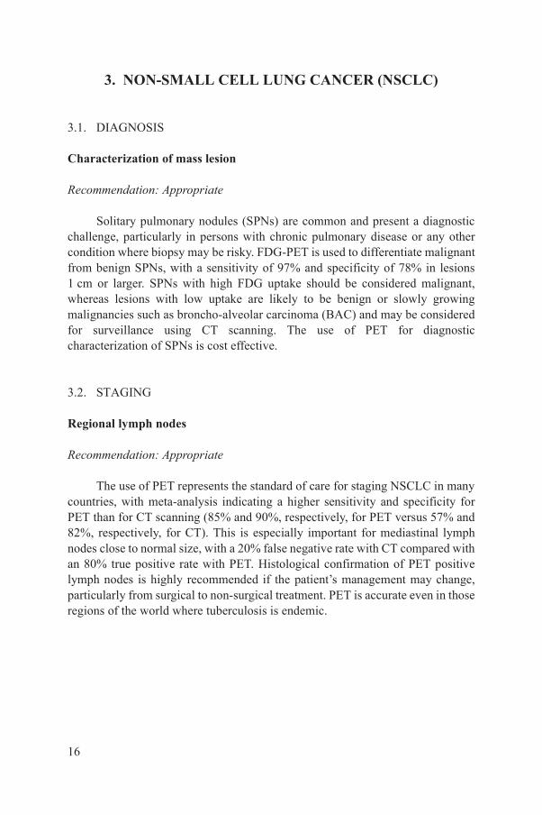
Solitary pulmonary nodules (SPNs) are common and present a diagnostic challenge, particularly in persons with chronic pulmonary disease or any other condition where biopsy may be risky. FDG-PET is used to differentiate malignant from benign SPNs, with a sensitivity of 97% and specificity of 78% in lesions 1 cm or larger. SPNs with high FDG uptake should be considered malignant, whereas lesions with low uptake are likely to be benign or slowly growing malignancies such as broncho-alveolar carcinoma (BAC) and may be considered for surveillance using CT scanning. The use of PET for diagnostic characterization of SPNs is cost effective.
3.2. STAGING
Regional lymph nodes
Recommendation: Appropriate
The use of PET represents the standard of care for staging NSCLC in many countries, with meta-analysis indicating a higher sensitivity and specificity for PET than for CT scanning (85% and 90%, respectively, for PET versus 57% and 82%, respectively, for CT). This is especially important for mediastinal lymph nodes close to normal size, with a 20% false negative rate with CT compared with an 80% true positive rate with PET. Histological confirmation of PET positive lymph nodes is highly recommended if the patient’s management may change, particularly from surgical to non-surgical treatment. PET is accurate even in those regions of the world where tuberculosis is endemic.
16
Distant metastases
Recommendation: Appropriate
Approximately one quarter of tumours initially staged as stage III prior to PET scanning are upstaged to stage IV following PET scanning. Brain metastases are not detected adequately using FDG-PET.
3.3. RESPONSE EVALUATION
Following neoadjuvant chemotherapy
Recommendation: Potentially appropriate
The PET response following neoadjuvant chemotherapy can be used to select patients with stage III tumours for subsequent surgical resection. If metastatic mediastinal lymph nodes show good response to chemotherapy, debulking or curative surgery may be considered. However, if there is poor response in mediastinal nodes, survival is very poor and patients probably should not undergo surgery.
Following definitive RT or chemoradiation
Recommendation: Inappropriate
Survival following definitive RT or chemoradiation is strongly predicted by PET, with improved survival in patients whose tumours show no uptake on post-treatment PET scans. This predictive value is much greater than that based on CT response. However, as this information does not change subsequent management, the use of PET for this purpose is not indicated.
During definitive RT or chemoradiation
Recommendation: Possibly appropriate
17
Some initial reports suggest that serial PET scans during a course of RT may be useful in determining the total RT dose. Tumours that fail to show a reduction in PET uptake during RT may be considered for a higher RT dose.
3.4. RESTAGING
End of therapy
Recommendation: Inappropriate
There is no rationale for the use of FDG-PET following completion of therapy.
Confirmed recurrence
Recommendation: Possibly appropriate
Although there are no data regarding the value of PET when recurrence has been confirmed, in a situation involving a solitary metastasis or local recurrence, restaging with PET may allow selection of appropriate therapy.
3.5. SUSPECTED RECURRENCE
Recommendation: Possibly appropriate
Data are lacking for this indication. However, there is a good rationale for the use of PET to confirm recurrence.
3.6. FOLLOW-UP
Recommendation: Inappropriate
While recurrence can probably be detected at an earlier point by PET than by clinical examination or another type of imaging, there is no evidence that patient management or survival would be affected.
18
3.7. RT PLANNING
Recommendation: Potentially appropriate
Many single centre reports, mostly on limited series of patients, indicate that the information available from PET scanning alters the size of RT treatment
fields in 27–100% of the cases. In most cases, the field size is increased to incorporate PET positive areas, while in some cases the field size is reduced in order to avoid unnecessary radiation to adjacent normal tissues, especially in the proximity of critical anatomic structures. To date there are no data showing an improvement in outcome.
BIBLIOGRAPHY
FLETCHER, J.W., et al., A comparison of the diagnostic accuracy of 18F-FDG PET and CT in the characterization of solitary pulmonary nodules, VA SNAP Cooperative Studies Group, J. Nucl. Med. 49 2 (2008) 179–185.
KIM, Y.K., et al., Mediastinal nodal staging of non-small cell lung cancer using integrated 18F-FDG PET/CT in a tuberculosis-endemic country: Diagnostic efficacy in 674 patients, Cancer 109 6 (2007) 1068–1077.
MacMANUS, M., HICKS, R.J., The use of positron emission tomography (PET) in the staging/evaluation, treatment, and follow-up of patients with lung cancer: A critical review, Int. J. Radiat. Oncol. Biol. Phys. 72 5 (2008) 1298–1306.
MacMANUS, M., et al., Use of PET and PET/CT for radiation therapy planning: IAEA expert report 2006–2007, Radiother. Oncol. 91 1 (2009) 85–94.
SAMSON, D.J., et al., Evidence for management of small cell lung cancer: ACCP evidence-based clinical practice guidelines, 2nd edn, Chest 132 3 (2007) 314–323.
YEN, R.F., et al., 18F-FDG PET for the lymph node staging of non-small cell lung cancer in a tuberculosis-endemic country: Is dual time point imaging worth the effort? Eur. J. Nucl. Med. Mol. Imaging 35 7 (2008) 1305–1315.
19

4. SMALL CELL LUNG CANCER (SCLC)
4.1. DIAGNOSIS
Characterization of mass lesion
Recommendation: Inappropriate
SCLC usually presents with a large central mass and concomitant hilo-mediastinal adenopathy; SCLC rarely presents with a peripheral mass. (In the rare event of SCLC presenting as an SPN, FDG-PET would be of value, as indicated for NSCLC.)
4.2. STAGING
Recommendation: Possibly appropriate
Management of SCLC is based on staging derived predominantly from CT findings. Although a number of reports indicate upstaging in approximately a quarter of the cases of limited stage SCLC, there are no data to indicate whether these patients should be managed as per limited stage or extensive stage disease.
4.3. RESPONSE EVALUATION
Recommendation: Inappropriate
As SCLC shrinks rapidly in response to effective treatment, it is unlikely that PET would contribute to the assessment of treatment response.
4.4. RESTAGING
20
Recommendation: Inappropriate
Although FDG-PET is likely to be more sensitive than CT in detecting sites of recurrent disease, recurrence is considered to be incurable and CT should be adequate for identifying recurrence.
4.5. SUSPECTED RECURRENCE
Recommendation: Possibly appropriate
The high FDG uptake of SCLC suggests that PET is a sensitive tool for identifying recurrence, although there are insufficient data indicating that PET alters clinical management.
4.6. FOLLOW-UP
Recommendation: Inappropriate
Recurrence of SCLC is considered to be incurable, with CT providing adequate detection of recurrence.
4.7. RT PLANNING
Recommendation: Possibly appropriate
It is likely that PET would have the same benefit for SCLC as has been demonstrated for NSCLC, resulting in a modification of the RT field definition for a high proportion of cases.
BIBLIOGRAPHY
BRADLEY, J.D., et al., Positron emission tomography in limited-stage small-cell lung cancer: A prospective study, J. Clin. Oncol. 22 16 (2004) 3248–3254.
ONITILO, A.A., ENGEL, J.M., DEMOS, J.M., MUKESH, B., Prognostic significance of 18F-fluorodeoxyglucose–positron emission tomography after treatment in patients with limited stage small cell lung cancer, Clin. Med. Res. 6 2 (2008) 72–77.
SAMSON, D.J., et al., Evidence for management of small cell lung cancer: ACCP evidence-
21
based clinical practice guidelines, 2nd edn, Chest 132 3 (2007) 314–323 (Review).
5. LYMPHOMA
5.1. DIAGNOSIS
Recommendation: Inappropriate
There is no rationale to support the use of FDG-PET for the diagnosis of lymphoma, since histology is needed to establish such a diagnosis.
5.2. STAGING
Recommendation: Appropriate
Owing to its superior sensitivity and specificity for most types of lymphoma, FDG-PET is appropriate for staging of Hodgkin’s disease (HD) and aggressive non-Hodgkin’s lymphomas (NHLs), but not for non-follicular low grade lymphomas. Since diffuse bone marrow involvement and small disease foci may be missed, FDG-PET cannot be recommended to replace bone marrow biopsy at initial staging.
A baseline FDG-PET scan is also indicated to assess FDG avidity of the tumour when subsequent evaluation of response to treatment with FDG-PET is planned.
5.3. RESPONSE EVALUATION
Recommendation: Appropriate
FDG-PET is the method of choice for the assessment of response to therapy in Hodgkin’s and non-Hodgkin’s lymphomas with pretreatment FDG avidity, and is superior to the CT based International Workshop Criteria. It helps to characterize residual masses, and the absence or persistence of FDG uptake even after fewer than three chemotherapy courses permits the separation of patients into favourable and unfavourable prognosis categories.
22
5.4. RESTAGING
Recommendation: Appropriate
The role of FDG-PET in restaging is equivalent to that in staging.
5.5. SUSPECTED RECURRENCE
Recommendation: Appropriate
FDG-PET is useful in selected patients for determining the nature of new masses. Positive foci require pathological confirmation.
5.6. FOLLOW-UP
Recommendation: Inappropriate
FDG-PET currently has no recognized role in the routine surveillance of patients treated for HD and NHL.
5.7. RT PLANNING
Recommendation: Inappropriate
There are no data available to support the use of PET for RT planning.
Note: The above recommendations also apply to primary central nervous system (CNS) lymphomas.
BIBLIOGRAPHY
BRUSAMOLINO, E., et al., Classical Hodgkin’s lymphoma in adults: Guidelines of the Italian Society of Hematology, the Italian Society of Experimental Hematology, and the Italian Group
23
for Bone Marrow Transplantation on initial work-up, management, and follow-up, Haematologica 94 4 (2009) 550–565.
CHESON, B.D., et al. Revised response criteria for malignant lymphoma, J. Clin. Oncol. 25(2007) 579–586.
ISASI, C.R., LU, P., BLAUFOX, M.D., A metaanalysis of 18F-2-deoxy-2-fluoro-D-glucose positron emission tomography in the staging and restaging of patients with lymphoma, Cancer 104 5 (2005) 1066–1074.
JOHNSTON, P.B., WISEMAN, G.A., MICALLEF, I.N., Positron emission tomography using F-18 fluorodeoxyglucose pre- and post-autologous stem cell transplant in non-Hodgkin’s lymphoma, Bone Marrow Transplant 41 (2008) 919–925.
JUWEID, M.E., et al., Use of positron emission tomography for response assessment of lymphoma: Consensus of the Imaging Subcommittee of the International Harmonization Project in Lymphoma, J. Clin. Oncol. 25 (2007) 571–578.
MIKHAEEL, N.G., Use of FDG-PET to monitor response to chemotherapy and radiotherapy in patients with lymphomas, Eur. J. Nucl. Med. Mol. Imaging 33 1 (2006) 22–26.
PAKOS, E.E., FOTOPOULOS, A.D., IOANNIDIS, J.P., 18F-FDG PET for evaluation of bone marrow infiltration in staging of lymphoma: A meta-analysis, J. Nucl. Med. 46 6 (2005) 958–963.
SCHAEFER, N.G., et al., Non-Hodgkin lymphoma and Hodgkin disease: Coregistered FDG PET and CT at staging and restaging — Do we need contrast-enhanced CT? Radiology 232(2004) 823–829.
TERASAWA, T., NIHASHI, T., HOTTA, T., NAGAI, H., 18F-FDG PET for posttherapy assessment of Hodgkin’s disease and aggressive non-Hodgkin’s lymphoma: A systematic review, J. Nucl. Med. 49 (2008) 13–21.
ZIJLSTRA, J.M., et al., 18F-fluoro-deoxyglucose positron emission tomography for post-treatment evaluation of malignant lymphoma: A systematic review, Haematologica 91 4 (2006) 522–529.
24
6. BREAST CANCER
6.1. DIAGNOSIS
Recommendation: Inappropriate
Multiple prospective studies have shown a low sensitivity (25%) for primary tumours 1 cm or smaller in diameter. The uptake of FDG in primary breast cancers is related to tumour size, histology and grade; more aggressive tumours usually have higher uptake than less aggressive ones. Other factors relevant to tumour biology also seem to influence the degree of FDG uptake and consequently the ability to detect the primary tumour by PET/CT.
6.2. STAGING
Axilla
Recommendation: Inappropriate
The sensitivity of FDG-PET is too low to correctly stage the axilla, as micrometastases may be missed. FDG-PET cannot replace sentinel node biopsy.
Distant metastases
Recommendation: Potentially appropriate
FDG-PET allows detection of extra-axillary nodes and distant metastases with higher sensitivity than other diagnostic imaging methods; an exception is brain metastases, where magnetic resonance imaging (MRI) is the method of choice. The relative role of bone scans using 99mTc compounds or FDG-PET in the detection of bone metastases remains undefined. Nevertheless, bone metastases from breast cancer tend to be osteolytic, and such lesions are known to be detected with higher sensitivity by FDG-PET than are sclerotic bone
25
metastases.
6.3. RESPONSE EVALUATION
Recommendation: Potentially appropriate
There is growing evidence that FDG-PET permits reliable response assessment after 1–3 cycles of chemotherapy in locally advanced and/or metastatic disease. This is an evolving role for PET-FDG in the management of breast cancer.
6.4. RESTAGING
End of therapy
Recommendation: Inappropriate
No data are available to support the use of FDG-PET in the restaging of breast cancer.
Confirmed recurrence
Recommendation: Potentially appropriate
Due to its high sensitivity for distant metastases, particularly nodal and skeletal metastases, FDG-PET is helpful in establishing the extent of recurrent disease.
6.5. SUSPECTED RECURRENCE
Recommendation: Potentially appropriate
There is a role for FDG-PET in the detection of recurrence, especially in patients with rising tumour markers. So far, however, prospective trials that also address the issues of management changes, outcome and cost efficiency are
26
lacking.
6.6. FOLLOW-UP
Recommendation: Inappropriate
No data are available, including from patients on long term therapy.
6.7. RT PLANNING
Recommendation: Possibly appropriate
Although only limited data are available, a rationale exists supporting the use of FDG-PET to define radiation fields for metastatic lesions.
BIBLIOGRAPHY
COOK, G.J., et al., Detection of bone metastases in breast cancer by 18FDG PET: Differing metabolic activity in osteoblastic and osteolytic lesions, J. Clin. Oncol. 16 10 (1998) 3375–3379.
COUTURIER, O., JERUSALEM, G., N’GUYEN, J.M., HUSTINX, R., Sequential positron emission tomography using [18F]fluorodeoxyglucose for monitoring response to chemotherapy in metastatic breast cancer, Clin. Cancer Res. 12 21 (2006) 6437–6443.
HODGSON, N.C., GULENCHYN, K.Y., Is there a role for positron emission tomography in breast cancer staging? J. Clin. Oncol. 26 5 (2008) 712–720.
ISASI, C.R., MOADEL, R.M., BLAUFOX, M.D., A meta-analysis of FDG-PET for the evaluation of breast cancer recurrence and metastases, Breast Cancer Res. Treat. 90 2 (2005) 105–112.
LAVAYSSIERE, R., CABEE, A.E., FILMONT, J.E., Positron Emission Tomography (PET) and breast cancer in clinical practice, Eur. J. Radiol. 69 1 (2009) 50–58.
27
7. MELANOMA
7.1. DIAGNOSIS
Recommendation: Inappropriate
The diagnosis of melanoma requires biopsy and histopathological examination. FDG-PET does not reliably distinguish between benign and malignant naevi, particularly for the small cutaneous lesions that usually characterize pigmented skin lesions.
7.2. STAGING
Stages I and II, low pretest probability of metastases
Recommendation: Inappropriate
PET is less sensitive than sentinel node biopsy for staging regional lymph nodes. In patients with low pretest probability of distant metastases, the sensitivity of PET for distant metastases has been reported to be low. Very small metastases are common in melanoma and may be beyond the resolution of PET, despite the usually high avidity of these tumours for FDG.
Stages I and II, high pretest probability of metastases
Recommendation: Appropriate
In patients with intermediate or high risk of distant metastases (melanoma of the head, neck and trunk, Breslow index >4 mm, ulceration, high mitotic rate), FDG-PET is appropriate for detecting potentially operable metastases.
Stage III or potential stage IV
28
Recommendation: Potentially appropriate
There is a role for FDG-PET in assessing locoregional or distant disease to guide appropriate therapy.

7.3. RESPONSE EVALUATION
Recommendation: Inappropriate
There are few data supporting the role of FDG-PET in assessing response to systemic therapy.
7.4. RESTAGING
End of treatment
Recommendation: Inappropriate
There is no rationale for the use of FDG-PET following completion of therapy.
Confirmed recurrence
Recommendation: Appropriate
FDG-PET is of value in distinguishing operable from non-operable recurrent disease. It should be noted that PET is less sensitive than MRI and CT in the detection of brain and lung metastases, respectively. Management changes are reported to occur in 22–34% of patients after PET scanning.
7.5. SUSPECTED RECURRENCE
Recommendation: Possibly appropriate
In the case of a lesion that is not readily amenable to biopsy, high uptake of FDG-PET is strongly suggestive of recurrent melanoma. There is an overlap with the role of FDG-PET in confirmed recurrence (see discussion above).
29

7.6. FOLLOW-UP
Recommendation: Inappropriate
There is no evidence that early detection of unsuspected metastases will influence patient outcome.
7.7. RT PLANNING
Recommendation: Inappropriate
There is no evidence that FDG-PET contributes to treatment planning.
BIBLIOGRAPHY
BELHOCINE, T.Z., et al., Role of nuclear medicine in the management of cutaneous malignant melanoma, J. Nucl. Med. 47 (2006) 957–967.
CONSTANTINIDOU, A., et al., Routine positron emission tomography and positron emission tomography/computed tomography in melanoma staging with positive sentinel node biopsy is of limited benefit, Melanoma Res. 18 1 (2008) 56–60.
DANCEY, A.L., MAHON, B.S., RAYATT, S.S., A review of diagnostic imaging in melanoma, J. Plast. Reconstr. Aesthet. Surg. 61 (2008) 1275–1283.
FLETCHER, J.W., et al. Recommendations on the use of 18F-FDG PET in oncology, J. Nucl. Med. 49 3 (2008) 480–508.
FRIEDMAN, K.P., WAHL, R.L., Clinical use of positron emission tomography in the management of cutaneous melanoma, Semin. Nucl. Med. 34 (2004) 242–253.
MIJNHOUT, G.S., et al., Systematic review of the diagnostic accuracy of (18)F-fluorodeoxyglucose positron emission tomography in melanoma patients, Cancer 91 8 (2001) 1530–1542.
30
PLEISS, C., RISSE, J.H., BIERSACK, H.J., BENDER, H., Role of FDG-PET in the assessment of survival prognosis in melanoma, Cancer Biother. Radiopharm. 22 6 (2007) 740–747.
8. OVARIAN CANCER
8.1. DIAGNOSIS
Recommendation: Inappropriate
Currently, there is no evidence of the value of FDG-PET in the initial diagnostic approach to ovarian cancer.
8.2. STAGING
Recommendation: Potentially appropriate
Although staging of ovarian cancers is usually performed surgically, the US National Oncologic PET Registry (NOPR) shows an impact of FDG-PET on intended management at initial staging of ovarian cancer in 16.1% of patients.
8.3. RESPONSE EVALUATION
Recommendation: Possibly appropriate
Relevant prospective studies are lacking.
8.4. RESTAGING
End of treatment
Recommendation: Possibly appropriate
Currently, there is no evidence of the value of FDG-PET in the restaging of ovarian cancer.
31
Confirmed recurrence
Recommendation: Potentially appropriate
According to the NOPR, the use of FDG-PET changed the intended management plan in 37.7% of the cases where it was used in restaging and in 44.5% of the cases where it was used in detection of recurrence.
8.5. SUSPECTED RECURRENCE
Recommendation: Appropriate
The number of patients in prospective controlled studies is small. Nevertheless, most studies show the diagnostic accuracy of FDG-PET, and particularly PET/CT, to be slightly superior to that of other imaging methods, in particular contrast-enhanced CT. In some studies, MRI was shown to be slightly more accurate; other studies found MRI and PET to be complementary for lesion characterization. In cases of peritoneal involvement, no currently used imaging method is sensitive enough to depict the full extent of the disease, as early proliferative peritoneal lesions are less than 1 mm thick.
8.6. FOLLOW-UP
Recommendation: Possibly appropriate
Currently, there is no evidence of the value of FDG-PET in follow-up of ovarian cancer, although a strong rationale exists for its use.
8.7. RT PLANNING
Recommendation: Inappropriate
32
RT has a very limited role in the management of ovarian carcinoma. When used palliatively, RT is directed at symptomatic masses identified by CT.
Note: Mucinous adenocarcinomas are usually non-FDG avid, and PET may therefore be inappropriate in this particular subgroup.
BIBLIOGRAPHY
CHUNG, H.H., et al., Role of [18F]FDG PET/CT in the assessment of suspected recurrent ovarian cancer: Correlation with clinical or histological findings, Eur. J. Nucl. Med. Mol. Imaging 34 4 (2007) 480–486.
HILLNER, B.E., et al., Impact of positron emission tomography/computed tomography and positron emission tomography (PET) alone on expected management of patients with cancer: Initial results from the National Oncologic PET Registry, J. Clin. Oncol. 26 13 (2008) 2155–2161.
KITAJIMA, K., et al., Diagnostic accuracy of integrated FDG-PET/contrast-enhanced CT in staging ovarian cancer: Comparison with enhanced CT, Eur. J. Nucl. Med. Mol. Imaging 35 10 (2008) 1912–1920.
MANGILI, G., et al., Integrated PET/CT as a first-line re-staging modality in patients with suspected recurrence of ovarian cancer, Eur. J. Nucl. Med. Mol. Imaging 34 5 (2007) 658–666.
SOUSSAN, M., et al., Impact of FDG PET-CT imaging on the decision making in the biologic suspicion of ovarian carcinoma recurrence, Gynecol. Oncol. 108 1 (2008) 160–165.
33

9. CANCER OF THE UTERUS AND CERVIX
9.1. DIAGNOSIS
Recommendation: Inappropriate
Currently, there is no evidence of the value of FDG-PET in the diagnosis of cancer of the uterus and cervix.
9.2. STAGING
Recommendation: Appropriate
In stage Ib–IV cervical cancer, FDG-PET is a valuable adjunct to conventional imaging methods, namely CT. Although MRI is the preferred method for evaluation of local extension, PET is superior for the evaluation of nodal involvement. The sentinel lymph node technique combined with surgical staging is more sensitive for local node involvement. In a recent NOPR evaluation, the use of PET changed the intended management plan in 14.1% of the cases where it was used in staging cancer of the uterus and in 9.1% of the cases where it was used in staging cancer of the cervix.
9.3. RESPONSE EVALUATION
Recommendation: Possibly appropriate
There is insufficient evidence to validate the usefulness of FDG-PET in assessing response to chemoradiation therapy, although persistent FDG avidity seems to be related to unfavourable outcome.
9.4. RESTAGING
34
End of therapy
Recommendation: Potentially appropriate
Persistence of FDG uptake seems to be related to unfavourable outcome.
Confirmed recurrence
Recommendation: Appropriate
There is evidence of the improved diagnostic accuracy of FDG-PET in restaging of these tumours. The NOPR study confirmed that the addition of FDG-PET changed the intended management plan in 30.5% of patients with uterine cancer and in 26.9% of patients with cervical cancer.
9.5. SUSPECTED RECURRENCE
Recommendation: Appropriate
According to the NOPR study, the impact of FDG-PET on detection of suspected recurrence resulted in a change of the intended management plan in 38.8% of patients with uterine carcinomas and in 35.9% of patients with cervical carcinomas.
9.6. FOLLOW-UP
Recommendation: Inappropriate
There are no data to support the use of FDG-PET in this setting.
9.7. RT PLANNING
Recommendation: Potentially appropriate
For locally advanced tumours, the detection by FDG-PET of metastasis in para-aortic lymph nodes may lead to modification of treatment fields. This is of particular importance in cervical cancer.
35
BIBLIOGRAPHY
GRIGSBY, P.W., SIEGEL, B.A., DEHDASHTI, F., Lymph node staging by positron emission tomography in patients with carcinoma of the cervix, J. Clin. Oncol. 19 17 (2001) 3745–3749.
GRIGSBY, P.W., et al., Posttherapy [18F] fluorodeoxyglucose positron emission tomography in carcinoma of the cervix: Response and outcome, J. Clin. Oncol. 22 11 (2004) 2167–2171.
HILLNER, B.E., et al., Relationship between cancer type and impact of PET and PET/CT on intended management: Findings of the National Oncologic PET Registry, J. Nucl. Med. 49 12 (2008) 1928–1935.
KITAJIMA, K., et al., Performance of FDG-PET/CT for diagnosis of recurrent uterine cervical cancer, Eur. Radiol. 18 10 (2008) 2040–2047.
LAI, C.H., YEN, T.C., CHANG, T.C., Positron emission tomography imaging for gynecologic malignancy, Curr. Opin. Obstet. Gynecol. 19 1 (2007) 37–41.
SUZUKI, R., et al., Validity of positron emission tomography using fluoro-2-deoxyglucose for the preoperative evaluation of endometrial cancer, Int. J. Gynecol. Cancer 17 4 (2007) 890–896.
36
10. HEAD AND NECK CANCERS
The following discussion does not include nasopharyngeal and thyroid cancers; these are discussed in separate sections.
10.1. DIAGNOSIS
Characterization of mass lesion
Recommendation: Inappropriate
The diagnosis of primary head and neck cancers is made on the basis of clinical examination, endoscopy with biopsies, and imaging with CT/MRI and/or ultrasound.
PET guided biopsy
Recommendation: Inappropriate
No data are available to suggest that FDG-PET improves imaging guided biopsy.
Cervical adenopathy with occult primary
Recommendation: Appropriate
The true positive rate for PET is approximately 30% where PET is performed when all other diagnostic tests are negative or when some other tests may have been positive. Small tumours (<5 mm) may be missed by PET.
10.2. STAGING
37
Recommendation: Potentially appropriate
Use of CT or MRI remains the standard of care for T and N staging in this setting. FDG-PET is accurate in detecting regional nodal disease, distant metastases and synchronous tumours.
10.3. RESPONSE EVALUATION
Recommendation: Appropriate
If performed 8–10 weeks after treatment, FDG-PET is accurate in detecting residual disease after chemotherapy alone or combined with RT. If performed earlier, false positive results due to inflammatory changes are possible. Persistently enlarged FDG negative nodes need to be clinically monitored.
10.4. RESTAGING
End of therapy
Recommendation: Appropriate
The role of FDG-PET in the restaging of head and neck cancers is the same as in response evaluation (see Section 10.3).
Confirmed recurrence
Recommendation: Potentially appropriate
FDG-PET is accurate in detecting regional nodal recurrence, distant metastases and second tumours.
10.5. SUSPECTED RECURRENCE
Recommendation: Appropriate
Since distortion of tissue structures following surgery and RT may limit the diagnostic abilities of anatomic imaging techniques, the use of PET to identify recurrences is appropriate if conventional methods of diagnosing recurrence are inconclusive.
38
10.6. FOLLOW-UP
Recommendation: Inappropriate
There is no evidence that FDG-PET is useful in patients who have already been treated and are without any evidence of disease.
10.7. RT PLANNING
Recommendation: Potentially appropriate
Data demonstrate that target volumes and doses may be modified on the basis of FDG-PET findings. In particular, FDG-PET is helpful for the inclusion or exclusion of lymph nodes in the radiation field, although no data on patient outcome are available.
BIBLIOGRAPHY
ISLES, M.G., McCONKEY, C., MEHANNA, H.M., A systematic review and meta-analysis of the role of positron emission tomography in the follow-up of head and neck squamous cell carcinoma following radiotherapy or chemoradiotherapy, Clin. Otolaryngol. 33 3 (2008) 210–222.
LANGO, M.N., MYERS, J.N., GARDEN, A.S., Controversies in surgical management of the node-positive neck after chemoradiation, Semin. Radiat. Oncol. 19 1 (2009) 24–28.
PORCEDDU, S.V., BURMEISTER, B.H., HICKS, R.J., Role of functional imaging in head and neck squamous cell carcinoma: Fluorodeoxyglucose positron emission tomography and beyond, Hematol. Oncol. Clin. North Am. 22 6 (2008) 1221–1238.
SHAH, G.V., WESOLOWSKI, J.R., ANSARI, S.A., MUKHERJI, S.K., New directions in head and neck imaging, J. Surg. Oncol. 97 8 (2008) 644–648.
39
WONG, R.J., Current status of FDG-PET for head and neck cancer, J. Surg. Oncol. 97 8 (2008) 649–652.
11. KIDNEY CANCER
11.1. DIAGNOSIS
Recommendation: Inappropriate
Currently, there is no evidence of the value of FDG-PET in the diagnosis of kidney cancer.
11.2. STAGING
Recommendation: Possibly appropriate
Although some studies suggest a potential role for FDG-PET in advanced kidney cancer, there are still insufficient data to support its use for routine staging.
11.3. RESPONSE EVALUATION
Recommendation: Inappropriate
Currently, there is no evidence of the value of FDG-PET in the assessment of treatment response.
11.4. RESTAGING
End of treatment
Recommendation: Inappropriate
Currently, there is no evidence of the value of FDG-PET in the restaging of
40
kidney cancer.
Confirmed recurrence
Recommendation: Potentially appropriate
Limited studies suggest that FDG-PET has good accuracy for the detection of unsuspected metastatic disease.
11.5. SUSPECTED RECURRENCE
Recommendation: Inappropriate
Currently, there is no evidence of the value of FDG-PET in detecting suspected recurrence of kidney cancer.
11.6. FOLLOW-UP
Recommendation: Inappropriate
Currently, there is no evidence of the value of FDG-PET in follow-up of kidney cancer.
11.7. RT PLANNING
Recommendation: Inappropriate
The placement of radiation fields is based on the presence of symptomatic gross disease, which is evident from results of conventional imaging.
41
BIBLIOGRAPHY
BOUCHELOUCHE, K., OEHR, P., Recent developments in urologic oncology: Positron emission tomography molecular imaging, Curr. Opin. Oncol. 20 3 (2008) 321–326.
GOLDFARB, C.R., et al., Radionuclide imaging in urology, Urol. Clin. North Am. 33 3 (2006) 319–328.
LAWRENTSCHUK, N., DAVIS, I.D., BOLTON, D.M., SCOTT, A.M., Positron emission tomography (PET), immuno-PET and radioimmunotherapy in renal cell carcinoma: A developing diagnostic and therapeutic relationship, BJU Int. 97 5 (2006) 916–922.
MUELLER-LISSE, U.G., et al., Staging of renal cell carcinoma, Eur. Radiol. 17 9 (2007) 2268–2277.
42
12. GERMINAL TUMOURS
12.1. DIAGNOSIS
Recommendation: Inappropriate
Currently, there is no evidence of the value of FDG-PET in the diagnosis of germinal tumours.
12.2. STAGING
Recommendation: Inappropriate
The negative predictive value is not high enough to avoid adjuvant therapies in the case of negative results.
12.3. RESPONSE EVALUATION
Recommendation: Possibly appropriate
FDG-PET is superior to CT, with a reported sensitivity of 59–89% and specificity of 92–100%. With the exception of mature teratoma, PET can distinguish residual tumour from necrosis and/or fibrosis.
12.4. RESTAGING
Recommendation: Inappropriate
Currently, there is no evidence of the value of FDG-PET in the restaging of germinal tumours.
43
12.5. SUSPECTED RECURRENCE
Recommendation: Possibly appropriate
In cases of equivocal CT findings and/or elevation of serum tumour markers, PET can be used to diagnose recurrence when other imaging techniques are not helpful.
12.6. FOLLOW-UP
Recommendation: Inappropriate
Currently, there is no evidence of the value of FDG-PET in follow-up of germinal tumours.
12.7. RT PLANNING
Recommendation: Inappropriate
RT has a minimal role in non-seminomatous germ cell tumours, and there are no data indicating that PET has an impact. For early stage seminomas, for which the patterns of failure are well described, there are no data to suggest that PET may influence radiation fields.
BIBLIOGRAPHY
BOUCHELOUCHE, K., OEHR, P., Recent developments in urologic oncology: Positron emission tomography molecular imaging, Curr. Opin. Oncol. 20 3 (2008) 321–326.
FANTI, S., et al., PET in genitourinary tract cancers, Q. J. Nucl. Med. Mol. Imaging 51 3 (2007) 260–271.
44
HEIDENREICH, A., THÜER, D., POLYAKOV, S., Postchemotherapy retroperitoneal lymph node dissection in advanced germ cell tumours of the testis, Eur. Urol. 53 2 (2008) 260–272.
SOHAIB, S.A., KOH, D.M., HUSBAND, J.E., The role of imaging in the diagnosis, staging, and management of testicular cancer, AJR Am. J. Roentgenol. 191 2 (2008) 387–395.
13. CANCER OF UNKNOWN PRIMARY (CUP)
13.1. DIAGNOSIS
Raised tumour markers
Recommendation: Possibly appropriate
For tumour types that are potential origins of the raised markers and that are generally FDG avid, PET-CT should be used if the conventional workup has failed to identify the primary tumour.
Metastases outside the neck
Recommendation: Potentially appropriate
A single-trial analysis comparing PET and CT in locating primary tumour in patients with cancer of unknown origin indicated that the sensitivity of PET-CT was 36% versus 15% for CT.
Metastases in the head and neck area
See the discussion of head and neck cancers in Section 10 of this report.
13.2. STAGING
Recommendation: Possibly appropriate
FDG-PET may be appropriate for evaluation of the extent of disease.
13.3. RESPONSE EVALUATION
45
Not applicable
13.4. RESTAGING
Not applicable
13.5. SUSPECTED RECURRENCE
Not applicable
13.6. FOLLOW-UP
Not applicable
13.7. RT PLANNING
Not applicable
BIBLIOGRAPHY
DONG, M.J., et al., Role of fluorodeoxyglucose-PET versus fluorodeoxyglucose-PET/computed tomography in detection of unknown primary tumor: A meta-analysis of the literature, Nucl. Med. Commun. 29 9 (2008) 791–802.
FREUDENBERG, L.S., et al., Cancer of unknown primary, Recent Results Cancer Res. 170(2008) 193–202.
KAYA, A.O., et al., Whole body 18F-FDG PET/CT imaging in the detection of primary tumours in patients with a metastatic carcinoma of unknown origin, Asian Pac. J. Cancer Prev. 9 4 (2008) 683–686.
KWEE, T.C., KWEE, R.M., Combined FDG-PET/CT for the detection of unknown primary tumors: Systematic review and meta-analysis, Eur. Radiol. 19 3 (2009) 731–744.
SÈVE, P., et al., The role of 2-deoxy-2-[F-18]fluoro-D-glucose positron emission tomography in disseminated carcinoma of unknown primary site, Cancer 109 2 (2007) 292–299.
46
14. COLORECTAL CANCER
14.1. DIAGNOSIS
Recommendation: Inappropriate
Any symptoms suggestive of colorectal cancer must be investigated by endoscopy, with biopsy of suspicious lesions. However, there are numerous cases where unsuspected and asymptomatic colorectal cancers have been detected on FDG-PET scans performed for other purposes.
14.2. STAGING
Recommendation: Potentially appropriate
FDG-PET is superior to other imaging modalities for detecting additional intrahepatic and extrahepatic metastases when a hepatic metastasis has been detected by CT or ultrasound, and may also be superior to those imaging techniques for detecting lymph node metastases. The use of FDG-PET in staging results in a change of treatment in approximately one quarter of the cases.
14.3. RESPONSE EVALUATION
Recommendation: Possibly appropriate
FDG-PET provides a sensitive assessment of the response to chemotherapy or chemoradiation that is superior to CT assessment. This may lead to a change from ineffective therapy.
14.4. RESTAGING
47
Recommendation: Appropriate
The common situations where restaging is required are (1) consideration of isolated local recurrence and (2) isolated hepatic metastases. The use of FDG-PET prior to hepatic resection changes management in approximately one third of the cases, mainly through identification of more extensive metastatic disease than
is shown with CT. The use of FDG-PET in this situation is cost effective. FDG-PET correctly identified resectable disease in 80% of the cases, and correctly identified unresectable, incurable disease in 90% of the cases. Therefore, surgical exploration should be undertaken when FDG-PET indicates resectable disease; conversely, surgery may be avoided when FDG-PET identifies extensive incurable disease. Care in interpretation of PET images is required following pre-operative chemotherapy, as hepatic metastases may be less evident.
14.5. SUSPECTED RECURRENCE
Recommendation: Appropriate
FDG-PET is valuable for determining the site or sites of recurrence when carcinoembryonic antigen (CEA) levels are rising and CT is non-diagnostic.
14.6. FOLLOW-UP
Recommendation: Possibly appropriate
FDG-PET provides evidence of pelvic recurrence earlier than CT scanning, with the potential for more effective local therapy.
14.7. RT PLANNING
Recommendation: Possibly appropriate
There are no data indicating a role for PET in assisting with the placement of radiation fields, although a strong rationale exists for its usefulness in this setting.
Note: GEPTs (gastro-entero-pancreatic tumours) and mucinous adenocarcinomas usually are not FDG avid, and FDG-PET may be inappropriate in this particular subgroup.
48
BIBLIOGRAPHY
CHUA, S.C., et al., The impact of 18F-FDG PET/CT in patients with liver metastases, Eur. J. Nucl. Med. Mol. Imaging 34 12 (2007) 1906–1914.
DIAS, A.R., NAHAS, S.C., CAMARGO, E.E., NAHAS, C.S., Recent evidences of the use of 18F-fluorodeoxyglucose positron emission tomography in the management of colorectal cancer, J. Surg. Educ. 64 2 (2007) 114–119.
HERBERTSON, R.A., et al., Established, emerging and future roles of PET/CT in the management of colorectal cancer, Clin. Radiol. 64 3 (2009) 225–237.
VIKRAM, R., IYER, R.B., PET/CT imaging in the diagnosis, staging, and follow-up of colorectal cancer, Cancer Imaging 8 A (2008) 46–51.
49
15. GASTRIC CARCINOMA
The following discussion refers to distal gastric cancers. Tumours involving the gastroesophageal junction are generally considered as distal oesophageal carcinomas.
15.1. DIAGNOSIS
Characterization of mass lesion
Recommendation: Inappropriate
There is no evidence that the addition of PET to endoscopy and biopsy improves diagnostic ability.
PET guided biopsy
Recommendation: Inappropriate
There are very limited data available to date. Normal gastric mucosa shows some level of physiological FDG uptake.
15.2. STAGING
Recommendation: Possibly appropriate
There are limited data on the value of FDG-PET in detecting nodal and metastatic disease.
15.3. RESPONSE EVALUATION
50
Recommendation: Possibly appropriate
FDG-PET may identify response to neoadjuvant therapy. There are, however, no data to determine the impact of PET on clinical outcome.
15.4. RESTAGING
Recommendation: Inappropriate
There are no data indicating a role for FDG-PET after the completion of definitive therapy.
15.5. SUSPECTED RECURRENCE
Recommendation: Inappropriate
There are no data indicating a role for FDG-PET.
15.6. FOLLOW-UP
Recommendation: Possibly appropriate
There are limited data indicating a role for FDG-PET.
15.7. RT PLANNING
Recommendation: Inappropriate
There are no data indicating a role for FDG-PET. Palliative RT is targeted at the CT defined mass; curative post-operative RT (usually with chemotherapy) is targeted at the surgical bed.
Note: Gastro-entero-pancreatic tumours (GEPTs) and mucinous adenocarcinomasusually are not FDG avid, and PET may be inappropriate in this particular subgroup.
51

BIBLIOGRAPHY
DASSEN, A.E., et al., FDG-PET has no definite role in preoperative imaging in gastric cancer, Eur. J. Surg. Oncol. 35 5 (2009) 449–455.
NAKAMOTO, Y., et al., Clinical value of whole-body FDG-PET for recurrent gastric cancer: A multicenter study, Jpn. J. Clin. Oncol. 39 5 (2009) 297–302.
SIM, S.H., et al., The role of PET/CT in detection of gastric cancer recurrence, BMC Cancer 9(2009) 73.
52

16. SARCOMAS (SOFT TISSUE AND BONE)
16.1. DIAGNOSIS
Characterization of mass lesion
Recommendation: Inappropriate
Although benign tumours generally exhibit less uptake than do sarcomas, there is considerable overlap, and some benign tumours have high avidity for FDG. Biopsy is required for diagnosis.
PET guided biopsy
Recommendation: Possibly appropriate
As sarcomas behave according to the highest grade of the tumour, and as treatment may change according to the tumour grade, the use of PET to identify the optimal biopsy site has a strong rationale, which has been confirmed by several reports.
16.2. STAGING
Recommendation: Possibly appropriate
Sarcomas have a particular propensity for initial metastatic spread to the lungs. High resolution CT is more effective than FDG-PET for detecting small lung metastases. However, PET may be more useful for extrapulmonary metastases. PET has also been shown to be more sensitive than bone scans using 99mTc labelled compounds for bone metastases from Ewing’s sarcoma.
16.3. RESPONSE EVALUATION
53
Recommendation: Possibly appropriate
There is considerable interest in the use of PET to monitor the response of osteosarcomas to neoadjuvant chemotherapy. The goal is early evaluation of response; in the event of poor response, the drug combinations can be changed.
16.4. RESTAGING
Recommendation: Possibly appropriate
The role of FDG-PET in the restaging of sarcomas is the same as in the initial staging of sarcomas.
16.5. SUSPECTED RECURRENCE
Recommendation: Possibly appropriate
Suspected recurrence will usually require biopsy for confirmation. However, as indicated above (see Section 16.1), FDG-PET may guide biopsy to the site most likely to yield a high grade component.
16.6. FOLLOW-UP
Recommendation: Possibly appropriate
FDG-PET may be useful for detecting recurrence at an early stage, when salvage surgery may be possible or less mutilating. FDG-PET has some additional advantages over CT and MRI, as PET is not affected by abnormal, post-surgical anatomy or metal prostheses.
16.7. RT PLANNING
Recommendation: Possibly appropriate
There are no reports indicating the use of PET to assist RT planning. However, there is a rationale to support the concept.
54
BIBLIOGRAPHY
EARY, J.F., PET imaging: Update on sarcomas, Oncology 21 2 (2007) 249–252.
TEWFIK, J.N., GREENE, G.S., Fluorine-18-deoxyglucose-positron emission tomography imaging with magnetic resonance and computed tomographic correlation in the evaluation of bone and soft-tissue sarcomas: A pictorial essay, Curr. Probl. Diagn. Radiol. 37 4 (2008) 178–188.
TONER, G.C., HICKS, R.J., PET for sarcomas other than gastrointestinal stromal tumors, Oncologist 13 2 (2008) 22–26.
VAN DE LUIJTGAARDEN, A.C., et al., Promises and challenges of positron emission tomography for assessment of sarcoma in daily clinical practice, Cancer Imaging 8 A (2008) 61–68.
55
17. PRIMARY TUMOURS OFTHE CENTRAL NERVOUS SYSTEM
17.1. DIAGNOSIS
Characterization of whether a mass lesion is low or high grade
Recommendation: Inappropriate
Although there is generally good correlation between FDG uptake and tumour grade, the high background in normal grey matter limits the ability to detect lesions with FDG-PET.
PET guided biopsy
Recommendation: Possibly appropriate
In selected cases, FDG-PET may be of value for identifying the most aggressive component within a lesion.
17.2. STAGING
Recommendation: Inappropriate
MRI provides excellent anatomic definition to determine the extent of the tumour.
17.3. RESPONSE EVALUATION
Recommendation: Possibly appropriate
There are few reports regarding the use of FDG-PET to assess the response
56
to multimodality therapy, although a strong rationale exists for its use.
17.4. RESTAGING
End of therapy
Recommendation: Inappropriate
There is no indication of a role for PET scanning following the completion of therapy.
Confirmed recurrence
Recommendation: Inappropriate
There is generally no requirement to further define the tumour using PET when recurrence has been confirmed.
17.5. SUSPECTED RECURRENCE
Recommendation: Possibly appropriate
PET may provide information additional to that provided by MRI or CT for detection of recurrence following resection. FDG-PET has also been used to distinguish radiation necrosis from recurrent tumour; however, there are conflicting results and the accuracy seems to be low.
17.6. FOLLOW-UP
Recommendation: Possibly appropriate
PET has been used for routine surveillance of untreated low grade gliomas to assess transformation to high grade lesions.
57
17.7. RT PLANNING
Recommendation: Possibly appropriate
FDG-PET currently has no role in defining radiation fields or doses. However, there is a rationale for using PET for dose escalation to the metabolically intense region within the tumour.
Note: For CNS lymphomas, see the discussion on lymphomas in Section 5 of this report.
BIBLIOGRAPHY
CHEN, W., Clinical applications of PET in brain tumors, J. Nucl. Med. 48 9 (2007) 1468–1481.
FISCHMAN, A.J., PET imaging of brain tumors, Cancer Treat. Res. 143 (2008) 67–92.
JADVAR, H., CONNOLLY, L.P., FAHEY, F.H., SHULKIN, B.L., PET and PET/CT in pediatric oncology, Semin. Nucl. Med. 37 5 (2007) 316–331.
PATIL, S., BIASSONI, L., BORGWARDT, L., Nuclear medicine in pediatric neurology and neurosurgery: Epilepsy and brain tumors, Semin. Nucl. Med. 37 5 (2007) 357–381.
ULLRICH, R.T., KRACHT, L.W., JACOBS, A.H., Neuroimaging in patients with gliomas, Semin. Neurol. 28 4 (2008) 484–494.
58
18. NASOPHARYNGEAL CARCINOMAS
18.1. DIAGNOSIS
Recommendation: Inappropriate
There are no data indicating a role for FDG-PET in the diagnosis of nasopharyngeal carcinomas.
18.2. STAGING
Recommendation: Appropriate
For both the N and M stages of the disease, FDG-PET provides incremental value over conventional imaging.
18.3. RESPONSE EVALUATION
Recommendation: Appropriate
If performed 8–10 weeks after treatment, FDG-PET is accurate in detecting residual disease. If performed earlier, there is a possibility of false positive results due to inflammatory changes. Persistently enlarged FDG negative nodes require watchful monitoring.
18.4. RESTAGING
End of therapy
Recommendation: Appropriate
59
See discussion on response evaluation.
Confirmed recurrence
Recommendation: Appropriate
Due to the high risk of distant disease, whole body imaging with FDG-PET is required to guide therapy.
18.5. SUSPECTED RECURRENCE
Recommendation: Potentially appropriate
When standard procedures are non-diagnostic, FDG-PET may identify the site(s) of recurrence.
18.6. FOLLOW-UP
Recommendation: Possibly appropriate
No data exist supporting the use of FDG-PET for follow-up, but a rationale exists, as early detection of local recurrence may permit curative treatment.
18.7. RT PLANNING
Recommendation: Possibly appropriate
PET may improve target volume delineation and identify involved lymph nodes of borderline size on structural imaging.
60

BIBLIOGRAPHY
CHIA-HUNG, K., et al., Detection of recurrent or persistent nasopharyngeal carcinomas after radiotherapy with 18-fluoro-2-deoxyglucose positron emission tomography and comparison with computed tomography, J. Clin. Oncol. 16 (1998) 3550–3555.
CHUA, M.L., et al., Comparison of 4 modalities for distant metastasis staging in endemic nasopharyngeal carcinoma, Head Neck 31 3 (2009) 346–354.
LIU, T., et al., FDG-PET, CT, MRI for diagnosis of local residual or recurrent nasopharyngeal carcinoma, which one is the best? A systematic review, Radiother. Oncol. 85 3 (2007) 327–335.
YEN, R.F., et al., Whole-body 18F-FDG PET in recurrent or metastatic nasopharyngeal carcinoma, J. Nucl. Med. 46 5 (2005) 770–774.
61

19. GASTROINTESTINAL STROMAL TUMOURS (GISTs)
19.1. DIAGNOSIS
Recommendation: Inappropriate
Gastrointestinal stromal tumours (GISTs) are usually diagnosed by endoscopy and/or biopsy.
19.2. STAGING
Recommendation: Appropriate
A baseline FDG-PET scan is necessary to determine tumour avidity for subsequent treatment and response evaluation.
19.3. RESPONSE EVALUATION
Recommendation: Appropriate
For FDG avid tumours, PET is highly recommended for response evaluation because of the ability to identify the early response to tyrosine kinase inhibitor therapy.
19.4. RESTAGING
End of therapy
Recommendation: Inappropriate
After complete surgical resection, PET is not indicated. In patients with
62
unresectable or residual disease, tyrosine kinase inhibitor therapy is continued unless intolerable toxicity occurs or resistance is documented.
Confirmed recurrence
Recommendation: Appropriate
An FDG-PET scan is necessary to determine FDG avidity of the recurrent tumour.
19.5. SUSPECTED RECURRENCE
Recommendation: Appropriate
FDG-PET is a sensitive procedure to determine possible recurrence(s), as the vast majority of GISTs are FDG avid.
19.6. FOLLOW-UP
Recommendation: Appropriate
In patients with persistent tumour, following incomplete resection of primary or recurrent tumour, FDG-PET is required to identify active disease.
19.7. RT PLANNING
Recommendation: Inappropriate
There are no data indicating a role for FDG-PET in RT planning for treatment of GISTs.
BIBLIOGRAPHY
BASU, S., et al., FDG-PET and PET/CT in the clinical management of gastrointestinal stromal tumor, Nucl. Med. Commun. 29 12 (2008) 1026–1039.
63
BENJAMIN, R.S., et al., We should desist using RECIST, at least in GIST, J. Clin. Oncol. 2513 (2007) 1760–1764.
VAN DEN ABBEELE, A.D., The lessons of GIST-PET and PET/CT: A new paradigm for imaging, Oncologist 13 2 (2008) 8–13.

20. PANCREATIC ADENOCARCINOMA
20.1. DIAGNOSIS
Recommendation: Potentially appropriate
When a pancreatic mass is detected by conventional imaging, the degree of FDG avidity may help distinguish benign from malignant lesions.
20.2. STAGING
Recommendation: Possibly appropriate
FDG-PET sensitivity is low for N staging but may be improved by the use of contrast enhanced PET/CT. For M staging, FDG-PET may complement conventional imaging modalities.
20.3. RESPONSE EVALUATION
Recommendation: Possibly appropriate
There is a rationale for the use of FDG-PET for the assessment of response to systemic therapy, but only limited data are available.
20.4. RESTAGING
End of therapy
Recommendation: Inappropriate
There are no data indicating a role for FDG-PET following completion of
64
therapy for pancreatic adenocarcinoma.

Confirmed recurrence
Recommendation: Inappropriate
There are no data indicating a role for FDG-PET in the restaging of pancreatic adenocarcinoma.
20.5. SUSPECTED RECURRENCE
Recommendation: Potentially appropriate
The degree of FDG avidity may help distinguish recurrence from post-treatment changes.
20.6. FOLLOW-UP
Recommendation: Inappropriate
There are no data indicating a role for FDG-PET in follow-up of pancreatic adenocarcinoma.
20.7. RT PLANNING
Recommendation: Possibly appropriate
FDG-PET data may be useful for target volume delineation and dose intensification.
Note: Gastro-entero-pancreatic tumours (GEPTs) usually are not FDG avid and are excluded from these recommendations.
65
BIBLIOGRAPHY
BANG, S., et al., The clinical usefulness of 18-fluorodeoxyglucose positron emission tomography in the differential diagnosis, staging, and response evaluation after concurrent chemoradiotherapy for pancreatic cancer, J. Clin. Gastroenterol. 40 10 (2006) 923–929.
PAKZAD, F., GROVES, A.M., ELL, P.J., The role of positron emission tomography in the management of pancreatic cancer, Semin. Nucl. Med. 36 3 (2006) 248–256.
SCHICK, V., et al., Diagnostic impact of 18F-FDG PET-CT evaluating solid pancreatic lesions versus endosonography, endoscopic retrograde cholangio-pancreatography with intraductal ultrasonography and abdominal ultrasound, Eur. J. Nucl. Med. Mol. Imaging 35 10 (2008) 1775–1785.
66
21. CHOLANGIO- AND GALLBLADDER CARCINOMAS
21.1. DIAGNOSIS
Recommendation: Possibly appropriate
FDG uptake may discriminate benign from malignant strictures of the biliary tract.
21.2. STAGING
Recommendation: Possibly appropriate
In limited series, FDG-PET is more accurate than CT scanning for defining the N and M stages of the disease.
21.3. RESPONSE EVALUATION
Recommendation: Possibly appropriate
No data are available, although a rationale exists for the use of FDG-PET in this setting with the use of chemotherapy to downstage tumours.
21.4. RESTAGING
End of therapy
Recommendation: Inappropriate
There are no data indicating a role for FDG-PET following completion of therapy for cholangio- and gallbladder carcinomas.
67
Confirmed recurrence
Recommendation: Inappropriate
Limited data are available; however, it is unlikely that PET detected recurrence would be amenable to curative treatment.
21.5. SUSPECTED RECURRENCE
Recommendation: Inappropriate
Limited data are available; however, it is unlikely that PET detected recurrence would be amenable to curative treatment.
21.6. FOLLOW-UP
Recommendation: Inappropriate
There are no data indicating a role in FDG-PET in follow-up of cholangio- and gallbladder carcinomas.
21.7. RT PLANNING
Recommendation: Inappropriate
There are no data indicating a role for FDG-PET in the planning of RT for cholangio- and gallbladder carcinomas.
68
BIBLIOGRAPHY
CORVERA, C.U., et al., 18F-fluorodeoxyglucose positron emission tomography influences management decisions in patients with biliary cancer, J. Am. Coll. Surg. 206 1 (2008) 57–65.
KIM, J.Y., et al., Clinical role of 18F-FDG PET-CT in suspected and potentially operable cholangiocarcinoma: A prospective study compared with conventional imaging, Am. J. Gastroenterol. 103 5 (2008) 1145–1151.
LI, J., et al., Preoperative assessment of hilar cholangiocarcinoma by dual-modality PET/CT, J. Surg. Oncol. 98 6 (2008) 438–443.
MOON, C.M., et al., Usefulness of 18F-fluorodeoxyglucose positron emission tomography in differential diagnosis and staging of cholangiocarcinomas, J. Gastroenterol. Hepatol. 23 5 (2008) 759–765.
NISHIYAMA, Y., et al., Comparison of early and delayed FDG PET for evaluation of biliary stricture, Nucl. Med. Commun. 28 12 (2007) 914–919.
SHUKLA, P.J., et al., Does PET-CT scan have a role prior to radical re-resection for incidental gallbladder cancer? HPB Oxford 10 6 (2008) 439–445.
69
22. OESOPHAGEAL CANCER
22.1. DIAGNOSIS
Characterization of mass lesion
Recommendation: Inappropriate
There is no evidence that the addition of FDG-PET improves the diagnostic accuracy of endoscopic ultrasound (EUS) and biopsy.
PET guided biopsy
Recommendation: Inappropriate
Only very limited data are available on the use of FDG-PET in PET guided biopsy of oesophageal cancer.
22.2. STAGING
Recommendation: Appropriate
There are several reports on the value of FDG-PET in detecting metastatic disease. The reported sensitivity varies, but it is always superior to that of CT. This feature is important, as upstaging usually indicates that radical surgery is inappropriate; it is also important for multimodality therapy.
22.3. RESPONSE EVALUATION
Recommendation: Potentially appropriate
FDG-PET may identify locoregional disease unresponsive to neoadjuvant
70
therapy and interval metastases prior to planned surgery (approximately 8–14% of cases). The endoscopic findings should be taken into consideration, as oesophagitis may mimic residual disease on a PET scan.
22.4. RESTAGING
Recommendation: Inappropriate
There are no data indicating a definite role for FDG-PET after completion of potentially curative therapy.
22.5. SUSPECTED RECURRENCE
Recommendation: Potentially appropriate
This recommendation is particularly relevant for lower stage tumours treated with local techniques that have recurred locally and remain amenable to potentially curative locoregional therapy.
22.6. FOLLOW-UP
Recommendation: Inappropriate
There are no data indicating a role for FDG–PET in follow-up of oesophageal cancer.
22.7. RT PLANNING
Recommendation: Potentially appropriate
FDG-PET findings have been used to modify target volumes. Insufficient data are available on clinical outcome.
71
BIBLIOGRAPHY
BRUZZI, J.F., et al., PET/CT of esophageal cancer: Its role in clinical management, Radiographics 27 6 (2007) 1635–1652.
LORDICK, F., et al., PET to assess early metabolic response and to guide treatment of adenocarcinoma of the oesophagogastric junction: The MUNICON phase II trial, Lancet Oncol. 8 9 (2007) 797–805.
SCHMIDT, M., et al., Mean and maximum standardized uptake values in [18F]FDG-PET for assessment of histopathological response in oesophageal squamous cell carcinoma or adenocarcinoma after radiochemotherapy, Eur. J. Nucl. Med. Mol. Imaging 36 5 (2009) 735–744.
ZHONG, X., et al., Using 18F-fluorodeoxyglucose positron emission tomography to estimate the length of gross tumor in patients with squamous cell carcinoma of the esophagus, Int. J. Radiat. Oncol. Biol. Phys. 73 1 (2009) 136–141.
72
23. THYROID CANCER
23.1. DIAGNOSIS
Recommendation: Inappropriate
No data are available. FDG avid incidental nodules need to be evaluated with ultrasound guided fine needle aspiration cytology (USG-FNAC).
23.2. STAGING
Recommendation: Inappropriate
No data are available to support the use of FDG-PET for the staging of thyroid cancer. For undifferentiated (anaplastic) cancer and for medullary thyroid cancers, PET is not useful for modifying treatment. Well differentiated tumours are usually non-FDG avid.
23.3. RESPONSE EVALUATION
Recommendation: Inappropriate
No data are available to support the use of PET to evaluate the response to treatment of thyroid cancer.
23.4. RESTAGING AND SUSPECTED RECURRENCE
Differentiated thyroid cancers
Recommendation: Appropriate
131
73
In patients with rising thyroglobulin (TG) levels and a negative I whole body scan, FDG-PET provides useful data. RhTSH stimulation may increase sensitivity.
Medullary thyroid cancers
Recommendation: Potentially appropriate
In patients with rising calcitonin or carcinoembryonic antigen (CEA) levels, FDG-PET may identify tumour foci amenable to surgical treatment.
23.5. FOLLOW-UP
Recommendation: Inappropriate
No data are available to support the use of PET for follow-up of thyroid cancer.
23.6. RT PLANNING
Recommendation: Inappropriate
No data are available to support the use of PET.
BIBLIOGRAPHY
GIRAUDET, A.L., et al., Imaging medullary thyroid carcinoma with persistent elevated calcitonin levels, J. Clin. Endocrinol. Metab. 92 11 (2007) 4185–4190.
LEBOULLEUX, S., SCHROEDER, P.R., SCHLUMBERGER, M., LADENSON, P.W., The role of PET in follow-up of patients treated for differentiated epithelial thyroid cancers, Nat. Clin. Pract. Endocrinol. Metab. 3 2 (2007) 112–121.
MIRALLIÉ, E., et al., Therapeutic impact of 18FDG-PET/CT in the management of iodine-negative recurrence of differentiated thyroid carcinoma, Surgery 142 6 (2007) 952–958.
74
PALMEDO, H., et al., Integrated PET/CT in differentiated thyroid cancer: Diagnostic accuracy and impact on patient management, J. Nucl. Med. 47 4 (2006) 616–624.
ROBBINS, R.J., LARSON, S.M., The value of positron emission tomography (PET) in the management of patients with thyroid cancer, Best Pract. Res. Clin. Endocrinol. Metab. 22 6 (2008) 1047–1059.

CONTRIBUTORS TO DRAFTING AND REVIEW
Bischof Delaloye, A. CHUV, Switzerland
Chiti, A. Istituto Clinico Humanitas, Italy
Dondi, M. International Atomic Energy Agency
Goh, A. Singapore General Hospital, Singapore
Macapinlac, H. M.D. Anderson Cancer Center,United States of America
Mariani, G. University of Pisa, Italy
Rangarajan, V. Tata Memorial Hospital, India
Stevens, G. Auckland Hospital, New Zealand
75

.
@������
��������� ���������������� ��������������������������������� �������������������������� ������������ ���������
����������������������������������� �������������������������������� ����� !"����������
������ �#��������� ��� ��$�����%&'�(� �������)�����*�+!,�*�-.-/��
+�������0�1%.�-�2/.3�4444�5�6�70�1%.�-�2/.3�44''��
���0����$��8������ ��������5�(���� �0�� �099���������� ��������
!� " �#:�������;��������$��������)��/3/��<=..23�<��������
+�������0�1-/�/�>-'�&-�3'�5�6�70�1-/�/�>-'�3'�&.��
���0��������������8������������5�(���� �0�� �099��������=��=���������
$���%�<������������ ����&>3.�6������<$��� � ��/33��;�������*#�/343%=&-&%��� ���
+�������0�.='33='%>=-&>4�5�6�70�.='33='%>=-&>3��
���0���� ��������8�����������5�(���� �0�� �099���������������
)������������?�!�������; ����.=>-%2�!��� ���)����" �����"� �����@.:�2:-��
+�������0�1%.-�4&>�/%%>�5�6�70�1%.-�4&>�4%%3��
���0���������� 8����������������5�(���� �0�� �099��������������������
$& ������������ ������!�����0�!���������������?������� ���!������ ����+����� ��� �� ������"��<�7�/.3-��<���?�
$'�$&���(�! $ ������!A�� �)�"���@�����$��-&4��.'3�/.�������2��
+�������0�1&/3�/%%3-�>-%&�5�6�70�1&/3�/'&'/�.%&%��
���0������8��������B�5�(���� �0�� �099������������B�
) � ��%��� �������@������������"�<"C�./'�D@������� ��.E��6��=33.3.�,������
+�������0�1->'�2�./.�&.�5�6�70�1->'�2�./.�&&>3��
���0���� ���8��� �����������5�(���� �0�� �099������� �����������
)���$�6���=�� ��>������:����������"��<�7�/>��6=4>2/.������!���7�.2��
+�������0�1--�.�&/�3.�&2�&2�5�6�70�1--�.�&/�3.�23�23��
���0������� 8������ ����5�(���� �0�� �099����������� ����
;�$����� � ��.&>�����������$?����2&/-%�!������!���7��
+�������0�1�--�.�&4�&3�%4�3/�5�6�7�1--�.�&4�&3�%4�3/��
���0��������$�����8�$��������5�(���� �0�� �099�����$��������
"��#��*��"=F���?��F�� ����=�����F���?��G��,�����,��?�� ���.3��#=>-..-�<�����
+�������0�1�&2�//'�2&�23�/3�5�6�70�1&2�//'�2&�23�/3����1&2�//'�2&�23�///��
���0���� ���?8���=$���?����5�(���� �0�� �099�������=$���?����
&��"��*;��� �����; ����<��������� ����"��<�7�./%��,=.%>%�<������ ��
+�������0�1-%�.�/>4�4444�5�6�70�1-%�.�/>4�4&4/�5����0������8��� ��������
�% ��������������G������.� �6�����#������,������.>��:�����,������*��?��<������� � ���*�����&33�33.���
+�������0�12.�//�//%.42/%9/4�5�6�70�12.�//�//%.42/'��
���0�����8$�������5�(���� �0�� �099��������������������
<�������/94/����������!������#���..3332��
+�������0�12.�..�/-/%'4'%��12.�..�/-/>4/%&�5�6�70�12.�..�/-/'.-.>��
���0�������8$����� �
�� *;������ ��� H���#� ��;������<����I���"�J��F��!������%���=/3.&%�*����
+�������0�1-2�3/�&'�2>�&>�>/����&'�2>�&>�%/�5�6�70�1-2�3/�&'�2>�&>�&'��
���0����8�������������5�(��� �0�����������������
+�(��*���B���!��������; ����.-=%�����������-��������!���=����+�����.3-=33/4��
+�������0�1'.�-�-/4>�'>'/�5�6�70�1'.�-�-/4>�234/��
���0�������8����B���������5�(���� �0�� �099��������B���������
��(�! $�,)�-,���@�� �������������� ���<�������#�� �� �����<�?��/���6�����/4>=.�K��?�:��=���?� ��!��=G�� ����.-4=.-3�
+�������0�13/�>'2�.4&3�5�6�70�13/�>'2�.4&%�5�(���� �0�� �099��������������
���&�� ��%�#��;���������� ���� ���������� ���<�F���*�������)�� ��� ��� �/3����;=4&'/�<A�,�������?����
+�������0�1-.�D3E�>-�>4&333&�5�6�70�1-.�D3E�>-�>4/2/2%��
���0������8��������������5�(���� �0�� �099������������������
*�� ������������� ���� �����@���������>3����"��<�7�.'>-��/433�!A�A�� ��������
+�������0�1-.�42-�%'&�&33�5�6�70�1-.�42-�%.>�%2'��
���0����8���������5�(���� �0�� �099�������������
�� ������A� �?�����$�����"��<�7�'-3��/.%3� A�;�����
+�������0�1-.�/>/�&->�...�5�6�70�1-.�/>/�&.>�'''��
���0������8��� ����5�(���� �0�� �099������� ����
����'�� ��%#��������� ��� ��$�����%&'�(� �������)�����*�+!,�*�-.-/����� �����
+�������0�1%.�-�2/.3�4444�5�6�70�1%.�-�2/.3�44''��
���0����$��8������ ��������5�(���� �0�� �099���������� ��������
� ,.�� �!�������$��A��B���������@�� ����$��/�� �=.>./�;���������
+�������0�1-'%�.�&-/�-.�&&�5�6�70�1-'%�.�/-3�.&�->��
���0����� ������8��������$�=B���5�(���� �0�� �099������������$�=B��9�$�B�
�(� �#L�B���� �� ���� ������9�:����<��$���-����=/'33%�*������
+�������0�1-&�2.�4'.�2&�'3�5�6�70�1-&�2.�>4>�>>�%-��
���0��������8��B����� �������������8��B����� ���������������8��B����� ����������8��B����� �����
(���� �0�� �099������B����� ������
�� ��%�- �"%,#+��� � ������"�H���; ����� ���� ���� �����?�������"�<�7�/2�����������)-�.�G���
+��������D������E0�1&&�'43�%33�>>>/�5�D��M����E0�1&&�/34�'4-�'-4/�5�6�70�1&&�/34�'4-�'/3-��
����D������E0������������8 ���������5�D��M����E0��������M����8 ���������5�(���� �0�� �099���� ���������
"�=�����������
#�;+���� ��<����(���������; ����-2���7������)���������� ����� �������@+.>�/�N��
���0����8��������������5�(���� �0�� �099������������������
<�������� �����$������ ��
��� ���� �; ������"��<�7�..2�� �$���?�� G.�&+���
+�������0�1&&�.&-'4&'...�5�6�70�1&&�.&-'4&''&&��
���0�������8��� ���� �����5�(���� �0�� �099������� ���� ����
�� ��%���� ,��#�� ���33&��)����#!/=3'>-��6�� ��$������ �&% �� ��� ������K�������K��.33.4��� ���
D��E�+�������0�1'33�/>-=2%&%����1/./�2%-='-3/�5�6�70�1/./�2%-=-&'2��
���0������ ���8�����?�5�(���� �0�� �099���������?�
�� ��%��������,)��#�� $�<������������ ����&>3.�6������<$���� � ��/33��;�������*#�/343%=&-&%�
+�������0�.='33='%>=-&>4�5�6�70�.='33='%>=-&>3��
���0���� ��������8�����������O�(���� �0�� �099���������������
)������������?�!�������; ����'./����� ����$����"?�������?���K��.-%%2��
+�������0�1'''�>>.�4&43�D �=����E�5�6�70�1'''�>%'�'>&%�D �=����E��
���0���������� 8����������������5�(���� �0�� �099��������������������
,�������/������������0������������������������������� �� �0�
#�1������������������2� ��������������0������3�����3��
F������� ���� ����!�� �����"�<�7�.33��.&33�F��������� ���
+�������0�1&-�.�/%33�//>/2�D���//>-3E�5�6�70�1&-�.�/%33�/2-3/�
���0����������� ���8������?�5�(���� �0�� �099����������?9�����
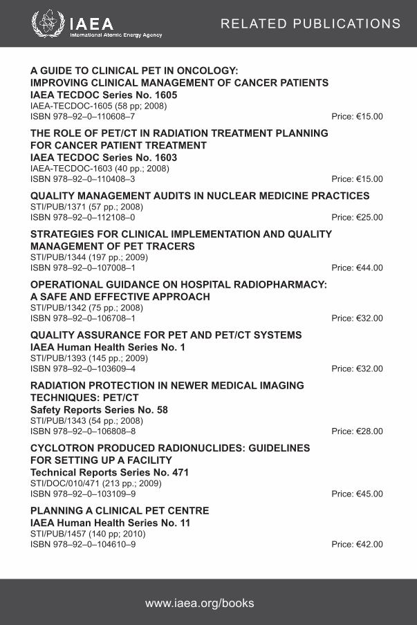
IAEA HUMAN HEALTH SERIES PUBLICATIONS
The mandate of the IAEA human health programme originates from Article II of its Statute, which states that the “Agency shall seek to accelerate and enlarge the contribution of atomic energy to peace, health and prosperity throughout the world”. The main objective of the human health programme is to enhance the capabilities of IAEA Member States in addressing issues related to the prevention, diagnosis and treatment of health problems through the development and application of nuclear techniques, within a framework of quality assurance.
Publications in the IAEA Human Health Series provide information in the areas of: radiation medicine, including diagnostic radiology, diagnostic and therapeutic nuclear medicine, and radiation therapy; dosimetry and medical radiation physics; and stable isotope techniques and other nuclear applications in nutrition. The publications have a broad readership and are aimed at medical practitioners, researchers and other professionals. International experts assist the IAEA Secretariat in drafting and reviewing these publications. Some of the publications in this series may also be endorsed or co-sponsored by international organizations and professional societies active in the relevant fields. There are two categories of publications in this series:
IAEA HUMAN HEALTH SERIESPublications in this category present analyses or provide information of an
advisory nature, for example guidelines, codes and standards of practice, and quality assurance manuals. Monographs and high level educational material, such as graduate texts, are also published in this series.
IAEA HUMAN HEALTH REPORTSHuman Health Reports complement information published in the IAEA Human
Health Series in areas of radiation medicine, dosimetry and medical radiation physics, and nutrition. These publications include reports of technical meetings, the results of IAEA coordinated research projects, interim reports on IAEA projects, and educational material compiled for IAEA training courses dealing with human health related subjects. In some cases, these reports may provide supporting material relating to publications issued in the IAEA Human Health Series.
All of these publications can be downloaded cost free from the IAEA web site:http://www.iaea.org/Publications/index.html
Further information is available from:Marketing and Sales UnitInternational Atomic Energy AgencyVienna International CentrePO Box 1001400 Vienna, Austria
Readers are invited to provide their impressions on these publications. Information may be provided via the IAEA web site, by mail at the address given above, or by email to:
RELATED PUBLICATIONS
www.iaea.org/books
A Guide to clinicAl pet in oncoloGy: improvinG clinicAl mAnAGement of cAncer pAtientsiAeA tecdoc series no. 1605IAEA-TECDOC-1605 (58 pp; 2008)ISBN 978–92–0–110608–7 Price: €15.00
the role of pet/ct in rAdiAtion treAtment plAnninG for cAncer pAtient treAtmentiAeA tecdoc series no. 1603IAEA-TECDOC-1603 (40 pp.; 2008)ISBN 978–92–0–110408–3 Price: €15.00
QuAlity mAnAGement Audits in nucleAr medicine prActicesSTI/PUB/1371 (57 pp.; 2008)ISBN 978–92–0–112108–0 Price: €25.00
strAteGies for clinicAl implementAtion And QuAlity mAnAGement of pet trAcersSTI/PUB/1344 (197 pp.; 2009)ISBN 978–92–0–107008–1 Price: €44.00
operAtionAl GuidAnce on hospitAl rAdiophArmAcy: A sAfe And effective ApproAchSTI/PUB/1342 (75 pp.; 2008)ISBN 978–92–0–106708–1 Price: €32.00
QuAlity AssurAnce for pet And pet/ct systemsiAeA human health series no. 1STI/PUB/1393 (145 pp.; 2009)ISBN 978–92–0–103609–4 Price: €32.00
rAdiAtion protection in newer medicAl imAGinG techniQues: pet/ctsafety reports series no. 58STI/PUB/1343 (54 pp.; 2008)ISBN 978–92–0–106808–8 Price: €28.00
cyclotron produced rAdionuclides: Guidelines for settinG up A fAcilitytechnical reports series no. 471STI/DOC/010/471 (213 pp.; 2009)ISBN 978–92–0–103109–9 Price: €45.00
plAnninG A clinicAl pet centreiAeA human health series no. 11STI/PUB/1457 (140 pp; 2010)ISBN 978–92–0–104610–9 Price: €42.00
10-06881_P1438_cover.indd 2 2010-04-22 11:25:23
IAEA HumAn HEAltH SErIESno. 9
Appropriate Use of FDG-PET for the Management
of Cancer Patients
INTERNATIONAL ATOMIC ENERGY AGENCYVIENNA
ISBN 978–92–0–101610–2ISSN 2075–3772
This book provides guidance on the value and appropriateness of the use of positron emission tomography (PET), either alone or in combination with computed tomography (CT) scanners using 2-fluoro-2-deoxy-D-glucose (FDG) labelled with fluorine-18, in the management of patients affected by cancer. The concept of appropriateness provides a tool for determining which diagnostic investigations and therapies should be implemented, with the overall aim of optimizing health resource allocation, recognizing not only the cost of the intervention but also the consequences of failure to implement innovations of proven effectiveness. The book includes clinical scenarios for FDG-PET/CT indications; in all, 21 different types of cancer are considered, with seven different possible indications each.
IAEA HumAn HEAltH SErIES
Appropriate U
se of FDG
-PET for the M
anagement of C
ancer Patients
IAEA HumAn HEAltH SErIES no. 9
10-06881_P1438_cover.indd 1 2010-04-22 11:25:23