Embed Size (px)
Citation preview
1
The IAC Standards and Guidelines for Pediatric Echocardiography
Accreditation
2 IAC Standards and Guidelines for Pediatric Echocardiography Accreditation (Published 6/1/2017, Revised 4/27/2018) ©2018 Intersocietal Accreditation Commission. All Rights Reserved.
Table of Contents All entries in Table of Contents are linked to the corresponding sections.
Introduction .................................................................................................................................................................................... 4 Part A: Organization ................................................................................................................................................... 5 Section 1A: Personnel and Supervision ......................................................................................................................................... 5
STANDARD – Medical Director ........................................................................................................................................... 5 STANDARD – Technical Director ........................................................................................................................................ 6 STANDARD – Medical Staff ................................................................................................................................................ 7 STANDARD – Technical Staff .............................................................................................................................................. 8 STANDARD – Support Services ........................................................................................................................................... 9
Section 1A: Personnel and Supervision Guidelines .................................................................................................................... 10 Section 2A: Facility ....................................................................................................................................................................... 11
STANDARD – Examination Areas ...................................................................................................................................... 11 STANDARD – Interpretation Areas .................................................................................................................................... 11 STANDARD – Storage ........................................................................................................................................................ 11 STANDARD – Instrument Maintenance .............................................................................................................................. 11
Section 2A: Facility Guidelines .................................................................................................................................................... 12 Section 3A: Examination Reports and Records ......................................................................................................................... 13
STANDARD – Records ....................................................................................................................................................... 13 STANDARD – Examination Interpretation and Reports ..................................................................................................... 13
Section 3A: Examination Reports and Records Guidelines ....................................................................................................... 16 Section 4A: Safety ......................................................................................................................................................................... 17
STANDARD – Patient and Facility Safety .......................................................................................................................... 17 Section 4A: Safety Guidelines ...................................................................................................................................................... 18 Section 5A: Administrative .......................................................................................................................................................... 19
STANDARD – Patient Confidentiality ................................................................................................................................ 19 STANDARD – Patient or Other Customer Complaints ....................................................................................................... 19 STANDARD – Primary Source Verification ....................................................................................................................... 19
Section 5A: Administrative Guidelines ........................................................................................................................................ 19 Section 6A: Multiple Sites (Fixed and/or Mobile) ...................................................................................................................... 20
STANDARD – Multiple Sites .............................................................................................................................................. 20 Section 6A: Multiple Sites (Fixed and/or Mobile) Guidelines .................................................................................................... 20 Bibliography .................................................................................................................................................................................. 21 Part B: Examinations and Procedures .................................................................................................................... 22 Section 1B: Pediatric Transthoracic Echocardiography Testing ............................................................................................. 22
STANDARD – Instrumentation ........................................................................................................................................... 22 STANDARD – Procedure Volumes ..................................................................................................................................... 23 STANDARD – Indications, Ordering Process and Scheduling ............................................................................................ 23 STANDARD – Techniques .................................................................................................................................................. 23 STANDARD – Components of the Transthoracic Echocardiogram .................................................................................... 24
Section 1B: Pediatric Transthoracic Echocardiography Testing Guidelines ........................................................................... 26 Bibliography .................................................................................................................................................................................. 27
STANDARD – Instrumentation ........................................................................................................................................... 28 STANDARD – Procedure Volumes ..................................................................................................................................... 28 STANDARD – Indications, Ordering Process and Scheduling ............................................................................................ 28 STANDARD – Training ...................................................................................................................................................... 29 STANDARD – Techniques .................................................................................................................................................. 29 STANDARD – Components of Transesophageal Echocardiograms .................................................................................... 30 STANDARD – Focused Pediatric TEE ................................................................................................................................ 31
Section 2B: Pediatric Transesophageal Echocardiography Testing Guidelines ....................................................................... 32 Bibliography .................................................................................................................................................................................. 33
3 IAC Standards and Guidelines for Pediatric Echocardiography Accreditation (Published 6/1/2017, Revised 4/27/2018) ©2018 Intersocietal Accreditation Commission. All Rights Reserved.
Introduction to IAC Standards for Fetal Echocardiography Testing ...................................................................................... 34 Section 3B: Fetal Echocardiography Testing ............................................................................................................................. 35
STANDARD – Instrumentation ........................................................................................................................................... 35 STANDARD – Procedure Volumes ..................................................................................................................................... 35 STANDARD – Indications, Ordering Process and Scheduling ............................................................................................ 35 STANDARD – Techniques .................................................................................................................................................. 36 STANDARD – Components of the Fetal Echocardiogram .................................................................................................. 36
Bibliography .................................................................................................................................................................................. 38 Part C: Quality Improvement .................................................................................................................................. 39 Section 1C: Quality Improvement Program ............................................................................................................................... 39
STANDARD – QI Program ................................................................................................................................................. 39 STANDARD – QI Oversight ............................................................................................................................................... 39
Section 1C: Quality Improvement Program Guidelines ............................................................................................................ 39 Section 2C: Quality Improvement Measures .............................................................................................................................. 40
STANDARD – QI Measures ................................................................................................................................................ 40 Section 2C: Quality Improvement Measures Guidelines ........................................................................................................... 41 Section 3C: Quality Improvement Meetings .............................................................................................................................. 42
STANDARD – QI Meetings ................................................................................................................................................ 42 Section 4C: Quality Improvement Documentation .................................................................................................................... 43
STANDARD – QI Documentation ....................................................................................................................................... 43
IAC Standards and Guidelines for Pediatric Echocardiography Accreditation (Published 6/1/2017, Revised 4/27/2018) 4 ©2018 Intersocietal Accreditation Commission. All Rights Reserved. Return to Table of Contents»
Introduction The Intersocietal Accreditation Commission (IAC) accredits imaging facilities specific to echocardiography. IAC accreditation is a means by which facilities can evaluate and demonstrate the level of patient care they provide. An echocardiography facility is defined as an entity located at one postal address, composed of at least one ultrasound instrument, a Medical Director and a Technical Director. There may be additional physicians and sonographers. An accredited pediatric echocardiography facility requires that the interpreting physicians and practicing sonographers be adequately trained and experienced to interpret and perform echocardiograms in children. Published documents recognize that echocardiography in pediatrics and congenital heart disease requires considerable training and expertise. Although published opinions vary with regard to the absolute numbers necessary for attaining and maintaining competence in echocardiography, all agree that numbers of studies performed or interpreted are helpful but not sufficient by themselves to assure clinical competence. It is recognized that many echocardiography facilities will perform echocardiograms on children and adults and it is likely in this setting that the pediatric echocardiograms will represent a minority of the studies performed in that facility. The intent of the accreditation process is two-fold. It is designed to recognize facilities that provide quality echocardiographic services. It is also designed to be used as an educational tool to improve the overall quality of the facility. The following are the specific areas of pediatric echocardiography for which accreditation may be obtained: • pediatric transthoracic • pediatric transesophageal • fetal These accreditation Standards and Guidelines are the minimum standards for accreditation of echocardiography facilities. Standards are the minimum requirements to which an accredited facility is held accountable. Guidelines are descriptions, examples, or recommendations that elaborate on the Standards. Guidelines are not required, but can assist with interpretation of the Standards. Standards are printed in regular typeface in outline form. Guidelines are printed in italic typeface in narrative form. Standards that are highlighted are content changes that were made as part of the June 1, 2017 revision. These Standards became effective on December 1, 2017. Facilities applying for accreditation after December 1, 2017 revision must comply with these new highlighted Standards. In addition to all Standards listed below, the facility, including all staff, must comply at all times with all federal, state and local laws and regulations, including but not limited to laws relating to licensed scope of practice, facility operations and billing requirements.
IAC Standards and Guidelines for Pediatric Echocardiography Accreditation (Published 6/1/2017, Revised 4/27/2018) 5 ©2018 Intersocietal Accreditation Commission. All Rights Reserved. Return to Table of Contents»
Part A: Organization
Section 1A: Personnel and Supervision
STANDARD – Medical Director 1.1A The Medical Director must be a licensed physician.
1.1.1A Medical Director Required Training and Experience The Medical Director must meet one of the following criteria:
1.1.1.1A Advanced Level of Expertise: High level of expertise in all aspects of pediatric
echocardiography. Physicians with this level of training are expected to be able to perform independently and to interpret echocardiograms in patients with all forms of congenital and acquired pediatric heart disease, and to supervise and train others. In addition to the core requirement of 150 studies, each advanced level physician must perform and interpret at least 200 additional pediatric transthoracic echocardiograms and review, or perform and interpret, another 200 pediatric echocardiograms. At least 50 must be done in infants one year of age or younger.
1.1.1.2A Three years of echocardiography practice experience with at least 1,800
echocardiogram/Doppler examination interpretations in infants, children and patients with congenital heart disease.
Comment: It is recognized that some facilities performing pediatric echocardiograms, particularly those that perform a majority of adult studies, will not achieve the above numbers. However, the individual Medical Director must possess the outlined experience, while it is not necessary that it be obtained at a single institution.
1.1.2A Medical Director Responsibilities
The Medical Director responsibilities include but are not limited to: 1.1.2.1A all clinical services provided and for the determination of the quality and
appropriateness of care provided;
1.1.2.2A supervising the entire operation of the facility as it relates to pediatric echocardiography or may delegate specific operations to associate directors and the Technical Director;
1.1.2.3A assuring compliance of the medical and technical staff to the Standards outlined in
this document and the supervision of their work; and
1.1.2.4A must be an active participant in the interpretation of studies performed in the facility.
1.1.3A Continuing Medical Education (CME) Requirements
1.1.3.1A The Medical Director must document at least 30 hours of CME relevant to
echocardiography over a period of three years. CME credits must be earned within the three-year period prior to application submission.
IAC Standards and Guidelines for Pediatric Echocardiography Accreditation (Published 6/1/2017, Revised 4/27/2018) 6 ©2018 Intersocietal Accreditation Commission. All Rights Reserved. Return to Table of Contents»
i. 20 hours must be Category 1 AMA ii. Other 10 echocardiography-related hours may be non-category I AMA (i.e.,
ASE CME) iii. 10 hours must be relevant to pediatric echocardiography
1.1.3.2A Yearly accumulated CME must be kept on file and available to IAC when requested. Comment: If the Medical Director has completed formal training as specified under 1.1.1.1A in the past three years, the CME requirement will be considered fulfilled.
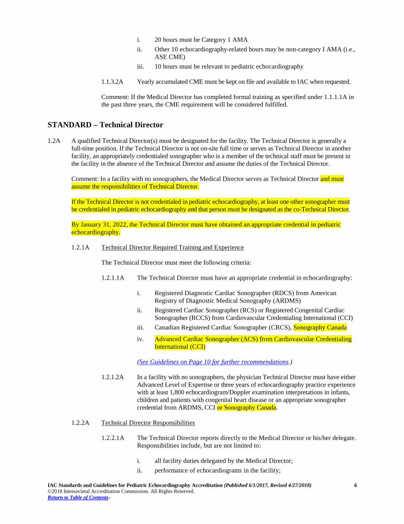
STANDARD – Technical Director 1.2A A qualified Technical Director(s) must be designated for the facility. The Technical Director is generally a
full-time position. If the Technical Director is not on-site full time or serves as Technical Director in another facility, an appropriately credentialed sonographer who is a member of the technical staff must be present in the facility in the absence of the Technical Director and assume the duties of the Technical Director. Comment: In a facility with no sonographers, the Medical Director serves as Technical Director and must assume the responsibilities of Technical Director. If the Technical Director is not credentialed in pediatric echocardiography, at least one other sonographer must be credentialed in pediatric echocardiography and that person must be designated as the co-Technical Director. By January 31, 2022, the Technical Director must have obtained an appropriate credential in pediatric echocardiography.
1.2.1A Technical Director Required Training and Experience
The Technical Director must meet the following criteria:
1.2.1.1A The Technical Director must have an appropriate credential in echocardiography:
i. Registered Diagnostic Cardiac Sonographer (RDCS) from American
Registry of Diagnostic Medical Sonography (ARDMS) ii. Registered Cardiac Sonographer (RCS) or Registered Congenital Cardiac
Sonographer (RCCS) from Cardiovascular Credentialing International (CCI) iii. Canadian Registered Cardiac Sonographer (CRCS), Sonography Canada
iv. Advanced Cardiac Sonographer (ACS) from Cardiovascular Credentialing International (CCI)
(See Guidelines on Page 10 for further recommendations.)
1.2.1.2A In a facility with no sonographers, the physician Technical Director must have either
Advanced Level of Expertise or three years of echocardiography practice experience with at least 1,800 echocardiogram/Doppler examination interpretations in infants, children and patients with congenital heart disease or an appropriate sonographer credential from ARDMS, CCI or Sonography Canada.
1.2.2A Technical Director Responsibilities
1.2.2.1A The Technical Director reports directly to the Medical Director or his/her delegate.
Responsibilities include, but are not limited to:
i. all facility duties delegated by the Medical Director; ii. performance of echocardiograms in the facility;
IAC Standards and Guidelines for Pediatric Echocardiography Accreditation (Published 6/1/2017, Revised 4/27/2018) 7 ©2018 Intersocietal Accreditation Commission. All Rights Reserved. Return to Table of Contents»
iii. general supervision of technical and ancillary staff, if applicable; iv. the delegation, when warranted, of specific responsibilities to the technical
staff and/or the ancillary staff; v. daily technical operation of the facility (e.g., staff scheduling, patient
scheduling, facility record keeping, etc.); vi. operation and maintenance of facility equipment; vii. compliance of the technical and/or ancillary staff to the Standards outlined
within this document; viii. working with the Medical Director, medical staff and technical staff to
ensure quality patient care; and ix. technical training.
1.2.3A Continuing Medical Education (CME) Requirements
1.2.3.1A The Technical Director must document at least 15 hours of echocardiography-
related CME over a period of three years. CME credits must be earned within the three-year period prior to application submission. (See Guidelines on Page 10 for further recommendations.)
1.2.3.2A Yearly accumulated CME must be kept on file and available to IAC when requested. Comment: The CME requirement will be considered fulfilled if the credential status of the Technical Director is currently active as a Registered Diagnostic Cardiac Sonographer (RDCS) from American Registry of Diagnostic Medical Sonography (ARDMS); or Registered Cardiac Sonographer (RCS) or Registered Congenital Cardiac Sonographer (RCCS) from Cardiovascular Credentialing International (CCI); Canadian Registered Cardiac Sonographer (CRCS), Sonography Canada; or Advanced Cardiac Sonographer (ACS) from CCI. Fifteen CME hours must be echocardiography-related, 10 hours must be relevant to pediatric echocardiography.
STANDARD – Medical Staff 1.3A All members of the medical staff must be licensed physicians.
1.3.1A Medical Staff Required Training and Experience The medical staff must meet one of the following criteria:
1.3.1.1A Advanced Level of Expertise: High level of expertise in all aspects of pediatric
echocardiography. Physicians with this level of training are expected to be able to perform independently and to interpret echocardiograms in patients with all forms of congenital and acquired pediatric heart disease, and to supervise and train others. In addition to the core requirement of 150 studies, each advanced level physician must perform and interpret at least 200 additional pediatric transthoracic echocardiograms and review, or perform and interpret, another 200 pediatric echocardiograms. At least 50 must be done in infants.
1.3.1.2A Core Level of Expertise: Basic set of technical and interpretive skills required for
graduation from a pediatric cardiology training program accredited by ACGME and includes four to six months of echocardiography, performance and interpretation at least 150 pediatric echocardiograms, including at least 50 in patients one year of age or younger, under the supervision of the facility director or other qualified staff pediatric cardiologist echocardiographer(s). Physicians with this level of expertise are expected to be able to perform and interpret TTEs in normal infants, children and adolescents, and in those with childhood heart disease with consultation as needed.
IAC Standards and Guidelines for Pediatric Echocardiography Accreditation (Published 6/1/2017, Revised 4/27/2018) 8 ©2018 Intersocietal Accreditation Commission. All Rights Reserved. Return to Table of Contents»
1.3.1.3A Three years of echocardiography practice experience with at least 450 echocardiogram/Doppler examination interpretations in infants, children and patients with congenital heart disease.
Comment: It is recognized that some facilities performing pediatric echocardiograms, particularly those that perform a majority of adult studies, will not achieve the above numbers. However, the individual pediatric medical staff member must have this experience, even if it is not achieved at a single institution.
1.3.2A Medical Staff Responsibilities
Medical staff responsibilities include but are not limited to:
1.3.2.1A The medical staff interprets and/or performs clinical studies.
1.3.3A Continuing Medical Education (CME) Requirements
1.3.3.1A The medical staff must document at least 15 hours of CME relevant to
echocardiography over a period of three years. CME credits must be earned within the three-year period prior to application submission.
i. 10 hours must be Category 1 AMA ii. Other five echocardiography-related hours may be non-category I AMA
(i.e., ASE CME) iii. 10 hours must be relevant to pediatric echocardiography
1.3.3.2A Yearly accumulated CME must be kept on file and available to IAC when
requested. Comment: If the medical staff has completed formal training as specified under 1.3.1.1A in the past three years, the CME requirement will be considered fulfilled.
STANDARD – Technical Staff
1.4A All members of the technical staff must be qualified sonographers.
1.4.1A Technical Staff Required Training and Experience The technical staff members must meet one of the following criteria:
1.4.1.1A An appropriate credential in echocardiography:
i. Registered Diagnostic Cardiac Sonographer (RDCS) from American
Registry of Diagnostic Medical Sonography (ARDMS) ii. Registered Cardiac Sonographer (RCS) or Registered Congenital Cardiac
Sonographer (RCCS) from Cardiovascular Credentialing International (CCI) iii. Canadian Registered Cardiac Sonographer (CRCS), Sonography Canada
iv. Advanced Cardiac Sonographer (ACS) from Cardiovascular Credentialing International (CCI)
(See Guidelines on Page 10 for further recommendations.)
IAC Standards and Guidelines for Pediatric Echocardiography Accreditation (Published 6/1/2017, Revised 4/27/2018) 9 ©2018 Intersocietal Accreditation Commission. All Rights Reserved. Return to Table of Contents»
1.4.1.2A Provisional Staff i. New graduates of a cardiac ultrasound program who are employed in an
accredited facility must obtain an appropriate credential within one year from the date of graduation. These individuals must be listed on the application as provisional technical staff who are eligible for credentialing, and must only work under appropriate supervision of a credentialed sonographer.
ii. Individuals employed in an accredited facility who are cross-training in echocardiography or working to fulfill clinical experience pre-requisites for a credentialing examination must obtain an appropriate credential within two years from the start date of training. These individuals must be listed on the application as provisional technical staff who are eligible for credentialing, and must only work under appropriate supervision of a credentialed sonographer.
1.4.2A Technical Staff Responsibilities Technical staff responsibilities include but are not limited to: 1.4.2.1A must report to the Technical Director; and
1.4.2.2A assumes the responsibilities specified by the Technical Director and, in general, is
responsible for the performance of clinical examinations and other tasks assigned.
1.4.3A Continuing Medical Education (CME) Requirements 1.4.3.1A The technical staff must document at least 15 hours of echocardiography-related
CME over a period of three years. CME credits must be earned within the three-year period prior to application submission. (See Guidelines on Page 10 for further recommendations.)
1.4.3.2A Yearly accumulated CME must be kept on file and available to IAC when requested. Comment: The CME requirement will be considered fulfilled if credential status of the technical staff member is currently active as a Registered Diagnostic Cardiac Sonographer (RDCS) from American Registry of Diagnostic Medical Sonography (ARDMS); or Registered Cardiac Sonographer (RCS) or Registered Congenital Cardiac Sonographer (RCCS) from Cardiovascular Credentialing International (CCI); Canadian Registered Cardiac Sonographer (CRCS), Sonography Canada; or Advanced Cardiac Sonographer (ACS) from CCI. Fifteen CME hours must be echocardiography-related; 10 hours must be relevant to pediatric echocardiography.
STANDARD – Support Services
1.5A Ancillary personnel (clerical, nursing, transport, etc.) necessary for safe and efficient patient care are provided. 1.5.1A Clerical and administrative support must be sufficient to ensure efficient operation and record
keeping.
1.5.2A Nursing and ancillary services sufficient to ensure quality patient care are available when necessary.
1.5.3A Supervision: The Medical Director must ensure that support services appropriate for and in the best interest of patient care are provided.
IAC Standards and Guidelines for Pediatric Echocardiography Accreditation (Published 6/1/2017, Revised 4/27/2018) 10 ©2018 Intersocietal Accreditation Commission. All Rights Reserved. Return to Table of Contents»
Section 1A: Personnel and Supervision Guidelines
1.2.1.1A A credential in pediatric echocardiography is preferred if the Technical Director will be performing
pediatric echocardiograms. 1.2.3.1A and 1.4.3.1A Technical Director and Technical Staff CME Requirements
Explanation: Echocardiography-related CME may be Category 1 AMA, or other approved non-category 1 credit including those credits designated as approved by organizations such as ASE, SDMS or ARRT that have content specific to echocardiography.
1.4.1.1A Comment: A credential in pediatric echocardiography is recommended.

IAC Standards and Guidelines for Pediatric Echocardiography Accreditation (Published 6/1/2017, Revised 4/27/2018) 11 ©2018 Intersocietal Accreditation Commission. All Rights Reserved. Return to Table of Contents»
Section 2A: Facility
STANDARD – Examination Areas 2.1A Examinations must be performed in a setting providing patient and technical staff safety, comfort and
privacy. 2.1.1A The adequate performance of an echocardiogram requires the proper positioning of the patient,
the echocardiographic system and the sonographer. For this reason, adequate spacing is required for inclusion of a patient bed, which allows for position changes, an echocardiographic imaging system and patient privacy. 2.1.1.1A It is understood that many echocardiographic studies are performed on a portable
basis, requiring performance of the studies in less than optimal conditions. All studies, regardless of the location, must be performed with adequate room for patient positioning and equipment use.
2.1.1.2A Patient privacy must be assured with the use of either appropriate curtains or
doors.
2.1.1.3A A sink and antiseptic soap must be readily available and used for hand washing in accordance with the infection control policy of the facility.
(See Guidelines on Page 12 for further recommendations.)
STANDARD – Interpretation Areas
2.2A Adequate designated space must be provided for the interpretation of the echocardiogram and the preparation of reports. (See Guidelines on Page 12 for further recommendations.)
STANDARD – Storage 2.3A Space permitted for storage of records and supplies must be sufficient for the patient volume of the facility.
STANDARD – Instrument Maintenance
2.4A Instrumentation used for diagnostic testing must be maintained in good operating condition. The accuracy of the data collected by ultrasound instruments is paramount in the interpretation and diagnostic utilization of the information collected. Guidelines for equipment maintenance include, but are not limited to, the following: 2.4.1A Recording of the method and frequency of maintenance of ultrasound instrumentation and
digitizing equipment.
2.4.2A Establishment of and adherence to a policy regarding routine safety inspections and testing of all facility electrical equipment.
2.4.3A Establishment of and adherence to an instrument cleaning schedule that includes routine cleaning of equipment parts, including filters and transducers, according to the specifications of the manufacturer. The cleaning schedule must be frequent enough to allow for accurate collection of data.
IAC Standards and Guidelines for Pediatric Echocardiography Accreditation (Published 6/1/2017, Revised 4/27/2018) 12 ©2018 Intersocietal Accreditation Commission. All Rights Reserved. Return to Table of Contents»
Section 2A: Facility Guidelines
2.1.1A Approximately 150 square feet is recommended for a transthoracic echocardiography examination
room. 2.2A Space should be provided for data evaluation, interpretation and discussion of the study with the
sonographer and/or referring physician as needed.
IAC Standards and Guidelines for Pediatric Echocardiography Accreditation (Published 6/1/2017, Revised 4/27/2018) 13 ©2018 Intersocietal Accreditation Commission. All Rights Reserved. Return to Table of Contents»
Section 3A: Examination Reports and Records
STANDARD – Records 3.1A Provisions must exist for the generation and retention of examination data for all echocardiograms performed.
3.1.1A A system for recording and archiving echocardiographic data (images, measurements and final
reports) obtained for diagnostic purposes must be in place.
3.1.2A A permanent record of the images and interpretation must be made and retained in accordance with applicable state or federal guidelines for medical records. Records for pediatric patients may need to be retained for a longer period of time than those of adult patients. Echocardiographic data, images and interpretations must be retrievable for comparison with new studies.
3.1.3A Studies must be archived in the original format that they were acquired. Archiving media
includes, but is not limited to:
3.1.3.1A videotape; and
3.1.3.2A digital storage – the facility must ensure that a sufficient portion of the examination can be archived in digital storage and that a secure back-up system is in place. Digital studies must include information consistent with that required for videotape acquisition, although fewer cardiac cycles are generally recorded.
(See Guidelines on Page 16 for further recommendations.
STANDARD – Examination Interpretation and Reports
3.2A Provisions must exist for the timely reporting of examination data. 3.2.1A There must be a policy in place for communicating critical results.
3.2.2A The findings of a STAT echocardiogram must be made available immediately by the
interpreting physician. Comment: Sonographer worksheets, comments (verbal or written) or electronic summary of findings must not be provided to anyone other than the interpreting physician. (See Guidelines on Page 16 for further recommendations.)
3.2.3A Preliminary reports can only be issued by a physician. There must be a policy in place for
communicating any significant changes between the preliminary and final reports. 3.2.4A Routine inpatient echocardiographic studies must be interpreted by a qualified physician within
24 hours of completion of the examination. Outpatient studies must be interpreted by the end of the next business day. The final verified (by the interpreting physician) signed report must be completed within 48 hours after interpretation. (See Guidelines on Page 16 for further recommendations.)
3.3A Echocardiography reporting must be standardized in the facility. All physicians interpreting
echocardiograms in the facility must agree on uniform diagnostic criteria and a standardized report format.1 3.3.1A The report must accurately reflect the content and results of the study. The report must include,
but may not be limited to:
IAC Standards and Guidelines for Pediatric Echocardiography Accreditation (Published 6/1/2017, Revised 4/27/2018) 14 ©2018 Intersocietal Accreditation Commission. All Rights Reserved. Return to Table of Contents»
3.3.1.1A Demographic Data: i. date of study; ii. name and/or identifier of the facility; iii. name and/or identifier of the patient; iv. date of birth and/or age of the patient; v. gender; vi. name of the performing sonographer and/or identifier; and vii. name of the ordering physician and/or identifier.
3.3.1.2A Clinical Data:
i. primary indication for the study; ii. patient height and weight for determination of BSA; and iii. blood pressure – systolic and diastolic blood pressure must be obtained on
or around the time of the study and displayed on the report. Comment: The information must be sufficient to allow for the identification and retrieval of previous studies on the same patient. Please note: The reporting requirements above (3.3.1.1A and 3.3.1.2A) are for pediatric TTE and TEE only. For the fetal requirements, please refer to Fetal Echocardiogram Report Components on page 15 of this document.
3.3.1.3A A summary of the results of the examination, including any pertinent positive and negative findings.
3.3.1.4A The final report must be completely typewritten, including the printed name of the interpreting physician. The final report must be reviewed, signed and dated manually or electronically by the interpreting physician. Electronic signatures must be password protected and indicate they are electronically recorded. Stamped signatures or signing by non-physician staff is unacceptable.
3.4A Pediatric Transthoracic Echocardiogram Report Components
3.4.1A The report must accurately reflect the content and results of the study. The report must comment
on whether a given dimension is normal or abnormal. If any structure is not well visualized this must be noted. The report text must be consistent with the quantitative and Doppler data. Where appropriate, this must include localization and quantification of abnormal findings. 3.4.1.1A The report for a complete study must include the following numerical data:
i. the measurements performed in the course of the examination and/or
interpretation; and ii. numerical data for transthoracic echocardiograms, must include, but not be
limited to (except where technically or anatomically unobtainable or not applicable in the clinical setting): • measurements of the left ventricular internal dimension or volume at
end-diastole; • left ventricular internal dimension or volume at end-systole; • left ventricular posterobasal free wall thickness and septal thickness or
left ventricular mass at end-diastole; • aortic root dimension; and

IAC Standards and Guidelines for Pediatric Echocardiography Accreditation (Published 6/1/2017, Revised 4/27/2018) 15 ©2018 Intersocietal Accreditation Commission. All Rights Reserved. Return to Table of Contents»
• additional measurements may be indicated and when performed must be included. Comment: Examples of exceptions to the above include: o right ventricular hypertension; o hypoplastic left heart syndrome; o tetralogy of Fallot; and o other pathology.
3.4.1.2A A report of the Doppler evaluation must include, but not be limited to:
i. the evaluation of peak and mean gradients (if stenosis is present); ii. degree of regurgitation; iii. peak tricuspid regurgitation velocity for estimation of right ventricular
systolic pressure where obtainable or applicable in the clinical setting; and iv. other pathology.
(See Guidelines on Page 16 for further recommendations.)
3.5A Pediatric Transesophageal Echocardiogram Report Components 3.5.1A The report must accurately reflect the content and results of the study. The report must include,
but may not be limited to: 3.5.1.1A The report text must include:
i. complications of procedure (yes or no).
3.5.1.2A The report text must comment on all structures evaluated in the examination and
must be consistent with the quantitative and Doppler data. Where appropriate, this must include location and quantification of abnormal findings.
(See Guidelines on Page 16 for further recommendations.)
3.6A Fetal Echocardiogram Report Components
3.6.1A The report must include, but may not be limited to:
3.6.1.1A Demographic Data:
i. date of study; ii. name and/or identifier of the facility; iii. name and/or identifier of the patient; iv. date of birth and/or age of the patient; v. name of the sonographer/physician performing the study; and vi. name of the ordering physician(s) and/or identifier.
3.6.1.2A Clinical Data:
i. primary indication for the study; ii. last menstrual period or estimated date of delivery; and iii. fetal number (if more than one).
IAC Standards and Guidelines for Pediatric Echocardiography Accreditation (Published 6/1/2017, Revised 4/27/2018) 16 ©2018 Intersocietal Accreditation Commission. All Rights Reserved. Return to Table of Contents»
3.6.1.3A The report of measurements must include but not be limited to: i. The measurements performed in the course of the examination where
normal values are known and/or interpretation appropriate to the clinical issue or area(s) of abnormality.
3.6.1.4A The report of the Doppler evaluation must include, but not be limited to: i. The Doppler values, normal and abnormal, obtained in the course of the
examination appropriate to the clinical issue or area(s) of abnormality.
3.6.1.5A The report text must include comments on: i. components of procedure (i.e., color flow Doppler, PW/CW Doppler); ii. all structures evaluated in the examination as specified in the IAC
Echocardiography Standards; and iii. the report text must be consistent with the quantitative and Doppler data.
Where appropriate, this must include localization and quantification of abnormal findings.
(See Guidelines below for further recommendations.)
Section 3A: Examination Reports and Records Guidelines
3.1.3A Archiving media:
i. Videotape: When utilizing videotape for archiving, at least 5-10 cardiac cycles of each portion of the M-Mode, 2-D and Doppler study should be recorded in real time. The location and method of measurements performed should also be archived.
ii. Digital storage: The number of cardiac cycles acquired must be sufficient to allow for adequate review).4
3.2.2A Suggested method for reporting life-threatening findings: Optimally, the interpreting physician in the
facility will call the appropriate physician. Alternatively, the sonographer may call the appropriate physician after conferring with the interpreting physician.
3.2.4A Comment: An interpretation can be in the form of paper, digital storage or an accessible voice system. 3.4.1.2A Pediatric Transthoracic Reports – If the examination is abbreviated for any reason (i.e., patient
uncooperativeness secondary to an unsedated exam) it should be noted in the report text. 3.5.1.2A Pediatric Transesophageal Reports – If any structure is not well visualized this should be noted. If the
examination is abbreviated for any reason it should be noted in the report text. 3.6.1.5A Fetal Reports – If any structure is not well visualized this should be noted.
IAC Standards and Guidelines for Pediatric Echocardiography Accreditation (Published 6/1/2017, Revised 4/27/2018) 17 ©2018 Intersocietal Accreditation Commission. All Rights Reserved. Return to Table of Contents»
Section 4A: Safety
STANDARD – Patient and Facility Safety 4.1A Patient and employee safety is ensured by written policies and procedures approved by the Medical
Director. 4.1.1A Personnel Safety Policy (Ergonomics) – A policy must be in place to address technical staff
safety, comfort and avoidance of work-related musculoskeletal disorders (MSD). (See Guidelines on Page 18 for further recommendations.)
4.1.2A Standard echocardiograms are safe to both patients and sonographers. Special echocardiographic procedures, such as transesophageal echocardiograms, sedated echocardiograms and stress echocardiograms, pose potential risks to the safety of the patient due to either their semi-invasive nature, or the physiologic stress placed on the cardiovascular system of the patient. For this reason, an echocardiography facility providing special echocardiographic procedures must have an emergency procedure plan and the following emergency supplies must be readily available for transesophageal and sedated echocardiograms:
4.1.2.1A a fully-equipped cardiac arrest cart (crash cart):
i. The size and dosage differences between pediatric and adult patients must
be recognized. ii. Pediatric dosing information and/or appropriate pediatric dose of
emergency medications must be available.
4.1.2.2A a defibrillator;
4.1.2.3A appropriate sized pediatric equipment for starting and maintaining intravenous access;
4.1.2.4A oxygen tanks or wall mounted oxygen sources with appropriately sized cannulae
and/or masks; and
4.1.2.5A suction equipment. 4.1.3A The facility must meet the standards set forth by the Occupational Safety and Health
Administration (OSHA) and by the Joint Commission (JC), where applicable.
4.1.4A The facility must have a written procedure in place for handling acute medical emergencies. 4.1.5A The facility must recognize the potential need for patient sedation in pediatrics to obtain an
adequate examination. Written policies must exist for the use of moderate sedation in children including but not limited to:
4.1.5.1A type of sedatives and appropriate dosing for age and size; and
4.1.5.2A monitoring of children during and after the examination.
(See Guidelines on Page 18 for further recommendations.)
IAC Standards and Guidelines for Pediatric Echocardiography Accreditation (Published 6/1/2017, Revised 4/27/2018) 18 ©2018 Intersocietal Accreditation Commission. All Rights Reserved. Return to Table of Contents»
Section 4A: Safety Guidelines
4.1A Sonographer Radiation Exposure: Sonographers may be exposed to significant levels of radiation from
patients who have both a nuclear test and an echocardiogram on the same day, and also from spending time in catheterization/hybrid laboratories. For this reason, it is recommended that facilities have a formal policy to address radiation safety for sonographers.6
4.1.1A Comment: For additional information regarding MSD, please visit:
www.cdc.gov/niosh/docs/wp-solutions/2006-148/ www.sdms.org/pdf/wrmsd2003.pdf

IAC Standards and Guidelines for Pediatric Echocardiography Accreditation (Published 6/1/2017, Revised 4/27/2018) 19 ©2018 Intersocietal Accreditation Commission. All Rights Reserved. Return to Table of Contents»
Section 5A: Administrative
STANDARD – Patient Confidentiality
5.1A All facility personnel must ascribe to professional principles of patient-physician confidentiality as legally required by federal, state, local or institutional policy or regulation.
STANDARD – Patient or Other Customer Complaints 5.2A There must be a policy in place outlining the process for patients or other customers to issue a
complaint/grievance in reference to the care/services they received at the facility and how the facility handles complaints/grievances.
STANDARD – Primary Source Verification 5.3A There must be a policy in place identifying how the facility verifies the medical education, training,
appropriate licenses and certifications of all physicians as well as, the certification and training of all technical staff members and any other direct patient care providers.
Section 5A: Administrative Guidelines
Sample documents are available for each of the required policies listed in Section 5A on the IAC Echocardiography website at intersocietal.org/echo/seeking/sample_documents_pediatric.htm.

IAC Standards and Guidelines for Pediatric Echocardiography Accreditation (Published 6/1/2017, Revised 4/27/2018) 20 ©2018 Intersocietal Accreditation Commission. All Rights Reserved. Return to Table of Contents»
Section 6A: Multiple Sites (Fixed and/or Mobile)
STANDARD – Multiple Sites
6.1A When testing is performed at more than one physical facility, the facility may be eligible to apply for a single accreditation as a multiple site facility. 6.1.1A All facilities have the same Medical Director.
6.1.2A All facilities have the same Technical Director. 6.1.3A Identical testing protocols are used at all sites. 6.1.4A Identical diagnostic criteria are used at all sites. 6.1.5A Quality Improvement (QI) must be evaluated for each site for all areas of testing performed at
the site. 6.1.6A Equipment of similar quality and capability must be used at all sites.
Section 6A: Multiple Sites (Fixed and/or Mobile) Guidelines
Facilities needing complete details on adding a multiple site should review the current IAC Policies and Procedures available on the IAC website at intersocietal.org/iac/legal/policies.htm.
IAC Standards and Guidelines for Pediatric Echocardiography Accreditation (Published 6/1/2017, Revised 4/27/2018) 21 ©2018 Intersocietal Accreditation Commission. All Rights Reserved. Return to Table of Contents»
Bibliography 1. ACC/AHA/AAP Recommendations for Training in Pediatric Cardiology - Task Force 2: Pediatric Training Guidelines for
Noninvasive Cardiac Imaging endorsed by the American Society of Echocardiography and the Society of Pediatric Echocardiography. Sanders, S., et al., J Am Coll of Cardiol, 2005;46(7);1384-1388. content.onlinejacc.org/article.aspx?articleid=1136952
2. Guidelines and Standards for Performance of a Pediatric Echocardiogram: A Report from the Task Force of the Pediatric Council
of the American Society of Echocardiography. Lai, W., et al., J Am Soc of Echocardiogr, 2006;19(12);1413-1430. www.onlinejase.com/article/S0894-7317(06)00936-9/fulltext
3. Guidelines for Monitoring and Management of Pediatric Patients During and After Sedation for Diagnosis and Therapeutic
Procedures Committee on Drugs for the American Academy of Pediatrics. Coté, C., et al., Pediatrics, 2006;118(6);2587-2602. pediatrics.aappublications.org/content/118/6/2587
4. Guidelines and Recommendations for Digital Echocardiography: A Report from the Digital Echocardiography Committee of the
American Society of Echocardiography. Thomas, J., et al., J Am Soc Echocardiogr, 2005;18(3):287-297. www.onlinejase.com/article/S0894-7317(05)00019-2/fulltext
5. Recommendations for Quantification Methods During the Performance of a Pediatric Echocardiogram: A Report from the
Pediatric Measurements Writing Group of the American Society of Echocardiography Pediatric and Congenital Heart Disease Council. Lopez, L., et al., J Am Soc Echocardiogr, 2010;23(5):465-495. www.onlinejase.com/article/S0894-7317(10)00266-X/fulltext
6. Radiation Safety for the Cardiac Sonographer: Recommendations of the Radiation Safety Writing Group for the Council on
Cardiovascular Sonography of the American Society of Echocardiography. McIlwain, E., et al., J Am Soc Echocardiogr, 2014;27(8):811-6. www.onlinejase.com/article/S0894-7317(14)00432-5/fulltext
7. Industry Standards for the Prevention of Work Related Musculoskeletal Disorders in Sonography: Developed Through A 2016
Consensus Conference Hosted by the Society of Diagnostic Medical Sonography. www.sdms.org/docs/default-source/Resources/industry-standards-for-the-prevention-of-work-related-musculoskeletal-disorders-in-sonography.pdf
IAC Standards and Guidelines for Pediatric Echocardiography Accreditation (Published 6/1/2017, Revised 4/27/2018) 22 ©2018 Intersocietal Accreditation Commission. All Rights Reserved. Return to Table of Contents»
Part B: Examinations and Procedures
Section 1B: Pediatric Transthoracic Echocardiography Testing
STANDARD – Instrumentation 1.1B Cardiac Ultrasound Systems
1.1.1B Ultrasound instruments utilized for diagnostic studies must include, at a minimum, hardware and software to perform: 1.1.1.1B M-Mode imaging;
1.1.1.2B 2-D imaging (the system must include harmonic capabilities);
1.1.1.3B spectral display for pulsed (PW) and continuous wave (CW) Doppler studies;
1.1.1.4B color flow imaging;
1.1.1.5B monitor or other display method of suitable size and quality for observation and
interpretation of all modalities; Comment: The display or DICOM header must identify the parent institution, the name of the patient, the date and time of the study. The ECG must also be displayed.
1.1.1.6B range or depth markers must be available on all displays;
1.1.1.7B capabilities to measure the distance between two points, an area on a 2-D image,
blood flow velocities, time intervals and peak and mean gradients from spectral Doppler studies;
1.1.1.8B transducers, which can provide adequate imaging across the wide range of depths
encountered in pediatrics, must be available.
i. Multiple imaging transducers, ranging from low frequency (2-2.5 MHz) to high frequency (7.5 MHz or higher) or a multi-frequency transducer which includes a range of frequencies specific to the clinical needs in pediatric echo.
ii. A transducer dedicated to the performance of non-imaging continuous wave Doppler must be available at each site.
1.1.1.9B an audible output must be present at the time of acquisition;
1.1.1.10B machines with some, but not all of the above, equipment may be used for limited
or directed echocardiographic examinations. However, machines utilized for complete diagnostic procedures must include all of the above listed capabilities.
(See Guidelines on Page 26 for further recommendations.)

IAC Standards and Guidelines for Pediatric Echocardiography Accreditation (Published 6/1/2017, Revised 4/27/2018) 23 ©2018 Intersocietal Accreditation Commission. All Rights Reserved. Return to Table of Contents»
STANDARD – Procedure Volumes 1.2B The annual procedure volume is sufficient to maintain proficiency in examination performance and
interpretation.
STANDARD – Indications, Ordering Process and Scheduling 1.3B Transthoracic echocardiography testing is performed for appropriate indications.1
1.3.1B Verification of the Indication – A process must be in place in the facility for obtaining and recording the indication. Before a study is performed, the indication must be verified and any additional information needed to direct the examination must be obtained.1
1.4B Echocardiography testing is appropriately ordered and scheduled.
1.4.1B Ordering Process – The echocardiogram order and requisition must clearly indicate the type of study to be performed (i.e., complete or limited), the reason(s) for the study and the clinical question(s) to be answered. The signed (electronic or handwritten) order/requisition must be present in the medical record of the patient.
1.4.2B Definition of Procedure Types and Protocols
1.4.2.1B Complete Study: A complete imaging study is one that defines the cardiac and visceral position and a complete segmental image analysis of the heart from multiple views and also defines the cardiac anatomy and physiology as fully as possible using imaging and Doppler modalities.
1.4.2.2B Limited Study: A limited study generally examines a specific region of interest of
the heart and/or addresses a defined clinical question. Limited studies are not sufficient if the patient with suspected congenital heart disease has never had a complete echocardiogram before.
1.4.3B Scheduling – Sufficient time must be allotted for each study according to the procedure type.
The performance time allotted for a complete (imaging and Doppler) pediatric transthoracic examination is 45 to 60 minutes from patient encounter to departure. Additional time may be required for complicated studies or sedated patients.
1.4.3.1B An urgent study must be performed in the next available time period.
1.4.3.2B A stat study must be performed as soon as possible, preempting routine studies.
1.4.3.3B Availability for Emergencies: Qualified personnel and equipment must be
available for urgent or stat studies outside normal working hours in inpatient facilities or where appropriate.
(See Guidelines on Page 26 for further recommendations.)
STANDARD – Techniques 1.5B Examination performance must include proper technique.
1.5.1B All procedures must be explained to the patient and/or parents or guardian.
1.5.2B Elements of study performance and quality include, but are not limited to:

IAC Standards and Guidelines for Pediatric Echocardiography Accreditation (Published 6/1/2017, Revised 4/27/2018) 24 ©2018 Intersocietal Accreditation Commission. All Rights Reserved. Return to Table of Contents»
1.5.2.1B optimizing patient position with careful attention to comfort and safety; Comment: This is particularly important in vulnerable patients such as critically ill neonates.
1.5.2.2B appropriate patient distraction such as movies or sedation utilizing institutional
protocols;
1.5.2.3B correct transducer selection for patient size;
1.5.2.4B optimization of equipment settings and display of ECG;
1.5.2.5B performance of a complete 2-D/M-Mode/Doppler imaging and hemodynamic examination according to the facility specific protocols that incorporate all views and imaging planes mandated by Standards 1.6.1.1B, 1.6.1.2B, 1.6.1.3B, 1.6.1.4B, 1.6.1.5B;
1.5.2.6B storage of all images and data; and
1.5.2.7B timely report generation and communication of results.
STANDARD – Components of the Transthoracic Echocardiogram 1.6B Transthoracic echocardiograms must be comprehensive and include standard components.
1.6.1B Components of the Examination – A protocol must be in place that defines the components of the standard examination. (See Guidelines on Page 26 for further recommendations.)
1.6.1.1B The complete examination, when applicable or available, must include the
following standard views when cardiac anatomy allows:
i. inferior and superior vena cava; ii. hepatic veins; iii. pulmonary veins; iv. right, left or single atrial morphology; v. atrial septum; vi. mitral, tricuspid or single atrioventricular valve morphology and function; vii. right, left or single ventricular morphology; viii. ventricular septum; ix. semilunar valve morphology and function; x. coronary arteries when visible; xi. ascending, transverse and descending aorta with demonstration of arch
sidedness and branching pattern; xii. main pulmonary artery and proximal branch pulmonary arteries; xiii. pericardium; xiv. measurements of the cardiac chambers and ventricular function where
standard measurements are available.
1.6.1.2B Complete Doppler Study: Includes spectral Doppler and/or color flow interrogation of all normal and abnormal flows within the heart including:
i. atrioventricular valves; ii. semilunar valves;
IAC Standards and Guidelines for Pediatric Echocardiography Accreditation (Published 6/1/2017, Revised 4/27/2018) 25 ©2018 Intersocietal Accreditation Commission. All Rights Reserved. Return to Table of Contents»
iii. atrial septum; iv. ventricular septum; v. great vessels.
1.6.1.3B Standard views for the complete examination of the anatomically normal heart
must include the following standard 2-D views (except where technically unobtainable):
i. parasternal long axis view; (including evaluation of the right ventricular
inflow and right ventricular outflow); ii. parasternal short axis views (including evaluation at the level of the aorta
and pulmonary valves, mitral, mid - papillary muscle level and apex); iii. apical four-chamber view; iv. apical long axis view; v. subcostal long axis view; vi. subcostal short axis view including evaluation of the SVC, IVC, hepatic
veins and descending aorta; vii. suprasternal long axis view; viii. suprasternal short axis view; ix. right parasternal view (when indicated).
1.6.1.4B The following 2-D or M-Mode measurements of the left heart (where appropriate):
i. left ventricular internal dimension or volume at end-diastole; ii. left ventricular internal dimension or volume at end-systole; iii. left ventricular posterobasal free wall thickness and ventricular septal
thickness at end-diastole or left ventricular mass; iv. aortic root dimension.
1.6.1.5B The following standard Doppler evaluations:
i. spectral Doppler interrogation and/or color mapping in at least two imaging
planes for all four valves; ii. tricuspid regurgitation velocity when available to estimate the systolic right
ventricular pressure in patients with anatomically normal hearts; iii. color mapping of the atrial and ventricular septa to exclude defects; iv. spectral Doppler interrogation and color mapping for the ventricular
outflow tracts and aortic arch; v. spectral Doppler interrogation and color mapping of the hepatic and
pulmonary veins may be helpful in some cases; vi. use of a dedicated non-imaging CW Doppler transducer to assess stenotic
valves or valvular regurgitation may be helpful in some cases.
Comment: These may be different in congenitally malformed and/or surgically repaired complex malformations and cases with abnormalities of cardiac position.
IAC Standards and Guidelines for Pediatric Echocardiography Accreditation (Published 6/1/2017, Revised 4/27/2018) 26 ©2018 Intersocietal Accreditation Commission. All Rights Reserved. Return to Table of Contents»
Section 1B: Pediatric Transthoracic Echocardiography Testing
Guidelines 1.1.1B Cardiac Ultrasound Systems
• Instrument settings to enable optimization of ultrasound contrast agents. • There should be a system setting to display low frequency Doppler filtering for tissue Doppler
display. 1.4.3B Scheduling
A routine study on an inpatient should be performed on the same working day as ordered, unless otherwise specified. Outpatient studies should be assigned priority as defined by the referring physician and/or the indication of the study.
1.6.1B Components of the Examination
For all imaging protocols, if any required view or Doppler signal cannot be adequately obtained, it should be recorded and labeled in order to demonstrate that it was attempted.
IAC Standards and Guidelines for Pediatric Echocardiography Accreditation (Published 6/1/2017, Revised 4/27/2018) 27 ©2018 Intersocietal Accreditation Commission. All Rights Reserved. Return to Table of Contents»
Bibliography 1. Guidelines and Standards for Performance of a Pediatric Echocardiogram: A Report from the Task Force of the Pediatric Council
of the American Society of Echocardiography. Lai, W., et al., J Am Soc of Echocardiogr, 2006;19(12);1413-1430. www.onlinejase.com/article/S0894-7317(06)00936-9/fulltext
2. Recommendations for Quantification Methods During the Performance of a Pediatric Echocardiogram: A Report from the Pediatric Measurements Writing Group of the American Society of Echocardiography Pediatric and Congenital Heart Disease Council. Lopez, L., et al., J Am Soc Echocardiogr, 2010;23(5):465-495. www.onlinejase.com/article/S0894-7317(10)00266-X/fulltext
3. ACC/AAP/AHA/ASE/HRS/SCAI/SCCT/SCMR/SOPE 2014 Appropriate Use Criteria for Initial Transthoracic Echocardiography
in Outpatient Pediatric Cardiology. Campbell, R., et al., J Am Coll of Cardiol, 2014;64(19):735-1097. www.onlinejacc.org/content/64/19/2039

IAC Standards and Guidelines for Pediatric Echocardiography Accreditation (Published 6/1/2017, Revised 4/27/2018) 28 ©2018 Intersocietal Accreditation Commission. All Rights Reserved. Return to Table of Contents»
Section 2B: Pediatric Transesophageal Echocardiography Testing
STANDARD – Instrumentation 2.1B Cardiac Ultrasound Systems
2.1.1B Ultrasound instruments utilized for pediatric transesophageal echocardiographic studies (TEEs)
must include the echocardiographic imaging system requirements, as outlined in the Section 1B: Pediatric Transthoracic Echocardiography Testing, STANDARD – Instrumentation.
2.2B Transesophageal Ultrasound Transducer
2.2.1B Transesophageal ultrasound transducers must be those manufactured for the ultrasound system
used in the facility.
2.2.2B Pediatric transesophageal ultrasound transducers must incorporate multiplane imaging capabilities where the patient is of sufficient size to allow such probe use. In cases where extremely small infants are examined, the use of a “mini” single plane TEE transducer may be appropriate.
2.2.3B Pediatric transesophageal ultrasound transducers must be small enough to be used in a safe and
prudent manner in infants and children and appropriate for their body weight.
2.2.4B Cleansing and Care of the TEE Transducer: The manufacturer’s guidelines must be followed for the appropriate care and cleansing of the TEE transducer and adhere to the appropriate infectious disease standards to prevent the transmission of disease. Effective December 31, 2015, the structural and electrical integrity of the transducer must be checked between each use, using an ultrasound transducer leakage tester. “Passed” or “Failed” must be documented in the routine TEE probe cleaning / maintenance log along with action taken if “Failed.”
STANDARD – Procedure Volumes 2.3B The annual procedure volume must be sufficient to maintain proficiency in examination performance and
interpretation.
STANDARD – Indications, Ordering Process and Scheduling 2.4B Transesophageal echocardiographic testing is performed for appropriate indications.1
2.4.1B Verification of the Indication – A process must be in place in the facility for obtaining and
recording the indication. Before a study is performed, the indication must be verified and any additional information, including pertinent clinical history, needed to direct the examination should be obtained. If the indication for the examination and/or clinical history are not clear, the physician performing the TEE must verify the clinical history and an appropriate indication before proceeding with the examination. (See Guidelines on Page 32 for further recommendations.)
2.5B Transesophageal echocardiographic studies are appropriately ordered and scheduled.
2.5.1B Ordering Process – The TEE order and/or requisition must clearly indicate the type of study to
be performed, reason(s) for the study and the clinical question(s) to be answered. The order/requisition must be present in the medical record of the patient.
IAC Standards and Guidelines for Pediatric Echocardiography Accreditation (Published 6/1/2017, Revised 4/27/2018) 29 ©2018 Intersocietal Accreditation Commission. All Rights Reserved. Return to Table of Contents»
2.5.2B Definition of Procedure Types and Protocols
2.5.2.1B A TEE examination is one that examines all of the cardiac chambers, valves and great vessels from multiple imaging planes, and then uses the information to completely define any recognized abnormalities. This examination must include appropriate Doppler interrogation of all cardiac valves and structures (e.g., pulmonary veins and atrial appendage) and provide any hemodynamic data felt to be of importance for patient care.
2.5.2.2B The TEE is an invasive examination performed using general anesthesia or moderate
sedation. The facility must demonstrate that all medical and technical staff routinely adhere to the global moderate sedation policies in place for the medical facility as required by the Joint Commission or other appropriate accrediting organizations.
2.5.3B Scheduling – Sufficient time must be allotted for each study according to the procedure type.
The performance time allotted for an uncomplicated, complete study (outside of the OR) is estimated to be 45 to 60 minutes, with an additional 15 to 30 minutes for complicated studies from patient encounter to departure. Sufficient time must be included in the scheduling process for adequate post-sedation monitoring. (See Guidelines on Page 32 for further recommendations.)
STANDARD – Training
2.6B Transesophageal echocardiography is an invasive examination, which, if performed incorrectly, can lead to serious harm to patients and therefore, must be performed by appropriately trained physicians.
2.6.1B A TEE facility requires that the performing physicians are adequately trained and experienced to
perform and interpret the study. All physicians performing TEE must meet published guidelines.1
2.6.2B All assisting sonographers and nurses must be adequately trained to assist in invasive procedures using moderate sedation or general anesthesia.
STANDARD – Techniques
2.7B Examination performance must include proper technique. 2.7.1B Elements of study performance and quality include, but are not limited to:
2.7.1.1B correct transducer selection;
2.7.1.2B atraumatic probe insertion and manipulation with maintenance of patient stability;
2.7.1.3B optimization of equipment settings and display of ECG;
2.7.1.4B performance of a transesophageal examination according to the facility specific
and appropriate protocol that incorporates all views and imaging planes mandated by the IAC Echocardiography Standards.
2.7.1.5B optimal and lesion specific utilization of appropriate imaging and Doppler
techniques and measurements; and
2.7.1.6B safe transducer removal, image storage and timely reporting of results.
IAC Standards and Guidelines for Pediatric Echocardiography Accreditation (Published 6/1/2017, Revised 4/27/2018) 30 ©2018 Intersocietal Accreditation Commission. All Rights Reserved. Return to Table of Contents»
STANDARD – Components of Transesophageal Echocardiograms 2.8B Transesophageal echocardiograms must be comprehensive and include standard components.
2.8.1B Technical Personnel – Due to the complexity of the TEE study, appropriate technical personnel
must be available to assist the performing physician. These personnel may include a sonographer and a nurse. The duties of these individuals include, but are not limited to:
2.8.1.1B preparing the patient for the test;
2.8.1.2B assisting the physician with the ultrasound equipment;
2.8.1.3B monitoring the patient during and after the examination; and
2.8.1.4B administration of anesthetic medication and airway management.
2.8.2B Preparation of the Patient – To perform TEE studies safely, appropriate safety guidelines must
be in place. Patients must have a functioning intravenous access in place. Cardiac monitoring with standard ECG telemetry leads must be utilized. Instrumentation to monitor the oxygen saturation of the patient before, during and after the examination must be available, as well as oxygen with appropriate delivery devices if needed.
2.8.3B Moderate Sedation – The facility must recognize the potential need for patient sedation in order
to obtain an adequate examination. During the use of moderate sedation there must be methods in place to assess the patient’s level of consciousness pre-procedure and throughout the procedure. All procedures must be explained to the patient and/or the parents or guardians of those unable to give informed consent. Consent must be obtained in a manner consistent with the rules and regulations required by the hospital or facility. Written policies must exist for the use of moderate sedation including but not limited to:
2.8.3.1B type of sedatives and appropriate dosing; and
2.8.3.2B monitoring during and after the examination.
2.8.4B Monitoring the Patient – During the procedure, the vital signs and medical stability of the
patient must be periodically evaluated and recorded. The development of instability in either the comfort or vital signs of the patient must be addressed by the performing physician and/or other attending staff providing sedation or anesthesia. Facility guidelines for the monitoring of patients who receive intravenous anesthetic agents are required. These written guidelines must be in place and available for all facilities where TEEs are performed. A list of peri-procedural complications must be maintained.
2.8.5B Recovery of the Patient – Prior to discharge from the TEE facility, the patient must be monitored
for a sufficient amount of time to assure that no complications have arisen either from the procedure or the medication administered. The patient and/or the family must be instructed on any post-procedure care that the physician feels is necessary. Information must be given that will allow them to contact the performing physician or physician on call should complications arise after patient discharge. A list of post-procedural complications must be maintained.
2.8.6B Components of the Examination – A protocol must be in place that defines the standard views and
components of a comprehensive TEE examination. Indications for performance of a TEE examination must be included. A complete TEE and TEE-Doppler examination includes standard views from multiple planes including views of all cardiac structures and selected extracardiac structures. (See Guidelines on Page 32 for further recommendations.)
2.8.7B The complete examination, when applicable or available, must include the following standard
views when cardiac anatomy allows:
IAC Standards and Guidelines for Pediatric Echocardiography Accreditation (Published 6/1/2017, Revised 4/27/2018) 31 ©2018 Intersocietal Accreditation Commission. All Rights Reserved. Return to Table of Contents»
2.8.7.1B multiple imaging planes and “4 chamber” equivalent scanning showing the right, left sided or single atrial and ventricular anatomy and function;
2.8.7.2B multiple imaging planes of the pulmonary venous connections bilaterally, with
appropriate Doppler;
2.8.7.3B multiple imaging planes of the connections of the proximal inferior and superior vena cavae and hepatic veins;
2.8.7.4B multiple imaging planes of the atrial septum, foramen ovale and the entire
ventricular septum, with appropriate Doppler;
2.8.7.5B multiple imaging planes of the right, left or single atrio-ventricular valves, with appropriate Doppler;
2.8.7.6B multiple imaging planes of the right, left or single ventricular outflow tracts;
2.8.7.7B short and long axis views of the aortic valve including coronary artery origins,
with appropriate Doppler;
2.8.7.8B longitudinal view of the pulmonic valve, with appropriate Doppler;
2.8.7.9B short and long axis views of the ascending, descending and transverse arch of the aorta and ductus arteriosus (if present) when possible;
2.8.7.10B short and long axis views of the main pulmonary artery and proximal portions of
the right and left pulmonary arteries;
2.8.7.11B gastric short axis and long axis views of ventricles and outflow tracts;
2.8.7.12B imaging of the pericardial space and pericardium; and
2.8.7.13B evaluation of extracardiac structures visualized.
STANDARD – Focused Pediatric TEE
2.9B It is recognized that many TEEs are performed in situations (i.e., in the OR or interventional catheterization suite) that may limit or prevent complete evaluation due to time constraints or are focused studies to answer specific clinical questions. The focused examination when applicable or available must include the following standard views when cardiac anatomy allows:
2.9.1B multiple imaging planes and “4 chamber” equivalent scanning showing the right, left or single
atrial and ventricular anatomy and function;
2.9.2B multiple imaging planes of the atrial septum, foramen ovale and the entire ventricular septum with appropriate Doppler;
2.9.3B multiple imaging planes of the right, left or single atrio-ventricular valves with appropriate Doppler;
2.9.4B multiple imaging planes of the right, left or single ventricular outflow tracts;
2.9.5B short and long axis views of the aortic valve with appropriate Doppler;
2.9.6B longitudinal view of the pulmonic valve with appropriate Doppler; and
2.9.7B gastric short axis and long axis views of ventricles and outflow tracts.

IAC Standards and Guidelines for Pediatric Echocardiography Accreditation (Published 6/1/2017, Revised 4/27/2018) 32 ©2018 Intersocietal Accreditation Commission. All Rights Reserved. Return to Table of Contents»
Section 2B: Pediatric Transesophageal Echocardiography Testing
Guidelines 2.4.1B Indications
In general, a TEE should be performed to answer clinical questions that cannot be answered by transthoracic imaging.
2.5.3B Scheduling
• An urgent or stat TEE study should be performed as soon as possible and may preempt other clinical facility activities.
• Availability for Emergencies: Qualified personnel and equipment should be available for urgent or stat studies outside of normal working hours in most tertiary inpatient facilities or where appropriate in other medical facilities offering TEE services.
2.8.6B Components of the Examination
The examination should be performed in a methodical fashion although the order of imaging plane acquisitions and Doppler may vary so as to answer the question at hand in an expeditious fashion. Although limited TEE examinations may have a role in specific clinical situations, a facility should generally perform comprehensive examinations routinely, due to the high yield of unexpected findings.

IAC Standards and Guidelines for Pediatric Echocardiography Accreditation (Published 6/1/2017, Revised 4/27/2018) 33 ©2018 Intersocietal Accreditation Commission. All Rights Reserved. Return to Table of Contents»
Bibliography
1. Indications and Guidelines for Performance of Transesophageal Echocardiography in the Patient with Pediatric Acquired or Congenital Heart Disease: A Report from the Task Force of the Pediatric Council of the American Society of Echocardiography. Ayres, N., et al., J Am Soc Echocardiogr, 2005;18(1):91-98. www.onlinejase.com/article/S0894-7317(04)01029-6/fulltext
IAC Standards and Guidelines for Pediatric Echocardiography Accreditation (Published 6/1/2017, Revised 4/27/2018) 34 ©2018 Intersocietal Accreditation Commission. All Rights Reserved. Return to Table of Contents»
Introduction to IAC Standards for Fetal Echocardiography Testing
The fetal echocardiogram is an important diagnostic imaging test, which is used to evaluate the fetal heart for the presence of structural or functional abnormalities. As with other aspects of medical care involving the fetus, it is inadequate to consider either the fetus or the mother as the sole patient. Rather, the maternal-fetal relationship must be carefully considered for the respective impact of both the mother and fetus on each other. It has been difficult within the practice of medical care to define personnel with the appropriate training and expertise to address the issues involved both in the care of the maternal-fetal patient as well as the often complex issues faced by the fetus after birth when complex problems or anomalies are present. Therefore, the training and practice of fetal echocardiography often requires a team approach, encompassing the skills of both the obstetrical and pediatric aspects of medical care. Perinatologists and specialists in maternal-fetal medicine in many instances have become very skilled at imaging fetal structures, including the heart, but often lack the knowledge and experience of the care and long-term management of the infant after birth. Conversely, the pediatric cardiologist and echocardiographer have an understanding of the anatomy and physiology of congenital heart problems, but often lack formal training in the issues surrounding the care of the mother and imaging other fetal structures. These Standards are being offered as an adjunctive accreditation to the pediatric transthoracic echocardiography facility accreditation. Therefore, an intimate relationship with a pediatric cardiology facility will be needed for this accreditation. However, the IAC recognizes the unique nature of fetal echocardiography and has allowed flexibility for a variety of practice situations.
IAC Standards and Guidelines for Pediatric Echocardiography Accreditation (Published 6/1/2017, Revised 4/27/2018) 35 ©2018 Intersocietal Accreditation Commission. All Rights Reserved. Return to Table of Contents»
Section 3B: Fetal Echocardiography Testing
STANDARD – Instrumentation
3.1B Cardiac Ultrasound Systems
3.1.1B Ultrasound instruments utilized for fetal echocardiographic studies must include the echocardiographic imaging system requirements, as outlined in Section 1B: Pediatric Transthoracic Echocardiography Testing, STANDARD – Instrumentation.
3.2B Fetal Ultrasound Transducer
3.2.1B Ultrasound transducers must be those manufactured for the ultrasound system of the facility.
3.2.2B M-Mode, Doppler, color-flow Doppler and image enlarging (zoom) are features which must be incorporated into each fetal ultrasound imaging system.
STANDARD – Procedure Volumes 3.3B The annual procedure volume is sufficient to maintain proficiency in examination performance and
interpretation.
STANDARD – Indications, Ordering Process and Scheduling 3.4B Fetal echocardiographic testing is performed for appropriate indications.1
3.4.1B Verification of the Indication – A process must be in place in the facility for obtaining and recording the indication. Before a study is performed, the indication must be verified and any additional information needed to direct the examination must be obtained.1
3.5B Fetal Echocardiography studies are appropriately ordered and scheduled.
3.5.1B Ordering Process – The echocardiogram order and requisition must clearly indicate the type of study to be performed, the reason(s) for the study and the clinical question(s) to be answered. The order/requisition must be present in the medical record of the patient.
3.5.2B Definition of Procedure Types and Protocols
3.5.2.1B A complete fetal echocardiographic study is one that examines all of the cardiac
chambers, valves, great vessels, septa and venous connections from multiple imaging planes. The examination must include an assessment of cardiac position, ventricular function, extracardiac fluid and heart rhythm. This study must include appropriate Doppler interrogation of all cardiac valves and structures and provide any hemodynamic data felt to be of importance for patient care.
3.5.2.2B A follow-up or repeat fetal echocardiographic study may contain all of the
elements found in a complete study, but is more often used to re-examine specific elements of cardiac structure or function or to evaluate the changes in the abnormal or suspected abnormal fetal heart which have may have occurred with fetal growth or to evaluate the effects of therapy (e.g., fetal arrhythmia treatment).
3.5.3B Scheduling – Sufficient time must be allotted for each study according to the procedure type. The
performance time for an uncomplicated, complete study, is estimated to be 30 to 60 minutes.
IAC Standards and Guidelines for Pediatric Echocardiography Accreditation (Published 6/1/2017, Revised 4/27/2018) 36 ©2018 Intersocietal Accreditation Commission. All Rights Reserved. Return to Table of Contents»
STANDARD – Techniques 3.6B Examination performance must include proper technique.
3.6.1B All procedures must be explained to the patient (mother) and/or parents or guardian.
3.6.2B Echocardiogram examinations of the fetal heart must examine all cardiac chambers and structures and areas of abnormality. The course and extent of disease must be documented.
3.6.3B Elements of study performance and quality include, but are not limited to:
3.6.3.1B optimizing patient position with careful attention to comfort and safety;
3.6.3.2B correct transducer selection for patient size;
3.6.3.3B optimization of equipment gain and display setting;
3.6.3.4B performance of an examination according to the facility specific and appropriate
protocol that incorporates all views and imaging planes mandated by the IAC Echocardiography Standards;
3.6.3.5B representative image storage of all images and data; and
3.6.3.6B timely report generation and communication of results.
STANDARD – Components of the Fetal Echocardiogram 3.7B Fetal echocardiograms must be comprehensive and include standard components.
3.7.1B Patient Preparation – To perform fetal echocardiography the patient (mother of the fetus) must be comfortably positioned and adequate privacy maintained.
3.7.2B Technical Personnel – These personnel may include a sonographer and a nurse. In addition to
assisting with or performing the sonographic examination, the duties of the technical personnel may also include, but are not limited to, preparing the patient for the test, assisting the physician with the ultrasound equipment, and monitoring the mother’s well-being and comfort during and after the examination.
3.7.3B Components of the Examination – Components of the standard fetal examination must be
defined for the facility. A technical protocol including the components of the examination must be written and adhered to in the facility. Indications for the performance of a fetal examination must be included.
3.7.3.1B A complete examination of the fetus includes standard views from multiple planes
including views of all cardiac structures and selected extracardiac structures. It is recognized that the orientation of the fetus does not always allow the examiner to obtain “standard” images as in transthoracic echocardiography. Therefore, imaging views may be defined by their functional components rather than their specific orientation.
3.7.4B The complete examination, when applicable or available, must include the following standard
views when cardiac anatomy allows:
3.7.4.1B presence of single or multiple gestations and the locations of fetus(s) relative to mother (and each other);
3.7.4.2B survey of fetal lie and position defining fetal orientation;
IAC Standards and Guidelines for Pediatric Echocardiography Accreditation (Published 6/1/2017, Revised 4/27/2018) 37 ©2018 Intersocietal Accreditation Commission. All Rights Reserved. Return to Table of Contents»
3.7.4.3B measurement of biparietal diameter (BPD), head circumference or other measures
for estimation of fetal size/gestational age;
3.7.4.4B fetal cardiac position and visceral situs;
3.7.4.5B measurement of chest and heart circumference and area for calculation of size ratios;
3.7.4.6B assessment of fetal heart rate and rhythm using appropriate M-Mode/Doppler
techniques;
3.7.4.7B short axis view of fetal umbilical cord vasculature with spectral Doppler evaluation of flow in the fetal umbilical vessels and ductus venosus;
3.7.4.8B imaging of the pericardial and pleural spaces, abdomen and skin for fluid or
edema;
3.7.4.9B imaging and Doppler/color flow Doppler interrogation of the systemic veins, their course and cardiac connection;
3.7.4.10B imaging and Doppler/color flow Doppler interrogation of the pulmonary veins,
their course and cardiac connection;
3.7.4.11B multiple imaging planes of the atria, atrial septum, foramen ovale, ductus arteriosus and ventricular septum, with appropriate Doppler assessment of flow direction and velocity;
3.7.4.12B multiple imaging planes of the atrioventricular (mitral and/or tricuspid) valves,
with appropriate Doppler evaluation;
3.7.4.13B “four-chamber” or equivalent and short axis views of the heart for assessment of cardiac chamber size and function;
3.7.4.14B assessment of ventricular outflow and semilunar valves including the ventriculo-
arterial connections with appropriate Doppler/color flow Doppler;
3.7.4.15B short and long axis views of the ascending, descending and transverse arch of the aorta and ductus arteriosus with appropriate Doppler/color flow Doppler;
3.7.4.16B short and long axis views of the main pulmonary artery and proximal portions of
the right and left pulmonary arteries; and
3.7.4.17B additional fetal examination elements, such as, evaluation of the middle cerebral blood flow Doppler for evidence of redistribution of flow (i.e., brain sparing) may also be performed at the discretion of the operator.

IAC Standards and Guidelines for Pediatric Echocardiography Accreditation (Published 6/1/2017, Revised 4/27/2018) 38 ©2018 Intersocietal Accreditation Commission. All Rights Reserved. Return to Table of Contents»
Bibliography 1. American Society of Echocardiography Guidelines and Standards for Performance of the Fetal Echocardiogram. Rychik, J.,
et al., J Am Soc Echocardiogr, 2004;17(7):803-810. www.onlinejase.com/article/S0894-7317(04)00382-7/fulltext
IAC Standards and Guidelines for Pediatric Echocardiography Accreditation (Published 6/1/2017, Revised 4/27/2018) 39 ©2018 Intersocietal Accreditation Commission. All Rights Reserved. Return to Table of Contents»
Part C: Quality Improvement
Section 1C: Quality Improvement Program
STANDARD – QI Program 1.1C The facility must have a written Quality Improvement (QI) program for all imaging procedures. The QI
program must include the QI measures outlined below but may not be limited to the evaluation and review of:
1.1.1C technical quality and, if applicable, safety of the imaging;
1.1.2C interpretive quality review;
1.1.3C report completeness and timeliness;
1.1.4C correlation.
STANDARD – QI Oversight 1.2C The Medical Director, staff and/or an appointed QI Committee must provide oversight to the QI program
including but not limited to review of the reports of QI evaluations and any corrective actions taken to address any deficiencies.
Section 1C: Quality Improvement Program Guidelines
Sample documents are available for each of the required QI components on the IAC Echocardiography website at intersocietal.org/echo/seeking/sample_documents_pediatric.htm.
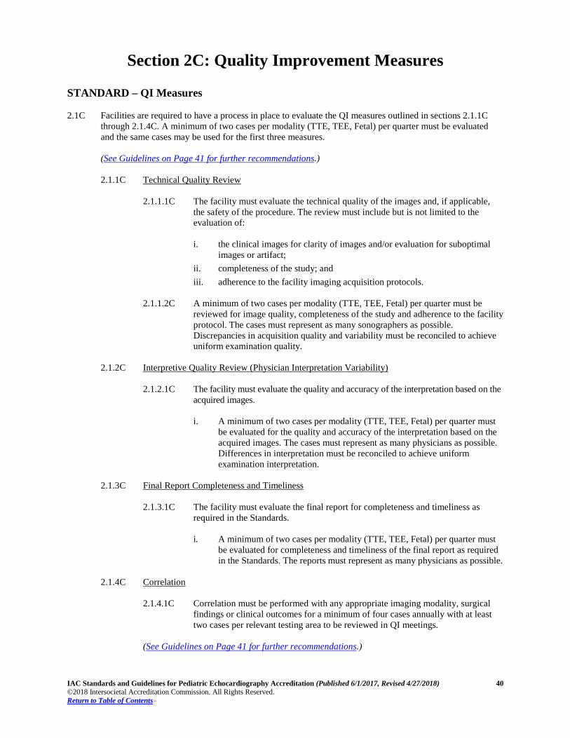
IAC Standards and Guidelines for Pediatric Echocardiography Accreditation (Published 6/1/2017, Revised 4/27/2018) 40 ©2018 Intersocietal Accreditation Commission. All Rights Reserved. Return to Table of Contents»
Section 2C: Quality Improvement Measures
STANDARD – QI Measures 2.1C Facilities are required to have a process in place to evaluate the QI measures outlined in sections 2.1.1C
through 2.1.4C. A minimum of two cases per modality (TTE, TEE, Fetal) per quarter must be evaluated and the same cases may be used for the first three measures. (See Guidelines on Page 41 for further recommendations.) 2.1.1C Technical Quality Review
2.1.1.1C The facility must evaluate the technical quality of the images and, if applicable,
the safety of the procedure. The review must include but is not limited to the evaluation of: i. the clinical images for clarity of images and/or evaluation for suboptimal
images or artifact; ii. completeness of the study; and iii. adherence to the facility imaging acquisition protocols.
2.1.1.2C A minimum of two cases per modality (TTE, TEE, Fetal) per quarter must be
reviewed for image quality, completeness of the study and adherence to the facility protocol. The cases must represent as many sonographers as possible. Discrepancies in acquisition quality and variability must be reconciled to achieve uniform examination quality.
2.1.2C Interpretive Quality Review (Physician Interpretation Variability) 2.1.2.1C The facility must evaluate the quality and accuracy of the interpretation based on the
acquired images. i. A minimum of two cases per modality (TTE, TEE, Fetal) per quarter must
be evaluated for the quality and accuracy of the interpretation based on the acquired images. The cases must represent as many physicians as possible. Differences in interpretation must be reconciled to achieve uniform examination interpretation.
2.1.3C Final Report Completeness and Timeliness 2.1.3.1C The facility must evaluate the final report for completeness and timeliness as
required in the Standards. i. A minimum of two cases per modality (TTE, TEE, Fetal) per quarter must
be evaluated for completeness and timeliness of the final report as required in the Standards. The reports must represent as many physicians as possible.
2.1.4C Correlation 2.1.4.1C Correlation must be performed with any appropriate imaging modality, surgical
findings or clinical outcomes for a minimum of four cases annually with at least two cases per relevant testing area to be reviewed in QI meetings.
(See Guidelines on Page 41 for further recommendations.)
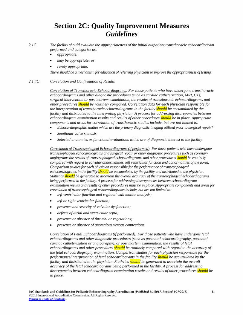
IAC Standards and Guidelines for Pediatric Echocardiography Accreditation (Published 6/1/2017, Revised 4/27/2018) 41 ©2018 Intersocietal Accreditation Commission. All Rights Reserved. Return to Table of Contents»
Section 2C: Quality Improvement Measures Guidelines
2.1C The facility should evaluate the appropriateness of the initial outpatient transthoracic echocardiogram
performed and categorize as: • appropriate; • may be appropriate; or • rarely appropriate. There should be a mechanism for education of referring physicians to improve the appropriateness of testing.
2.1.4C Correlation and Confirmation of Results Correlation of Transthoracic Echocardiograms: For those patients who have undergone transthoracic
echocardiograms and other diagnostic procedures (such as cardiac catheterization, MRI, CT), surgical intervention or post mortem examination, the results of transthoracic echocardiograms and other procedures should be routinely compared. Correlation data for each physician responsible for the interpretation of transthoracic echocardiograms in the facility should be accumulated by the facility and distributed to the interpreting physician. A process for addressing discrepancies between echocardiogram examination results and results of other procedures should be in place. Appropriate components and areas for correlation of transthoracic studies include, but are not limited to: • Echocardiographic studies which are the primary diagnostic imaging utilized prior to surgical repair* • Semilunar valve stenosis • Selected anatomies or functional evaluations which are of diagnostic interest to the facility Correlation of Transesophageal Echocardiograms (if performed): For those patients who have undergone transesophageal echocardiograms and surgical repair or other diagnostic procedures such as coronary angiograms the results of transesophageal echocardiograms and other procedures should be routinely compared with regard to valvular abnormalities, left ventricular function and abnormalities of the aorta. Comparison studies for each physician responsible for the performance of transesophageal echocardiograms in the facility should be accumulated by the facility and distributed to the physician. Statistics should be generated to ascertain the overall accuracy of the transesophageal echocardiograms being performed in the facility. A process for addressing discrepancies between echocardiogram examination results and results of other procedures must be in place. Appropriate components and areas for correlation of transesophageal echocardiograms include, but are not limited to: • left ventricular function and regional wall motion analysis; • left or right ventricular function; • presence and severity of valvular dysfunction; • defects of atrial and ventricular septa; • presence or absence of thrombi or vegetations; • presence or absence of anomalous venous connections.
Correlation of Fetal Echocardiograms (if performed): For those patients who have undergone fetal echocardiograms and other diagnostic procedures (such as postnatal echocardiography, postnatal cardiac catheterization or angiography), or post mortem examination, the results of fetal echocardiograms and other procedures should be routinely compared with regard to the accuracy of the fetal echocardiography examination. Comparison studies for each physician responsible for the performance/interpretation of fetal echocardiograms in the facility should be accumulated by the facility and distributed to the physician. Statistics should be generated to ascertain the overall accuracy of the fetal echocardiograms being performed in the facility. A process for addressing discrepancies between echocardiogram examination results and results of other procedures should be in place.

IAC Standards and Guidelines for Pediatric Echocardiography Accreditation (Published 6/1/2017, Revised 4/27/2018) 42 ©2018 Intersocietal Accreditation Commission. All Rights Reserved. Return to Table of Contents»
Section 3C: Quality Improvement Meetings
STANDARD – QI Meetings
3.1C Quality Improvement (QI) Meetings 3.1.1C The facility must have a minimum of two QI meetings per year, one of which is to review the
results of the QI analyses and any additional QI-related topics.
3.1.2C All staff must participate in at least one meeting per year.
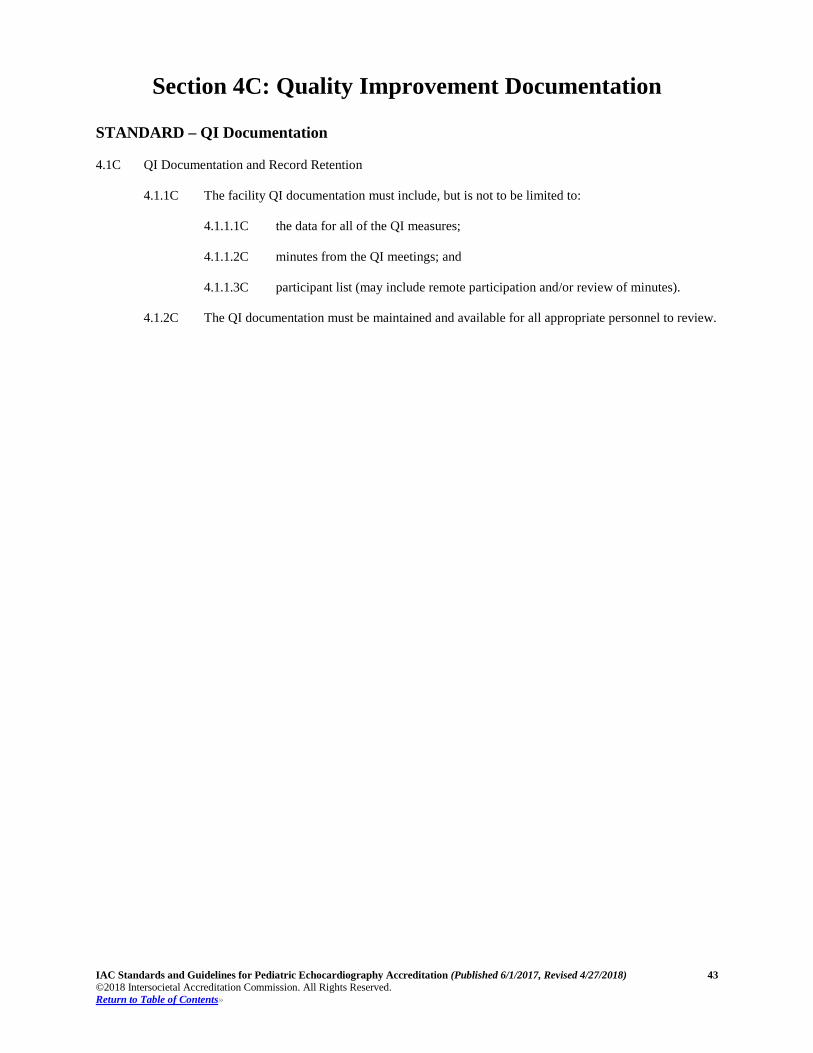
IAC Standards and Guidelines for Pediatric Echocardiography Accreditation (Published 6/1/2017, Revised 4/27/2018) 43 ©2018 Intersocietal Accreditation Commission. All Rights Reserved. Return to Table of Contents»
Section 4C: Quality Improvement Documentation
STANDARD – QI Documentation
4.1C QI Documentation and Record Retention 4.1.1C The facility QI documentation must include, but is not to be limited to:
4.1.1.1C the data for all of the QI measures;
4.1.1.2C minutes from the QI meetings; and
4.1.1.3C participant list (may include remote participation and/or review of minutes).
4.1.2C The QI documentation must be maintained and available for all appropriate personnel to review.