Embed Size (px)
Citation preview
I would feel more optimistic about a bright future for man if he spent less time proving that he can outwit Nature and more time tasting her sweetness and respecting her seniority.
—E. B. White, “Coon Tree,” 1977
Dr.S.Chakravarty, MD
Chemistry of Carbohydrates
CARBOHYDRATES !!! WHAT COMES TO YOUR MIND ??
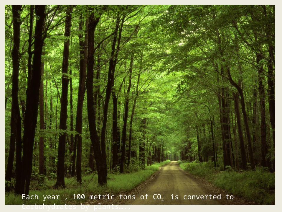
Each year , 100 metric tons of CO2 is converted to Carbohydrates by plants…..
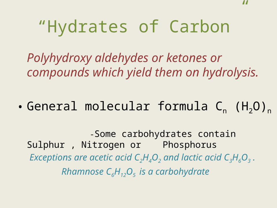
“Hydrates of Carbon”
Polyhydroxy aldehydes or ketones or compounds which yield them on hydrolysis.
• General molecular formula Cn (H2O)n
-Some carbohydrates contain Sulphur , Nitrogen or Phosphorus
Exceptions are acetic acid C2H4O2 and lactic acid C3H6O3 .
Rhamnose C6H12O5 is a carbohydrate
Functions of Carbohydrates
• Main sources of ENERGY in body (4kcal/g) – RBCs and Brain cells have an absolute requirement of carbohydrates .
• Storage form of energy (starch and glycogen)
• Excess carbohydrate is converted to fat.
• Glycoproteins and glycolipids are components of cell membranes and receptors.
• Structural basis of many organisms .e.g . Cellulose in plants ,exoskeleton of insects , cell wall of microbes, mucopolysaccharides and ground substance in higher organisms.
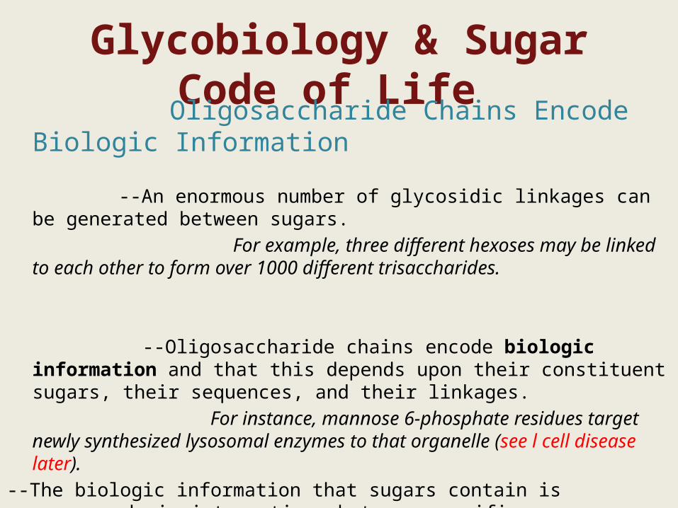
Glycobiology & Sugar Code of Life
Oligosaccharide Chains Encode Biologic Information
--An enormous number of glycosidic linkages can be generated between sugars. For example, three different hexoses may be linked to each other to form
over 1000 different trisaccharides.
--Oligosaccharide chains encode biologic information and that this depends upon their constituent sugars, their sequences, and their linkages.
For instance, mannose 6-phosphate residues target newly synthesized lysosomal enzymes to that organelle (see l cell disease later).
--The biologic information that sugars contain is expressed via interactions between specific sugars, either free or in glycoconjugates, and proteins (such as lectins; see below) or other molecules
These interactions lead to changes of cellular activity.
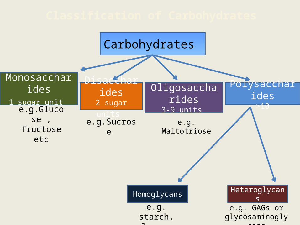
Classification of Carbohydrates
Carbohydrates
Disaccharides2 sugar units
Oligosaccharides3-9 units
Monosaccharides1 sugar unit Polysaccharides
>10
HeteroglycansHomoglycans
e.g.Glucose , fructose etc e.g.Sucrose e.g. Maltotriose
e.g. starch, glycogen
e.g. GAGs or glycosaminoglycans
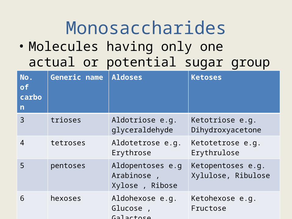
Monosaccharides• Molecules having only one actual or potential
sugar group .No. of carbon
Generic name Aldoses Ketoses
3 trioses Aldotriose e.g. glyceraldehyde
Ketotriose e.g. Dihydroxyacetone
4 tetroses Aldotetrose e.g.Erythrose
Ketotetrose e.g.Erythrulose
5 pentoses Aldopentoses e.gArabinose , Xylose , Ribose
Ketopentoses e.g.Xylulose, Ribulose
6 hexoses Aldohexose e.g. Glucose , Galactose , Mannose
Ketohexose e.g. Fructose
7 heptoses Aldoheptose: Glucoheptose
Ketoheptose e.g Sedoheptulose
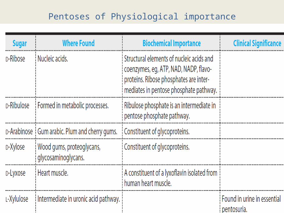
Pentoses of Physiological importance
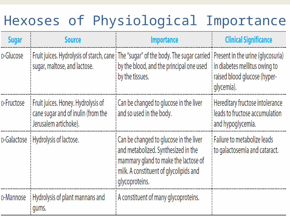
Hexoses of Physiological Importance
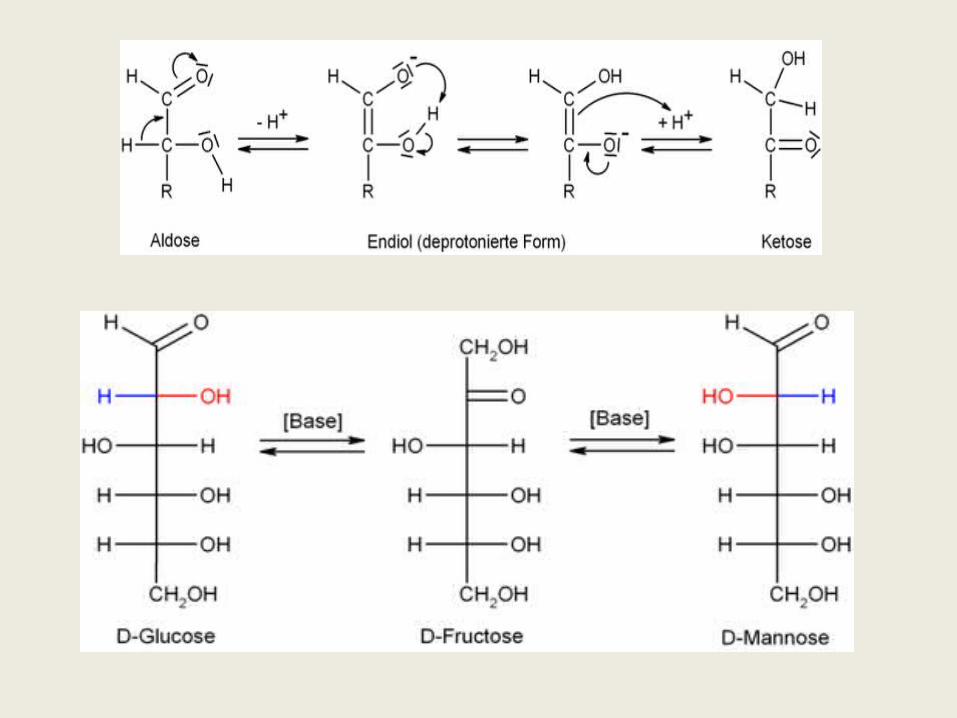
Enediol formation :-
• In mild alkaline solutions, carbohydrates containing a free sugar group (aldehyde or ketone) will tautomerise to form enediols , where two hydroxyl groups are attached to the double-bonded carbon atoms .
• Since enediols are powerful reducing agents in alkaline medium. When oxidising agents like cupric ions are present , sugars form a mixture of carboxylic acids by breaking at the double bonds.
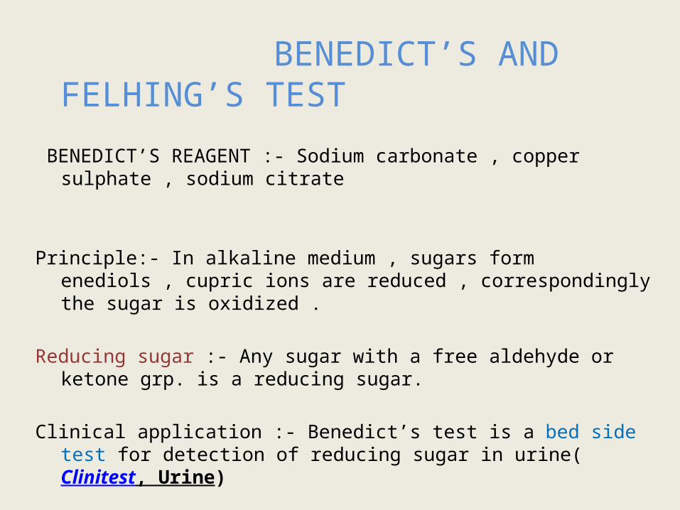
BENEDICT’S AND FELHING’S TEST
BENEDICT’S REAGENT :- Sodium carbonate , copper sulphate , sodium citrate
Principle:- In alkaline medium , sugars form enediols , cupric ions are reduced ,
correspondingly the sugar is oxidized .
Reducing sugar :- Any sugar with a free aldehyde or ketone grp. is a reducing sugar.
Clinical application :- Benedict’s test is a bed side test for detection of reducing sugar in urine(Clinitest, Urine)
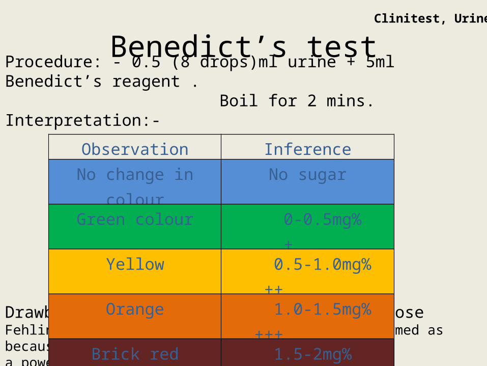
Benedict’s testProcedure: - 0.5 (8 drops)ml urine + 5ml Benedict’s reagent . Boil for 2 mins.Interpretation:-
Drawback – test is not specific for glucose Fehling’s test :- no intermediate colors are formed as because over there a powerful reducing agent KOH is used.
Observation Inference
No change in colour No sugar
Green colour 0-0.5mg% +
Yellow 0.5-1.0mg% ++
Orange 1.0-1.5mg% +++
Brick red 1.5-2mg% ++++
Clinitest, Urine
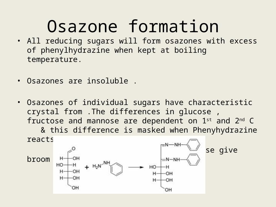
Osazone formation • All reducing sugars will form osazones with excess of phenylhydrazine
when kept at boiling temperature.
• Osazones are insoluble .
• Osazones of individual sugars have characteristic crystal from .The differences in glucose , fructose and mannose are dependent on 1st and 2nd C & this difference is masked when Phenyhydrazine reacts with these two carbons .
So, Glucose , Fructose and Mannose give broom shaped osazones.
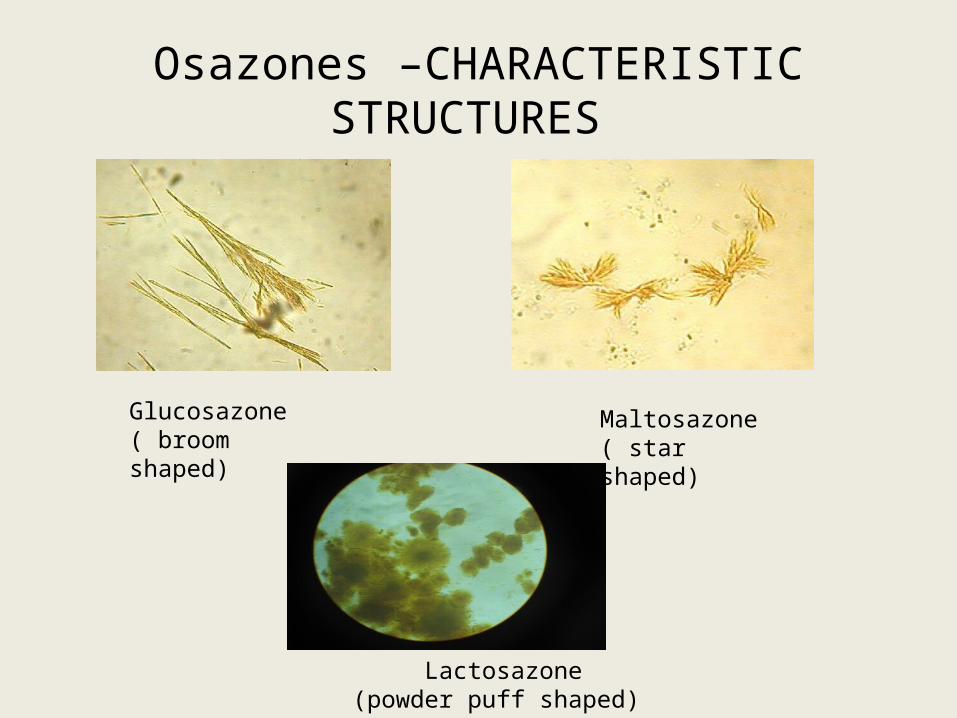
Osazones –CHARACTERISTIC STRUCTURES
Glucosazone( broom shaped)
Maltosazone( star shaped)
Lactosazone(powder puff shaped)
4)Reduction to form alcohols When treated with reducing agents such as sodium amalgam,
hydrogen can reduce sugars . Aldose yields corresponding alcohol .Ketoses form two alcohols because of appearance of new asymmetric carbon in this process.
E.g.D-Glucose D-Sorbitol D-Fructose D-Mannitol
Sorbitol and Mannitol are used to identify bacterial bacterial colonies.
Mannitol is used to reduce intracranial pressure by forced diuresis.
The osmotic effect of sorbitol and dulcitol produces changes in tissues when they accumulate in abnormal amounts. E.g cataract
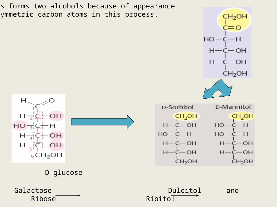
Galactose Dulcitol and Ribose Ribitol
Ketoses forms two alcohols because of appearance of asymmetric carbon atoms in this process.
D-fructose
D-glucose
Glycosides
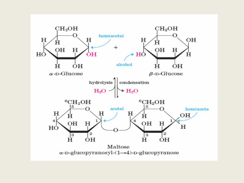
• When the hemi-acetal group (hydroxyl grp of the anomeric C ) of a monosaccharide is condensed with an alcohol or phenol grp , it is called as a glycoside .The non-carbohydrate grp is called aglycone.
• Glycosides are non –reducing (WHY ?)but they may be hydrolyzed by boiling with dilute acids.
• - glycosides are hydrolyzed by maltase from yeast , while beta-glycosides are hydrolyzed by Emulsin from almonds .So enzyme hydrolysis affords a method to distinguish b/w two forms .
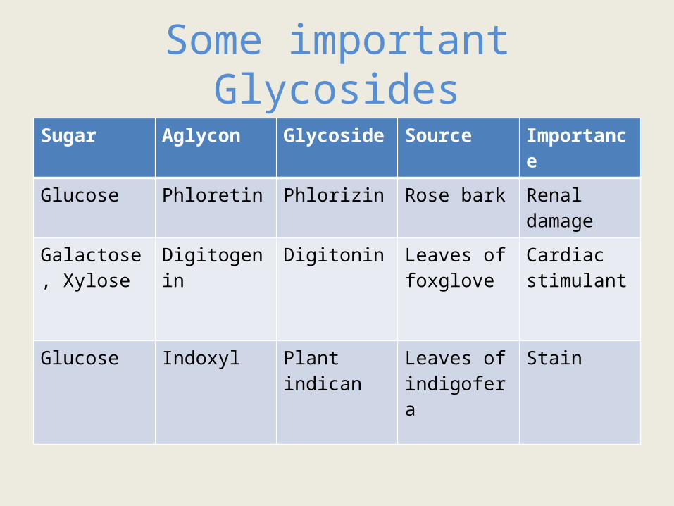
Some important GlycosidesSugar Aglycon Glycoside Source Importance
Glucose Phloretin Phlorizin Rose bark Renal damage
Galactose , Xylose
Digitogenin Digitonin Leaves of foxglove
Cardiac stimulant
Glucose Indoxyl Plant indican Leaves of indigofera
Stain
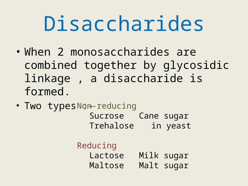
Disaccharides• When 2 monosaccharides are combined
together by glycosidic linkage , a disaccharide is formed.
• Two types :-Non-reducing
Sucrose Cane sugarTrehalose in yeast
ReducingLactose Milk sugarMaltose Malt sugar
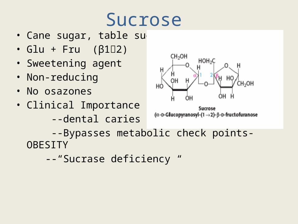
Sucrose • Cane sugar, table sugar• Glu + Fru (β12)• Sweetening agent• Non-reducing• No osazones• Clinical Importance :- --dental caries --Bypasses metabolic check points- OBESITY --“Sucrase deficiency “
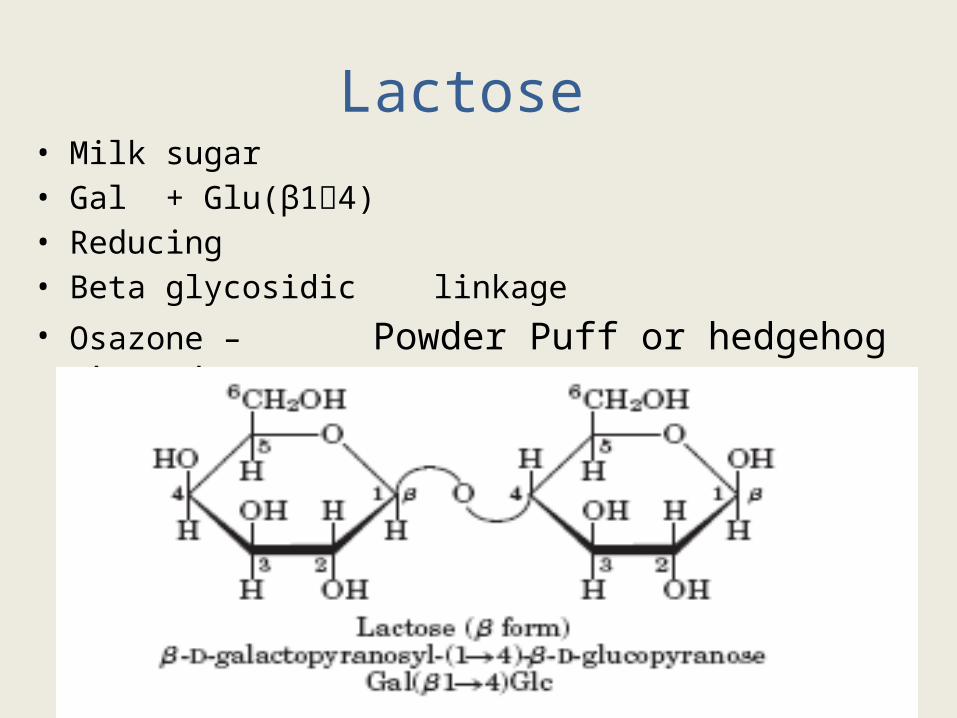
Lactose • Milk sugar• Gal + Glu(β14)• Reducing • Beta glycosidic linkage• Osazone – Powder Puff or hedgehog shaped
Lactose Intolerance
• Deficiency of enzyme lactase in brush border epithelium
• Primary – only in adults , absence of a lactase persistence allele
• Congenital- children( autosomal recessive) • Secondary - Also common in acute gastroenteritis ,
Abdominal cramps , pain , distension and diarrhoea. Treatment :- Restriction of dairy products Soy milk can be used for children
USMLE
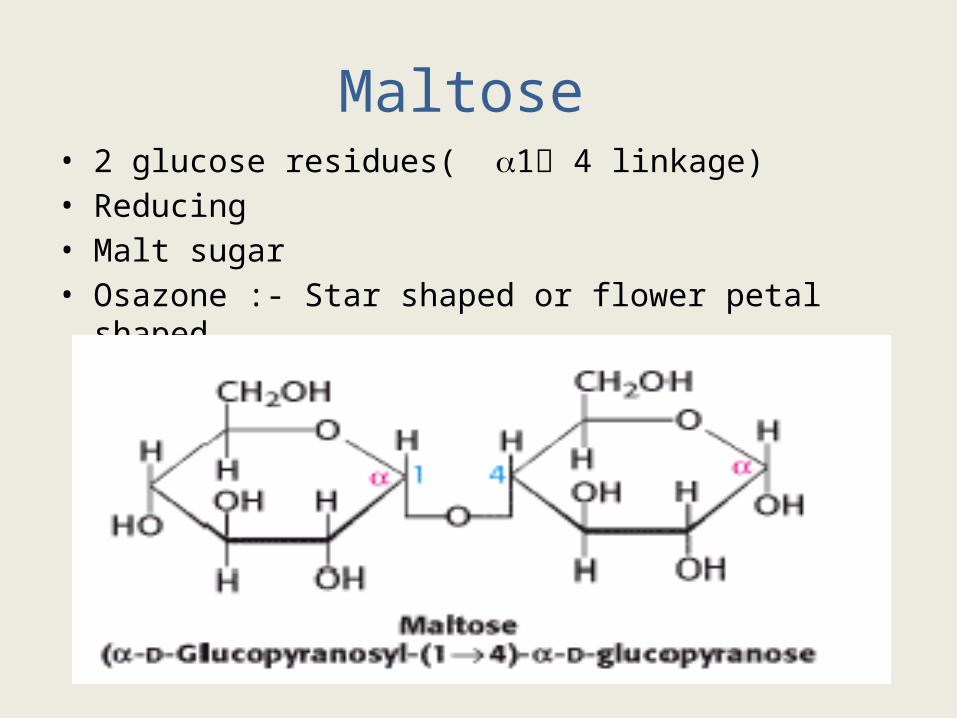
Maltose • 2 glucose residues( 1 4 linkage) • Reducing• Malt sugar• Osazone :- Star shaped or flower petal shaped
Lactulose
• Synthetic disaccharide of Galactose and Fructose
• Poorly absorbed from the gastrointestinal tract
• Used in the treatment of hepatic encephalopathy
• Metabolized by the colonic bacteria to acidic products CAUSES PURGATION
• Promotes the excretion of ammonia in feces as protonated ammonium ions
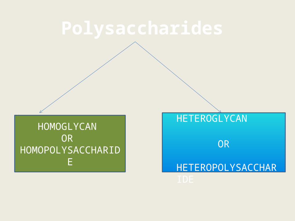
Polysaccharides
HOMOGLYCAN OR
HOMOPOLYSACCHARIDE
HETEROGLYCAN OR
HETEROPOLYSACCHARIDE
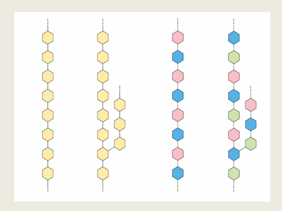
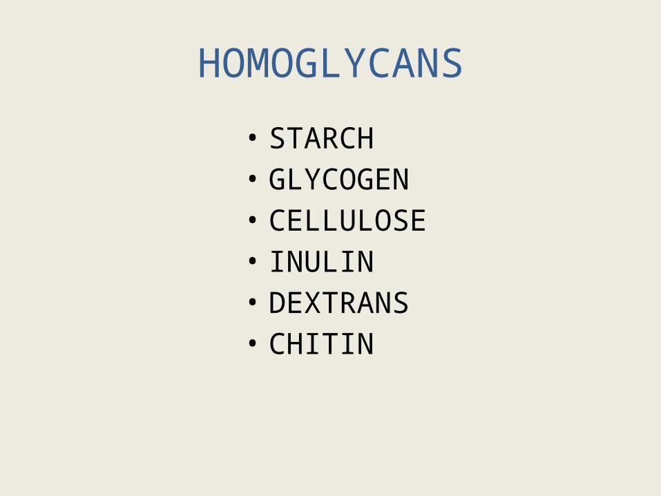
HOMOGLYCANS
• STARCH• GLYCOGEN• CELLULOSE• INULIN• DEXTRANS• CHITIN
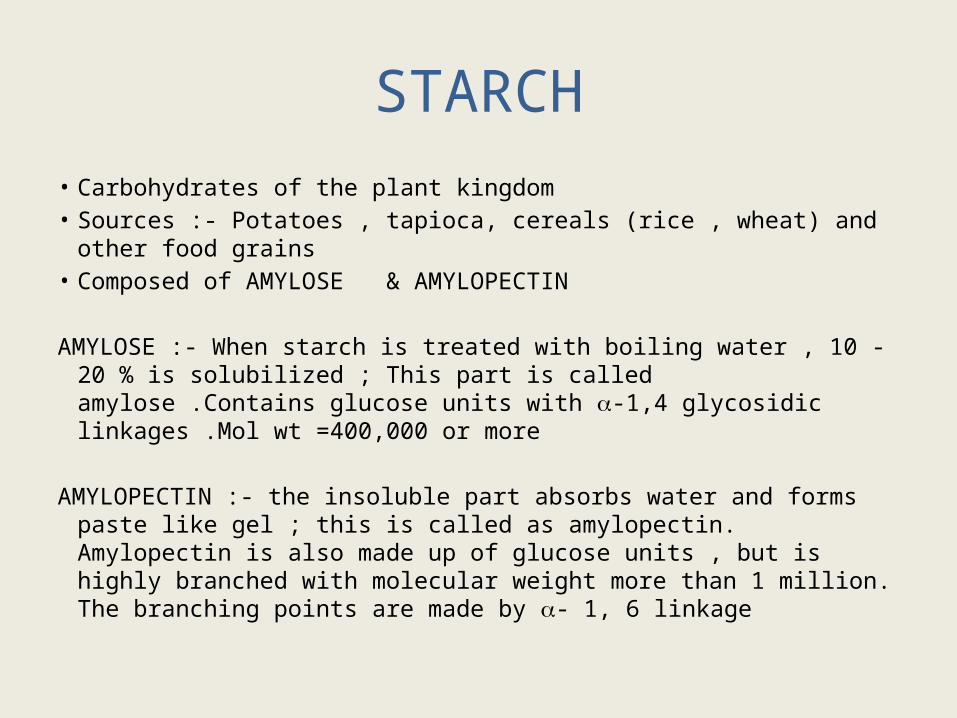
STARCH• Carbohydrates of the plant kingdom • Sources :- Potatoes , tapioca, cereals (rice , wheat) and other food
grains • Composed of AMYLOSE & AMYLOPECTIN
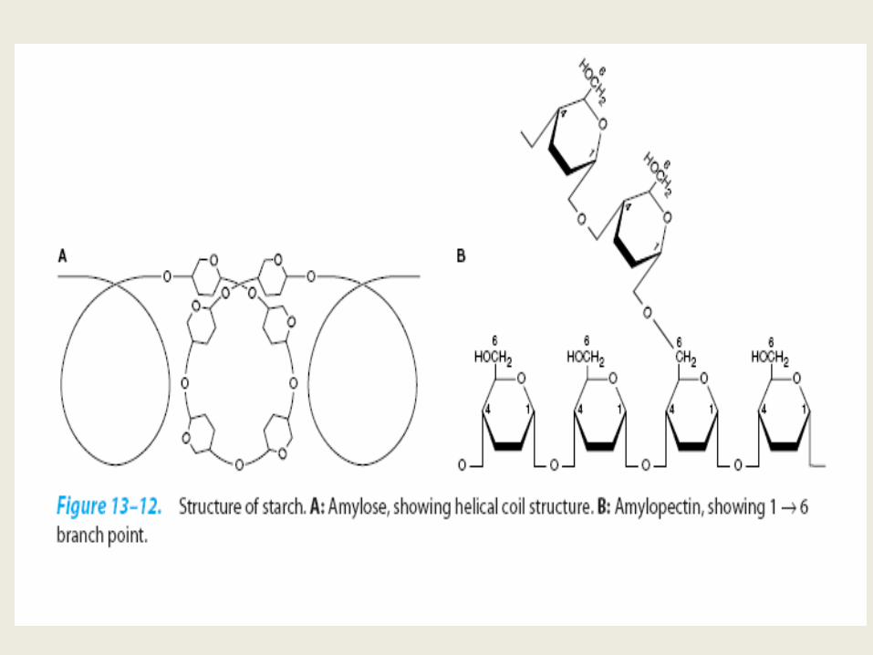
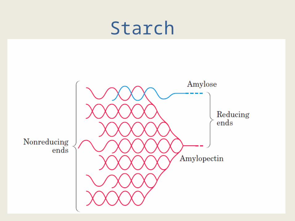
AMYLOSE :- When starch is treated with boiling water , 10 -20 % is solubilized ; This part is called amylose .Contains glucose units with -1,4 glycosidic linkages .Mol wt =400,000 or more
AMYLOPECTIN :- the insoluble part absorbs water and forms paste like gel ; this is called as amylopectin. Amylopectin is also made up of glucose units , but is highly branched with molecular weight more than 1 million. The branching points are made by - 1, 6 linkage
Starch
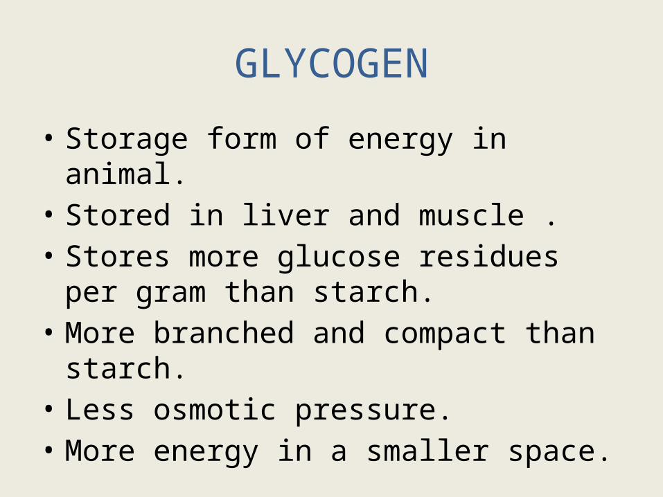
GLYCOGEN
• Storage form of energy in animal.• Stored in liver and muscle .• Stores more glucose residues per gram than
starch.• More branched and compact than starch.• Less osmotic pressure. • More energy in a smaller space.
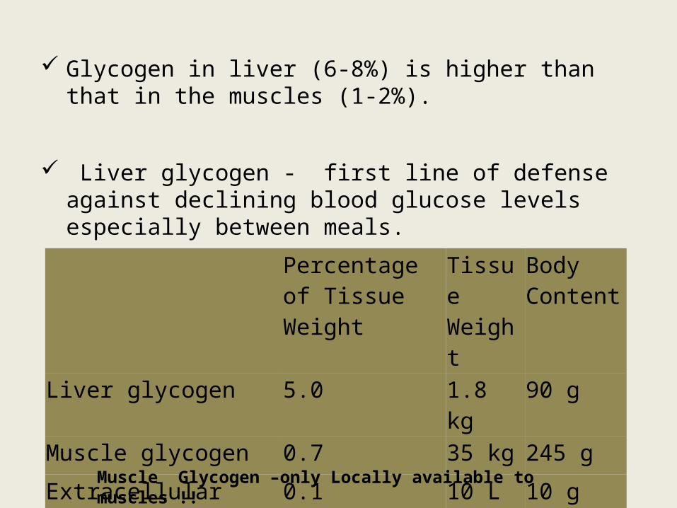
Glycogen in liver (6-8%) is higher than that in the muscles (1-2%).
Liver glycogen - first line of defense against declining blood glucose levels especially between meals.
Percentage of Tissue Weight
Tissue Weight
Body Content
Liver glycogen 5.0 1.8 kg 90 g
Muscle glycogen 0.7 35 kg 245 g
Extracellular glucose 0.1 10 L 10 g
Muscle Glycogen –only Locally available to muscles !!
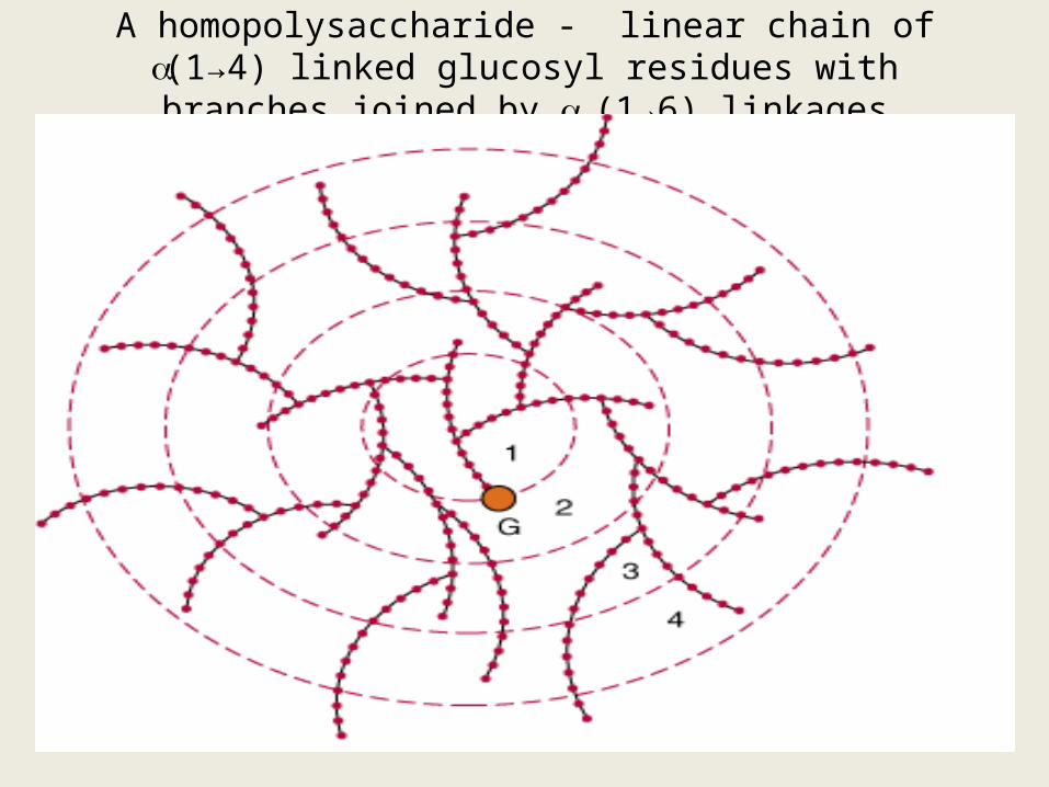
A homopolysaccharide - linear chain of (1→4) linked glucosyl residues with branches joined by
(1→6) linkages
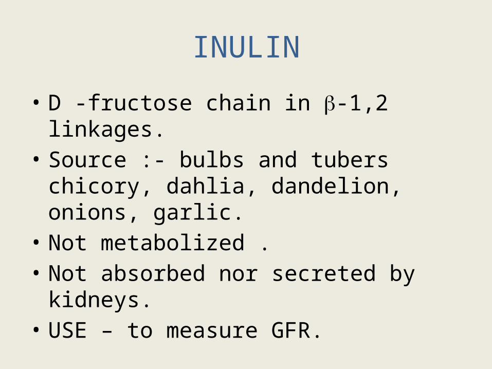
INULIN
• D -fructose chain in -1,2 linkages. • Source :- bulbs and tubers chicory, dahlia,
dandelion, onions, garlic.• Not metabolized .• Not absorbed nor secreted by kidneys.• USE – to measure GFR.
DEXTRANS• Highly branched homoglycan containing Glu residues in 1-6, 1-4 and 1-3
linkages.
• Produced by microbes.
• Mol. wt :- 1-4 million.
• Colloidal solution, Low osmotic effects, slow disintegration and utilization, slow elimination from the body
• As large sized , they will not move out of vascular compartment so used as plasma expanders.
• In hypovolemic shock, given intravenously increases blood volume.

Heteroglycans
Mucopolysaccharides or GAG
--- [ URONIC ACID + AMINO SUGAR]---n
Acetylated amino sugars, sulfate and carboxyl groups may also be present
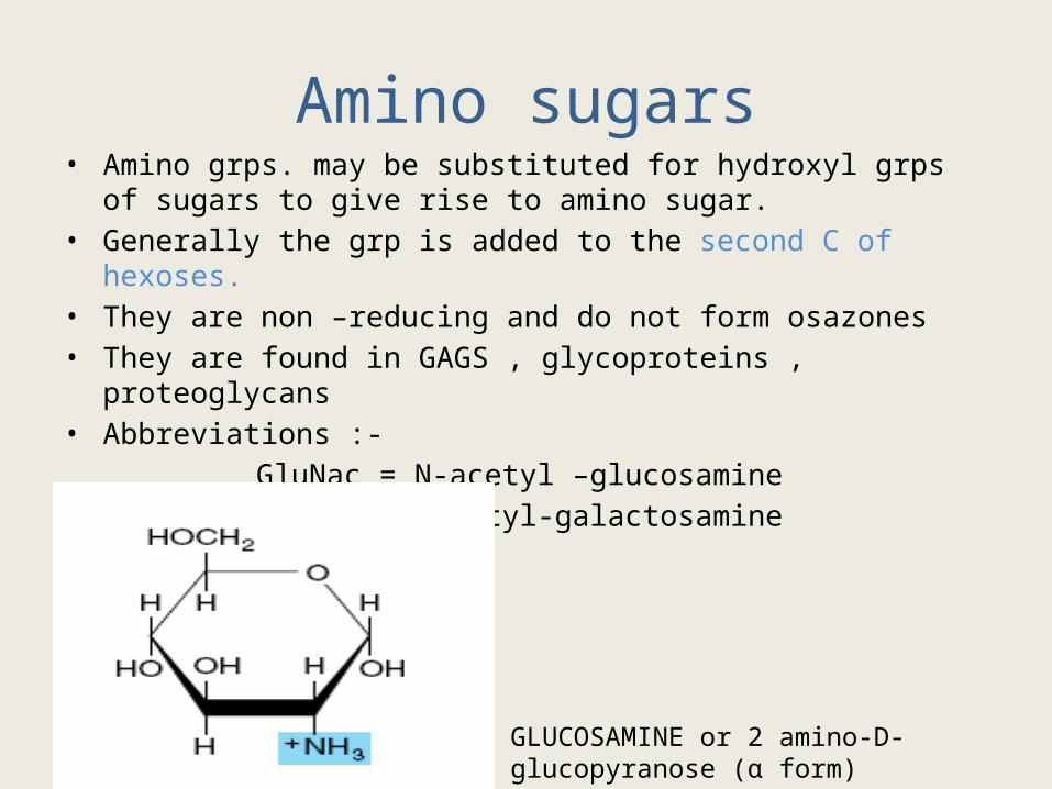
Amino sugars• Amino grps. may be substituted for hydroxyl grps of sugars to give rise to
amino sugar.• Generally the grp is added to the second C of hexoses.• They are non –reducing and do not form osazones• They are found in GAGS , glycoproteins , proteoglycans • Abbreviations :- GluNac = N-acetyl –glucosamine GalNac =N-acetyl-galactosamine
GLUCOSAMINE or 2 amino-D-glucopyranose (α form)
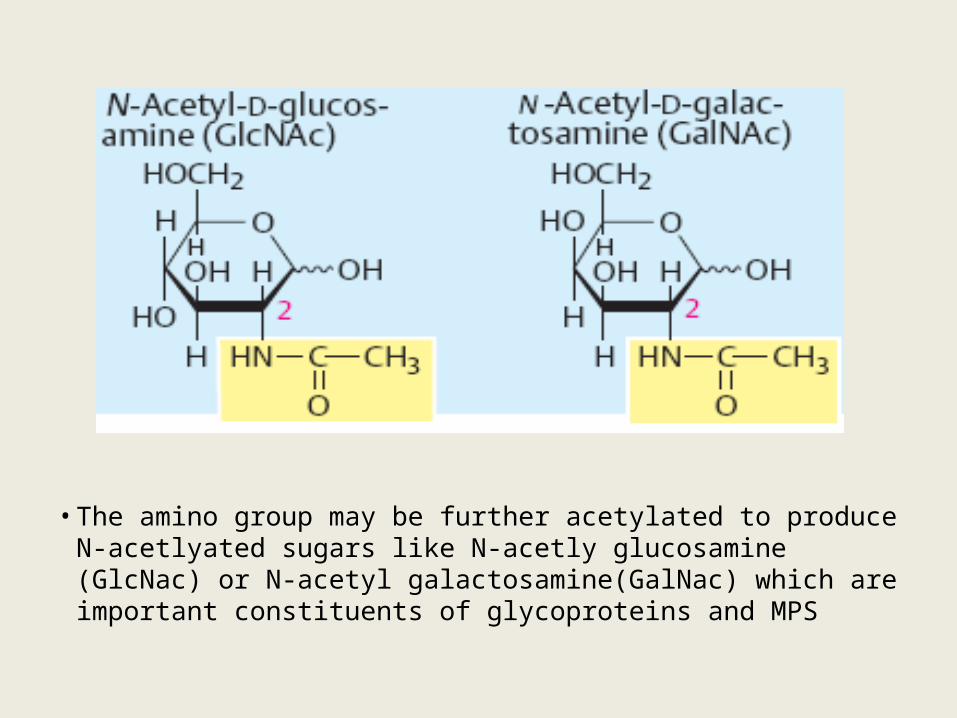
• The amino group may be further acetylated to produce N-acetlyated sugars like N-acetly glucosamine (GlcNac) or N-acetyl galactosamine(GalNac) which are important constituents of glycoproteins and MPS
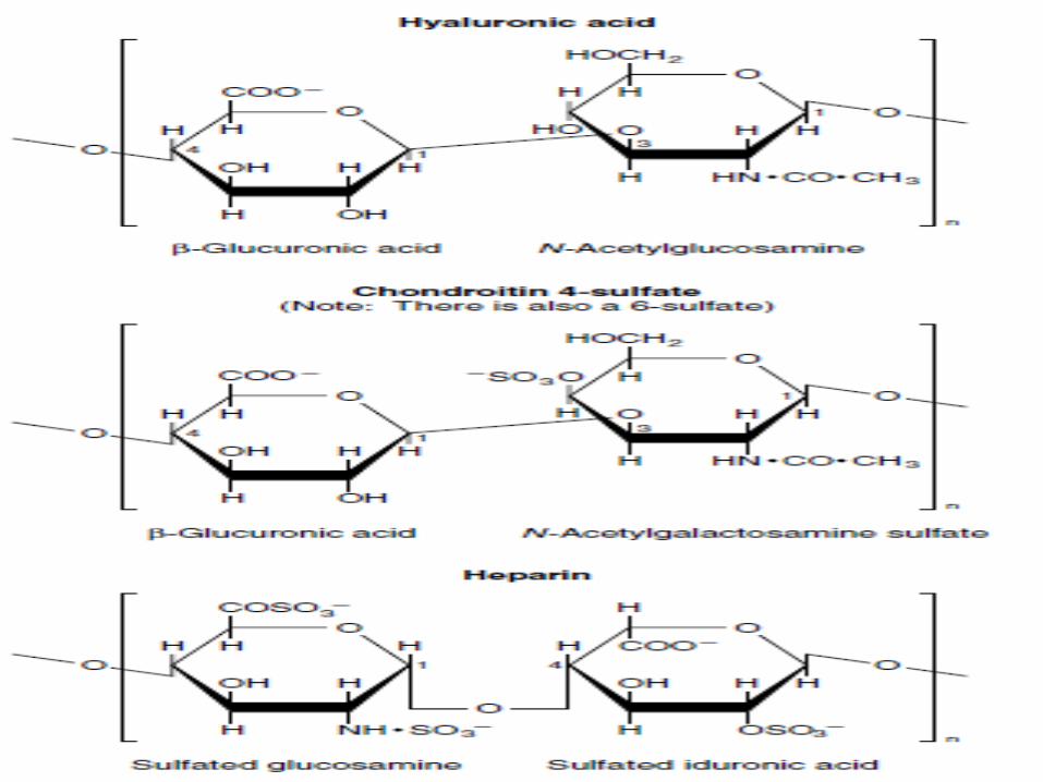
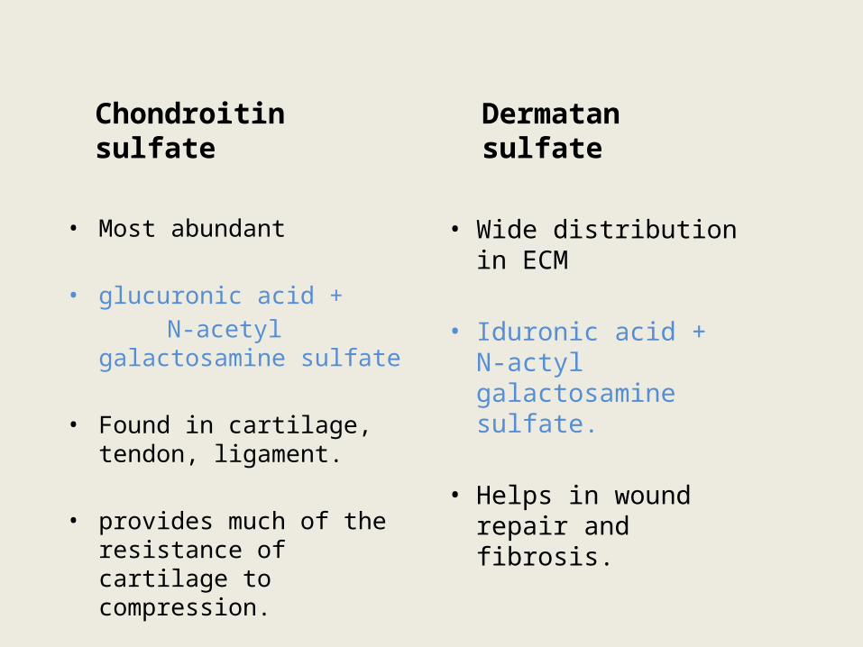
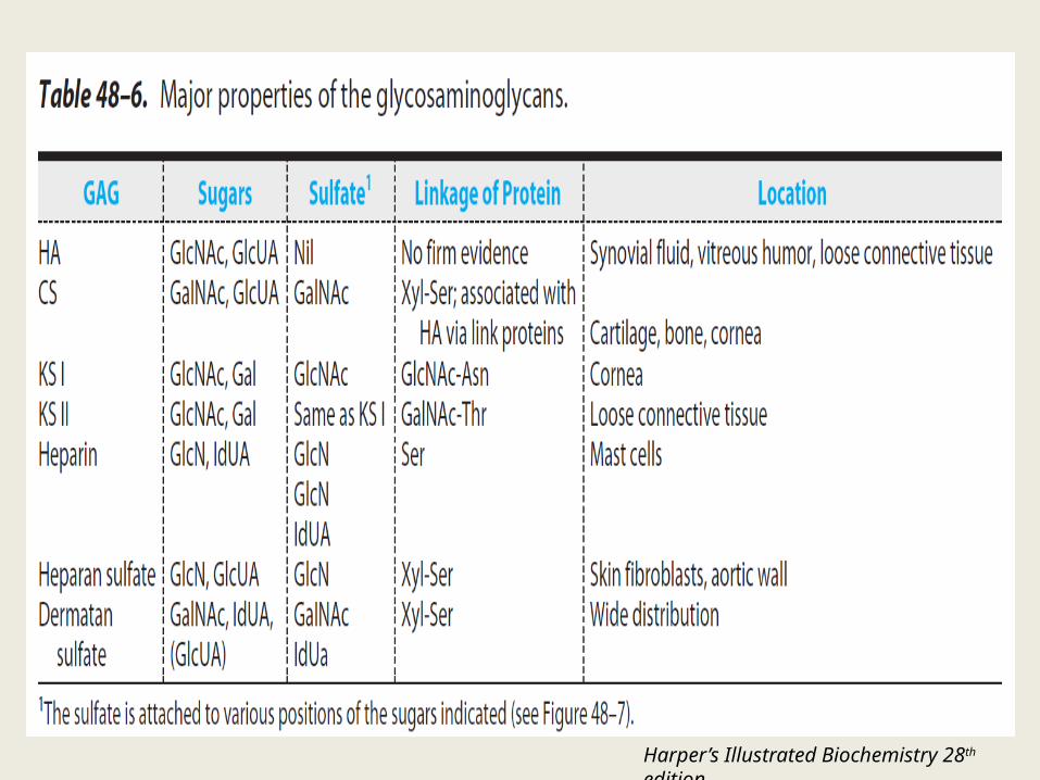
Chondroitin sulfate
• Most abundant
• glucuronic acid + N-acetyl galactosamine
sulfate
• Found in cartilage, tendon, ligament.
• provides much of the resistance of cartilage to compression.
Dermatan sulfate
• Wide distribution in ECM
• Iduronic acid + N-actyl galactosamine sulfate.
• Helps in wound repair and fibrosis.
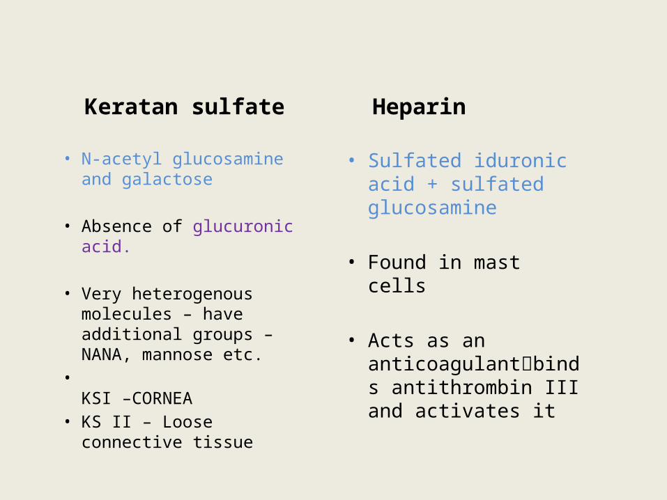
Keratan sulfate
• N-acetyl glucosamine and galactose
• Absence of glucuronic acid.
• Very heterogenous molecules – have additional groups – NANA, mannose etc.
•KSI –CORNEA
• KS II – Loose connective tissue
Heparin
• Sulfated iduronic acid + sulfated glucosamine
• Found in mast cells
• Acts as an anticoagulantbinds antithrombin III and activates it
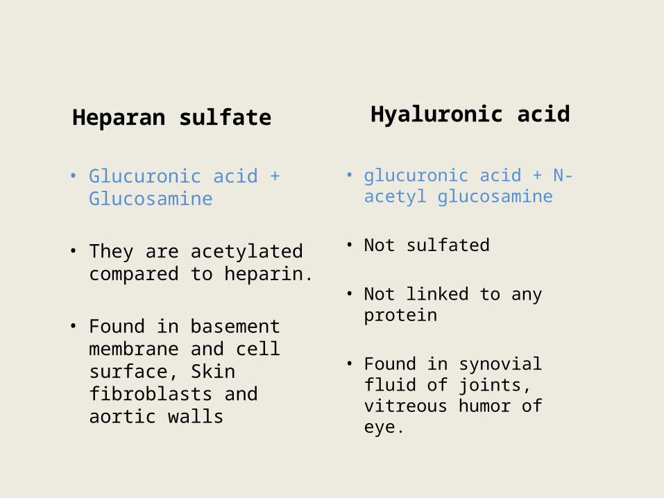
Heparan sulfate
• Glucuronic acid + Glucosamine
• They are acetylated compared to heparin.
• Found in basement membrane and cell surface, Skin fibroblasts and aortic walls
Hyaluronic acid
• glucuronic acid + N-acetyl glucosamine
• Not sulfated
• Not linked to any protein
• Found in synovial fluid of joints, vitreous humor of eye.
Relationship between GAG structure and function
• strong negative charges -- > ( POLYANIONS ) (-COO- and -OSO3-) cause molecule to fan outwards and repel adjacent molecules
• Surrounded by a shell of hydration.
• Slippery consistency – similar to magnets.
• Act like cushions – compressible but spring back after the pressure is removed (sponge effect).
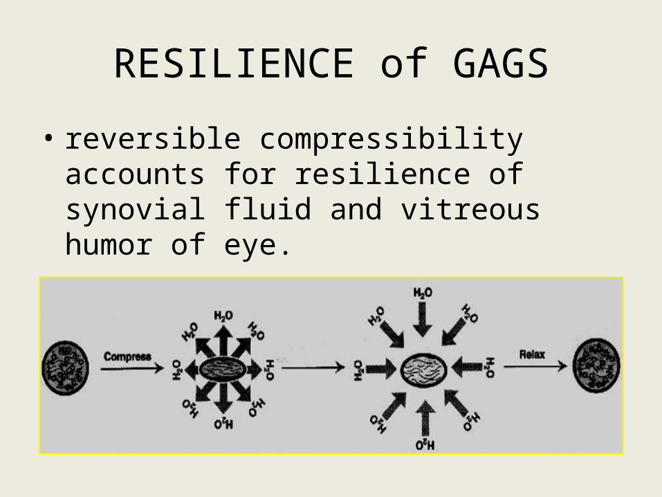
RESILIENCE of GAGS
• reversible compressibility accounts for resilience of synovial fluid and vitreous humor of eye.
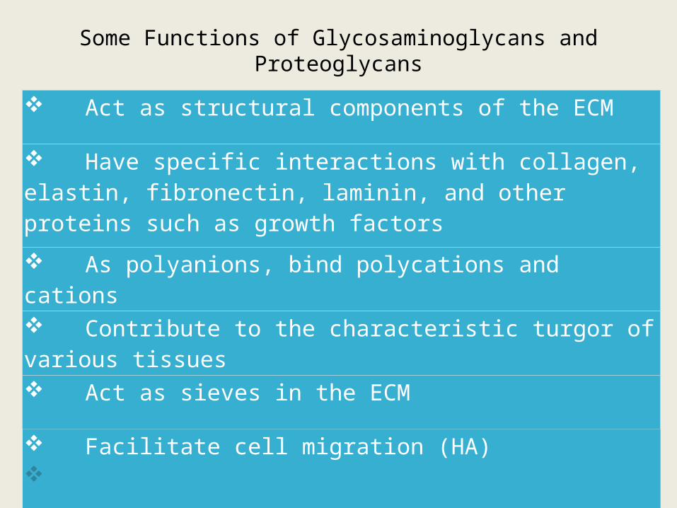
Some Functions of Glycosaminoglycans and Proteoglycans
Act as structural components of the ECM
Have specific interactions with collagen, elastin, fibronectin, laminin, and other proteins such as growth factors
As polyanions, bind polycations and cations
Contribute to the characteristic turgor of various tissues
Act as sieves in the ECM
Facilitate cell migration (HA) contd..
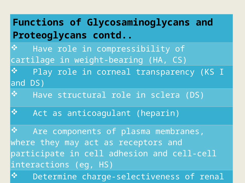
Functions of Glycosaminoglycans and Proteoglycans contd.. Have role in compressibility of cartilage in weight-bearing (HA, CS) Play role in corneal transparency (KS I and DS)
Have structural role in sclera (DS)
Act as anticoagulant (heparin)
Are components of plasma membranes, where they may act as receptors and participate in cell adhesion and cell-cell interactions (eg, HS) Determine charge-selectiveness of renal glomerulus (HS)
Are components of synaptic and other vesicles (eg, HS)
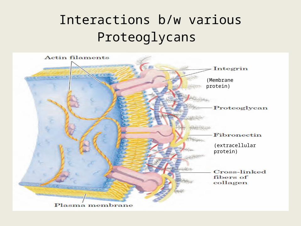
Interactions b/w various Proteoglycans
(Membrane protein)
(extracellular protein)
Harper’s Illustrated Biochemistry 28th edition
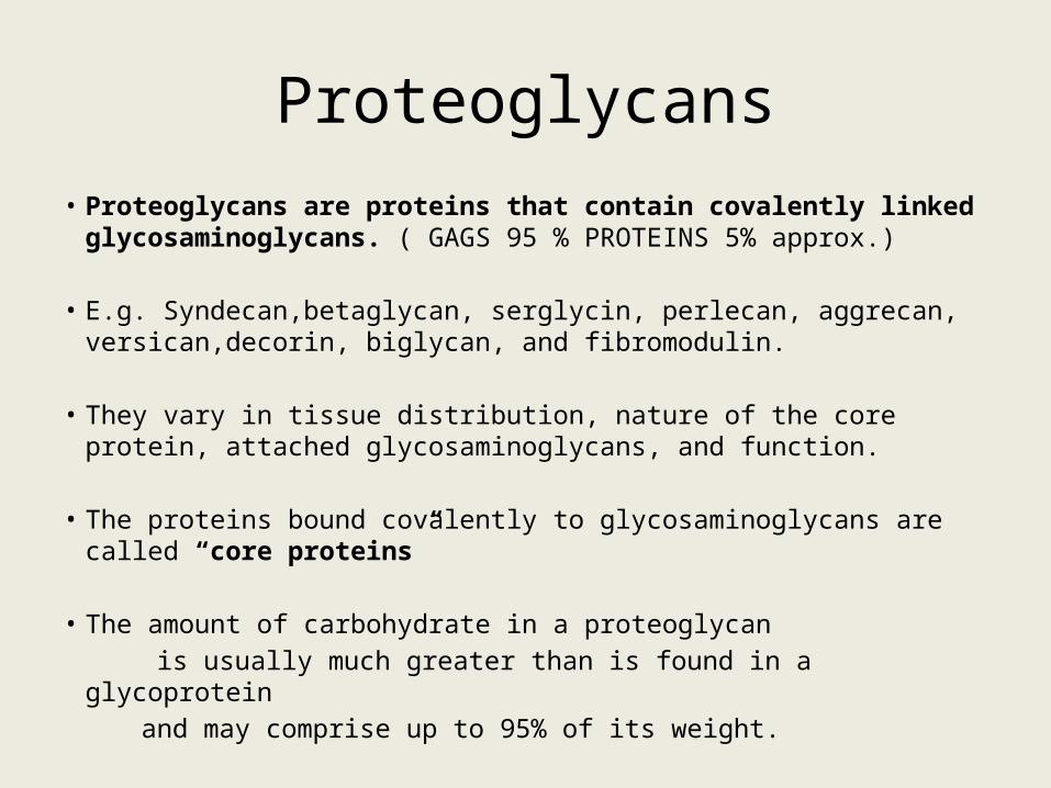
Proteoglycans• Proteoglycans are proteins that contain covalently linked
glycosaminoglycans. ( GAGS 95 % PROTEINS 5% approx.)
• E.g. Syndecan,betaglycan, serglycin, perlecan, aggrecan, versican,decorin, biglycan, and fibromodulin.
• They vary in tissue distribution, nature of the core protein, attached glycosaminoglycans, and function.
• The proteins bound covalently to glycosaminoglycans are called “core proteins”
• The amount of carbohydrate in a proteoglycan is usually much greater than is found in a glycoprotein and may comprise up to 95% of its weight.

Darkfield electron micrograph of a proteoglycan aggregate
• Bottle brush appearance
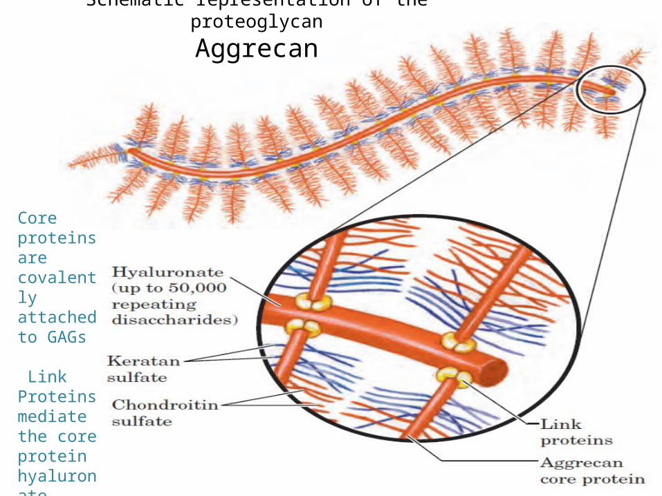
Schematic representation of the proteoglycan
Aggrecan
Core proteins are covalently attached to GAGs
Link Proteins mediate the core protein hyaluronate interaction
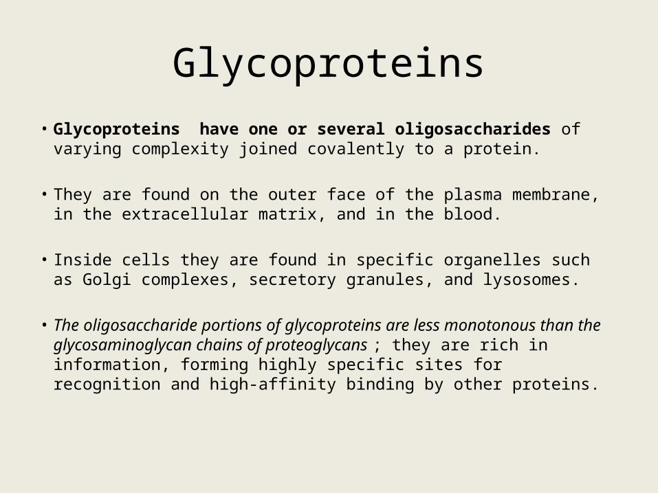
Glycoproteins• Glycoproteins have one or several oligosaccharides of varying
complexity joined covalently to a protein.
• They are found on the outer face of the plasma membrane, in the extracellular matrix, and in the blood.
• Inside cells they are found in specific organelles such as Golgi complexes, secretory granules, and lysosomes.
• The oligosaccharide portions of glycoproteins are less monotonous than the glycosaminoglycan chains of proteoglycans ; they are rich in information, forming highly specific sites for recognition and high-affinity binding by other proteins.
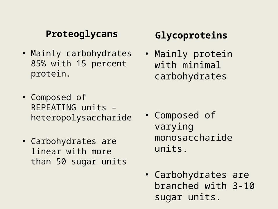
Proteoglycans
• Mainly carbohydrates 85% with 15 percent protein.
• Composed of REPEATING units – heteropolysaccharide
• Carbohydrates are linear with more than 50 sugar units
Glycoproteins
• Mainly protein with minimal carbohydrates
• Composed of varying monosaccharide units.
• Carbohydrates are branched with 3-10 sugar units.
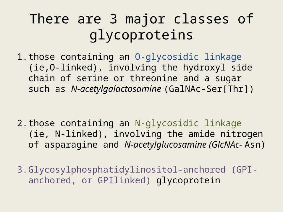
There are 3 major classes of glycoproteins
1. those containing an O-glycosidic linkage (ie,O-linked), involving the hydroxyl side chain of serine or threonine and a sugar such as N-acetylgalactosamine (GalNAc-Ser[Thr])
2. those containing an N-glycosidic linkage (ie, N-linked), involving the amide nitrogen of asparagine and N-acetylglucosamine (GlcNAc- Asn)
3. Glycosylphosphatidylinositol-anchored (GPI-anchored, or GPIlinked) glycoprotein
Glycolipids
• Glycolipids are membrane lipids in which the hydrophilic head groups are oligosaccharides, which, as in glycoproteins, act as specific sites for recognition by carbohydrate- binding proteins
Degradation of GAGs:
• Degraded by lysosomes.
• Enzymes – acid hydrolases.
• Extracelluar GAGs are brought inside by phagocytosis.
Lysosomal storage disorders:
• LSDs are a group of about 40 rare inherited disorders that are characterized by lysosomal dysfunction leading to abnormal accumulations of substances inside of lysosomes.
• Disorders are usually caused by the deficiency of one of the Lysosomal enzyme.
Classification:• MAIN GROUPS :-
– Mucopolysaccharidoses
– Mucolipidoses
– Sphingolipidoses
• Others :-
– Pompe’s disease (Glycogen storage disorder type II)
– Wolman disease( Lysosomal acid lipase deficiency)
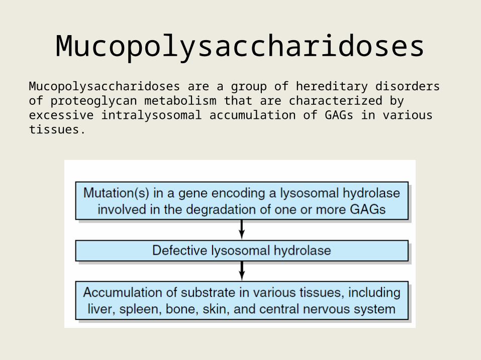
MucopolysaccharidosesMucopolysaccharidoses are a group of hereditary disorders of proteoglycan metabolism that are characterized by excessive intralysosomal accumulation of GAGs in various tissues.
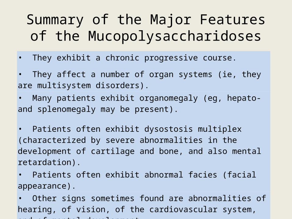
Summary of the Major Features of the Mucopolysaccharidoses
• They exhibit a chronic progressive course.
• They affect a number of organ systems (ie, they are multisystem disorders).
• Many patients exhibit organomegaly (eg, hepato- and splenomegaly may be present).
• Patients often exhibit dysostosis multiplex (characterized by severe abnormalities in the development of cartilage and bone, and also mental retardation).
• Patients often exhibit abnormal facies (facial appearance).
• Other signs sometimes found are abnormalities of hearing, of vision, of the cardiovascular system, and of mental development.

Hurler syndrome: MPS-1
• Enzyme def: α-L – Iduronidase
• Accumulation : Heparan and Dermatan sulfate
• Key features: corneal clouding, mental retardation, micrognathia, coarsening of facial features with macroglossia, inguinal and abdominal hernias, retinal degeneration.
• Death due to accumulation in coronary arteries.
• Can be treated before 18 months – BM transplant.
USMLE
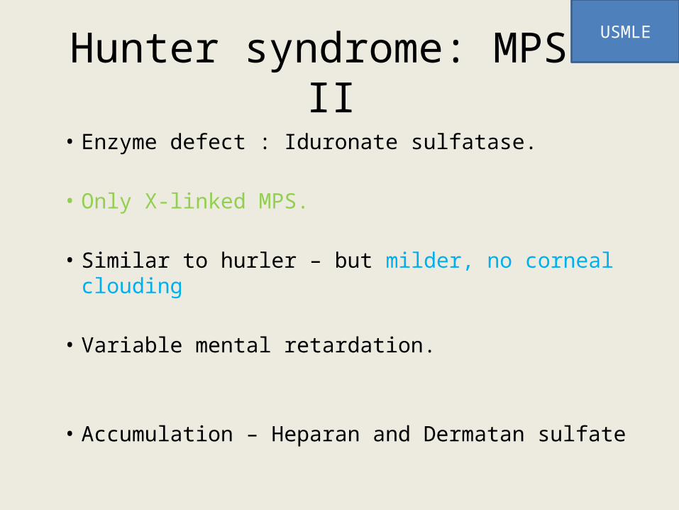
Hunter syndrome: MPS-II
• Enzyme defect : Iduronate sulfatase.
• Only X-linked MPS.
• Similar to hurler – but milder, no corneal clouding
• Variable mental retardation.
• Accumulation – Heparan and Dermatan sulfate
USMLE
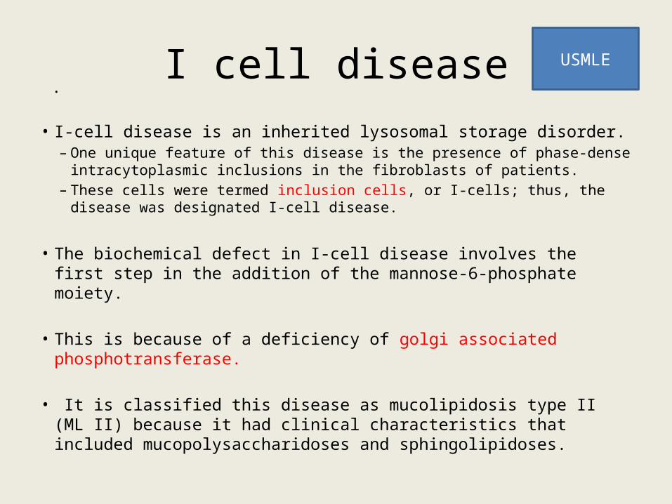
I cell disease• I-cell disease is an inherited lysosomal storage disorder.
– One unique feature of this disease is the presence of phase-dense intracytoplasmic inclusions in the fibroblasts of patients.
– These cells were termed inclusion cells, or I-cells; thus, the disease was designated I-cell disease.
• The biochemical defect in I-cell disease involves the first step in the addition of the mannose-6-phosphate moiety.
• This is because of a deficiency of golgi associated phosphotransferase.
• It is classified this disease as mucolipidosis type II (ML II) because it had clinical characteristics that included mucopolysaccharidoses and sphingolipidoses.
.
USMLE
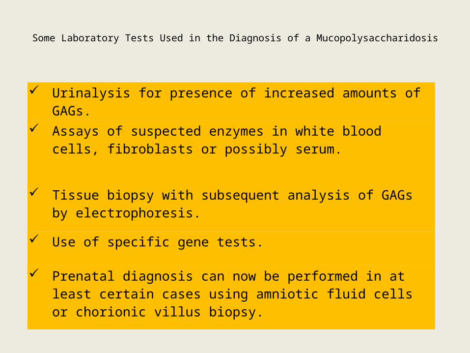
Some Laboratory Tests Used in the Diagnosis of a Mucopolysaccharidosis
Urinalysis for presence of increased amounts of GAGs.
Assays of suspected enzymes in white blood cells, fibroblasts or possibly serum.
Tissue biopsy with subsequent analysis of GAGs by electrophoresis.
Use of specific gene tests.
Prenatal diagnosis can now be performed in at least certain cases using amniotic fluid cells or chorionic villus biopsy.
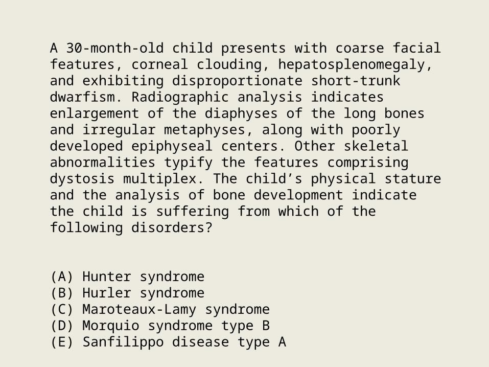
A 30-month-old child presents with coarse facial features, corneal clouding, hepatosplenomegaly, and exhibiting disproportionate short-trunk dwarfism. Radiographic analysis indicates enlargement of the diaphyses of the long bones and irregular metaphyses, along with poorly developed epiphyseal centers. Other skeletal abnormalities typify the features comprising dystosis multiplex. The child’s physical stature and the analysis of bone development indicate the child is suffering from which of the following disorders?
(A) Hunter syndrome(B) Hurler syndrome(C) Maroteaux-Lamy syndrome(D) Morquio syndrome type B(E) Sanfilippo disease type A

Thank you