Embed Size (px)
Citation preview
Autoimmune and allergic diseases, whichare marked by immune responses that are
polarized to either end of the T helper type 1(TH1)-TH2 spectrum, place a considerableburden on humanity. On the TH1 side, a vari-ety of organ-specific autoimmune disorders(including rheumatoid arthritis, type I dia-betes mellitis, inflammatory bowel disease
Marsha Wills-Karp, Yasmine Belkaid & Christopher
L. Karp are in the Divisions of Immunobiology and
Molecular Immunology, Cincinnati Children’s Hospital
Medical Center, Cincinnati, Ohio 45208, USA.
e-mail: [email protected]
I-Tim-izing the pathways of counter-regulationMarsha Wills-Karp, Yasmine Belkaid & Christopher L Karp
Tim-3, a member of the T cell immunoglobulin mucin family, is expressed by TH1 cells. Analysis of Tim-3–Tim3 ligand signalingnow shows this pathway is intimately involved in the counter-regulation of T helper type 1 immune responses.
N E W S A N D V I E W S
target cells. The cells were able to release inter-feron-γ, however, by the constitutive secretorypathway. Given that perforin deficiency resultsin severe immunodeficiency in humans andmice6, it is likely that the HPS2-associatedimmunodeficiency is related more to dimin-ished immune cytotoxicity than to CD1-related effects.
HPS2 is but one of many HPS syndromes,which feature defects of melanosomes,platelet-dense granules and lysosomes, result-ing in albinism, bleeding, accumulation ofintracellular ceroid (aggregated, oxidizedlipids) and pulmonary fibrosis. More than 15different mouse strains manifest a type ofalbinism-associated HPS-like syndrome, eachdue to the deletion of a different gene. Six ofthe gene defects have been described in HPS,but only HPS2 features immunodeficiency.Immunodeficiency and albinism also occur inGriscelli syndrome and Chediak-Hagashisyndrome. Patients with Griscelli syndromehave mutations in RAB27A, which encodes aRas-like GTPase involved in microtubule-and actin-dependent intracellular vesiclemovement. Children with Griscelli syn-drome develop a lethal T lymphocyte– andmacrophage-activation disease. Chediak-Hagashi syndrome results from mutation inthe gene encoding a protein called lysosomal-trafficking regulator (which also interactswith microtubules), and features defective Tand natural killer cell cytotoxicity as well as
delayed generation of peptide–MHC class IIcomplexes7.
The link between genotype and pheno-type is rarely a simple affair. If patients withHPS2 cannot use cytotoxic granules, why istheir immunodeficiency less severe than thatassociated with perforin deficiency, Griscellisyndrome or Chediak-Hagashi syndrome?Also, they do not have the problems withuncontrolled T cell and macrophage activa-tion seen in Griscelli syndrome8, and they donot suffer from the severe infections anduncontrolled lymphocyte proliferation ofperforin-deficient patients. One reason forthis could be that undetected residual AP-3function lessens the severity of HPS2. Clarket al. propose that there are defects in otherimmune cells that make use of lytic granules.Natural killer cells, also impaired in per-forin-deficient patients, and neutrophils areobvious subjects of further investigation. Afurther intriguing possibility is that antigenpresentation is impaired in dendritic cells,which are essential in initiating T cellresponses. Although the trafficking of MHCmolecules seems to occur independently ofAP-3 in most cell types9,10, immature den-dritic cells accumulate MHC molecules in anintracellular compartment and release themonly after maturation11. AP-3 could possiblybe involved in this process.
There is also a possible AP-3 link to the traf-ficking of viral membrane proteins. Human
immunodeficiency virus, for example, repli-cates in macrophages and CD4+ T cells, andgp160 uses the regulated secretory pathwayused by Fas ligand in CD4+ T cells12. Thispathway is similar to that followed by the Tcell activation molecule CTLA4. CTLA4 sort-ing is impaired in patients with Chediak-Hagashi syndrome. Could patients withdisorders in lysosomal or endosomal sortingshow resistance to viruses that coopt this path-way for their maturation? Variability in thesegenes may contribute to host fitness againstinfections, particularly in heterozygotes with afunctional allele. The torrid pace of researchpromises frequent advances in our knowledgeregarding adaptor protein complexes and reg-ulated secretion.
1. Blott, E.J. & Griffiths, G.G. Nat. Rev. Mol. Cell Biol. 3,122–131 (2002).
2. Clark, R. et al. Nat. Immunol. 4, 1111–1120 (2003).3. Esteban C. et al. Mol. Cell 3, 11-21, (1999).4. Bonifacino, J.S. & Lippincott-Schwartz, J. Nat. Rev.
Mol. Cell Biol. 4, 409 (2003).5. Sugita, M. et al. Immunity 16, 697–706 (2000).6. Stepp, S.E. et al. Science 286, 1957–1959 (1999).7. Faigle, W. et al. J. Cell Biol. 141, 1121–1134 (1998).8. Stepp, S.E., Mathew, P.A., Bennett, M., de Saint-Basile,
G. & Kumar, V. Immunol. Today 21, 254–256 (2000).9. Caplan, S., Dell’Angelica, E.C., Gahl, W.A. &
Bonifacino, J.S. Immunol. Lett. 72, 113–117 (2000).10. Sevilla, L.M., Richter, S.S. & Miller, J. Cell. Immunol.
210, 143–153 (2001).11. Chow, A., Toomre, D., Garrett, W. & Mellman, I.
Nature 418, 988–994 (2002).12. Miranda, L.R., Schaefer, B.C., Kupfer, A., Hu, Z. &
Franzusoff, A. Proc. Natl. Acad. Sci. USA 99,8031–8036 (2002).
1050 VOLUME 4 NUMBER 11 NOVEMBER 2003 NATURE IMMUNOLOGY
and multiple sclerosis) are associated withaberrant TH1 responses to self antigens. Overthe past 15 years, considerable experimentaland theoretical effort has gone into trying to understand the molecular mechanismsunderlying the development of TH1-polarized responses. Much has been learnedin this focus on T cell differentiation. Antigendose and affinity, the identity and differentia-tion state of the antigen-presenting cell, thecharacter of costimulation, the local cytokinemilieu, and specific transcription factors haveall been shown to influence TH polarization.An increasing amount of effort has also goneinto understanding the mechanisms that hold
such responses in check, modulating thedegree of T cell polarization and the overallvigor of the T cell response. Receptors (suchas CTLA-4), cytokines (such as interleukin10) and specialized cells (CD4+CD25+
T cells), separately and together, have come tothe fore in these studies. In this issue ofNature Immunology, two groups of investiga-tors provide substantial evidence that interac-tions between Tim-3, a recently discoveredprotein belonging to the T cell immunoglob-ulin mucin family of proteins, and its ligandare important in the counter-regulation ofTH1 immune responses1,2. These data maywell explain the linkage of the chromosomalregions occupied by the gene encoding Tim-3
©20
03 N
atu
re P
ub
lish
ing
Gro
up
h
ttp
://w
ww
.nat
ure
.co
m/n
atu
reim
mu
no
log
y
N E W S A N D V I E W S
used to begin addressing its in vivo function.In experimental autoimmune encephalo-myelitis, a TH1-dependent model of multiplesclerosis, antibodies to Tim-3 led to hypera-cute, atypical disease, with increased num-bers of neutrophils and activated macro-phages at sites of disease, greater demyelina-tion and higher mortality6. These data wereintriguing, but the ligand(s) and mecha-nism(s) of action remained to be identified.The authors of the present papers con-structed Tim-3–immunoglobulin (Ig) fusionproteins to address these issues. Use of Tim-3–Ig in flow cytometry of mononuclear cellsfrom lymphoid organs showed that Tim-3ligands (Tim-3Ls) were present on restingCD4+ T cells (naive, memory and CD25+
regulatory cells) and a subset of CD11c+ andCD11b+ splenocytes (whether the latter rep-resent dendritic cells or macrophages isunclear). Both TH1 and TH2 clones expressTim-3L. However, stimulation of CD4+
T cells leads to Tim-3L down-regulation,except on T regulatory cells.
These authors also pursued the mecha-nism with a variety of highly polarized, TH1-driven models. In all models tested, abroga-tion of Tim-3–Tim-3L interactions led toheightened TH1 responses: blockade of Tim-3–Tim-3L during immunization with anti-gen in complete Freund’s adjuvant led tohyperproliferation of TH1 cells and up-regulation of TH1 cytokine release, and abro-gated the development of tolerance to coad-minstered high-dose soluble antigen;
adoptive transfer of diabetogenic T cells intorecipient mice that had been treated withTim-3–Ig or antibody to Tim-3 accelerateddiabetes onset in the recipient mice; andcoadministration of Tim-3–Ig blocked thedevelopment of islet cell allograft toleranceinduced by two different protocols: donor-specific transfusion plus antibody to CD40ligand, and CTLA-4–Ig. These resultsdemonstrate that Tim-3–Tim-3L interac-tions have an important counter-regulatoryfunction during potent TH1-mediatedimmune responses. The available data pointto involvement ‘downstream’ of the TH1 dif-ferentiation process.
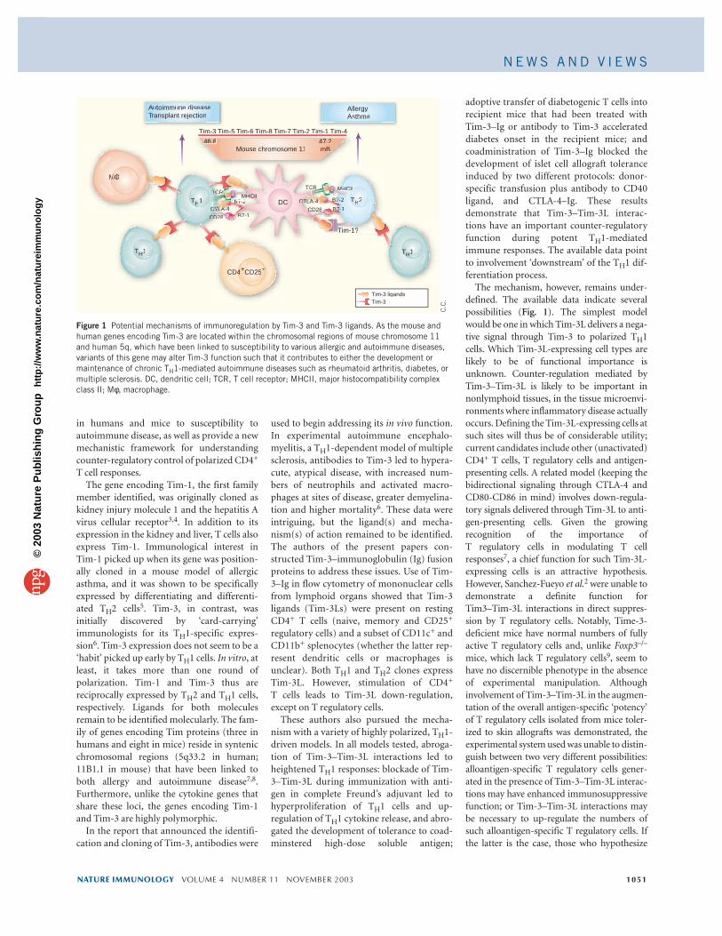
The mechanism, however, remains under-defined. The available data indicate severalpossibilities (Fig. 1). The simplest modelwould be one in which Tim-3L delivers a nega-tive signal through Tim-3 to polarized TH1cells. Which Tim-3L-expressing cell types arelikely to be of functional importance isunknown. Counter-regulation mediated byTim-3–Tim-3L is likely to be important innonlymphoid tissues, in the tissue microenvi-ronments where inflammatory disease actuallyoccurs. Defining the Tim-3L-expressing cells atsuch sites will thus be of considerable utility;current candidates include other (unactivated)CD4+ T cells, T regulatory cells and antigen-presenting cells. A related model (keeping thebidirectional signaling through CTLA-4 andCD80-CD86 in mind) involves down-regula-tory signals delivered through Tim-3L to anti-gen-presenting cells. Given the growingrecognition of the importance of T regulatory cells in modulating T cellresponses7, a chief function for such Tim-3L-expressing cells is an attractive hypothesis.However, Sanchez-Fueyo et al.2 were unable todemonstrate a definite function forTim3–Tim-3L interactions in direct suppres-sion by T regulatory cells. Notably, Time-3-deficient mice have normal numbers of fullyactive T regulatory cells and, unlike Foxp3–/–
mice, which lack T regulatory cells9, seem tohave no discernible phenotype in the absenceof experimental manipulation. Althoughinvolvement of Tim-3–Tim-3L in the augmen-tation of the overall antigen-specific ‘potency’of T regulatory cells isolated from mice toler-ized to skin allografts was demonstrated, theexperimental system used was unable to distin-guish between two very different possibilities:alloantigen-specific T regulatory cells gener-ated in the presence of Tim-3–Tim-3L interac-tions may have enhanced immunosuppressivefunction; or Tim-3–Tim-3L interactions maybe necessary to up-regulate the numbers ofsuch alloantigen-specific T regulatory cells. Ifthe latter is the case, those who hypothesize
NATURE IMMUNOLOGY VOLUME 4 NUMBER 11 NOVEMBER 2003 1051
in humans and mice to susceptibility toautoimmune disease, as well as provide a newmechanistic framework for understandingcounter-regulatory control of polarized CD4+
T cell responses.The gene encoding Tim-1, the first family
member identified, was originally cloned askidney injury molecule 1 and the hepatitis Avirus cellular receptor3,4. In addition to itsexpression in the kidney and liver, T cells alsoexpress Tim-1. Immunological interest inTim-1 picked up when its gene was position-ally cloned in a mouse model of allergicasthma, and it was shown to be specificallyexpressed by differentiating and differenti-ated TH2 cells5. Tim-3, in contrast, was initially discovered by ‘card-carrying’immunologists for its TH1-specific expres-sion6. Tim-3 expression does not seem to be a‘habit’ picked up early by TH1 cells. In vitro, atleast, it takes more than one round ofpolarization. Tim-1 and Tim-3 thus are reciprocally expressed by TH2 and TH1 cells,respectively. Ligands for both moleculesremain to be identified molecularly. The fam-ily of genes encoding Tim proteins (three inhumans and eight in mice) reside in syntenicchromosomal regions (5q33.2 in human;11B1.1 in mouse) that have been linked toboth allergy and autoimmune disease7,8.Furthermore, unlike the cytokine genes thatshare these loci, the genes encoding Tim-1and Tim-3 are highly polymorphic.
In the report that announced the identifi-cation and cloning of Tim-3, antibodies were
Figure 1 Potential mechanisms of immunoregulation by Tim-3 and Tim-3 ligands. As the mouse andhuman genes encoding Tim-3 are located within the chromosomal regions of mouse chromosome 11and human 5q, which have been linked to susceptibility to various allergic and autoimmune diseases,variants of this gene may alter Tim-3 function such that it contributes to either the development ormaintenance of chronic TH1-mediated autoimmune diseases such as rheumatoid arthritis, diabetes, ormultiple sclerosis. DC, dendritic cell; TCR, T cell receptor; MHCII, major histocompatibility complexclass II; Mφ, macrophage.
Autoimmune diseaseTransplant rejection
AllergyAsthma
TTHH1 TTH2
CD44++CD255+++
DC
CIIIMMHMHCHMTCCRCR
CTCTCTLA-4 2B77--2
CCCD28 B77--1B77B77--BBB
HCICIIIIMHTCRTTTCRTT
CCCTLACTC A-4AA 4ATLA 44A 4TLACT A-4TLACTC 442B7-2B7 2B7 2BB
CCCCD28C -1B7-
446.8 47.2mBMouse chromosome 11
Tim-3 Tim-5 Tim-6 Tim-8 Tim-7 Tim-2 Tim-1 Tim-4Tim 3 Tim 5 Tim 6 Tim 8 Tim 7 Tim 2 Tim 1 Tim 4
C.C
.
OOMMOMO
TH1 TTH1
1?Tim-1
Tim-3 ligandsTim-3
©20
03 N
atu
re P
ub
lish
ing
Gro
up
h
ttp
://w
ww
.nat
ure
.co
m/n
atu
reim
mu
no
log
y
Integrins are well known for their ability tofacilitate adhesion between cells and between
cells and the extracellular matrix. In theimmune system, the leukocyte function-associated antigen 1 (LFA-1, also known asCD11a/CD18 and αLβ2) integrin is particu-larly prominent in promoting the interactionbetween a naive T cell and an antigen-presenting cell (APC) that is necessary foreffective T cell activation. Inhibitory LFA-1-specific antibodies block interactions betweenT cells and APCs, and consequently disrupt T cell activation. In addition, LFA-1 forms theperipheral ring of the immunological synapse,the specialized structure that forms between T cells and APCs during T cell activation1,2.Extensive analysis of other integrins expressedon adherent cells has shown that instead ofserving merely as passive ‘strips of cellularVelcro’, integrin-mediated adhesion also leadsto signaling events that cooperatively regulatecellular responses mediated by growth factorreceptors3. Yet in the world of immunology,the idea that LFA-1 might also participate in T cell activation responses by transducing sig-nals has been met with some resistance. Thisskepticism regarding LFA-1 signaling in T cellsis due in part to lack of clear biochemical evi-
Yoji Shimizu is at the Department of Laboratory
Medicine and Pathology, Center for Immunology
and Cancer Center, University of Minnesota
Medical School, MMC 334/31, Church St. SE,
Minneapolis, MN 55455.
e-mail: [email protected]
LFA-1: more than just T cell VelcroYoji Shimizu
The LFA-1 integrin is known to mediate adhesion between T cells and antigen-presenting cells. New evidence, however, isprovided for its role in transmitting signals that promote T cell activation and differentiation.
N E W S A N D V I E W S
that T regulatory cells develop in the peripheryas normal sequelae of immune responses willbe pardoned for suspecting that the involve-ment of Tim-3–Tim-3L is far ‘upstream’ of Tregulatory cells in this model.
Finally, both groups focused on TH1-dri-ven processes. No compelling data have yetbeen presented that preclude the possibility ofa counter-regulatory function for Tim-3L inTH2 responses. Again, the complex matrix ofinteractions of CTLA-4 and CD28 with CD80and CD86 provides a possible guide for theo-
retical imagination here: Tim-3Ls may wellinteract with another receptor, possibly Tim-1, on TH2 cells.
The location of the genes encoding Tim-1and Tim-3 at a locus associated with suscepti-bility to TH1- and TH2-mediated diseases hasexciting implications for our growing under-standing of the immunopathogenesis of theseills. Although much remains to be learnedabout the molecular mechanisms underlyingthe (counter-) regulation of T cell responsesby Tim family members, the current reports
represent a considerable step forward in ourunderstanding of these molecules.
1. Sabatos, C.A. et al. Nat. Immunol. 4, 1102–1110(2003).
2. Sanchez-Fueyo, A. et al. Nat. Immunol. 4, 1093–1101(2003).
3. Han, W.K. et al. Kidney Int. 62, 237-244 (2002).4. Kaplan, G. et al. EMBO J. 15, 4292–4296 (1996).5. McIntire, J.J. et al. Nat. Immunol. 2, 1109–1116
(2001).6. Monney, L. et al. Nature 415, 536-541 (2002).7. Shevach, E.M. Nat. Rev. Immunol. 6, 389-400 (2002).8. Kuchroo, V.K. Nat. Immunol. 3, 454–462 (2003).9. Khattri, R. et al. J. Immunol. 1, 6312–6320 (2001).
1052 VOLUME 4 NUMBER 11 NOVEMBER 2003 NATURE IMMUNOLOGY
dence that this integrin can transmit signals inthe absence of signals initiated by the T cellreceptor (TCR) and coreceptors such as CD28.In this issue of Nature Immunology, Perez et al.4
provide some much-needed insight into thebiochemical events that occur when LFA-1 isengaged during T cell activation. These studiesprovide compelling evidence that LFA-1 canindeed transmit signals that promote T cellactivation and differentiation.
The idea that LFA-1 might do more thanjust keep the T cell and APC together is basedlargely on experiments demonstrating thatTCR stimulation together with the LFA-1 ligand ICAM-1 is sufficient to induce T cellproliferation in vitro5. Studies of this kind,however, have been unable to distinguishbetween two potential functions for LFA-1 inT cell signaling: quantitative enhancement ofTCR signaling through the promotion ofmore effective interaction between the T celland the TCR signal (whether an immobilizedantibody to CD3 (anti-CD3) or an APC), orqualitative modulation of T cell activationthrough the generation of unique LFA-1-dependent signals. Although these models arenot mutually exclusive, evidence in support ofa distinctive signaling function for LFA-1 haslagged far behind our understanding of howsignaling by other integrins regulates prolifer-ation and differentiation of anchorage-dependent cells such as fibroblasts3. Perez etal. used biochemical approaches and powerfulmultidimensional flow cytometry techniquesto more closely examine LFA-1-mediated sig-naling events in human T cells. Their findings
demonstrate that engagement of LFA-1 by asoluble form of ICAM-2, one of several LFA-1counter-receptors, results in two key phos-phorylation events, both of which are depen-dent on activation of the δ isoform of proteinkinase C (PKC-δ; Fig. 1). One is phosphoryla-tion of serine 745 in the cytoplasmic domainof the LFA-1 β2 integrin (CD18) subunit,which results in the release of Jun-activationdomain–binding protein 1 (JAB-1). This pro-tein was initially identified as a transcriptionalcoactivator that interacts with the c-Jun andJunD nuclear proteins6, which themselvesinteract with Fos proteins to form the activa-tor protein 1 (AP-1) transcriptional regulatorycomplex. More recently, JAB-1 was identifiedas an LFA-1-interacting protein that translo-cates to the nucleus after LFA-1 engagement 7.Consistent with these previous studies, Perezet al. demonstrate that c-Jun phosphorylationis induced by LFA-1 engagement on T cellsand is inhibited by JAB-1-specific antagonistpeptides. Furthermore, LFA-1 engagementenhances AP-1 activity after stimulation ofhuman T cells with anti-CD3 and anti-CD28,indicating involvement of JAB-1 in promotingLFA-1-dependent enhancement of AP-1 activ-ity through c-Jun phosphorylation.
As with other integrins, Perez et al. showthat LFA-1 engagement by ICAM-2 or ICAM-3 also induces phosphorylation of mitogen-activated protein kinase (MAPK). Activationof MAPK by LFA-1 requires a second PKC-dependent phosphorylation event initiated byLFA-1 engagement, which is phosphorylationof cytohesin-1, a guanine nucleotide–exchange
©20
03 N
atu
re P
ub
lish
ing
Gro
up
h
ttp
://w
ww
.nat
ure
.co
m/n
atu
reim
mu
no
log
y