Embed Size (px)
Citation preview


• I seek protection of ALLAH against the rejected Shaitan
• I seek refugee in ALLAH against the accused Shaitan
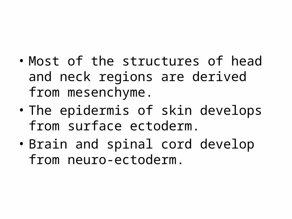
• Most of the structures of head and neck regions are derived from mesenchyme.
• The epidermis of skin develops from surface ectoderm.
• Brain and spinal cord develop from neuro-ectoderm.
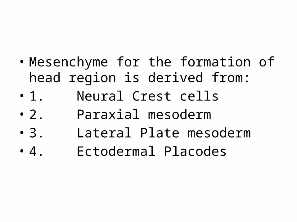
• Mesenchyme for the formation of head region is derived from:
• 1. Neural Crest cells• 2. Paraxial mesoderm• 3. Lateral Plate mesoderm• 4. Ectodermal Placodes

• Neural Crest cells form• Facial and pharyngeal arch skeletal structures
and all other tissues in these regions including cartilage, bone, dentin, tendon, dermis, pia mater, arachnoid mater, sensory neurons, glandular stroma and neurons of fifth, seventh, ninth and tenth cranial sensory ganglia.

• Paraxial mesoderm forms• Floor of skull and a small portion of occipital
region, voluntary muscles of craniofacial region, dermis and connective tissue in the dorsal region of head and meninges caudal to prosencephalon

• Lateral Plate mesoderm forms• Laryngeal cartilages and connective tissue in
this region• • Ectodermal Placodes forms• Neurons of fifth, seventh, ninth and tenth
cranial sensory ganglia
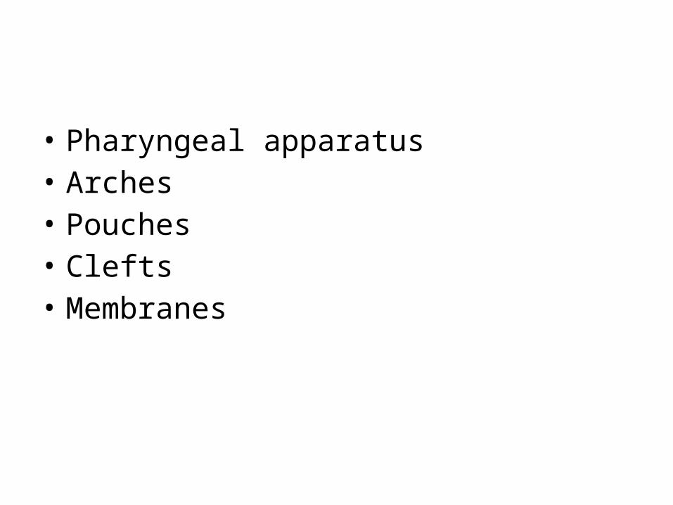
• Pharyngeal apparatus• Arches• Pouches• Clefts• Membranes

• Most of the structures of head and neck regions are derived from mesenchyme.
• Obviously the epidermis of skin develops from surface ectoderm.
• Brain and spinal cord develop from neuro-ectoderm.
• Mesenchyme for the formation of head region is derived from:–Neural Crest cells–Paraxial mesoderm– Lateral Plate mesoderm– Ectodermal Placodes
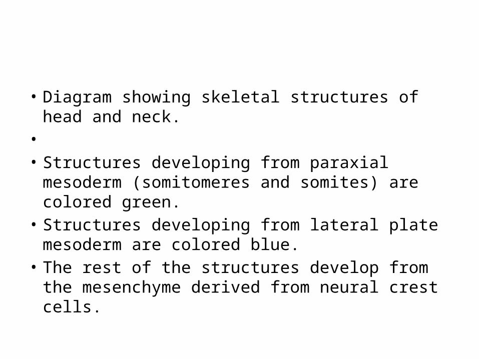
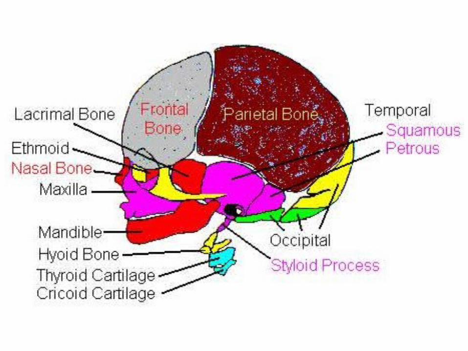
• Diagram showing skeletal structures of head and neck.
• • Structures developing from paraxial mesoderm
(somitomeres and somites) are colored green. • Structures developing from lateral plate
mesoderm are colored blue. • The rest of the structures develop from the
mesenchyme derived from neural crest cells.
• Neural Crest cells form• Facial and pharyngeal arch skeletal structures
and all other tissues in these regions including cartilage, bone, dentin, tendon, dermis, pia mater, arachnoid mater, sensory neurons, glandular stroma and neurons of fifth, seventh, ninth and tenth cranial sensory ganglia.
• Paraxial mesoderm forms• Floor of skull and a small portion of occipital
region, voluntary muscles of craniofacial region, dermis and connective tissue in the dorsal region of head and meninges caudal to prosencephalon
• Lateral Plate mesoderm forms • Laryngeal cartilages and connective tissue in
this region• • Ectodermal Placodes forms• Neurons of fifth, seventh, ninth and tenth
cranial sensory ganglia
• The pharyngeal or branchial apparatus contributes greatly to the formation of head and neck.
• Pharyngeal Apparatus consists of: • Pharyngeal or branchial arches • Pharyngeal or branchial pouches • Pharyngeal or branchial clefts or grooves • Pharyngeal or branchial membranes
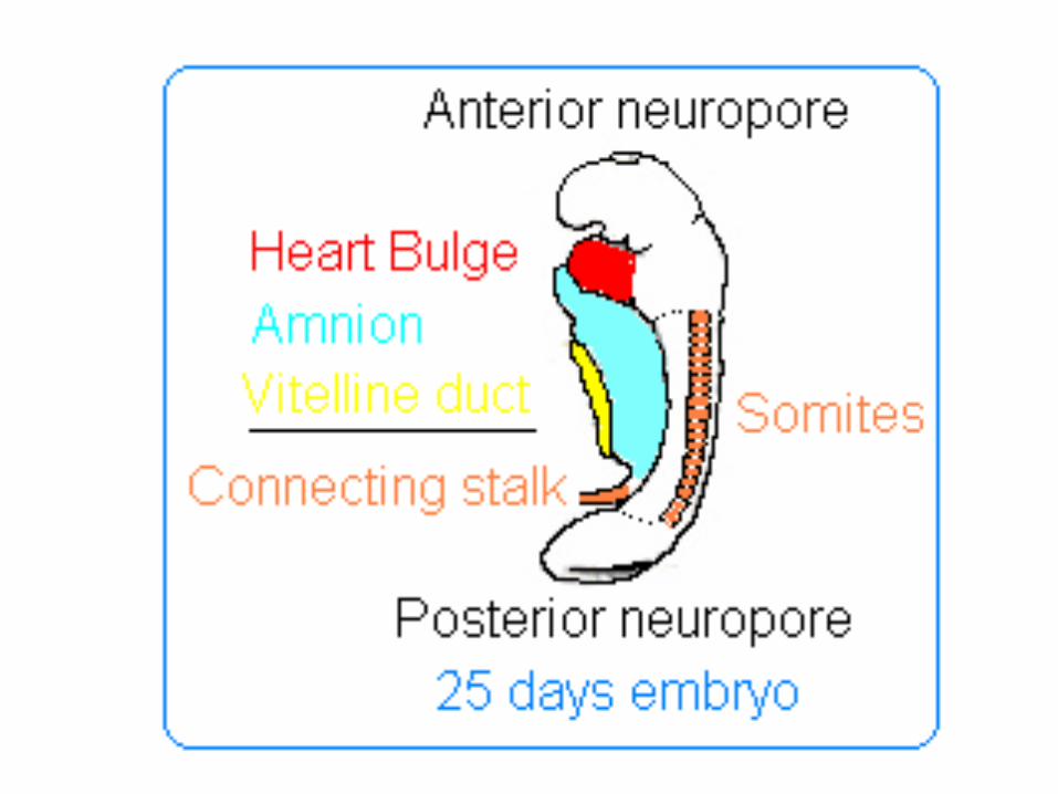
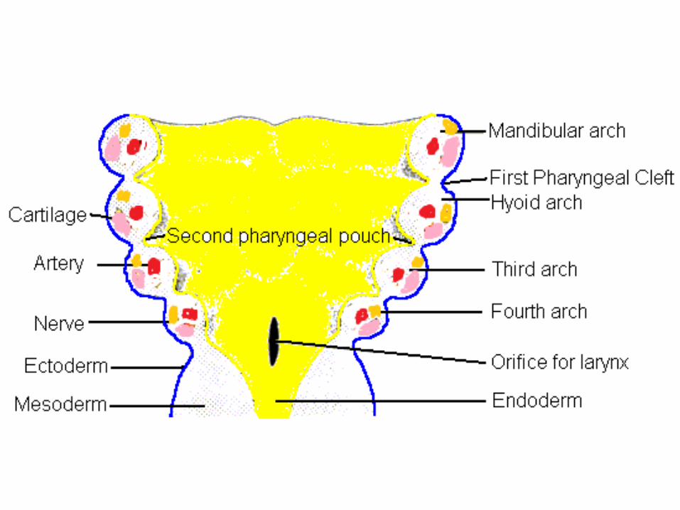
• In humans, they develop during the fourth week in utero as a series of mesodermal outpouchings on the left and right sides of the developing pharynx
• These grow and join in the ventral midline. The first arch, is the first to form, separates the mouth pit or stomodeum from the pericardium.
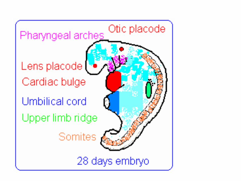
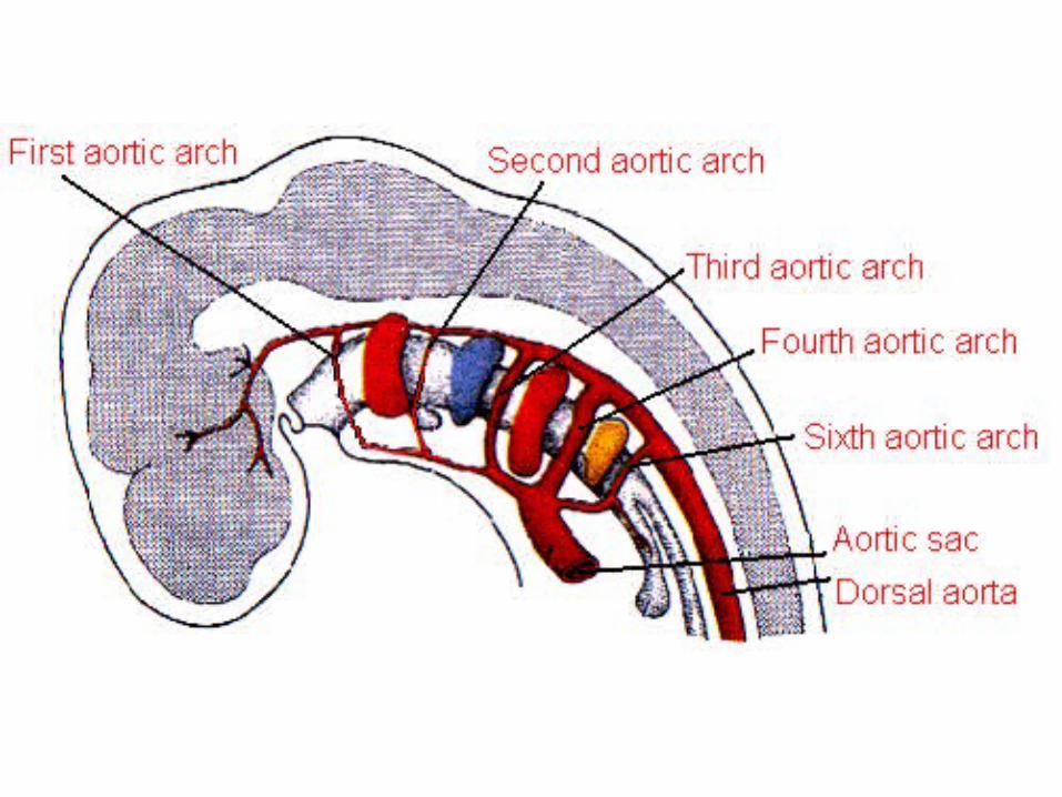
• By differential growth the neck elongates and new arches form, so the pharynx has six arches ultimately.
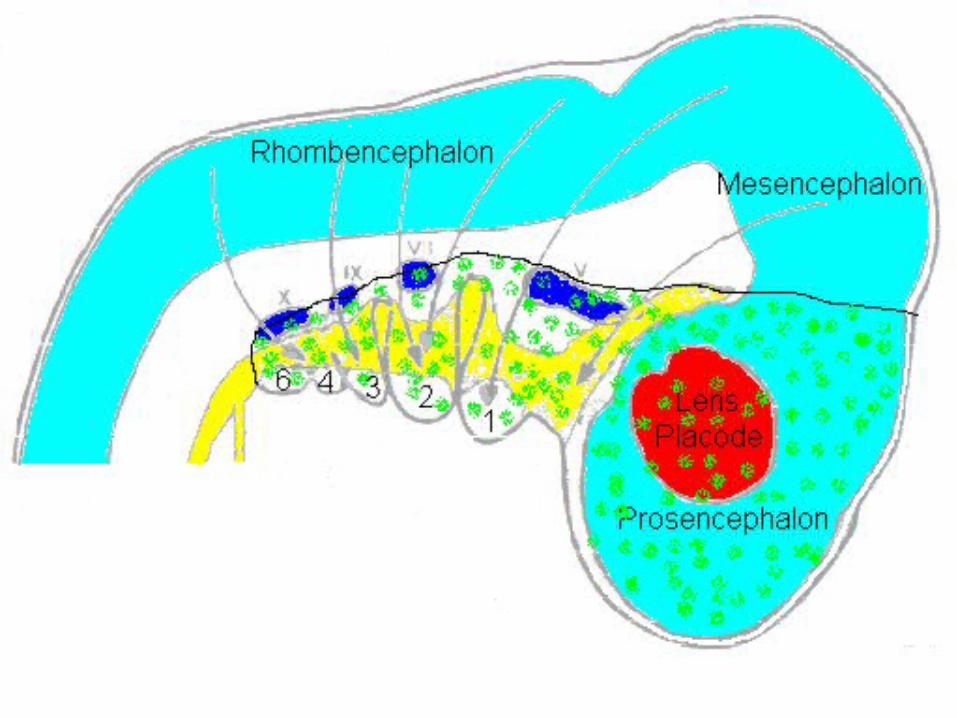
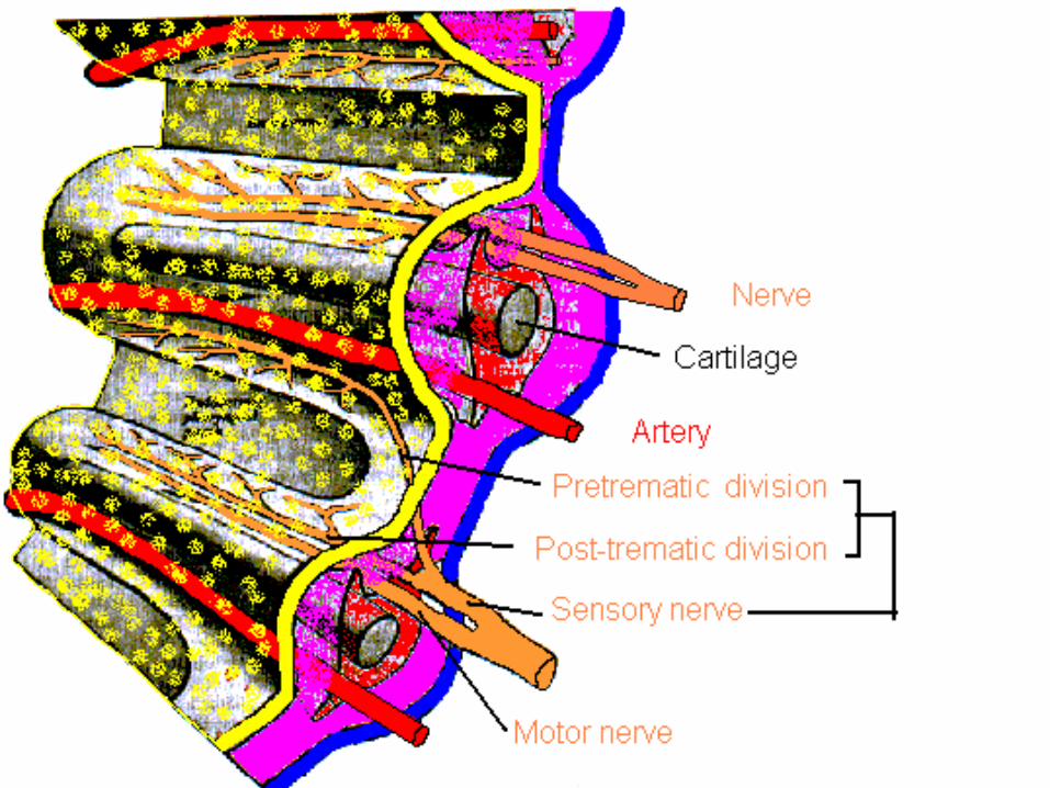
• Each pharyngeal arch has a cartilaginous stick, a muscle component which differentiates from the cartilaginous tissue, an artery, and a cranial nerve.
• Each of these is surrounded by mesenchyme. Arches do not develop simultaneously, but instead possess a "staggered" development.
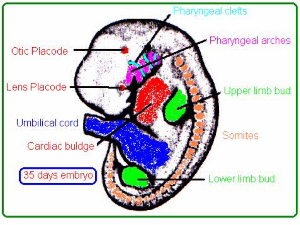
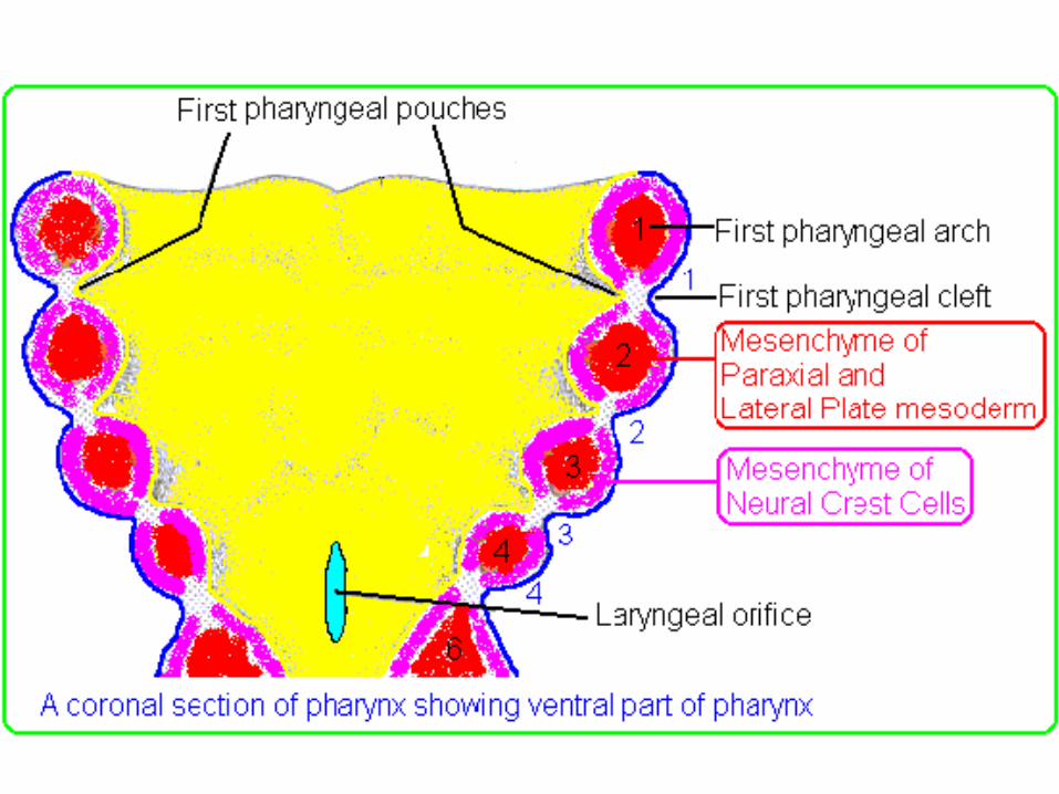
• Pharyngeal pouches (or branchial pouches) form on the endodermal side between the arches, and pharyngeal grooves (or clefts) form from the lateral ectodermal surface of the neck region to separate the arches
• The pouches line up with the clefts, and these thin segments become gills in fish.
• In mammals the endoderm and ectoderm not only remain intact, but continue to be separated by a mesoderm layer.
• More is known about the fate of the first arch than the remaining four.
• The first three contribute to structures above the larynx, while the last two contribute to the larynx and trachea.
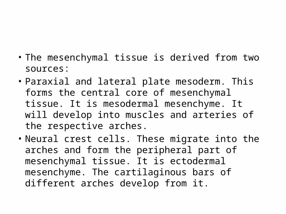
• The mesenchymal tissue is derived from two sources: • Paraxial and lateral plate mesoderm. This forms the
central core of mesenchymal tissue. It is mesodermal mesenchyme. It will develop into muscles and arteries of the respective arches.
• Neural crest cells. These migrate into the arches and form the peripheral part of mesenchymal tissue. It is ectodermal mesenchyme. The cartilaginous bars of different arches develop from it.
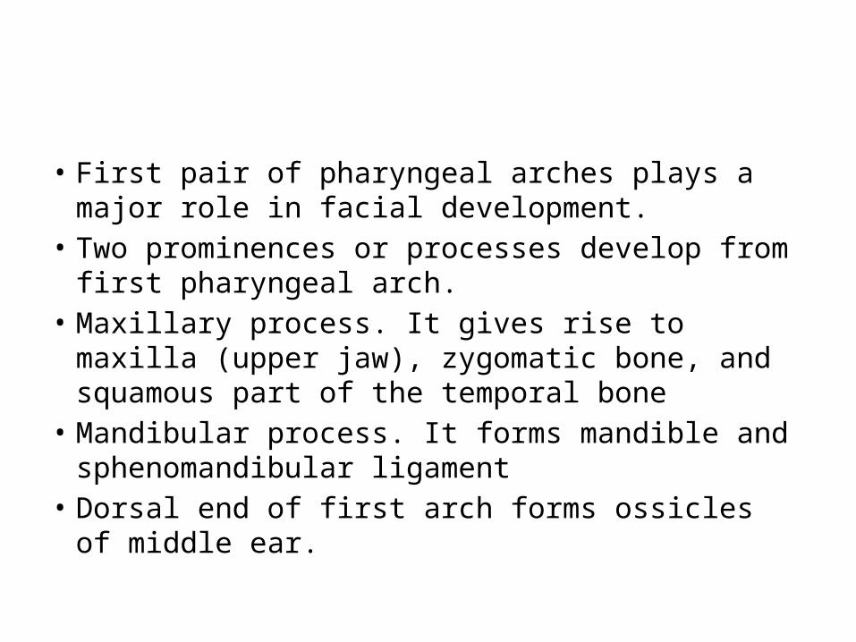
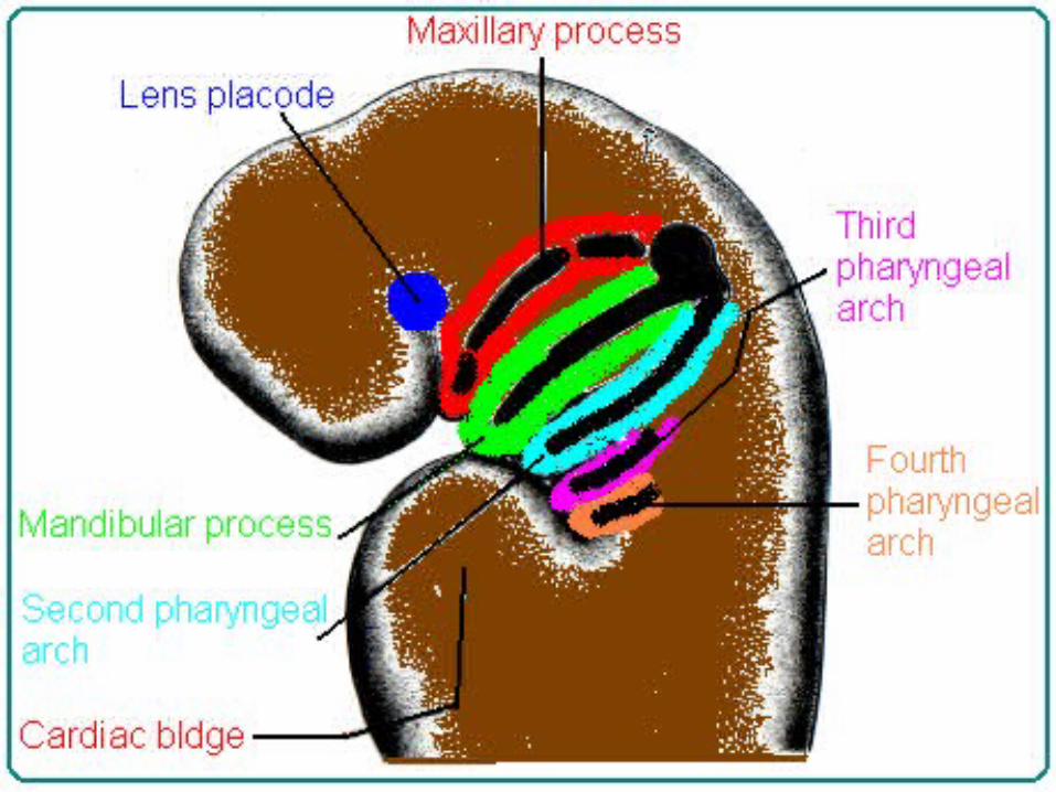
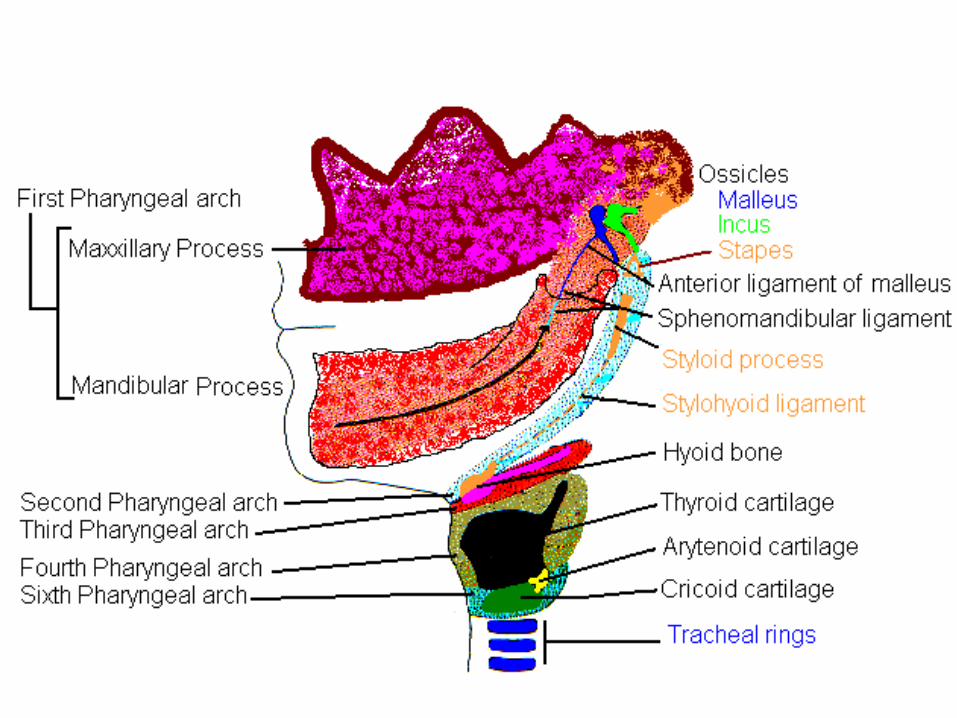
• First pair of pharyngeal arches plays a major role in facial development.
• Two prominences or processes develop from first pharyngeal arch.
• Maxillary process. It gives rise to maxilla (upper jaw), zygomatic bone, and squamous part of the temporal bone
• Mandibular process. It forms mandible and sphenomandibular ligament
• Dorsal end of first arch forms ossicles of middle ear.
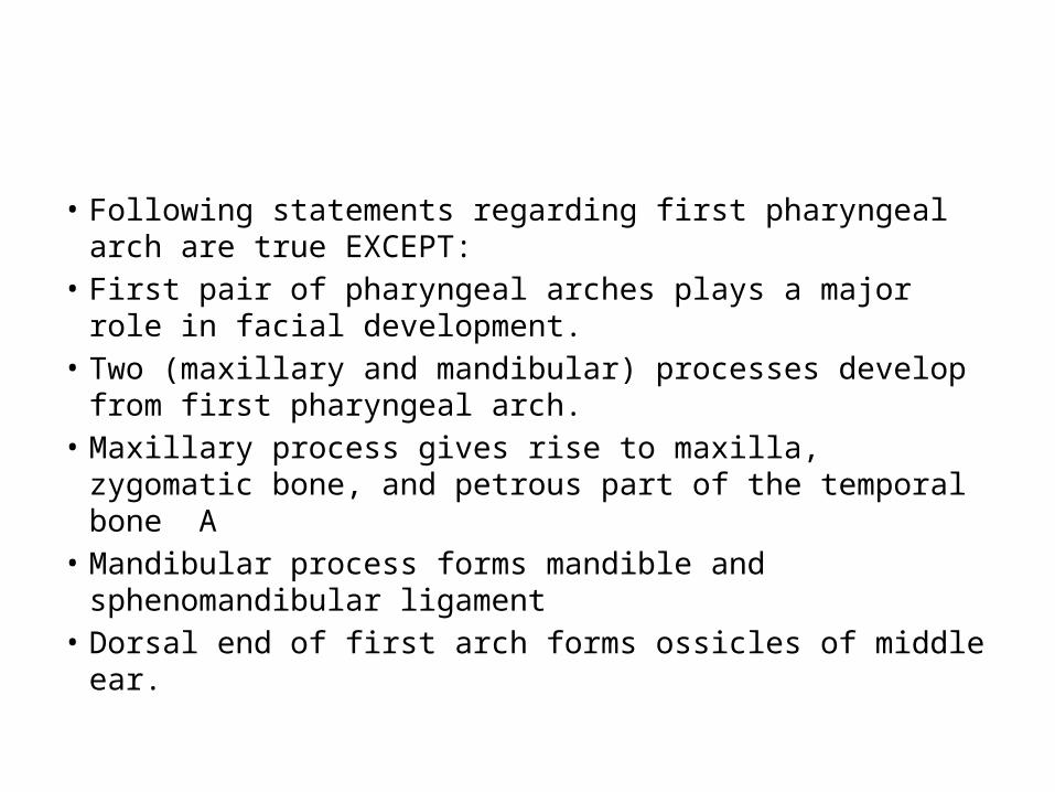
• Following statements regarding first pharyngeal arch are true EXCEPT:
• First pair of pharyngeal arches plays a major role in facial development.
• Two (maxillary and mandibular) processes develop from first pharyngeal arch.
• Maxillary process gives rise to maxilla, zygomatic bone, and petrous part of the temporal bone A
• Mandibular process forms mandible and sphenomandibular ligament
• Dorsal end of first arch forms ossicles of middle ear.
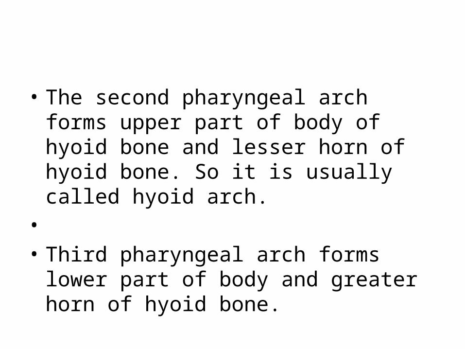
• The second pharyngeal arch forms upper part of body of hyoid bone and lesser horn of hyoid bone. So it is usually called hyoid arch.
• • Third pharyngeal arch forms lower part of
body and greater horn of hyoid bone.
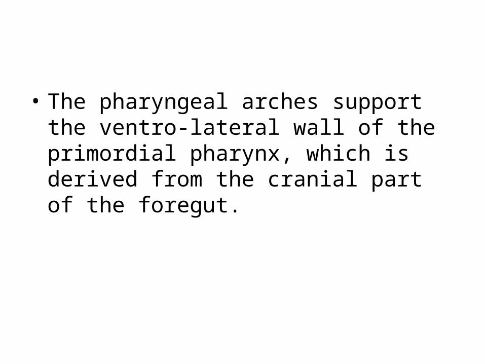
• The pharyngeal arches support the ventro-lateral wall of the primordial pharynx, which is derived from the cranial part of the foregut.
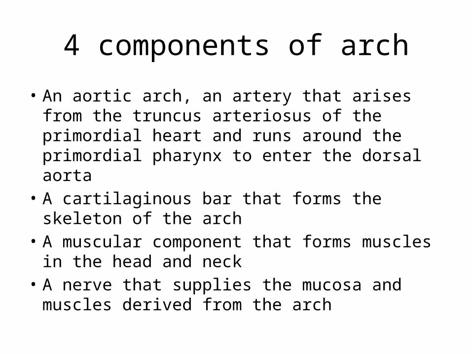
4 components of arch
• An aortic arch, an artery that arises from the truncus arteriosus of the primordial heart and runs around the primordial pharynx to enter the dorsal aorta
• A cartilaginous bar that forms the skeleton of the arch
• A muscular component that forms muscles in the head and neck
• A nerve that supplies the mucosa and muscles derived from the arch

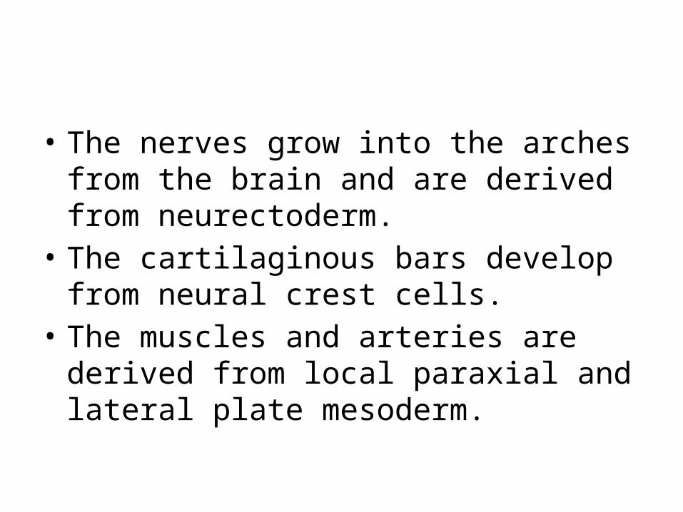
• The nerves grow into the arches from the brain and are derived from neurectoderm.
• The cartilaginous bars develop from neural crest cells.
• The muscles and arteries are derived from local paraxial and lateral plate mesoderm.
• The muscles of different arches do not always attach to the bony or cartilaginous components of their own arch but sometimes migrate into surrounding regions.
• The muscular components of each arch carry their own nerve, and wherever the muscle cells migrate, they carry their cranial nerve components with them. Thus the origin of these muscles can always be traced, since their nerve supply comes from the arch of origin.
• The dorsal end of the first arch cartilage (Meckel’s cartilage) is closely related to the developing ear and forms two middle ear ossicles (malleus and incus) and anterior ligament of malleus.
• The middle part of the cartilage regresses, but its perichondrium forms sphenomandibular ligament.
• Ventral part of the first arch cartilages form the horseshoe-shaped primordium of the mandible and, by keeping pace with its growth, guide its early morphogenesis (Hall, 1982).
• Each half of the mandible develops lateral to and in close association with Meckel’s cartilage.
• The cartilage disappears as the mandible develops by intramembranous ossification.
• Some endochondral ossification occurs in the median plane of the chin and in the mandibular condyle.
• The dorsal end of the second arch cartilage (Reichert cartilage), also closely related to the developing ear, ossifies to form the stapes of the middle ear and the styloid process of the temporal bone.
• The part of the cartilage between the styloid process and hyoid bone regresses; its perichondrium forms the stylohyoid ligament.
• The ventral end of the second arch cartilage ossifies to form the lesser cornu or horn of hyoid bone and the superior part of the body of the hyoid bone.
• The third arch cartilage, located in the ventral part of the arch, ossifies to form the greater cornu and the inferior part of the body of the hyoid bone.
• • The fourth and sixth arch cartilages fuse to
form the laryngeal cartilages, except for the epiglottis.
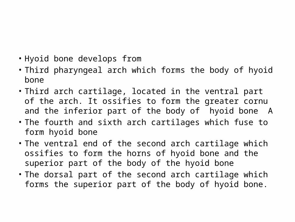
• Hyoid bone develops from• Third pharyngeal arch which forms the body of hyoid bone• Third arch cartilage, located in the ventral part of the arch. It
ossifies to form the greater cornu and the inferior part of the body of hyoid bone A
• The fourth and sixth arch cartilages which fuse to form hyoid bone
• The ventral end of the second arch cartilage which ossifies to form the horns of hyoid bone and the superior part of the body of the hyoid bone
• The dorsal part of the second arch cartilage which forms the superior part of the body of hyoid bone.
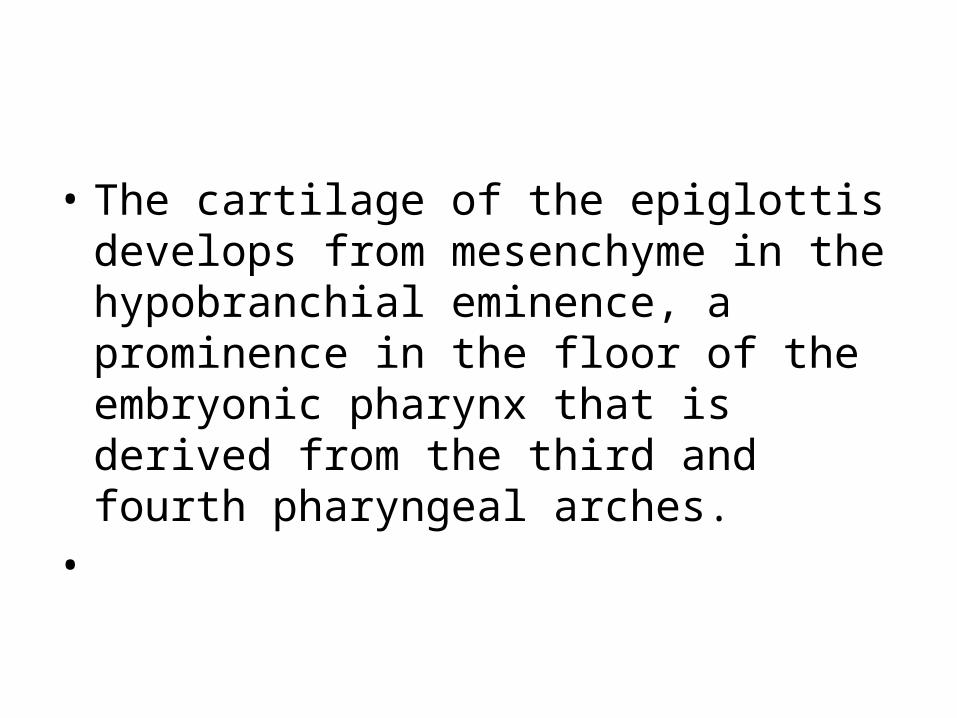
• The cartilage of the epiglottis develops from mesenchyme in the hypobranchial eminence, a prominence in the floor of the embryonic pharynx that is derived from the third and fourth pharyngeal arches.
•
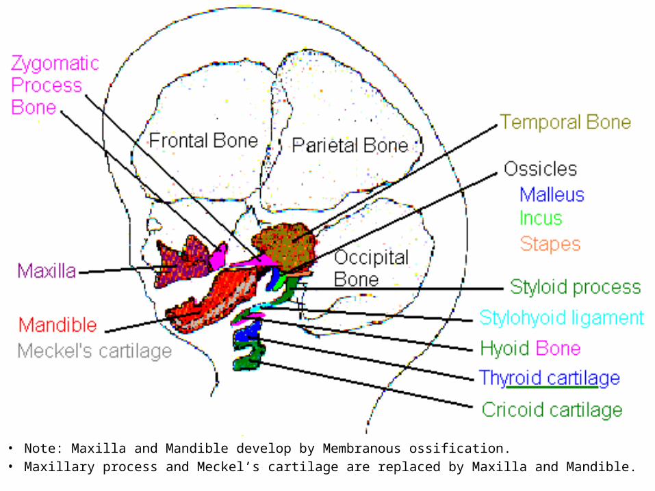
• Note: Maxilla and Mandible develop by Membranous ossification.• Maxillary process and Meckel’s cartilage are replaced by Maxilla and Mandible.
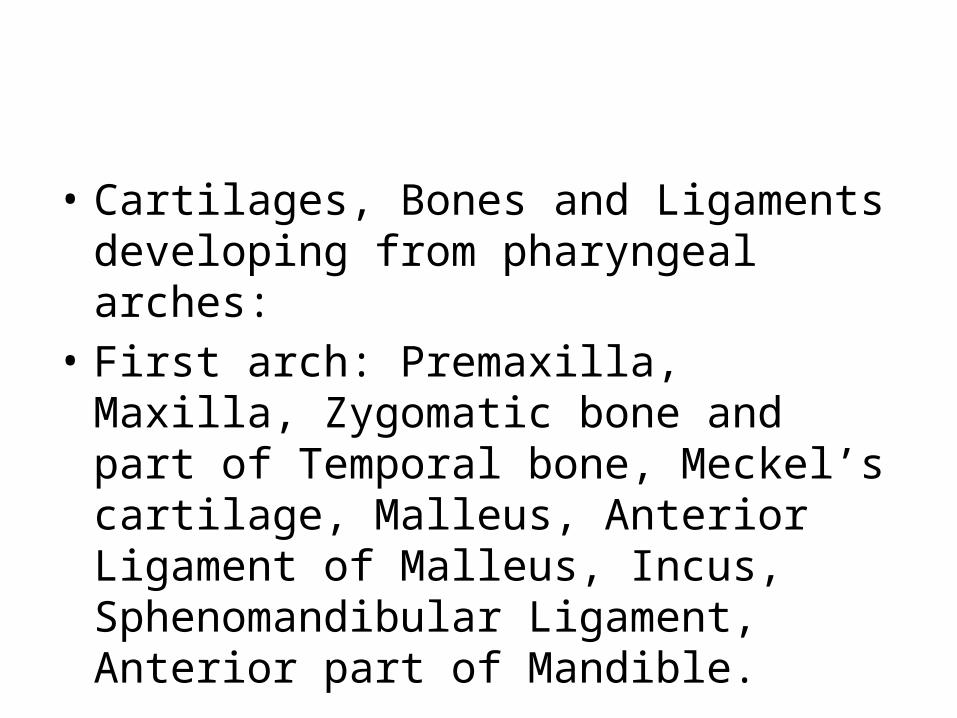
• Cartilages, Bones and Ligaments developing from pharyngeal arches:
• First arch: Premaxilla, Maxilla, Zygomatic bone and part of Temporal bone, Meckel’s cartilage, Malleus, Anterior Ligament of Malleus, Incus, Sphenomandibular Ligament, Anterior part of Mandible.
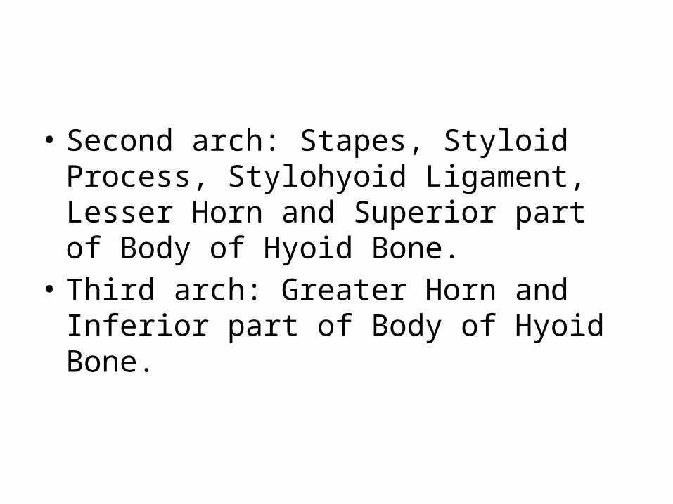
• Second arch: Stapes, Styloid Process, Stylohyoid Ligament, Lesser Horn and Superior part of Body of Hyoid Bone.
• Third arch: Greater Horn and Inferior part of Body of Hyoid Bone.
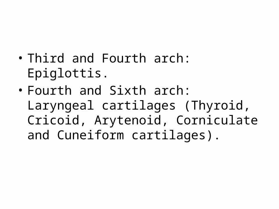
• Third and Fourth arch: Epiglottis.• Fourth and Sixth arch: Laryngeal cartilages
(Thyroid, Cricoid, Arytenoid, Corniculate and Cuneiform cartilages).
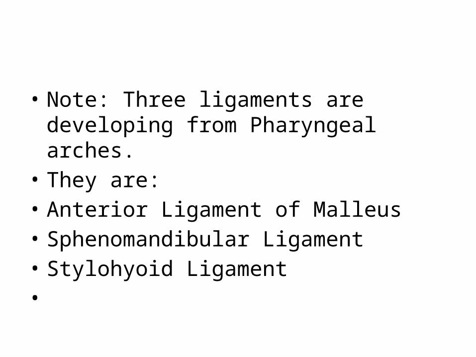
• Note: Three ligaments are developing from Pharyngeal arches.
• They are:• Anterior Ligament of Malleus• Sphenomandibular Ligament• Stylohyoid Ligament•
Muscles
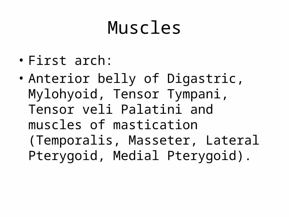
• First arch: • Anterior belly of Digastric, Mylohyoid, Tensor
Tympani, Tensor veli Palatini and muscles of mastication (Temporalis, Masseter, Lateral Pterygoid, Medial Pterygoid).
• The muscles developing from the first arch are• Posterior belly of Digastric A • Mylohyoid• Tensor Tympani • Tensor veli Palatini • Temporalis • Masseter
• Second arch: • Stepedius, Stylohyoid, Posterior belly of
Digastric and Muscles of Facial expression (Buccinator, Auricularis, Frontalis, Platysma, Orbicularis oris, Orbicularis occuli etc.).
• Third arch: Stylopharyngeus.• Fourth arch: Cricothyroid, Levator veli Palatini
and Constrictors of Pharynx.• Sixth arch: Intrinsic muscles of Larynx and
Striated muscles of esophagus
• Each arch is supplied by its own cranial nerve. The special visceral efferent (branchial) components of the cranial nerves supply muscles derived from the pharyngeal arches.
• Because mesenchyme from the pharyngeal arches contributes to the dermis and mucous membranes of the head and neck, these areas are supplied with special visceral afferent nerves.
• The facial skin is supplied by trigeminal (fifth cranial) nerve. However, only its caudal two branches (maxillary and mandibular) supply the derivatives of the first pharyngeal arch.
• Trigeminal nerve is the principle sensory nerve of head and neck and is the motor nerve for the muscles of mastication. Its sensory branches innervate the face, teeth, and mucous membranes of the nasal cavities, palate, mouth, and tongue.
• Facial nerve (seventh cranial) nerve supplies second pharyngeal arch.
• Glossopharyngeal (ninth cranial) nerve supplies third pharyngeal arch.
• Vagus (tenth cranial) nerve supplies fourth and sixth arches.
• The fourth arch is supplied by the superior laryngeal branch of vagus and the sixth arch by its recurrent laryngeal branch.
• The nerves of the second to sixth arches, however, innervate the mucous membranes of the tongue, pharynx, and larynx.
•
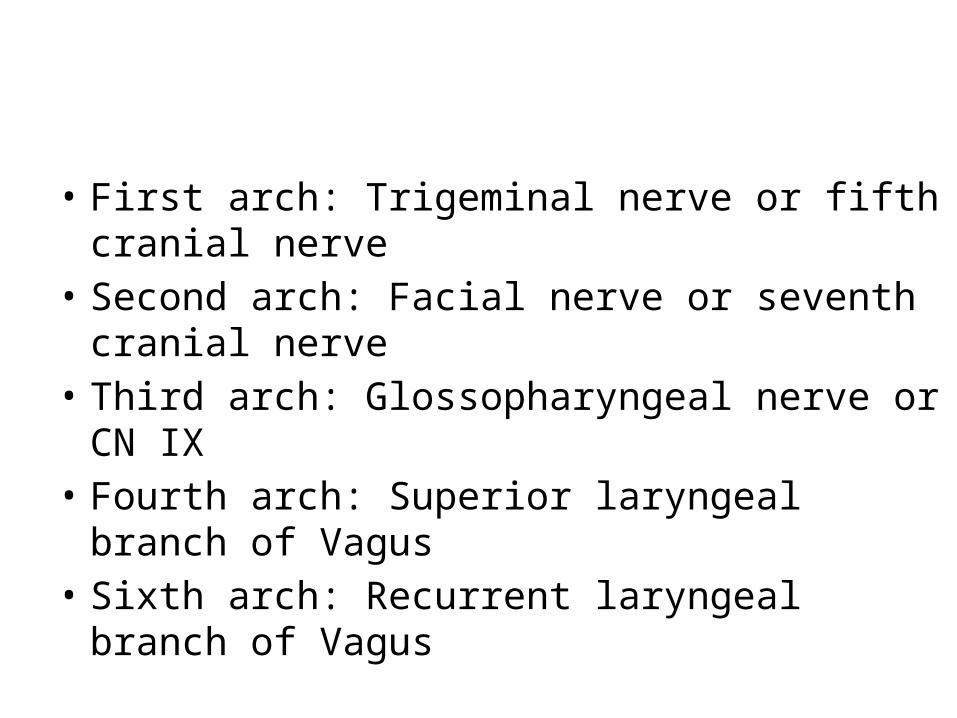
• First arch: Trigeminal nerve or fifth cranial nerve • Second arch: Facial nerve or seventh cranial
nerve • Third arch: Glossopharyngeal nerve or CN IX• Fourth arch: Superior laryngeal branch of Vagus • Sixth arch: Recurrent laryngeal branch of Vagus


![[Shaitan]Casting_slides [Compatibility Mode]11](https://img.pdfslide.us/doc/110x75/577d26531a28ab4e1ea0e0fd/shaitancastingslides-compatibility-mode11.jpg)