Embed Size (px)
Citation preview
volume 13 Number 2 1985 Nucleic Acids Research
Distinct characteristics of loop sequences of two Drosophila fold back transposable elements
Heidi L.Brierley* and S.Steven Potter
Department of Molecular Biology and Biochemistry, Wesleyan University, Middletown, CT 06457,USA
Received 10 September 1984; Revised and Accepted 18 December 1984
ABSTRACTA few foldback (FB) transposable elements have, between their long
terminal inverted repeats, central loop sequences which have been shown to bedifferent from FB inverted repeat sequences. We have investigated loopsequences from two such FB elements by analyzing their genomic distributionand sequence conservation and, in particular, by determining if they arenormally associated with FB elements. One of these FB loop sequences seemsto be present in a few conserved copies found adjacent to FB inverted repeatsequences, suggesting that it represents an integral component of some FBelements. The other loop sequence is less well-conserved and not usuallyassociated with FB inverted repeats. This sequence is a member of anotherfamily of transposable elements, the HB family, and was found inserted in anFB element only by chance. We compare the complete DNA sequences of two HBelements and examine the ends of four HB elements.
INTR0DUCTIOM
Transposable elements, units of DNA which can move to new locations in
the genome, have been studied in many procaryotic and eucaryotic systems (for
reviews see 1, 2). In Drosophila melanogaster several families of these
elements have been characterized (3) including the foldback (FB) family (4).
Foldback elements are among the most interesting transposable elements both
because of the many activities associated with them and because of their
unusual structures. They can, of course, insert into genes and inactivate
them. These mutations are particularly interesting because they revert at a
high frequency by precise excision of both the element and one copy of theDZL
target site duplication (5). Also, study of the w mutation (6,7) and
derivatives of the w mutatiion (8) suggest that FB elements can generate
rearrangements of adjacent DNA . Furthermore, two FB elements can cooperate
to move large blocks of DMA around the genome (9).
Structurally, the FB elements have long inverted terminal repeats and
sometimes a central loop between the repeats. The lengths of these repeats
and loops vary from element to element. We have previously shown that the
i IRL Press Limited, Oxford, England. 485
Downloaded from https://academic.oup.com/nar/article-abstract/13/2/485/1245114by gueston 12 April 2018
Nucleic Acids Research
inverted repeats carry a peculiar organization of sequences with highly
conserved complex sequences at the termini which gradually change into simple
31 base-pair (bp) tandem repeats as one moves toward the center of the
element (10, 11).
The FB loops are perhaps even more unusual. Restriction mapping data
suggest that often a loop structure seen by electron microscopy is simply
caused by the conjunction of a longer inverted repeat with a shorter one in
the same FB element (10). The excess tandem repeats from the longer end
constitute the loop structure. Very few FB elements have been demonstrated
to carry loop sequences that are different from FB inverted repeat sequences
(U, 7).
We have investigated the nature of the loop sequences contained in two
such FB elements. One of these elements is the FB which Levis et al (7)c
associated with white crimson (w ), an unstable allele of the white locus; wec
designate this element FBw . Levis et al (7) had previously shown that thec
loop of FBw was not homologous to FB inverted repeats or to the loop of FBA.
The other FB element reported on is FB4; its DNA sequence revealed an
interesting structure within the loop (11). There is an open reading frame
with characteristic gene punctuation signals in a region flanked by short
inverted repeats. The presence of this structure raised questions concerning
the conservation of this potential gene region and its distribution in the
genome. Is this structure an integral part of certain FB elements or is it
an independent transposable element? We investigated each of the two FB loop
sequences with respect to its genomlc distribution and its association with
FB inverted repeat sequences to determine if the sequence was usually between
FB inverted repeats or if it was found there only by chance.c
Our data suggest that the FBw loop sequences are indeed integral
components of some FB elements and not themselves independent elements. Some
interesting observations concerning the structure and evolution of FB
inverted repeat sequences also came from this study. Our investigation of
the FB4 loop sequence, however, indicates that it represents a member of an
independent family of transposable elements which we designate HB elements.
Many other homologous members have been found without any associated FB
inverted repeat sequences. FB4, therefore, is an unusual composite structure
with one transposable element (an HB) inserted into another (an FB). In this
report we compare the complete sequences of two HB elements and analyze the
ends of four HB elements. Furthermore, we describe an example in which
internal HB sequence is repeated in a flanking region near an HB element.
486
Downloaded from https://academic.oup.com/nar/article-abstract/13/2/485/1245114by gueston 12 April 2018
Nucleic Acids Research
MATERIALS AND METHODS
Materials
Enzymes were purchased from New England Biolabs. Isotopes were from New
England Nuclear. Nitrocellulose f i l t e r s were from Schleicher and Schuell.
Agarose was from Sigma, and acrylamide was from Biorad.
DNA Preparation and F i l t e r Hybridization
The construction of the recombinant pBR322-Drosophila melanogaster DNA
library and the preparation of the f i l t e r s for screening th is l ibrary have
been previously described (12, 13). Plasmid DNA was prepared by the rapid
boil method of Holmes and Quigley (14) with previously described
modifications (15). Restr ic t ion s i t e mapping was done by size analysis of
segments generated by single and double digests . DNA segments were prepared
by using low-melting agarose as previously described (10). Transfer of DNA
from agarose gels to n i t rocel lu lose f i l t e r s was done in the manner of
Southern (16). Sometimes one gel was used to make two f i l t e r s by placing
additional n i t roce l lu lose and b lo t te r paper below the gel and eliminating the
20X SSC supply (17).
DNA segments were nick translated in the manner of Rigby et al (18)32
using [ - PJdNTP from NEN. The labelled DNA was e i ther passed over aSephadex G50 column or ethanol precipi tated in the presence of 0.2 M sodium
ace ta te . F i l t e r s were presoaked in IX Denhardt's solut ion (19) (IX = 0.02%
polyvinylpyrrolidone, 0.02% bovine serum albumin, 0.02% f i co l l ) and 4X SSCP
(IX = 0.015 M sodium c i t r a t e , 0.12 M NaCl, 0.02 M sodium phosphate (pH 7))o
for at leas t 4 hours at 65 C. The nick-translated DNA was allowed tohybridize to the f i l t e r s in IX Denhardt's, 4X SSCP, and 0.1 mg/ml sonicated
osalmon sperm DNA for 15-24 hours at 65 C with ag i ta t ion . The f i l t e r s were
washed extensively in IX Denhardt's and 4X SSCP, with multiple wash changes,o o
at 65 C with gentle ag i ta t ion . The second to las t wash was done at 55 C, and
the f inal wash was done in 2X SSC (IX = 0.015 M sodium c i t r a t e , 0.15 M NaCl)
at room temperature.
DNA Sequence Analysis32
DNA segments were 3 ' end labelled with [ - PJdNTP by using the largefragment of E. co l l DNA Pol I (20) in the manner of Levis et al (21). DNA
sequencing was done by the procedure of Maxam and Gilbert (22); the chemical
reactions for G, T+C, C, and A>C were used. 75 cm long gels of 4-20?
acrylamide were used. The Stanford University SEO computer program was used
in the sequence ana lys i s .
487
Downloaded from https://academic.oup.com/nar/article-abstract/13/2/485/1245114by gueston 12 April 2018
Nucleic Acids Research
E =ua > io aco
t „if 1wo a
I ; ; i if0 a a. v> w o .1 i I i ma
E a "a u "u 3 E
i i r
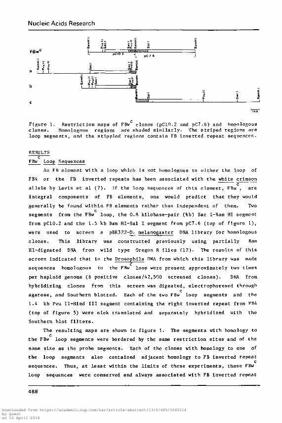
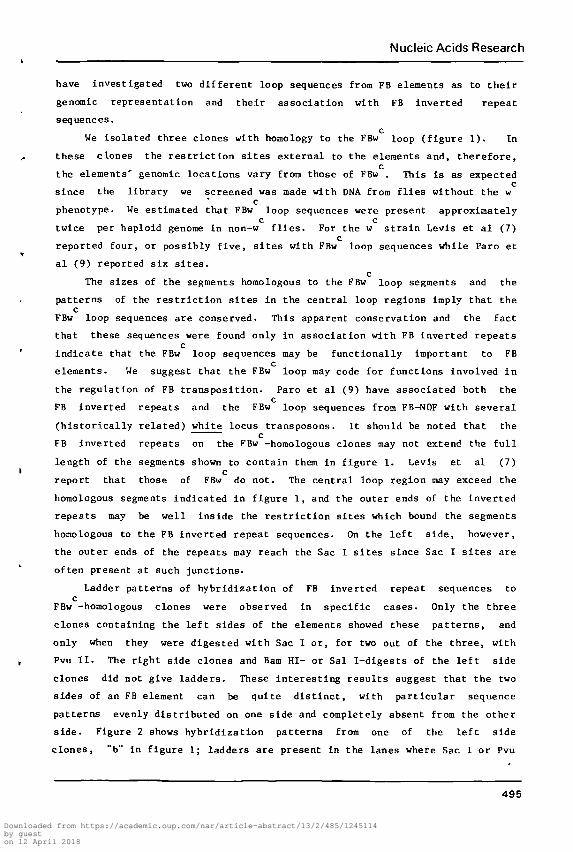
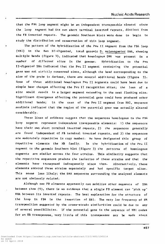
Figure 1. Restr ic t ion maps of FBw clones (pC10.2 and pC7.6) and homologousclones. Homologous regions are shaded s imilar ly . The s tr iped regions areloop segments, and the stippled regions contain FB inverted repeat sequences.
RESULTSc
FBw Loop Sequences
An FB element with a loop which is not homologous to either the loop of
FB4 or the FB inverted repeats has been associated with the white crimson_
a l l e l e by Levis et a l (7 ) . If the loop sequences of th i s element, FBw , are
in tegra l components of FB elements, one would predict that they would
generally be found within FB elements rather than independent of them. Twoc
segments from the FBw loop, the 0.8 kilobase-pair (kb) Sac I-Bam HI segmentfrom pC10.2 and the 1.5 kb Bam Hi-Sal I segment from pC7.6 (top of figure 1),
were used to screen a pBR322-J£1 melanogaster DNA l ibra ry for homologous
clones. This l ibrary was constructed previously using pa r t i a l l y Bam
Hi-digested DNA from wild type Oregon R f l i e s (17.). The resu l t s of th i s
screen indicated that in the Drosophila DNA from which this l ibrary was madec
sequences homologous to the FBw loop were present approximately two timesper haploid genome (6 positive clones/42,500 screened c lones) . DNA fromhybridizing clones from this screen was digested, electrophoresed through
cagarose, and Southern b lo t ted . Each of the two FBw loop segments and the
1.4 kb Pvu II-Hind I I I segment containing the r ight inverted repeat from FB4
(top of figure 5) were nick translated and separately hybridized with the
Southern blot f i l t e r s .
The resul t ing maps are shown in figure 1. The segments with horaology toc
the FBw loop segments were bordered by the same r e s t r i c t i o n s i t e s and of the
same size as the probe segments. Each of the clones with homology to one of
the loop segments also contained adjacent homology to FB inverted repeatc
sequences. Thus, at leas t within the l imits of these experiments, these FBw
loop sequences were conserved and always associated with FB inverted repeat
488
Downloaded from https://academic.oup.com/nar/article-abstract/13/2/485/1245114by gueston 12 April 2018
Nucleic Acids Research
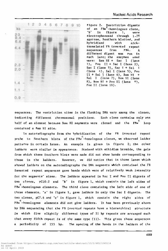
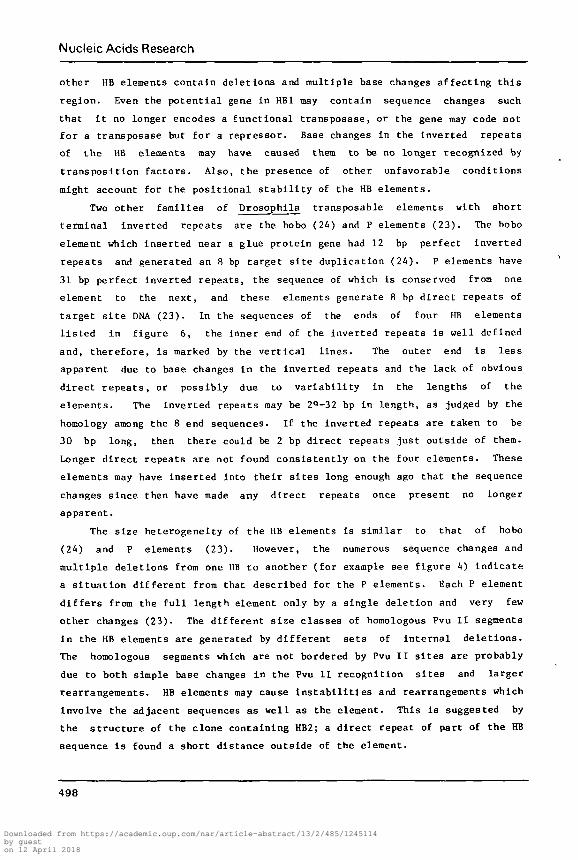
Figure 2. Restriction digestsof an FBw -homologous clone,"b" in figure 1, wereelectrophoresed through 1.27.agarose, Southern blotted, andhybridized with nick-translated FB inverted repeatsequences from FB4. Adifferent digest was run ineach lane; the enzymes usedwere: Bam HI + Sac I (lane1), Pvu II + Sac I (lane 2),Sac I (lane 3), Sac I + Sal I(lane 4), Sal I (lane 5), PvuII + Sal I (lane 6), Bam HI +
~ Sal I (lane 7), Bam HI (lane
1 A 8), Bam HI + Pvu II (lane «),Pvu II (lane 10).
1.1 -
0.9 -
sequences. The restriction sites in the flanking DNA vary among the clones,
indicating different chromosomal positions. Each clone contains only onec
half of an element because Bam HI segments were cloned and the FBw loop
contained a Bam HI site.
In autoradiographs from the hybridization of the FB inverted repeat
probe to Southern blots of the FBw -homologous clones, we observed ladder
patterns in certain lanes. An example is given In figure 2; the other
ladders were similar in appearance. Stained with ethidium bromide, the gels
from which these Southern blots were made did not show bands corresponding to
those in the ladders. However, we did notice that in those lanes which
showed ladders on the autoradiographs the DNA segments which contained the FB
inverted repeat sequences gave bands which were of relatively weak Intensity
for the segments' sizes. The ladders appeared in Sac I and Pvu II digests of
two clones, pC10.2 and "b" In figure 1, which contain the left sides ofc
FBw -homologous elements. The third clone containing the left side of one of
these elements, "a" in figure 1, gave ladders in only the Sac I digests. The
two clones, pC7.6 and "c" in figure 1, which contain the right sides ofc
FBw -homologous elements did not give ladders. It has been previously shown
by DNA sequencing that the FB inverted repeats have a hierarchical structure
in which five slightly different types of 31 bp repeats are arranged such
that every fifth repeat is of the same type (11). This gives these sequences
a periodicity of 155 bp. The spacing of the bands In the ladders of the
489
Downloaded from https://academic.oup.com/nar/article-abstract/13/2/485/1245114by gueston 12 April 2018
Nucleic Acids Research
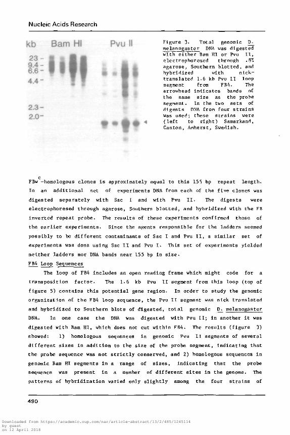
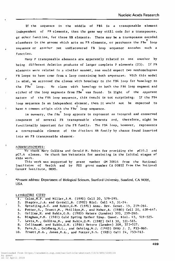
k b B a m HI P v u II Flgure 3- Total senora ic ^melanogaster DNA was digestedwith either Bam III or Pvu I I ,electrophoresed through .3%
X5 I I B̂ I ^B A agarose, Southern blotted, and6.6 " f V | | * " H W hybridized with nick-A A — I I F̂ translated 1.6 kb Pvu I I loop
• * • segment from FB4. Thearrowhead indicates bands ofthe same size as the probesegment. In the two sets of
t.»3 ~ digests PNA from four strainsp Q— was used; these strains were
^fc 4 (left to right) Samarkand,Canton, Amherst, Swedish.
Bam HI1 1 ^B H
I I• *
cFBw -homologous clones is approximately equal to this 155 bp repeat length.
In an additional set of experiments DNA from each of the five clones was
digested separately with Sac I and with Pvu I I . The digests were
electrophoresed through agarose, Southern blotted, and hybridized with the FB
inverted repeat probe. The results of these experiments confirmed those of
the ear l ier experiments. Since the agents responsible for the ladders seemed
possibly to be different contaminants of Sac I and Pvu I I , a similar set of
experiments was done using Sac I I and Pvu I . This set of experiments yielded
neither ladders nor DNA bands near 155 bp in size.
FB4 Loop Sequences
The loop of FBA includes an open reading frame which might code for a
transposition factor. The 1.6 kb Pvu II segment from this loop (top of
figure 5) contains this potential gene region. In order to study the genomic
organization of the FBA loop sequence, the Pvu I I segment was nick translated
and hybridized to Southern blots of digested, total genomic D. melanogaster
DNA. In one case the DNA was digested with Pvu I I ; in another i t was
digested with Bam HI, which does not cut within FB4. The results (figure 3)
showed: 1) homologous sequences in genomic Pvu I I segments of several
different sizes in addition to the size of the probe segment, indicating that
the probe sequence was not s t r ic t ly conserved, and 2) homologous sequences in
genomic Bam HI segments in a range of sizes, indicating that the probe
sequence was present in a number of different s i tes in the genome. The
patterns of hybridization varied only slightly among the four strains of
490
Downloaded from https://academic.oup.com/nar/article-abstract/13/2/485/1245114by gueston 12 April 2018
Nucleic Acids Research
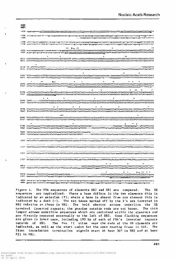
HB2HB1
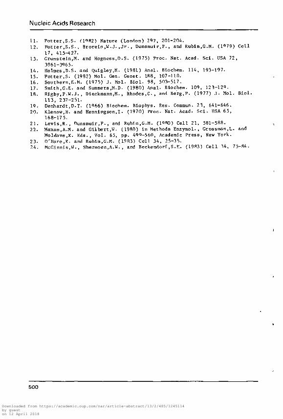
t-230 tttaatattaatattt>gctcg*a*aaeatattgccat*gtgcc«t8caa*|ccaga»««*c«aaggatcg*cacataactcc:tttcagtaaggttaat£-I SB . . . t i< i tn t t t c | i c t | i c cc i ccc t t t i i i t cc i i t t i t t c t c | t t tgccc<[cc t t ta
-130 cttttgcatcftgggacctacagctcgttctt*ttct**tactccflcgagtaacaggtatta*atttgt«c«a**ccgctgctataa«ggg*tataaacaa-130 • •atteatttctaacgtttftcccacccttcaaaatttgttttgtaagatgtggcgecaatteagatattttaggatcgBCgMtagaageacttacttat
• • Pvu I I • * *- 0 30 a t a c a t g a a a g a g t t a a a a t a a a c t tgcggAAATACACCXCxCTXCACAAAAAXACCACXCCGAAGCCAACTAAGTAATACAAACCXATTTTXCCAXCCX
0 1 7 0 TT-ATTTTCATTCTT TCAATATTTu l / I XXXAxxXACATTCXXGAGGAAAAXCACAAXXXXXAGG CxGxTCATAACAAXAGCAGXGxCTCGxXCXGACCAACGXAAAGXCCCGAAAXwAXCAAXAxxx
0785
ios;
I 1S7
I3S7
155
+00•00
Figure 4. The DMA sequences of elements HB2 and HB1 are compared. The HBsequences are capi tal ized. Where a base differs in the two elements th i s i sindicated by an as ter isk (*); where a base is absent from one element th is i sindicated by a dash ( - ) . The ten bases marked off by the i ' s are inverted inHB2 re la t ive to those in HB1. The bold shorter arrows underline the HBterminal inverted repeats ; the precise outside ends are not known. The thinlonger arrows underline sequences which are contained within the elements andare di rect ly repeated externally to the left of HB2. Some flanking sequencesare given in lower case, including 100 bp of each of FB4's inverted repeatsoutside of HB1. The Pvu I I s i t e s near the ends of the HB elements areindicated, as well as the s t a r t codon for the open reading frame in HB1. Thef i r s t t ranslat ion termination signals s t a r t at base 360 in HB2 and at base831 in HB1.
491
Downloaded from https://academic.oup.com/nar/article-abstract/13/2/485/1245114by gueston 12 April 2018
Nucleic Acids Research
D. melanogaster that were tested (figure 3).
The FB4 Pvu II loop segment was also used to select an homologous clone
from a previously constructed pBR322-D. melanogaster DNA library (12). On
this selected clone the primary homology was localized to a 1.0 kb Pvu II
segment, instead of a 1.6 kb one as in FB4. The restriction maps differed
outside this Pvu II segment also, and this second clone did not contain
homology to FB inverted repeat sequences.
The sequence of the entire FB4 element had been determined previously
(11) so, in order to more closely analyze the relationship of the two
homologous clones, the 1.0 kb Pvu II segment from the second was sequenced.
Much of the two Pvu II segments Is homologous. Short inverted repeats flank
the homologous regions within each of the Pvu II segments. These homologous
elements with short inverted repeats have been designated HB1 (inside FB4)
and HB2. A comparison of the sequences of HB1 and HB2 is given in figure 4.
The primary difference is that one large section (493 bp) and several smaller
ones present in the HB1 sequence are deleted from the HB2 sequence. There
are also many individual base differences between the two sequences. One
such change introduces a stop signal in HB2 only 48 bases after the start
signal; the open reading frame in HB 1 continues for an additional 396
bases.
To further investigate the nature of the association of HB sequences
with FB inverted repeat sequences and the possible relationship of HBc
sequences with the FBw loop sequences, we used the 1.0 kb Pvu II segment
from HB2 (figure 5) to thoroughly screen the pBR322-D^ melanogaster DNA
library. The results of this screen Indicated that sequences with homology
to this segment were present in approximately 20 copies per haploid genome
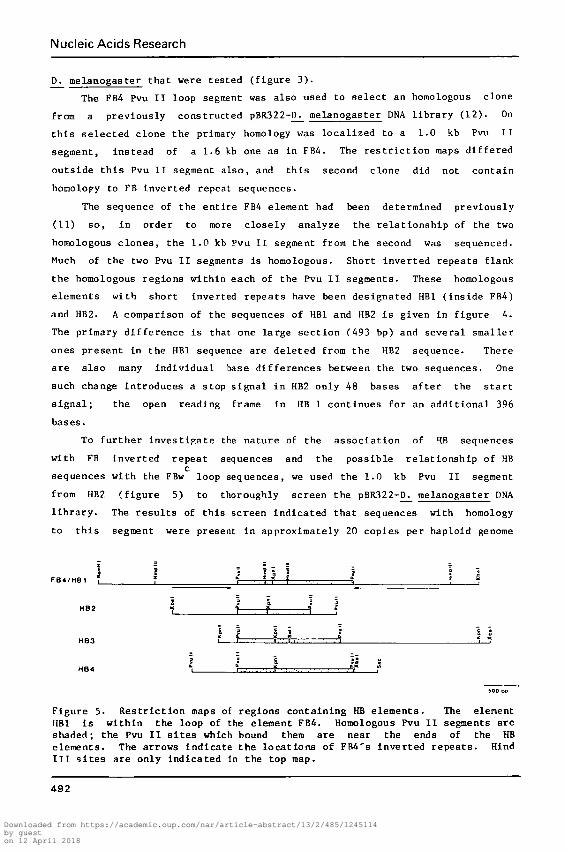
FB4/HB1 i
H 8 2
HB4
II
a.
I
lr—
7
r—
I
1 i !
f
I I
I
• iI
I
i
is s
1 X
1 i
Figure 5. Restriction maps of regions containing RB elements. The elementHB1 is within the loop of the element FB4. Homologous Pvu II segments areshaded; the Pvu II sites which bound them are near the ends of the HBelements. The arrows indicate the locations of FB4's inverted repeats. HindIII sites are only indicated in the top map.
492
Downloaded from https://academic.oup.com/nar/article-abstract/13/2/485/1245114by gueston 12 April 2018
Nucleic Acids Research
(40 posit ive clones/42,500 screened clones). DNA from randomly selected
hybridizing clones was prepared, digested, electrophoresed through agarose,
and Southern b lo t ted . Quadruplicate f i l t e r s were used in four separate
hybridizations with four nick-translated segments. These probes were the 1.0
kb Pvu I I segment from HB2 (figure 5) , the 1.4 kb Pvu II-Hind I I I segment
containing the right inverted repeat from FB4 (figure 5), the 0.8 kb Sac
I-Bam HI segment from pC10.2 (figure I ) , and the 1.5 kb Bam Hi-Sal I segmentc
from pC7.6 (figure 1); these l a s t two segments were from the FBw loop.The Pvu I I segment hybridized to 23 selected clones, but the FB inverted
repeat segment hybridized to none of these 23 new clones. This indicated
that HB sequences were generally not associated with FB inverted repeatc
sequences. The segments from the FBw loop did not hybridize to any of thec
selected clones. This suggested that the loop sequences from FB4 and FBw
were not related.
DNA from several of these new homologous clones was digested,
electrophoresed through agarose, and Southern blotted. These clones were
restriction mapped, and the Southern blot filters were used in a
hybridization to localize the regions of primary homology to the 1.0 kb Pvu
II segment. On many of the analyzed clones the regions of homology are Pvu
II segments with approximate sizes of 1.0, 1.4, and 1.6 kb. We have
sequenced parts of two of these new clones to better understand the ends of
the homologous regions. It should be noted that on a few clones the HB
homologous Pvu II segments were larger than 1.6 Kb, but restriction mappings
and Southern blot hybridizations showed that these HB elements were no longer
bordered by Pvu II restriction sites and these clones were not studied
further. Restriction maps of the regions containing four HB elements with
homologous Pvu II segments are given in figure 5. Elements HB3 and HB4 were
shown by sequencing to have short terminal inverted repeats. The sequences
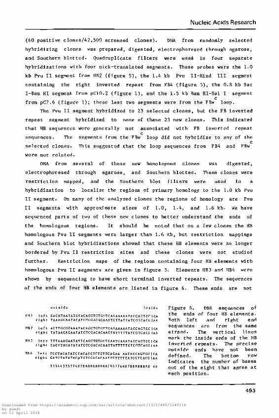
of the ends of four HB elements are listed in figure 6. These ends are not
HBl left GACATAATATCTACACCTCTCTTCACAAAAATAOCACTCr|CAright TAAAGCAATACATCTCGACACAACTCTTATTATCCTCACGIAA
HB2 left ACTTCCGCAAATACACCTCTCTTCACAAAAATAGCACTGC|CAright TATAAGAGAATATCTCGACACAACTATTTTTATCCTCACC IAA
HB3 left TTTAAAGAATATTCACCTGTCTTCAATGAAATACCAGTGCIGAright CACCTACATATATCTCGACACAACTATTTTTGTCGTrACG fAA
HB4 left TCCTACATATCTACACCTGTCTTCAGAA AATAGCAGTGCI HAright CATCTATATACATCTCGACACAATTCTTTTTATCCTCACCIAA
35344335774B78B8888S8R87R577688788888888 44
Figure 6. DNA sequences ofthe ends of four HB elements.Both left and right endsequences are from the samestrand. The ver t ica l l inesmark the inside ends of the HBinverted repeats . The preciseoutside ends have not beendefined. The bottom rowindicates the number of basesout of the eight that agree a teach posit ion.
4 9 3
Downloaded from https://academic.oup.com/nar/article-abstract/13/2/485/1245114by gueston 12 April 2018
Nucleic Acids Research
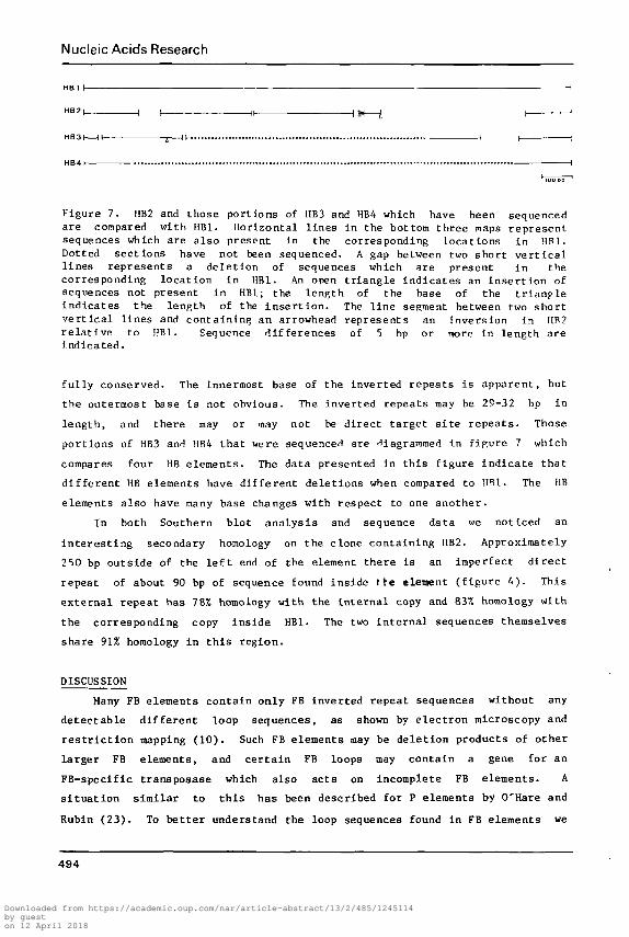
Figure 7. HB2 and those portions of IIB3 and HB4 which have been sequencedare compared with HB1. Horizontal l ines in the bottom three maps representsequences which are also present in the corresponding locations in HR1.Dotted sections have not been sequenced. A gap between two short ver t ica llines represents a deletion of sequences which are present in thecorresponding location in HB1. An open tr iangle indicates an inser t ion ofsequences not present in HB1; the length of the base of the t r tanpleindicates the length of the inser t ion . The l ine segment between two shortver t ica l lines and containing an arrowhead represents an inversion in HB2re la t ive to HB1. Sequence differences of 5 bp or more in length areindicated.
fully conserved. The innermost base of the inverted repeats is apparent, but
the outermost base is not obvious. The inverted repeats may be 25-32 bp in
length, and there may or may not be direct target s i t e repeats . Those
portions of HB3 and HB4 that were sequencer! are diagrammed In figure 7 which
compares four HB elements. The data presented in this figure indicate that
different HB elements have different deletions when compared to HB1. The HB
elements also have many base changes with respect to one another.
In both Southern blot analysis and sequence data we noticed an
interes t ing secondary horaology on the clone containing IIB2. Approximately
2 50 bp outside of the l e f t end of the element there is an imperfect direct
repeat of about 90 bp of sequence found inside t re element (figure 4) . This
external repeat has 78% homology with the internal copy and 83% homology with
the corresponding copy inside HB1. The two in ternal sequences themselves
share 91% horaology in th is region.
DISCUSSION
Many FB elements contain only FB inverted repeat sequences without any
detectable different loop sequences, as shown by electron microscopy and
r e s t r i c t i o n mapping (10). Such FB elements may be deletion products of other
larger FB elements, and cer ta in FB loops may contain a gene for an
FB-specific transposase which also acts on incomplete FB elements. A
s i tua t ion similar to th i s has been described for P elements by O'Hare and
Rubin (23). To be t te r understand the loop sequences found in FB elements we
494
Downloaded from https://academic.oup.com/nar/article-abstract/13/2/485/1245114by gueston 12 April 2018
Nucleic Acids Research
have investigated two different loop sequences from FB elements as to their
genomic representation and their association with FB inverted repeat
sequences.
We isolated three clones with homology to the FBw loop (figure 1). In
these clones the restriction sites external to the elements and, therefore,c
the elements' genomic locations vary from those of FBw . This is as expectedc
since the library we screened was made with DNA from flies without the wc
phenotype. We estimated that FBw loop sequences were present approximatelyc c
twice per haploid genome in non-w flies. For the w strain Levis et al (7)c
reported four, or possibly five, sites with FBw loop sequences while Paro et
al (9) reported six sites.c
The sizes of the segments homologous to the FBw loop segments and the
patterns of the restriction sites in the central loop regions imply that thec
FBw loop sequences are conserved. This apparent conservation and the fact
that these sequences were found only in association with FB inverted repeatsc
indicate that the FBw loop sequences may be functionally important to FBc
elements. We suggest that the FBw loop may code for functions involved in
the regulation of FB transposition. Paro et al (9) have associated both thec
FB inverted repeats and the FBw loop sequences from FB-NOF with several
(historically related) white locus transposons. It should be noted that thec
FB inverted repeats on the FBw -homologous clones may not extend the full
length of the segments shown to contain them in figure 1. Levis et al (7)c
report that those of FBw do not. The central loop region may exceed the
homologous segments indicated in figure 1, and the outer ends of the inverted
repeats may be well inside the restriction sites which bound the segments
homologous to the FB inverted repeat sequences. On the left side, however,
the outer ends of the repeats may reach the Sac I sites since Sac I sites are
often present at such junctions.
Ladder patterns of hybridization of FB inverted repeat sequences toc
FBw -homologous clones were observed in specific cases. Only the three
clones containing the left sides of the elements showed these patterns, and
only when they were digested with Sac I or, for two out of the three, with
Pvu II. The right side clones and Bam HI- or Sal I-dlgests of the left side
clones did not give ladders. These interesting results suggest that the two
sides of an FB element can be quite distinct, with particular sequence
patterns evenly distributed on one side and completely absent from the other
side. Figure 2 shows hybridization patterns from one of the left side
clones, "b" in figure 1; ladders are present in the lanes where Sac I or Pvu
495
Downloaded from https://academic.oup.com/nar/article-abstract/13/2/485/1245114by gueston 12 April 2018
Nucleic Acids Research
II was included and are absent from the other lanes. The DNA of the ladder
bands which hybridized to FB inverted repeat DNA must have come from the
primary segment containing the FB inverted repeat. On the gels from which
the Southern blots were made the small amount of DNA in each ladder band was
not visible, but the absence of the total of this DNA from the the primary FB
inverted repeat-containing band was observable. This FB inverted repeat DNA
band was less intense than expected when compared with the other DNA bands in
the same lane. Evidently partial digestion of some of the FB inverted repeat
sequences occurred, and because of the tandem repeat construction of these
sequences, this generated ladders of restriction segments.
The specificity of this phenomenon indicates that different agents, with
different recognition sites, contaminating the enzymes Sac I and Pvu II were
responsible since one clone ("a" in figure 1) was sensitive to one of these
agents and not to the other. The enzymes Sac I and Pvu II themselves do not
seem to be responsible, since conditions of 100-fold over digestion failed to
reduce the ladders to monomer size. We tested the obvious possible
contaminants Sac II and Pvu I, but the results implied that these were not
responsible for the ladders. Perhaps other as yet uncharacterized
contaminating enzymes were Involved.
FB inverted repeat sequences have been found to have a complex structure
which some distance in from the outside ends is made up of tandem 31 bp
direct repeats. Slightly different types of these direct repeats are
organized in sets of five such that a 155 bp repeat length may be established
(11). Taq I sites with this 155 bp spacing have been noted in FB inverted
repeats (10), and in the FBw -homologous clones the ladder bands have
approximately this spacing.
The specificity observed for the ladders also indicates that there are
differences among FB inverted repeats, even between two FB Inverted repeatsc c
on the same element (FBw ). The left Inverted repeat of FBw shows the
ladder pattern in Sac I or Pvu II digests, whereas the right one does not.
This implies that the inverted repeats are generated individually and do not
often exchange information by recombining with each other. This contrasts
with the previous study of an FB4 rearrangement which indicated that
"flip-flop" recombination between inverted repeats of a single element could
occur (15). However, FBA, with its composite construction, might represent a
special case.
When the potential gene region was discovered on the loop of FB4, one
proposal was that this might be an FB transposase gene. Another proposal was
496
Downloaded from https://academic.oup.com/nar/article-abstract/13/2/485/1245114by gueston 12 April 2018
Nucleic Acids Research
that the FB4 loop segment might be an independent transposable element since
the loop segment had its own short terminal inverted repeats, distinct from
the FB inverted repeats. The genomic Southern blots were done to begin to
study the distribution and conservation of this loop segment.
The pattern of the hybridization of the Pvu II segment from the FB4 loop
(HB1) to the Bam Hi-digested, total genomic D^ melanogaster DNA, showing
multiple bands (figure 3), indicated that homologous DNA was present in a
number of different sites in the genome. Hybridization to the Pvu
II-digested DNA indicated that the Pvu II segment containing the potential
gene was not strictly conserved since, although the band corresponding to the
size of the probe is darkest, there are several additional bands (figure 3).
Some of these additional homologous Pvu II segments could have been due to
simple base changes affecting the Pvu II recognition sites; the loss of a
site would result in a larger segment extending to the next flanking site.
Significant divergence affecting the potential gene could have produced other
additional bands; in the case of the Pvu II segment from HB2, sequence
analysis indicated that the region of the potential gene was actually altered
considerably.
Three lines of evidence suggest that the sequences homologous to the FB4
loop segment represent independent transposable elements: 1) the sequences
have their own short terminal inverted repeats, 2) the sequences generally
are found independent of FB terminal inverted repeats, and 3) the sequences
are moderately repetitive in copy number. We have designated this group of
repetitive elements the HB family. In the hybridization of the Pvu II
segment to the genomic Southern blot (figure 3) the patterns of homologous
segments are similar across the four strains. This similarity suggests that
the repetitive sequences predate the isolation of these strains and that the
elements have transposed infrequently since then. Alternatively, these
elements entered these strains separately and had specific target sites.
This seems less likely; the DNA sequences surrounding the analyzed elements
are not obviously related.
Although two FB elements apparently can mobilize other segments of DNA
between them (9), there is no evidence that a single FB element can "pick up"
DNA between its inverted repeats. The best explanation for the structure of
the loop in FB4 is the insertion of HB1. The very low frequency of HB
transposition suggested by the cross-strain similarities could be due to any
of several possibilities. If the potential gene in the sequence of HB1 codes
for an HB transposase, very little of this transposase may be made since
497
Downloaded from https://academic.oup.com/nar/article-abstract/13/2/485/1245114by gueston 12 April 2018
Nucleic Acids Research
other HB elements contain deletions and multiple base changes affecting this
region. Even the potential gene in HB1 may contain sequence changes such
that it no longer encodes a functional transposase, or the gene may code not
for a transposase but for a repressor. Base changes in the inverted repeats
of the HB elements may have caused them to be no longer recognized by
transposition factors. Also, the presence of other unfavorable conditions
might account for the positional stability of the HB elements.
Two other families of Drosophila transposable elements with short
terminal inverted repeats are the hobo (24) and P elements (23). The hobo
element which inserted near a glue protein gene had 12 bp perfect inverted
repeats and generated an 8 bp target site duplication (24). P elements have
31 bp perfect inverted repeats, the sequence of which is conserved from one
element to the next, and these elements generate R bp direct repeats of
target site DNA (23). In the sequences of the ends of four HB elements
listed in figure 6, the inner end of the inverted repeats is well defined
and, therefore, is marked by the vertical lines. The outer end is less
apparent due to base changes in the inverted repeats and the lack of obvious
direct repeats, or possibly due to variability in the lengths of the
elements. The inverted repeats may be 2Q-32 bp in length, as judged by the
homology among the 8 end sequences. If the inverted repeats are taken to be
30 bp long, then there could be 2 bp direct repeats just outside of them.
Longer direct repeats are not found consistently on the four elements. These
elements may have inserted into their sites long enough ago that the sequence
changes since then have made any direct repeats once present no longer
apparent.
The size heterogeneity of the HB elements is similar to that of hobo
(24) and P elements (23). However, the numerous sequence changes and
multiple deletions from one HB to another (for example see figure 4) indicate
a situation different from that described for the P elements. Each P element
differs from the full length element only by a single deletion and very few
other changes (23). The different size classes of homologous Pvu II segments
in the HB elements are generated by different sets of internal deletions.
The homologous segments which are not bordered by Pvu II sites are probably
due to both simple base changes in the Pvu II recognition sites and larger
rearrangements. HB elements may cause instabilities and rearrangements which
involve the adjacent sequences as well as the element. This is suggested by
the structure of the clone containing HB2; a direct repeat of part of the HB
sequence is found a short distance outside of the element.
498
Downloaded from https://academic.oup.com/nar/article-abstract/13/2/485/1245114by gueston 12 April 2018
Nucleic Acids Research
If the sequence in the middle of FB4 is a transposable element
independent of FB elements, then the gene may still code for a transposase,
or other function, for these HB elements. There may be a transposase encodedc
elsewhere in the genome which acts on FB elements, or perchance the FBw loop
sequence or another yet undiscovered FB loop sequence encodes such a
function.
Many P transposable elements are apparently related to one another by
being different deletion products of larger complete P elements (23). If FB
elements were related in a similar manner, one would expect two nonhomologous
FB loops to have come from a loop containing both sequences. With this model
in mind, we screened the clones with horaology to the FB4 loop for homology toc
the FBw loop. No clone with homology to both the FB4 loop segment andc
either of the loop segments from FBw was found. In light of the apparent
nature of the FB4 loop sequence, this result is not surprising. If the FB4
loop sequence is an independent element, then it would not be expected toc
have a common origin with the FBw loop sequence.
In summary, the FBw loop appears to represent an integral and conserved
component of several FB transposable elements and, therefore, might be
functionally important for the FB family. The FB4 loop, however, represents
a transposable element of the distinct HB family by chance found inserted
into an FB transposable element.
ACKNOWLEDGMENTS:We thank Mary Collins and Gerald M. Rubin for providing the pC10.2 and
pC7.6 clones. We thank Dan Weinstein for assisting in the initial stages ofthis work.
This work was supported by grant number GM 30816 from the NationalInstitutes of Health and by PHS grant number CA 00852 from the NationalCancer Institute, DHHS.
•Present address: Department of Biological Sciences, Stanford University, Stanford, CA 94305,USA
LITERATURE CITED1. Calos.M.P. and Miller ,J .M. (1980) Cell 20, 579-595.2. Shapiro,J.A. and Cordell.B. (1982) Biol. Cell 43, 31-54.3. Spradling.A.C. and Rubin,CM. (1981) Annu. Rev. Genet. 15, 219-264.4. P o t t e r , S . , Truett .M., Ph i l l ips .M. , andMaher.A. (1980) Cell 20, 639-647.5. Collins,M. and Rubin,G.M. (1983) Nature (London) 303, 259-260.6. Blngham.P.M. (1981) Cold Spring Harbor Symp. Quant. Biol . 45, 519-525-7. Levis.R., Collins,M., and Rubin,G.M. (1982) Cell 30, 551-565.8. CollinsnW. and Rubin,G.M. (1984) Nature (London) 308, 323-327.9. Paro.R., Goldberg,M.L. , and Gehring.W.J. ( l q83) EMBO J . 2, P53-86O.
10. Truett.M.A., Jones.R.A., and Po t t e r ,S .S . (1981) Cell 24, 753-763.
499
Downloaded from https://academic.oup.com/nar/article-abstract/13/2/485/1245114by gueston 12 April 2018
Nucleic Acids Research
11. Pot te r ,S .S . (1982) Nature (London) 297, 201-204.12. Po t t e r ,S .S . , Broreln.W.J. ,Jr . , Dunsrauir.P., and Rubin.G.M. (1179) Cell
17, 415-427.13. Grunsteln.M. and Hogness,D.S. (1975) Proc. Nat. Acad. Sci. USA 72,
3961-3965.14. Holmes,D.S. and Quigley.M. (1981) Anal. Biochem. 114, 193-197.15. Pot te r ,S . (1982) Mol. Gen. Genet. 188, 107-110.16. Southern.E.M. (1975) J . Mol. Biol. 98, 503-517.17. Smith,G.E. and Summers.M.D. (1980) Anal. Biochem. 109, 123-129.18. Rigby.P.W.J., Dieckmann.M., Rhodes,C., and Berg,P. (1977) J . Mol. Biol.
113, 237-251.19. Denhardt.D.T. (1966) Biochem. Biophys. P.es. Commun. 23, 641-646.20. Klenow.H. and Henningsen.I. (1970) P roc Nat. Acad. Sci. USA 65,
168-175.21. Levis.R., Dunsmuir,P., and Rubin.G.M. (1980) Cell 21, 581-588.22. Maxam.A.M. and Gilbert,W. (1980) in Methods Enzymol. , Grossman,L. and
Moldave.K. Eds., Vol. 65, pp. 499-560, Academic Press, New York.23. O'Hare.K. and Rubin.G.M. (1983) Cell 34, 25-35.24. McGinnis.W., Shermoen.A.W., and Beckendorf,S.K. (1983) Cell 34, 75-84.
500
Downloaded from https://academic.oup.com/nar/article-abstract/13/2/485/1245114by gueston 12 April 2018