Embed Size (px)
Citation preview

1
Hysterosalpingogram Confirmation for
Essure® Permanent Birth Control
Device Placement and Occlusion
Jaime Hartley
Radiologic Technology
The Ohio State University
2012

2
Table of Contents
Abstract Chapter One
• The Problem
Chapter Two
• Review of Literature
• The Objective
• Hypotheses
• Methodology Chapter Three
• Results
• Discussion
• Limitations and Suggestions for Further Research
• Conclusion
• Glossary References

3
Abstract
The Essure® hysteroscopic sterilization system is a minimally invasive
technique that involves the placement of micro-inserts into the fallopian tubes.
These micro-inserts contain polyethylene terephthalate (PET) fibers, which cause
a benign inflammatory response, occlude the fallopian tubes, and cause
permanent sterilization. The United States Food and Drug Administration (FDA)
requires that the patient undergo follow-up hysterosalpingography three months
after device placement to prove device retention, determine the location of the
inserts, and ensure tubal occlusion.
Many imaging professionals are not properly educated on the procedures
and images required in the manufacturer recommended follow-up
hysterosalpingography protocol. In this study, data from these examinations at a
large, mid-west university hospital were analyzed, and descriptive statistics about
the frequency of images obtained and complications noted in the radiologist’s
reports are presented. The data was also evaluated for trends relating to the
technical quality of the images.
Of 428 images obtained in 130 sample exams, 74 exams included a
preliminary scout image, 55 exams included a minimal fill image, 88 exams
included a partial fill image, 119 exams included a total fill image, 46 exams
included a right magnification image, and 46 exams included a left magnification
Seventy percent of the cases included 2,3, or 4 images, and about 12% of cases
only included one image for the radiologist to evaluate. However, only 9 reports

4
contained mentions of limitations in diagnosis due to the number or quality of the
images provided for interpretation.
Twenty-two percent of the cases included fundal images of the uterus.
Parts of the reproductive anatomy including the vagina, uterus, and cervix were
clipped off in 28% of the cases, and in a further 18%, the gynecologist failed to
remove the speculum prior to exposure, resulting in incomplete visualization of
reproductive anatomy. In 9% of the exams, one of more of the images was
considered blurry during this secondary analysis. The most frequent technical
error committed during these exams was failure to use a lead marker to denote
anatomic side; 52% of cases were not marked with a lead marker.

5
Chapter One
The Problem
There are many different methods of birth control available for women of
reproductive age. Their indications for use vary based many factors including
patient age, method permanency, and associated costs. Some of the most
common and inexpensive forms of temporary birth control include barrier
methods. This category includes both male and female condoms, diaphragms,
and cervical caps, and these methods can be up to 85% effective if used
properly.1 Another popular method of contraception is the use of hormones to
temporarily cease ovulation. These hormones can be administered by an
injection, an implanted device, or a pill that is ingested daily. When taken as
directed, these methods can be up to 99% effective.1 When women no longer
wish to have the option to become pregnant, a permanent method of birth control
can be considered. There are several types of permanent female sterilization
procedures. Both tubal ligation and hysterectomy procedures are surgical in
nature and require the use of general anesthesia. However, these forms of birth
control are over 99% effective in the prevention of pregnancy.1
One of the only permanent birth control systems currently approved by the
United States Food and Drug Administration (FDA) is the Essure® hysteroscopic
sterilization system.2 According to Conceptus, the manufacturer of this device,
this is a minimally invasive, transvaginal procedure which is usually done in an
outpatient setting without the use of general anesthesia.1 A small, flexible coil,
called a micro-insert, is placed into each fallopian tube. These micro-inserts are

6
comprised of a stainless steel inner coil, a Nitinol expanding outer coil, and
polyethylene terephthalate (PET) fibers that are wound into the inner coil. Each
micro-insert is extended into the uterus through a disposable delivery system
provided by Conceptus (Figure 1).1
Figure 1 – Essure® Delivery System (Conceptus Inc.)
The Essure® coil is attached to the delivery wire of this disposable delivery
system. Prior to device placement, the coil is wound-down and sheathed by a
flexible catheter (Figure 2).1
Figure 2 – Coil Appearance Before Placement (Conceptus Inc.)
If intravenous sedation is not required, micro-insert placement is
performed as an outpatient procedure at the physician’s office. Before the
Essure® delivery system is introduced, the physician places a speculum into the
vagina to allow access to the cervix. A sterile hysteroscope is then placed
through the cervix and into the uterine cavity. This hysteroscope employs the use
of a small camera and is used to position the device at one of the fallopian tube

7
ostium.2 The Essure® delivery system is advanced through the sealing cap on
the hysteroscope working channel, and the physician rotates the thumbwheel
portion of the delivery system to place the Essure® device in the proximal portion
of the fallopian tube (Figure 3).1
Figure 3 – Essure® Delivery System Inserted Through Sealing Cap of Hysteroscope (Conceptus Inc.)
Optimal positioning of the coils is marked when the device spans the utero-tubal
junction; ideally, three to eight outer coils should trail into the uterus.1 This
placement is desirable because the portion of the device trailing into the uterine
cavity aids in device anchoring.1 After the device is deployed, the outer coil
expands to mold to the varied diameters of the fallopian tube and anchors the
device in place (Figure 4).1
Figure 4 – Coil Appearance After Placement (Conceptus Inc.)
The PET fibers of the inner coil induce a benign inflammatory response and
eventual fibrosis of the intramural tubal lumen, thereby aiding in device retention
and causing tubal occlusion.1,2,3

8
On the day of device placement, complications arising from the Essure®
device include band detachment, vasovagal responses, pain, cramping, nausea
and vomiting, and dizziness.1 Other complications that may arise within a year of
placement have been recorded, such as cramps, generalized pain, back pain,
headache, and dysmenorrhea.1,4 Some patient’s may develop an infection or
salpingitis, and in some cases, become pregnant if device placement is not
optimal or the patient does not comply with alternative birth control methods until
a three month follow-up hysterosalpingogram (HSG) has been performed.1,2
However, during the phase II and pivotal trials, reliance on the device for
occlusion was 97% and zero pregnancies were recorded when proper device
placement was obtained.1,2
Because of the minimally invasive nature of this procedure, this technique
has steadily increased in popularity since its introduction in November of 2002.5
There are no abdominal incisions or scars, and women are usually able to return
to daily activities with a high rate of patient tolerance and satisfaction.6 In
addition, this form of birth control does not require the use of hormones, which
increases its appeal as an alternative form of contraception.6
As part of the FDA approval guidelines, the Essure® package labeling
includes a requirement for a three month follow-up hysterosalpingogram to prove
device retention, determine the location of the inserts, and ensure the occlusion
of the fallopian tubes.1,5
The most common technique for doing HSG examinations is the Kidde
technique. With the patient in the lithotomy position, a sterile speculum is

9
inserted into the vagina to provide clear visualization of the cervix. A tenaculum is
placed on the anterior cervical lip, from which the patient may experience slight
cramping.7 An acorn tip is attached to a Kidde cannula, and the tubular end of
this tip is inserted into the external cervical os. This tightly seals the external os
and prevents contrast leakage into the vagina.(Figure 5).7
Figure 5 – Kidde Cannula Placement During HSG (Lindheim et al) – HSG procedure; the acorn tip is sitting within the cervix and identified by the red arrow.
As recommended by the manufacturer protocol, prior to contrast
introduction, the physician should obtain a scout image with the uterus in a true
anteroposterior position to visualize the Essure® device presence and
placement.1 For all images, traction on the tenaculum or rotation of the patient
into an oblique position may be necessary to better depict the uterine anatomy in
a true anteroposterior position.7 The iodinated contrast medium is then injected
at a slow rate using steady, low pressure to minimize uterine cramping.1,7 The
physician should monitor contrast filling of the uterus with fluoroscopy, and obtain
a spot image with minimal contrast fill. This image depicts an adequate cervical
10
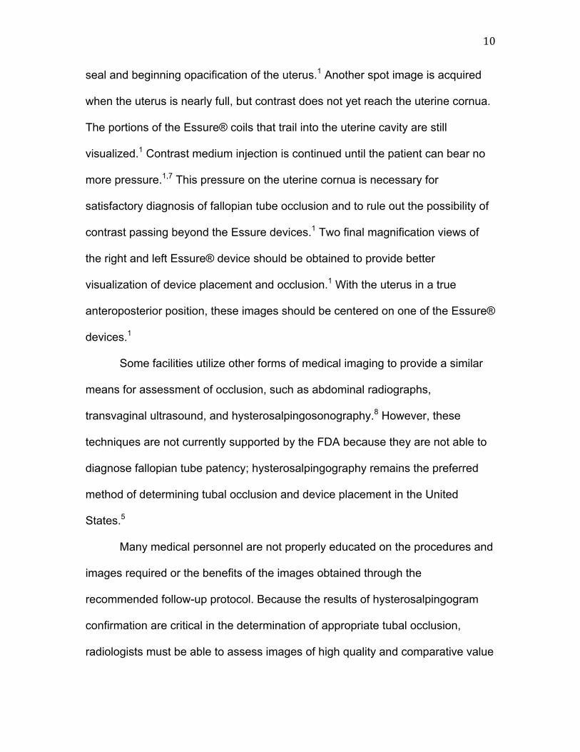
seal and beginning opacification of the uterus.1 Another spot image is acquired
when the uterus is nearly full, but contrast does not yet reach the uterine cornua.
The portions of the Essure® coils that trail into the uterine cavity are still
visualized.1 Contrast medium injection is continued until the patient can bear no
more pressure.1,7 This pressure on the uterine cornua is necessary for
satisfactory diagnosis of fallopian tube occlusion and to rule out the possibility of
contrast passing beyond the Essure devices.1 Two final magnification views of
the right and left Essure® device should be obtained to provide better
visualization of device placement and occlusion.1 With the uterus in a true
anteroposterior position, these images should be centered on one of the Essure®
devices.1
Some facilities utilize other forms of medical imaging to provide a similar
means for assessment of occlusion, such as abdominal radiographs,
transvaginal ultrasound, and hysterosalpingosonography.8 However, these
techniques are not currently supported by the FDA because they are not able to
diagnose fallopian tube patency; hysterosalpingography remains the preferred
method of determining tubal occlusion and device placement in the United
States.5
Many medical personnel are not properly educated on the procedures and
images required or the benefits of the images obtained through the
recommended follow-up protocol. Because the results of hysterosalpingogram
confirmation are critical in the determination of appropriate tubal occlusion,
radiologists must be able to assess images of high quality and comparative value

11
so that their reports can be as consistent and accurate as possible. Therefore,
Medical facilities should adopt the recommended post-Essure®
hysterosalpingogram protocol provided by the manufacturer to guarantee
standardized results and analysis.5

12
Chapter One
Review of Literature
Conceptus, the manufacturer of the Essure® female sterilization system,
conducted two separate clinical trials to assess the safety and effectiveness of
the Essure® system.1 Of 745 women included in both the phase II and pivotal
trials, placement of at least one Essure® coil was achieved in 682 women.
Bilateral placement was successful in 94.6% of cases.1 Nineteen of 476 patients
within the phase II trial experienced wither perforation or expulsion of one or
more Essure® devices. This represents 3.99% of the population of the study.1
Within one year of device placement, 127 of 476 patients experienced some sort
of pain associated with device placement, including severe menstrual cramping.1
Based on the research conducted in the phase II and pivotal trials,
Conceptus recommends a set protocol for the confirmation hysterosalpingogram
that is required by the FDA. This HSG confirms tubal occlusion and allows the
patient to rely on the devices for contraception. This protocol calls for a “low-flow,
low pressure [hysterosalpingogram] performed three months post Essure®
placement”1 to demonstrate correct placement of the micro-inserts and to assess
the occlusion of the fallopian tubes. A minimum of six still radiographs should be
obtained during the procedure, beginning with a scout image with the uterus in a
true anteroposterior position to visualize the Essure® device presence and
placement.1 After this image is assessed, a series of three radiographs
demonstrating the increasing fill of the uterine cavity are captured. The fifth and

13
sixth required radiographs are magnified views of the uterine cornua to determine
exact location of the micro-inserts.1
Protocol Image
Image Name Structures Visualized/ Image Importance
Image 1 Scout Device presence and placement
Image 2 Anteroposterior Minimal Fill adequate cervical seal and beginning opacification of the uterus
Image 3 Anteroposterior Partial Fill Contrast does not yet reach the uterine cornua; The portions of the Essure® coils that trail into the uterine cavity are still visualized
Image 4 Anteroposterior Total Fill Maximum pressure on the uterine cornua; Necessary for satisfactory diagnosis of fallopian tube occlusion and to rule out the possibility of contrast passing beyond the Essure devices
Image 5 Anteroposterior Magnified Right Essure® Device
Larger and more detailed visualization of the devices; determination of the relationship of the coil markers to the uterotubal junction
Image 6 Anteroposterior Magnified Left Essure® Device
Larger and more detailed visualization of the devices; determination of the relationship of the coil markers to the uterotubal junction
Figure 6 – Conceptus-Recommended Protocol Images
Each of these radiographs should clearly show the entire uterine cavity
silhouette with the uterine cornua maximally distended.1 In addition, the
fluoroscopy procedures should be completed with the beam entering the patient

14
as close to anteroposterior as possible. Failure to meet these minimum
requirements could result in repetition of the necessary images, which would
increase radiation dose, or misdiagnosis of tubal occlusion. This misdiagnosis
could lead the patient to rely on the Essure® devices for contraception when one
or more fallopian tubes is patent, possibly resulting in unintended pregnancy.1
The company suggested protocol also describes the ideal location of the
micro-inserts; this positioning is evident when the inner coil crosses the utero-
tubal junction. The distal end of the inner coil should sit within the fallopian tube
with less than fifty percent of the coil extending proximally into the uterine cavity.
(Figure 6).1
` Figure 7 – Device Placement 1) Device spans the utero-tubal junction. 2) Distal end of the inner coil sits within the fallopian tube. 3) <50% of the coil extends into the uterine cavity.
Conceptus suggests that if this optimal positioning is not successful, the
patient should not rely on the Essure® device for contraception. Improper
placement could result in unintentional pregnancy even if occlusion is apparent
during the follow-up HSG due to potential device expulsion.1 When assessing
occlusion, the radiologist evaluates the presence of contrast in the fallopian tubes

15
distal to the micro-insert location. The tube is occluded satisfactorily when
contrast solution is not visible past the cornua. Patients are advised not to rely on
the micro-inserts for contraception if contrast is seen past the distal end of the
coils or in the peritoneal cavity.1
In a six-year review of the Essure® hysteroscopic sterilization technique,
the effectiveness, safety, and complications reported with the use of the device
were analyzed.2 The hysteroscopic procedure involves the transcervical
implantation of coiled, double-layered micro-insert devices in the fallopian tubes.
This examination is generally performed without the use of anesthesia in an
outpatient setting.2 Post-placement, the white polyethylene terephthalate fibers
surrounding the coils trigger the benign growth of tissue into the fallopian tube,
causing occlusion and preventing future pregnancies.2 Three months after the
Essure® procedure, a hysterosalpingogram is obtained to determine bilateral coil
placement and complete tubal occlusion.2 This three month waiting period was
highly subjective when the system was first implemented, as there was little
factual evidence to support the hypothesized rate of tissue growth necessary to
occlude the tubes. However, studies have confirmed that up to 99% of patients
show evidence of successful tubal occlusion on the three month
hysterosalpingogram, even though some patients may be fully occluded within
one to two months.2 Long-term adverse effects are limited; one year post-device
placement, 99% of women rated their comfort level at good to excellent.2 From
1998 to 2007, Conceptus received 169 reports of unintended pregnancies, which
equates to 0.1% relative to device sales during that time. In addition, 44% of

16
these unintended pregnancies can be attributed to patient and physician
noncompliance with the follow-up HSG.2
A study completed in 2005 was designed to determine the efficacy of
hysterosalpingography examinations in determining tubal occlusion.9 Thirty-two
subjects were identified as having Essure® devices placed between April 2003
and February 2004. Of these patients, only nineteen had undergone a three
month follow-up hysterosalpingogram due to patient noncompliance.9 In addition,
it is stated that no set protocol was used when obtaining images during the
course of this study.9 Of the women who received the three month follow-up
examination, the number of exposures per patient varied greatly, ranging from
one spot film to five spot films with an average of 2.4 per examination. The
number and types of radiographs that were obtained were chosen according to
the preference of the radiologist analyzing the images.9 Almost half of the
patients included in the study received only one anteroposterior image. For these
patients, those physicians who reviewed the images agreed that additional
oblique images would have improved their evaluation of device placement and
degree of tubal occlusion.9
A different study utilized a retrospective chart review of eighty-three
patients that underwent Essure® device placement in a particular facility from
January 2003 to June 2007.5 It addressed the potential complications that may
arise after Essure® device placement. Device movement, nonocclusion, tubal
perforation, and structural malfunctions of the coils can be detected on the three
month hysterosalpingogram.5 However, of the 79 patients included in the sample,

17
only 12.7% received a follow-up HSG, which the researchers note to be
alarmingly low. One of 36 patients who received this HSG had unsatisfactory
device placement according to the manufacturer recommendations. However,
occlusion was confirmed for this patient.5 This device placement could
predispose the patient to device expulsion. Therefore, this patient was counseled
to not rely on the device for contraception.5 There was one documented
pregnancy in a patient who was noncompliant and did not receive a follow-up
HSG.5
Alternative methods to determine device placement are discussed within
this study, including pelvic radiology and ultrasonography. However, these
imaging modalities are only able to determine device placement and do not prove
tubal occlusion.5 Therefore, the Food and Drug Administration has not approved
these imaging modalities for Essure® confirmation; hysterosalpingography
remains the required three month confirmation assessment.5
Lipman and Famuyide discussed hysterosalpingography and its role in
Essure® placement and tubal occlusion confirmation. The hysterosalpingogram
required in the Essure® protocol differs significantly from a hysterosalpingogram
to evaluate infertility; the confirmation test requires much less pressure and
produces minimal discomfort compared to a traditional HSG.10 Therefore,
radiologists must be aware of the appearance of the Essure® coils before and
after insertion. The importance of following the recommended protocol set by the
Essure® manufacturer and providing detailed radiology reports are also noted.10
The benefits of confirmation hysterosalpingography should be emphasized with

18
the patient, noting that the procedure is not finished until this follow-up
examination is completed. This procedure is critical because complications
caused by placement failure can be diagnosed through evaluation of a
hysterosalpingogram.10

19
The Objective
Purpose of this study:
Compare hysterosalpingogram examinations for confirmation of Essure®
placement and fallopian tube occlusion in terms of:
1. the number of images obtained versus the recommended protocol
2. mentions of limitations in diagnosis within radiologists’ reports
3. instances of technical problems in the images provided for
evaluation
Benefits of my research:
Although other facilities and companies have conducted trials to assess
the effectiveness of the Essure® device and its resulting complications, most of
these studies have very limited sample sizes. My sample of 130 patients will be
the largest of any study that I have come across through my research. The
results of hysterosalpingogram confirmation are critical in the determination of
tubal occlusion, and radiologists must be able to assess images of high quality
and comparative value to guarantee standardized results and analysis. Because
many imaging professionals are not properly educated on the procedures and
images required in the manufacturer recommended follow-up protocol, my study
will better illustrate the need for standardization of imaging procedures.

20
Hypotheses
1. The average number of images obtained during the three month follow-up
HSG will not be significantly different than the number of images that are
required in the Essure® protocol.
2. There will be no occurrences of reported technical difficulties or limitations
due to the number of images provided for radiologist interpretation.
3. There will be no occurrences of technical errors within the images, including
fundal views, blurred images, lack of a marker noting anatomic side, presence
of a speculum within the reproductive anatomy, and/or cut off reproductive
anatomy.

21
Methodology
This study was conducted as a mixed-methods, retrospective analysis of
hysterosalpingography examinations performed for post-Essure® placement
between January 2008 and December 2010. Radiologist records for 130 patients
were analyzed to identify the number of images obtained and the reporting of
limitations or complications resulting from the number of images captured. A
secondary analysis of the images by the author was conducted to report
technical issues such as fundal positioning, blurred images, presence of a lead
marker within the radiographic field, presence of a speculum within the
reproductive anatomy, or cut off of reproductive anatomy. The criteria for an
optimal HSG image that was referenced for this secondary analysis was based
on information provided in Hysterosalpingography: a Text and Atlas.
The sample period from January 2008 to December 2010 was chosen as
a convenience sample; beginning in 2008 the prevalence of Essure® follow-up
HSGs at this facility has grown, and the idea for this research study was initially
proposed in early 2011. No subjects or examinations were excluded.
After IRB approval was obtained, the images and radiologist reports for
each patient were initially collected and de-identified. The de-identified records
were evaluated by the author, and a spreadsheet was used to record the
presence or absence of each image suggested by the Essure® protocol,
limitations noted in the radiologists’ reports, and any technical errors noticed in
the secondary analysis of the images. Key words searched for when noting
limitations reported by the radiologists were “limited,” “sub-optimal,”

22
“nondiagnostic,” and “un-interpretable.” The presence or absence of each
variable was independently recorded as (1, present) or (0, absent) in the
spreadsheets below.
Figure 8 – Data Collection Spreadsheet 1
Figure 9 – Data Collection Spreadsheet 2
Descriptive statistics were calculated to determine the mean number of
images included in all of the studies, the frequency of noted limitations in the
radiologists’ reports, and the frequency of technical errors in the images.

23
Chapter Three
Results
Of 428 images obtained in 130 sample exams, 74 exams included a
preliminary scout image, 55 exams included a minimal fill image, 88 exams
included a partial fill image, 119 exams included a total fill image, 46 exams
included a right magnification image, and 46 exams included a left magnification
image. Even though the totals for both the left and right magnification views
equaled 46, those examinations which included one magnification view did not
necessarily include the other (Figure 7).
Figure 10 – Image Frequency
Across all examinations, the mean number of images acquired was 3.29.
Sixteen cases included only one image, 26 cases included 2 images, 34 cases
included 3 images, 27 cases included 4 images, 12 cases included 5 images,
and 15 cases included all 6 images. Seventy percent of the cases included 2,3,

24
or 4 images. However, about 12% of cases only included one image for the
radiologist to evaluate (Figures 8 and 9).
Image 11 – Number of Images Per Examination
Image 12 – Comparison of the Number of Images Per Examination
Only 9 reports contained mentions of limitations in diagnosis due to the
number or quality of the images provided for interpretation. This represents 7% of
12.31%
20.00%
26.15%
20.77%
9.23% 11.54% 1 2 3 4 5 6

25
the sample population. In four of these reports, the radiologist stated that air-
bubbles within the uterine cavity limited the view of the cornua. Three stated sub-
optimal visualization of the uterus due to the failure to place the uterus in a true
anteroposterior position. One report noted that the evaluation of the devices and
potential free spill was limited because only one image was provided for
interpretation. Only one report stated that the study was non-diagnostic due to
the quality of the images obtained.
Following evaluation of the radiologists’ reports, the author conducted a
secondary image analysis that was independent of the radiologists’ findings.
Within this review, it was found that twenty-two percent (29/130) of the cases
included fundal images of the uterus. The uterus will naturally lie in a fundal
position unless the gynecologist uses a tenaculum to pull down the inferior uterus
into a true anteroposterior position. It was also found that parts of the
reproductive anatomy including the vagina, uterus, and cervix were clipped off in
28% (36/130) of the cases, and in a further 18% (23/130), the gynecologist failed
to remove the speculum prior to exposure, resulting in incomplete visualization of
reproductive anatomy. In 9% (12/130) of the exams, one of more of the images
was considered blurry during this secondary analysis. The most frequent
technical error committed during these exams was failure to use a lead marker to
denote anatomic side; 52% (68/130) of cases were not marked with a lead
marker.

26
Figure 9 – Technical Considerations
Free spill was diagnosed in five of the 130 patients included in this study.
This free spill is characteristic of tubal patency, and these patients were
counseled to rely on alternative forms of birth control for another three months
until tubal patency is reevaluated at a six month HSG. This rate of tubal patency
at the three month period (3.86%) is slightly higher than the national average of
3%. Three of the patients who exhibited free spill from one of their fallopian tubes
received three images during their three month follow-up HSG. One patient
received two images, and one patient received four images.

27
Discussion
Research Question 1 – Will the average number of images obtained during
the three month follow-up HSG be significantly different than the number of
images that are required in the Essure® protocol?
Most studies (12%) provided a total fill image for the radiologist to
interpret. This image is the most diagnostic of the images included in the protocol
because this volume of contrast provides maximum pressure on the Essure®
devices and the fallopian tubes.2 If any contrast is going to extravasate into the
peritoneum, it is usually during this phase of the examination. Patients are
advised not to rely on the micro-inserts for contraception if contrast is seen past
the distal end of the coils or in the peritoneal cavity, and failure to diagnose this
free spill and tubal patency could result in unintended pregnancies.1
The right and left magnification views were included the least frequently of
all of the protocol images. These images are important because they allow the
radiologist to visualize larger and more detailed views of the devices and to
determine the relationship of the coil markers to the uterotubal junction. If
extravasation is missed on the total fill image, it still may exist and be visible only
on these magnification views.
The mean number of images provided for radiologist interpretation per
examination was 3.29. This value is clearly much lower than the 6 images
suggested by the manufacturer.1 In only 12% of the examinations were all six
images provided for radiologist interpretation, and 12% of examinations
contained only one image.
28
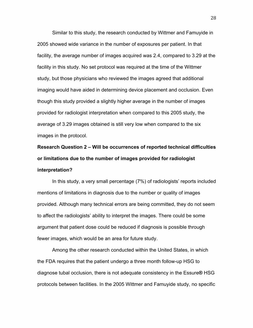
Similar to this study, the research conducted by Wittmer and Famuyide in
2005 showed wide variance in the number of exposures per patient. In that
facility, the average number of images acquired was 2.4, compared to 3.29 at the
facility in this study. No set protocol was required at the time of the Wittmer
study, but those physicians who reviewed the images agreed that additional
imaging would have aided in determining device placement and occlusion. Even
though this study provided a slightly higher average in the number of images
provided for radiologist interpretation when compared to this 2005 study, the
average of 3.29 images obtained is still very low when compared to the six
images in the protocol.
Research Question 2 – Will be occurrences of reported technical difficulties
or limitations due to the number of images provided for radiologist
interpretation?
In this study, a very small percentage (7%) of radiologists’ reports included
mentions of limitations in diagnosis due to the number or quality of images
provided. Although many technical errors are being committed, they do not seem
to affect the radiologists’ ability to interpret the images. There could be some
argument that patient dose could be reduced if diagnosis is possible through
fewer images, which would be an area for future study.
Among the other research conducted within the United States, in which
the FDA requires that the patient undergo a three month follow-up HSG to
diagnose tubal occlusion, there is not adequate consistency in the Essure® HSG
protocols between facilities. In the 2005 Wittmer and Famuyide study, no specific

29
protocol was followed for each examination, and an average of only 2.4 images
was provided for radiologist interpretation per patient.
In this study, it was shown that these Essure® follow-up HSGs are still
fairly infrequent 10 years after this birth control system’s FDA approval. With only
130 Essure® HSGs performed within a two year period, they comprise a very
small portion of the total workflow for this large mid-west university hospital.
Standardization of the images between examinations could assist the radiologists
in making a complete and accurate diagnosis of tubal occlusion because they
would be evaluating images of consistent appearance and technical quality.
Research Question 3 – Will there be occurrences of technical errors within
the images, including fundal views, blurred images, lack of a marker noting
anatomic side, presence of a speculum within the reproductive anatomy,
and/or cut off reproductive anatomy?
Fundal views of the uterus provide incomplete visualization of the
anatomy, which could lead to missed diagnoses of fibroids or polyps. The
gynecologist must remove the speculum and employ the use of a tenaculum to
pull the uterus down into a true anteroposterior position prior to imaging in order
to produce an image of optimal diagnostic quality. During the author’s evaluation
of the sample images, the position of the uterus was compared to the positioning
in examples of optimal HSG images from Hysterosalpingography: a Text and
Atlas.7 Those images in which the uterus was considered to be in a fundal rather
than a true anteroposterior position were included in the 22% of examinations
with fundal images.

30
Failure to place a lead marker in the radiation field to denote correct
anatomic side could lead to incorrect diagnosis of side of free spill. The facility
held an educational meeting about the importance of consistency between
examinations in 2010; during this meeting, a Conceptus representative educated
the imaging staff on the Essure® device and the proper procedures for the
company recommended protocol for the follow-up HSG. A policy regarding the
mandatory use lead markers has been added since the time of this study to
require the use of a lead marker for every examination. Compliance with this
policy could be evaluated at a later date.
Most of the literature available on the Essure® system at the time of this
study concentrated on the rate of follow-up HSG compliance among Essure®
patients. The results of this study cannot be compared to these measures, as
only those patients who actually received the follow-up HSG were examined. In
addition, there is limited research on these devices within the United States.
Although more information and study results are available from foreign sources,
other countries allow alternative follow-up imaging techniques and are difficult to
compare to the results of this study.

31
Limitations and Recommendations for Further Research
One possible limitation to this study is the fact that the results can only be
applied to a single medical facility because other facilities may employ a different
protocol for the three month follow-up HSGs. The follow-up HSG is required by
the FDA in order for the patient to rely on the devices for contraception. However,
the protocol provided by Conceptus is merely a recommendation. Variations
among protocols at different facilities make it hard to apply this study’s findings to
other facilities that perform Essure® follow-up HSGs. Standardization of the use
of the Conceptus recommended protocol across all facilities would be ideal so
these examinations can be compared.
In addition, cases were interpreted by a single radiologist, and not all of
the cases were read by the same radiologist. Therefore, there was no way to
compare the radiologists’ evaluations. This study relies on the reporting of
limitations by only one radiologist per case, and because this radiologist was not
the same for all cases, variations among reporting thoroughness and style could
affect the frequency of stated limitations. Many cases provided images in which
the author found technical issues, but the radiologist provided a reading and
diagnosis.
A single senior radiography student completed the secondary evaluations
of the images for technical limitations, which could be considered an additional
limitation to this study. If a secondary evaluator had reviewed the images, the
results of the two evaluations could have been compared to increase the validity
of the results for this set of variables.

32
The software associated with the fluoroscopic equipment used in this
study allows images to be deleted before they are sent to the picture archival and
communications system (PACS). Although imaging personnel have been
instructed to save all images and send them to PACS for radiologist
interpretation, the possibility that some images not deemed diagnostic could be
deleted must be considered.
The limitations of the study’s IRB approval do not allow for a longitudinal
evaluation of patients, and there is no way to track complications missed during
the follow-up HSGs. This factor would increase the importance of my findings
that an average of 3.29 images were provided for interpretation. This would be
an area that could lead to further research with these procedures.

33
Conclusions
Many technical errors are being committed during the follow-up HSG
examinations for Essure® placement. The number and quality of images
provided for radiologist interpretation varied widely from examination to
examination. At this major mid-west university hospital, radiologic technologists
are responsible for fluoroscopy image acquisition during HSG examinations.
However, there does not seem to be proper consistency between the images
captured from examination to examination.
In 2010, a Conceptus representative held a meeting at this facility about
the appearance and function of the Essure® devices as well as the importance of
adherence to the manufacturer suggested protocol. However, more education
about the importance of consistency is necessary to ensure optimal quality
images for diagnosis. Each follow-up HSG for Essure® placement and occlusion
should include the same six images from the manufacturer suggested protocol,
with limited artifacts or technical compromises.
Regardless of the number of images acquired, diagnosis was possible in
all of the sample cases. This could warrant further research to determine if
patient dose can be lowered through the reduction of images required in the
protocol.
Further research could also be conducted on improvements in HSG
examinations done for Essure® placement since the 2010 interventional meeting.
In addition, other variables, such as the gynecologist and imaging personnel

34
conducting the examination or patient demographics, could be included to further
understand the causes of variation between examinations.

35
Glossary
hysterosalpingogram (HSG) – a nonsurgical method for evaluating uterotubal pathology, in which radiographic contrast is instilled transcervically in the uterine cavity and fallopian tubes followed by fluoroscopic examination as a means of defining shape and size of the uterine cavity and tubal patency anteroposterior – from front to back of the anatomy of interest, for example, during a chest x-ray the back is placed against the receptor and the x-ray tube is in front of the patient intramural tubal lumen – within the walls of the fallopian tube lining
uterine cornua - the portion of the uterus to which the intramural section of the fallopian tube connects, forms a horn shape utero-tubal junction – the connection of the fallopian tube and uterus, occurs at the uterine cornua nitinol – alloy of nickel and titanium
polyethylene terephthalate - synthetic resin made by copolymerizing ethylene glycol and terephthalic acid, widely used to make polyester fibers, causes a benign inflammatory response in the fallopian tubes vasovagal - temporary fall in blood pressure, with pallor, fainting, sweating, and nausea, as a result of stress salpingitis – inflammation of a fallopian tube, can be related to pelvic inflammatory disease dysmennorhea – menstrual pain, painful periods, dysfunctional uterine bleeding

36
References
1. Conceptus. Essure® Permanent Birth Control by Conceptus; Instructions for Use.
2. Connor VF. Essure®: a review six years later. The Journal of Minimally Invasive Gynecology. 2009;16:282-290.
3. Ubeda A, Labastida R, Dexeus S. Essure®: a new device for hysteroscopic
tubal sterilization in an outpatient setting. Fertility and Sterility. 2004;82(1). 4. Sinha D, Kalathy V, Gupta JK, Clark, TJ. The feasibility, success and patient
satisfaction associated with outpatient hysteroscopic sterilisation. BJOG: An International Journal of Obstetrics & Gynaecology. 2007;114(6):676-683.
5. Shavell VI, Abdallah ME, Diamond MP, Kmak DC, Berman JM. Post-Essure®
hysterosalpingography compliance in a clinic population. The Journal of Minimally Invasive Gynecology. 2008;15:431-434.
6. Heredia F, Cos R, Torrabadella L, Cayuela E. Radiological control of Essure®
placements. Gynecol Surg. 2004;1:201-203. 7. Ott DJ, Fayez JA, Zagoria RJ. Hysterosalpingography: a Text and Atlas. 2nd
ed. Baltimore, Md. Williams and Wilkins; 1998. 8. Hurskainen R, Hovi SL, Gissler M, et al. Hysteroscopic tubal sterilization: a
systematic review of the Essure® system. Fertility and Sterility. 2010;94(1). 9. Wittmer MH, Famuyide AO, Creedon DJ, Hartman RP. Hysterosalpingography
for assessing efficacy of Essure® microinsert permanent birth control device. American Journal of Roentgenology. 2006;187:955-958.
10. Famuyide AO, Lipman JC. Hysteroscopic sterilization: what you need to
know about HSG confirmation. The Journal of Family Practice. October 2008;8(10)(suppl)