Embed Size (px)
Citation preview
Hypoxia, hyperoxia, ischemia,and brain necrosis
O. Miyamoto, MD, PhD; and R.N. Auer, MD, PhD
Article abstract—Background: Human brains show widespread necrosis when death occurs after coma due to cardiacarrest, but not after hypoxic coma. It is unclear whether hypoxia alone can cause brain damage without ischemia. Therelationship of blood oxygenation and vascular occlusion to brain necrosis is also incompletely defined. Methods: We usedphysiologically monitored Wistar rats to explore the relationship among arterial blood oxygen levels, ischemia, and brainnecrosis. Hypoxia alone (PaO2 5 25 mm Hg), even at a blood pressure (BP) of 30 mm Hg for 15 minutes, yielded nonecrotic neurons. Ischemia alone (unilateral carotid ligation) caused necrosis in 4 of 12 rats, despite a PaO2 . 100 mm Hg.To reveal interactive effects of hypoxia and ischemia, groups were studied with finely graded levels of hypoxia at a fixedBP, and with controlled variation in BP at fixed PaO2. In separate series, focal ischemic stroke was mimicked withtransient middle cerebral artery (MCA) occlusion, and the effect of low, normal, and high PaO2 was studied. Results:Quantitated neuropathology worsened with every 10 mm Hg decrement in BP, but the effect of altering PaO2 by 10 mmHg was not as great, nor as consistent. Autoradiographic study of cerebral blood flow with 14C-iodoantipyrine revealed nohypoxic vasodilatation during ischemia. In the MCA occlusion model, milder hypoxia than in the first series (PaO2 546.5 6 1.4 mm Hg) exacerbated necrosis to 24.3 6 4.7% of the hemisphere from 16.6 6 7.0% with normoxia (PaO2 5120.5 6 4.1 mm Hg), whereas hyperoxia (PaO2 5 213.9 6 5.8 mm Hg) mitigated hemispheric damage to 7.50 6 1.86%.Cortical damage was strikingly sensitive to arterial PaO2, being 12.8 6 3.1% of the hemisphere with hypoxia, 7.97 64.63% with normoxia, and only 0.3 6 0.2% of the hemisphere with hyperoxia (p , 0.01), and necrosis being eliminatedcompletely in 8 of 10 animals. Conclusions: Hypoxia without ischemia does not cause brain necrosis but hypoxia exacer-bates ischemic necrosis. Hyperoxia potently mitigates brain damage in this MCA occlusion model, especially in neocortex.Key words: Hypoxia—Ischemia—Brain—Necrosis—Coma—Blood pressure—Cerebral blood flow.
NEUROLOGY 2000;54:362–371
It is common but incorrect in clinical neuroscience touse the term “hypoxic/ischemic brain damage,” relat-ing or equating hypoxia and ischemia pathophysi-ologically.1 Profound arterial hypoxia is usually seenin young patients with respiratory obstruction suchas asthma, anaphylaxis, epiglottitis, croup, orbronchiolitis,2-4 whereas brain ischemia is usuallyseen in older patients with atherosclerosis of the cor-onary or carotid arteries. Ischemia rarely compli-cates the hypoxia seen in young patients, andpatients with stroke usually do not have accompany-ing systemic hypoxia.
Hypoxia is also distinct from ischemia pathophysi-ologically. In hypoxic hypoxia (i.e., hypoxia producedby lowering inspired oxygen content), cerebral bloodflow (CBF) is actually increased,5 whereas by defini-tion, CBF is lowered during ischemia. Hypoxia im-pairs delivery of only oxygen. Glucose and othersubstances are still supplied by the blood, and meta-
bolic products, such as lactate and H1 ions, continueto be removed by the augmented blood flow. Mosthumans subjected to acute hypoxia have a normalEEG.6 Hypoxia often does not cause neurologic defi-cits in humans exposed to severe (PaO2 , 30 mmHg; , 4 kPa) hypoxia4,7 or extreme (PaO2 , 20 mmHg; , 2.7 kPa) hypoxia.2,3 Autopsy examination aftersevere hypoxia has failed to show necrosis or otherneuropathologic changes,4 correlating with the lackof permanent neurologic deficit after coma due topure hypoxia.
Experimentally as well as clinically, pure hypoxicinsults fail to cause necrotizing brain damage. If sys-tolic blood pressure was maintained over 65 mm Hg,no damage was seen from hypoxic hypoxia in cats.8
As opposed to ischemia, hypoxia does not lead torelease of excitatory amino acids.9 Histotoxic hypoxiausing sulfide or cyanide yields no brain necrosis un-less profound and prolonged collapse of thesystemiccirculation occurs.10,11 Together, these controlled ex-perimental results throw into question the entireconcept of purely hypoxic brain damage in all itsforms, be it hypoxic or histotoxic.
The role of the oxygen molecule in ischemic braindamage is also unclear. For over two decades,
Additional material related to this article can be found on the NeurologyWeb site. Go to www.neurology.org and then scroll down the Table ofContents for the January 25 issue to find the title link for this article.
From the Departments of Basic Sports Medicine and Anatomy (Dr. Miyamoto), Kagawa Medical University, Kagawa, Japan; and the Departments ofPathology & Laboratory Medicine and Clinical Neurosciences (Dr. Auer), University of Calgary, Alberta, Canada.Supported by a grant from the Heart and Stroke Foundation of Canada to R.N.A. O.M. is supported by the Ministry of Education of Japan.Received February 19, 1999. Accepted in final form August 27, 1999.Address correspondence and reprint requests to Dr. R.N. Auer, Departments of Pathology & Laboratory Medicine and Clinical Neurosciences, University ofCalgary, 3330 Hospital Drive N.W., Calgary, Alberta, Canada T2N 4N1; e-mail: [email protected]
362 Copyright © 2000 by the American Academy of Neurology
oxygen-derived free radicals have been proposed toplay a role in cerebral ischemia, especially duringthe reperfusion phase.12 Purely hyperoxic brain dam-age does exist at normal atmospheric pressures inthe neonate,13 but hyperbaric pressures are requiredto produce hyperoxic necrosis in the adult animal.14
Artificially high arterial oxygen tension might theo-retically exacerbate ischemic damage by augmentedproduction of oxygen-derived free radicals in the bor-der zone of focal ischemia, or at any brain locationduring reperfusion.15
We used two models to study the effects of varyingarterial blood oxygenation separately from ischemiausing focal ischemia of the middle cerebral artery(MCA), and studied high as well as normal and lowarterial oxygen tensions in relation to quantitatedbrain necrosis.
Methods. A total of 184 male Wistar rats (Charles RiverBreeding Center, St. Constant, Quebec) weighing 250 to350 grams were used—135 in the Levine (bilateral carotidocclusion plus hypertension) series, 19 in the autoradio-graphic measurement of CBF, and 30 in the MCA oxygen-ation experiments. Animals were housed in cages withsawdust-covered floors and kept on a 12:12-hour light-darkcycle at 24 °C. Rat chow and water were accessible adlibitum. All animals were treated in accordance with theguidelines of the Canadian Council on Animal Care.
Rats in all series were anesthetized with 4% halothanein 60% N2O and 40% O2, intubated (PE-240 tubing) underthe guidance of a fiber optic light source (Intralux 4000;Volpi, Switzerland), and ventilated on a Starling type ven-tilator (Harvard Apparatus, Bournemouth, England) with1% halothane in 60% N2O and 40% O2. The tail artery wascannulated (PE-50 tubing) and connected to a pressuretransducer (Gould P50). Blood pressure (BP) readings weredigitized, displayed, and recorded every 5 seconds by com-puter. Arterial pH, PaCO2, and PaO2 were intermittentlysampled with a blood gas analyzer (model 1304, Instru-mentation Laboratories, Milan, Italy). Blood glucose wasalso measured pre and post hypoxia and/or ischemia. Alateral tail vein was cannulated and connected to a com-pact syringe pump (Harvard model 975, South Natick, MA)infusing 0.9% normal saline at an infusion rate of 0.84mL/hour. The bipolar interhemispheric EEG was moni-tored (figure 1) and intermittently recorded. Rectal tem-perature was monitored continuously and kept close to 37°C with a thermistor-regulated servo-controlled heatingblanket (Harvard, Edenbridge, Kent, England), and a60-W heating lamp was maintained symmetrically posi-tioned above the skull. After surgery, the halothane wasdiscontinued and the rats were immobilized with suxam-ethonium chloride (Sigma, St. Louis, MO).
Levine series. The rats in these series (n 5 135) weredivided into hypoxia only, hypoxia with ischemia, and is-chemia only groups.
To induce hypoxia only, after ;15 minutes, the inspiredoxygen was lowered to give an arterial PaO2 of 25 mm Hg.Tidal volume was adjusted when necessary throughout theexperiment to maintain PaCO2 in the normal range. Tocounteract systemic acidosis, a peripheral IV infusion ofsodium bicarbonate (1 M, 0.84 mL/hour) was given duringhypoxia to maintain blood pH . 6.9. Hypoxia led immedi-
ately to a marked reduction in BP. Because of the knownsensitivity of ischemic damage to small differences in BP,16
the mean arterial BP (MABP) was fixed at 30 mm Hg for15 minutes (see figure 1) by adjusting the fractional in-spiratory oxygen concentration (FiO2). Hypoxia wasstopped by reoxygenation with 100% O2, and rats wereextubated when spontaneous breathing was strong andconstant. Arterial blood was sampled before, during, andafter hypoxia.
To study hypoxia and ischemia simultaneously, ligationof the right common carotid artery was combined 1 daylater with reduction in inspired oxygen content to achievethe desired PaO2, and simultaneous exsanguination asneeded to clamp MABP to the desired level without the useof pharmacologic agents. Two series (table 1) were done,one varying MABP at a constant PaO2 and the secondvarying PaO2 at a clamped MABP. In one series of fivegroups, hypoxia was induced to a PaO2 of 25 mm Hg bylowering the FiO2, and when this induced the MABP tospontaneously fall to 30, 40, 50, 60, or 70 mm Hg, the FiO2
was adjusted to maintain the desired MABP for 15 min-utes. In a separate series of four groups, hypoxia was in-duced to a PaO2 of 25, 35, or 45 mm Hg, or . 100 mm Hg(normoxia), by adjusting the FiO2. The PaO2 5 25,MABP 5 30 mm Hg group was a member of both series, asshown in table 1. MABP was kept at 30 mm Hg for 15minutes by regulating the FiO2 in the PaO2 25, 35, and 45mm Hg groups, or by rapid withdrawal or reinfusion ofblood via the central venous catheter in the PaO2 . 100mm Hg group (8.4 6 0.3 mL blood withdrawn). In thePaO2 25, 35, and 45 mm Hg groups, blood withdrawal wasalso performed as in the PaO2 . 100 mm Hg group (6.0 60.5 mL blood withdrawn), if MABP did not fall to 30 mmHg within 45 minutes during hypoxia. After this period oftime, the shed blood was reinfused, and rats were reoxy-genated with 100% oxygen. The rats were extubated andreturned to their cages after recovery from anesthesia.
In all groups, the animals were allowed access to foodand water in the postoperative period. After 1 week, theywere anesthetized with 4% halothane in 40% O2 and 60%N2O, and perfused transcardially with 4% phosphate-buffered formaldehyde solution. The brains were removedon the following day and cut into 5-mm slices, processed ingraded ethanols and xylol, and embedded in paraffin.
Each brain was serially sectioned (8 mm thick) at 500-mmintervals with a sliding microtome (model Hn 40, Reichert-Jung), from a rostral limit of bregma 12.2 mm to bregma26.8 mm caudally. Animals that died spontaneously (seeResults) before the 1-week survival period were excludedfrom analysis. The sections were stained with hematoxylinand eosin, and examined under a light microscope.
To quantitate brain damage, the infarcted area on sec-tions of each of 18 coronal levels was traced using micro-computer-based digital image analysis (Jandel videoanalysis software JAVA, version 1.41, Jandel ScientificInc., San Rafael, CA). Because significant atrophy was alsopresent in the ischemic hemisphere, total tissue loss wascalculated by summing the areas of cortical necrosis, stria-tal necrosis, and atrophy (difference between the hemi-spheres). Volumes of hemispheric tissue loss weredetermined by integrating the areas of damage in the an-teroposterior dimension, from the rostral to the caudal sec-tion. Interindividual variation in brain size was controlled
January (2 of 2) 2000 NEUROLOGY 54 363
for by dividing the volume of tissue loss by the contralat-eral hemispheric volume, expressing damage as a percent16
rather than as raw data in mm3. The hippocampus wassampled at six coronal levels encompassing the entire sep-
totemporal axis, and the percentage of necrotic neurons inCA1 was calculated. All data were expressed as mean 6standard error (SEM). Physiologic variables (tables 1 and2) were analyzed using one-way analysis of variance(ANOVA) followed by Scheffe test for individual groupcomparisons. The percentages of tissue volume loss andCA1 necrotic neurons were analyzed using the Kruskal-Wallis test followed by the Mann-Whitney U test, and byarcsine transformation followed by ANOVA.
Autoradiographic measurement of CBF. Nineteenmale Wistar rats were divided into four groups: normalcontrol (n 5 4), hypoxia only (n 5 5), ischemia only (n 5 6),and hypoxia with ischemia (n 5 4). Hypoxia, ischemia, andthe combination of the two were produced by methodsidentical to those described above for the histologic series,except that the PaO2 was regulated to 35 mm Hg in thehypoxia and hypoxia with ischemia groups. By the meth-ods described above, MABP was controlled to '30 mm Hg,except in controls. Local CBF (lCBF) was measured using4-iodo-14C-antipyrine (14C-IAP) as described by Sakuradaet al.17 Briefly, femoral venous and arterial lines wereplaced to allow injection and sampling, respectively, of ra-dioisotope for CBF measurement. Fifty mCi of 14C-IAP (50mCi/mmol, Amersham Laboratories, Buckinghamshire,England) in 0.5 mL of 0.9% saline was infused via thefemoral vein over a period of 30 seconds by a Harvard 944infusion pump after at least 10 minutes of stable MABP at30 mm Hg. Arterial blood samples were taken every 5seconds to assess 14C activity. Immediately after a circula-tion time of 30 seconds in the normoxia and ischemiagroups, or 60 seconds in the hypoxia and hypoxia withischemia groups (due to their slower circulation), the ratswere decapitated. Brains were removed rapidly and frozenin isopentane cooled to 260 °C with dry ice, and stored in afreezer at 270 °C until prepared for autoradiography. Atthat time, brains were cut into 20-mm sections in a cryo-stat (Frigocut 2800, Leica Instruments, Germany) andwere exposed to x-ray film (Hyperfilm bmax, Amersham)for 7 days, together with 14C-methyl methacrylate stan-dard (range 31 to 874 nCi/g). Plasma 14C radioactivity wasmeasured with a scintillation counter (Beckman LS 6800series, liquid scintillation system, Beckman Instruments,Irvine, CA). The density of the autoradiograms was mea-sured using an image analysis system (JAVA version 1.41,Jandel Scientific Inc.) via computer-based densitometry.LCBF was calculated by using a tissue-blood partition co-efficient of 0.8 and the equation of Sakurada et al.17
Oxygenation in MCA ischemia. A total of 30 maleWistar rats were equally divided into normoxia (40% O2 inventilation gas), hypoxia (PaO2 controlled to 45 mm Hg byvarying the FiO2), and hyperoxia (100% O2 in inspired gas)groups. Core body temperature and ipsilateral temporalismuscle temperature were monitored and maintained at37.5 °C using a thermistor-regulated servo-controlled heat-ing blanket and overhead lamp during MCA occlusion.
MCA occlusion was done as previously reported.16 The3-0 nylon suture was advanced until a feeling of faintresistance was encountered. PaO2 was adjusted to the de-sired levels in each group. MABP was also controlled to 60mm Hg. This was done by rapid withdrawal or reinfusionof blood via the central venous catheter in the hyperoxiaand normoxia groups (5.5 6 0.4 and 5.1 6 0.5 mL, respec-tively), and by adjusting the FiO2 in the hypoxia group.
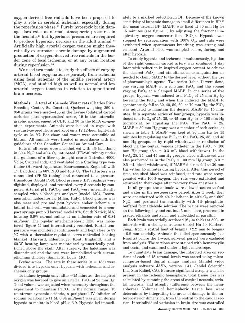
Figure 1. Recording of mean arterial blood pressure(MABP) and EEG before, during, and after the insult.(A) From an animal with hypoxia only, the typical imme-diate drop in MABP is seen. When MABP fell to 30 mmHg, it was maintained at 30 mm Hg for 15 minutes byadjusting oxygen content and exsanguination/reinfusion ofshed blood as necessary, delivering a 15-minute combinedinsult. Reoxygenation was done with 100% oxygen. (B)Representative 5-second strips of EEG sampled before hyp-oxia, and during hypoxia with hypotension to 30 mm Hg.Some slowing of EEG frequencies occurs, with high ampli-tude d waves during hypoxia. Note that isoelectricity of theinterhemispheric EEG is not seen.
364 NEUROLOGY 54 January (2 of 2) 2000
Suxamethonium chloride was injected IV to immobilizeanimals. After 80 minutes occlusion, the nylon suture wasremoved and the external carotid artery was tied perma-nently. MABP and PaO2 were restored to preischemic lev-els by reinfusion of blood and adjusting the FiO2,respectively. Halothane was discontinued and the animalswere allowed to awaken.
One week after MCA occlusion, rats were perfused-fixedand the brain was removed, paraffinized, and serially sec-tioned (8 mm thick) at 500-mm intervals with a sliding mic-rotome. The sections were stained with hematoxylin-eosin,and polygons for necrosis in the neocortex and in subcortical
structures (white matter, deep gray) were traced separately,as were the areas of the ipsilateral and contralateral hemi-spheres. Brain tissue loss was then quantified as mm3 byintegrating necrosis and atrophy in the third, orthogonalaxis. Volumes were finally normalized to the opposite hemi-sphere to control for variation in absolute brain size, beingexpressed as % of the hemisphere.
Results. Physiologically monitored/controlled Levinepreparation. Severe hypoxia caused immediate cardiachypotension (see figure 1), and preliminary experimentsrevealed that it was very difficult for rats to survive hyp-
Table 1 Groups—physiologic variables
VariableHypoxia
only
Hypoxia with ischemia
Groups varying MABP Both series* Groups varying PaO2
BP (mm Hg) 30 70 60 50 40 30 30 30 30
PaO2 (mm Hg) 25 25 25 25 25 25 35 45 .100 mm Hg†
n 13 5 10 9 8 9 15 9 12
Preischemia
MABP (mm Hg) 100.8 6 3.1 116.4 6 6.6 107.3 6 3.4 105.2 6 3.8 106.6 6 3.2 98.6 6 2.2 103.7 6 2.5 102.1 6 3.7 100.8 6 2.8
PaO2 (mm Hg) 148.5 6 4.4 133.8 6 9.0 149.2 6 6.1 173.3 6 6.3 158.7 6 7.0 126.6 6 9.9 151.5 6 5.3 146.4 6 9.6 121.0 6 6.5
PaCO2 (mm Hg) 33.8 6 1.1 38.6 6 2.2 37.9 6 0.9 35.4 6 1.0 36.5 6 0.7 34.8 6 1.4 35.5 6 1.0 39.4 6 0.9 36.4 6 0.7
pH 7.39 6 0.01 7.40 6 0.03 7.42 6 0.01 7.53 6 0.02 7.43 6 0.02 7.39 6 0.02 7.42 6 0.02 7.40 6 0.01 7.37 6 0.01
Hematocrit (%) 44.0 6 1.2 44.0 6 1.4 45.9 6 1.2 45.8 6 1.0 46.4 6 0.9 48.2 6 1.2 4.0 6 0.8 45.0 6 1.1 46.4 6 0.5
Body temperature (°C) 37.5 6 0.1 37.5 6 0.1 37.5 6 0.1 37.5 6 0.1 37.5 6 0.1 37.5 6 0.1 37.4 6 0.1 37.5 6 0.1 37.5 6 0.1
Glucose (mM) 9.6 6 0.6 10.1 6 0.8 11.3 6 2.0 10.4 6 1.1 11.6 6 0.4 11.9 6 1.4 11.1 6 0.5 10.2 6 0.3 11.4 6 0.6
Values are mean 6 SEM. p Values derived from one-way ANOVA followed by Scheffe test.
* This group is a member of both series (varying MABP and varying PaO2).† Inspired oxygen content was constant during experiment.
Table 2 Physiologic variables in the cerebral blood flow study
VariableNormal(n 5 4)
Hypoxia only(n 5 5)
Ischemia only(n 5 6)
Hypoxia withischemia (n 5 4)
Prehypoxia/ischemia
MABP (mm Hg) 112.2 6 4.6 105.2 6 5.2 94.1 6 5.3 114.6 6 3.6
PaO2 (mm Hg) 152.8 6 9.2 131.8 6 8.5 139.7 6 4.5 120.3 6 18.3
PaCO2 (mm Hg) 36.9 6 2.0 39.7 6 1.6 35.5 6 1.8 36.7 6 1.5
pH 7.39 6 0.01 7.40 6 0.02 7.41 6 0.02 7.40 6 0.02
Hematocrit (%) 43.8 6 1.1 45.0 6 1.0 44.2 6 0.9 45.0 6 2.3
Glucose (mM) 12.2 6 1.9 10.6 6 1.2 12.3 6 1.2 12.9 6 1.9
During control of MABP
MABP (mm Hg) — 33.0 6 1.8* 34.1 6 1.2* 33.9 6 2.1*
PaO2 (mm Hg) — 35.4 6 2.5* — 33.0 6 1.5*
PaCO2 (mm Hg) — 34.7 6 3.3 — 34.2 6 1.2
pH — 7.29 6 0.02 — 7.28 6 0.04
Values are mean 6 SEM. p Values derived from one-way ANOVA followed by Scheffe test.
* p , 0.001 Compared with normal group.
MABP 5 mean arterial blood pressure.
January (2 of 2) 2000 NEUROLOGY 54 365
oxia at PaO2 levels ,20 mm Hg owing to immediate car-diac death. Forty-four rats died within 2 days of the insult;10 of these were in the hypoxia only group, where mortal-ity was 43%. The rest were from hypoxia with ischemiagroups, and died likely due to delayed cardiogenic shock8 (5 ofthese 44 died following crescendo seizures). In the variableischemia groups, at a fixed PaO2 of 25 mm Hg, the mortalitywas 0%, 9%, 47%, 36%, and 53% at MABP 70, 60, 50, 40, and30 mm Hg, respectively. In the groups with a fixed MABPof 30 mm Hg, the mortality was 29%, 18%, and 17% atPaO2 . 100, 45, and 35 mm Hg, respectively.
Physiologic variables are shown in table 1. MABP waswell controlled to the desired values by adjusting the FiO2.The PaO2 levels during control of MABP and the durationof hypoxia showed no differences among groups.
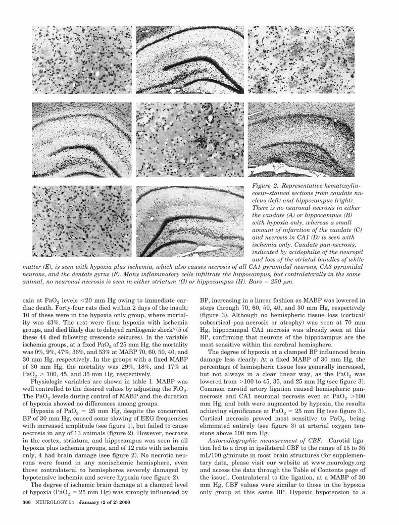
Hypoxia of PaO2 5 25 mm Hg, despite the concurrentBP of 30 mm Hg, caused some slowing of EEG frequencieswith increased amplitude (see figure 1), but failed to causenecrosis in any of 13 animals (figure 2). However, necrosisin the cortex, striatum, and hippocampus was seen in allhypoxia plus ischemia groups, and of 12 rats with ischemiaonly, 4 had brain damage (see figure 2). No necrotic neu-rons were found in any nonischemic hemisphere, eventhose contralateral to hemispheres severely damaged byhypotensive ischemia and severe hypoxia (see figure 2).
The degree of ischemic brain damage at a clamped levelof hypoxia (PaO2 5 25 mm Hg) was strongly influenced by
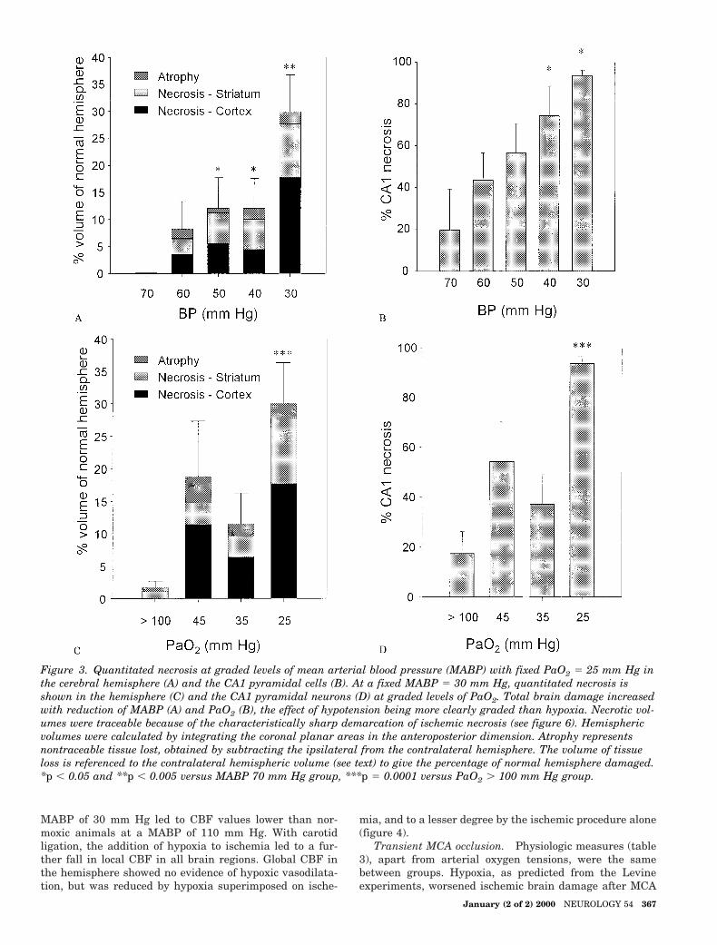
BP, increasing in a linear fashion as MABP was lowered insteps through 70, 60, 50, 40, and 30 mm Hg, respectively(figure 3). Although no hemispheric tissue loss (cortical/subcortical pan-necrosis or atrophy) was seen at 70 mmHg, hippocampal CA1 necrosis was already seen at thisBP, confirming that neurons of the hippocampus are themost sensitive within the cerebral hemisphere.
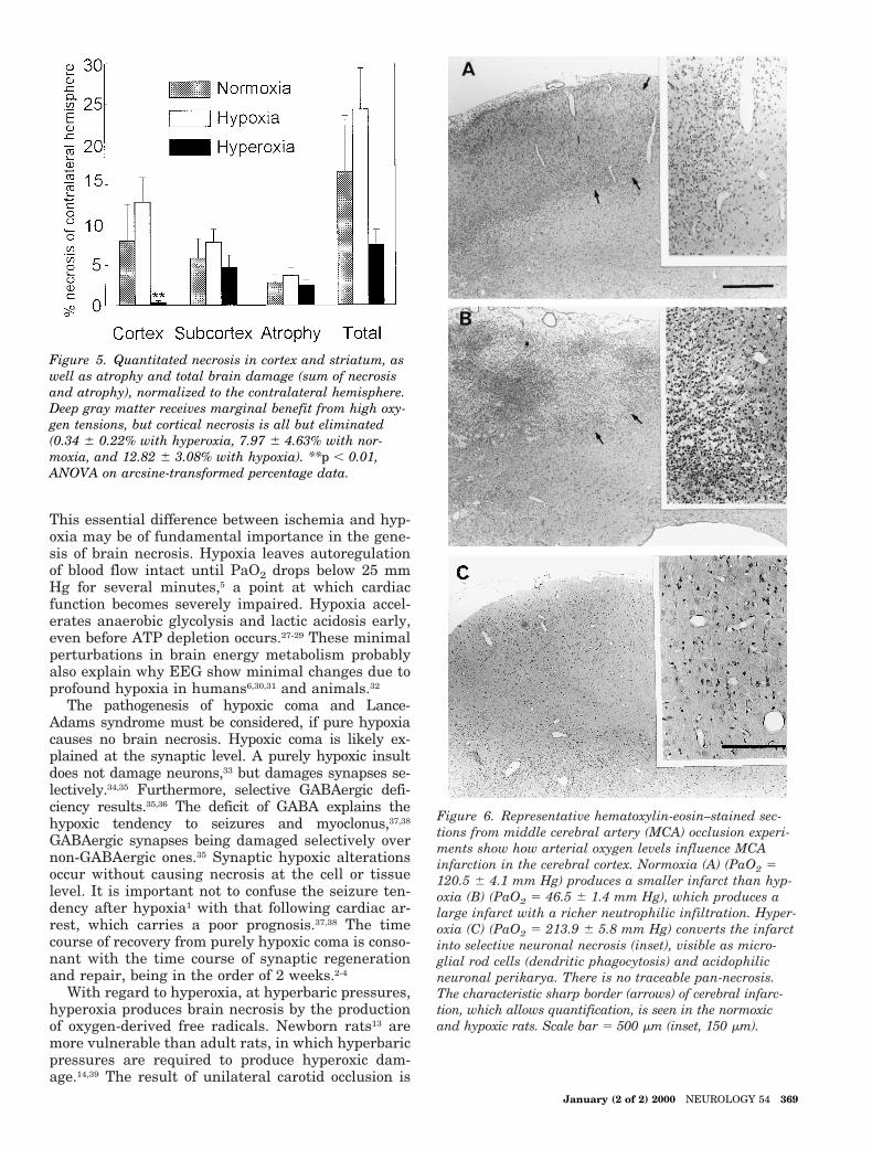
The degree of hypoxia at a clamped BP influenced braindamage less clearly. At a fixed MABP of 30 mm Hg, thepercentage of hemispheric tissue loss generally increased,but not always in a clear linear way, as the PaO2 waslowered from .100 to 45, 35, and 25 mm Hg (see figure 3).Common carotid artery ligation caused hemispheric pan-necrosis and CA1 neuronal necrosis even at PaO2 .100mm Hg, and both were augmented by hypoxia, the resultsachieving significance at PaO2 5 25 mm Hg (see figure 3).Cortical necrosis proved most sensitive to PaO2, beingeliminated entirely (see figure 3) at arterial oxygen ten-sions above 100 mm Hg.
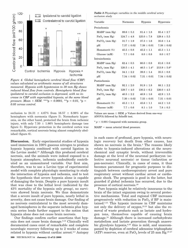
Autoradiographic measurement of CBF. Carotid liga-tion led to a drop in ipsilateral CBF to the range of 15 to 35mL/100 g/minute in most brain structures (for supplemen-tary data, please visit our website at www.neurology.organd access the data through the Table of Contents page ofthe issue). Contralateral to the ligation, at a MABP of 30mm Hg, CBF values were similar to those in the hypoxiaonly group at this same BP. Hypoxic hypotension to a
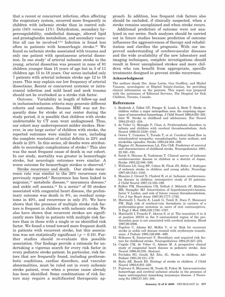
Figure 2. Representative hematoxylin-eosin–stained sections from caudate nu-cleus (left) and hippocampus (right).There is no neuronal necrosis in eitherthe caudate (A) or hippocampus (B)with hypoxia only, whereas a smallamount of infarction of the caudate (C)and necrosis in CA1 (D) is seen withischemia only. Caudate pan-necrosis,indicated by acidophilia of the neuropiland loss of the striatal bundles of white
matter (E), is seen with hypoxia plus ischemia, which also causes necrosis of all CA1 pyramidal neurons, CA3 pyramidalneurons, and the dentate gyrus (F). Many inflammatory cells infiltrate the hippocampus, but contralaterally in the sameanimal, no neuronal necrosis is seen in either striatum (G) or hippocampus (H). Bars 5 250 mm.
366 NEUROLOGY 54 January (2 of 2) 2000
MABP of 30 mm Hg led to CBF values lower than nor-moxic animals at a MABP of 110 mm Hg. With carotidligation, the addition of hypoxia to ischemia led to a fur-ther fall in local CBF in all brain regions. Global CBF inthe hemisphere showed no evidence of hypoxic vasodilata-tion, but was reduced by hypoxia superimposed on ische-
mia, and to a lesser degree by the ischemic procedure alone(figure 4).
Transient MCA occlusion. Physiologic measures (table3), apart from arterial oxygen tensions, were the samebetween groups. Hypoxia, as predicted from the Levineexperiments, worsened ischemic brain damage after MCA
Figure 3. Quantitated necrosis at graded levels of mean arterial blood pressure (MABP) with fixed PaO2 5 25 mm Hg inthe cerebral hemisphere (A) and the CA1 pyramidal cells (B). At a fixed MABP 5 30 mm Hg, quantitated necrosis isshown in the hemisphere (C) and the CA1 pyramidal neurons (D) at graded levels of PaO2. Total brain damage increasedwith reduction of MABP (A) and PaO2 (B), the effect of hypotension being more clearly graded than hypoxia. Necrotic vol-umes were traceable because of the characteristically sharp demarcation of ischemic necrosis (see figure 6). Hemisphericvolumes were calculated by integrating the coronal planar areas in the anteroposterior dimension. Atrophy representsnontraceable tissue lost, obtained by subtracting the ipsilateral from the contralateral hemisphere. The volume of tissueloss is referenced to the contralateral hemispheric volume (see text) to give the percentage of normal hemisphere damaged.*p , 0.05 and **p , 0.005 versus MABP 70 mm Hg group, ***p 5 0.0001 versus PaO2 . 100 mm Hg group.
January (2 of 2) 2000 NEUROLOGY 54 367
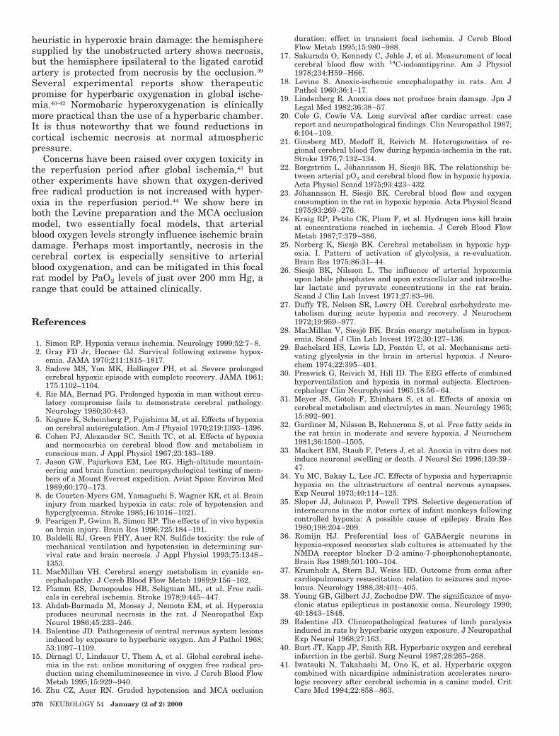
occlusion to 24.31 6 4.67% from 16.57 6 6.98% of thehemisphere with normoxia (figure 5). Normobaric hyper-oxia, on the other hand, protected the brain from ischemicinjury, with only 7.50 6 1.86% hemispheric damage (seefigure 5). Hyperoxic protection in the cerebral cortex wasremarkable, cortical necrosis being almost completely abol-ished (figure 6).
Discussion. Early experimental studies of hypoxiaused immersion in 100% gaseous nitrogen to producehypoxic hypoxia combined with carotid ligation inthe Levine model.18 Although this produced cerebralnecrosis, and the animals were indeed exposed to ahypoxic atmosphere, ischemia undoubtedly contrib-uted as an unmonitored variable. Our first aim,therefore, was to use a Levine type of experimentalmodel with complete physiologic monitoring to studythe interaction of hypoxia and ischemia, and to testthe hypothesis that severe hypoxia alone is capableof damaging the brain. Despite a pure hypoxic insultthat was close to the lethal level (indicated by the43% mortality of the hypoxia only group), no survi-vors showed brain necrosis. We thus demonstratethat a severe insult of pure hypoxia, nearly lethal inseverity, does not cause brain damage. Our finding ofno necrosis contralateral to the most severely dam-aged hemispheres in the separate ischemia plus hyp-oxia series lends further support to the notion thathypoxia alone does not cause brain necrosis.
Our findings confirm earlier assertions that hyp-oxia does not cause brain damage.9,19 Clinically welldocumented cases exist of remarkable and completeneurologic recovery following up to 2 weeks of comarelated to hypoxia without cardiac arrest.2,3 Autopsy
in such cases of profound, pure hypoxia, with neuro-logic recovery but death from other causes, hasshown no necrosis in the brain.4 The reasons likelyrelate to hypoxia-induced alterations at the neuro-chemical and synaptic levels, without irreversibledamage at the level of the neuronal perikaryon (se-lective neuronal necrosis) or tissue (infarction orpan-necrosis). Clinically, in cases of coma, it thusbecomes paramount for prognostic purposes to dis-tinguish between cardiorespiratory arrest and purerespiratory arrest without cardiac arrest or cardio-genic shock. The prognosis is good in the latter, butguarded if global ischemia has occurred, due to thepresence of cortical necrosis.20
Pure hypoxia might be relatively innocuous to thebrain of the intact organism owing to several protec-tive mechanisms. Total and regional CBF increasesprogressively with reduction in PaO2 if BP is main-tained.5,21 This hypoxic increase in CBF maintainsnot only the delivery of oxygen,22,23 but also the re-moval of waste products such as lactate and hydro-gen ions, themselves capable of causing braindamage.24 Although there is increased carbohydratemetabolism and lactate generation through anaero-bic glycolysis hypoxia,6,25 this is mild, and unaccom-panied by depletion of cerebral adenosine triphosphate(ATP) reserves, even at PaO2 levels of 25 mm Hg.25-27
Figure 4. Global hemispheric cerebral blood flow (CBF)values calculated as arithmetic means of all structuresmeasured. Hypoxia with hypotension to 30 mm Hg showsreduced blood flow from controls. Hemispheric blood flowipsilateral to carotid occlusion is depressed, with no in-crease in CBF with superadded hypoxia, at the same bloodpressure. Mean 6 SEM. ***p 5 0.0001, **p 5 0.01, *p 50.05 versus control.
Table 3 Physiologic variables in the middle cerebral arteryocclusion study
Variable Normoxia Hypoxia Hyperoxia
Preischemia
MABP (mm Hg) 89.6 6 5.3 81.4 6 1.8 85.4 6 2.7
PaO2 (mm Hg) 124.7 6 4.8 125.0 6 7.8 129.4 6 5.5
PaCO2 (mm Hg) 31.7 6 1.9 37.3 6 1.6 35.6 6 1.4
pH 7.37 6 0.02 7.38 6 0.01 7.38 6 0.02
Hematocrit (%) 45.5 6 0.8 45.2 6 1.1 45.3 6 1.1
Glucose (mM) 7.7 6 0.6 8.0 6 0.8 8.0 6 0.7
Intraischemia
MABP (mm Hg) 62.4 6 0.5 60.0 6 0.8 61.6 6 0.8
PaO2 (mm Hg) 120.5 6 4.1 46.5 6 1.4* 213.9 6 5.8*
PaCO2 (mm Hg) 34.1 6 2.2 39.8 6 1.4 35.5 6 0.8
pH 7.34 6 0.02 7.31 6 0.01 7.34 6 0.02
Postischemia
MABP (mm Hg) 92.1 6 2.0 91.9 6 1.8 88.1 6 2.2
PaO2 (mm Hg) 129.7 6 4.0 130.2 6 6.2 126.0 6 4.5
PaCO2 (mm Hg) 40.5 6 2.2 40.9 6 1.6 42.9 6 1.5
pH 7.30 6 0.02 7.33 6 0.01 7.31 6 0.01
Hematocrit (%) 45.3 6 1.1 45.6 6 1.1 44.2 6 1.0
Glucose (mM) 7.7 6 0.6 8.1 6 1.0 7.8 6 0.3
Values are mean 6 SEM. p Values derived from one-wayANOVA followed by Scheffe test.
* p , 0.001 Compared with normoxia group.
MABP 5 mean arterial blood pressure.
368 NEUROLOGY 54 January (2 of 2) 2000
This essential difference between ischemia and hyp-oxia may be of fundamental importance in the gene-sis of brain necrosis. Hypoxia leaves autoregulationof blood flow intact until PaO2 drops below 25 mmHg for several minutes,5 a point at which cardiacfunction becomes severely impaired. Hypoxia accel-erates anaerobic glycolysis and lactic acidosis early,even before ATP depletion occurs.27-29 These minimalperturbations in brain energy metabolism probablyalso explain why EEG show minimal changes due toprofound hypoxia in humans6,30,31 and animals.32
The pathogenesis of hypoxic coma and Lance-Adams syndrome must be considered, if pure hypoxiacauses no brain necrosis. Hypoxic coma is likely ex-plained at the synaptic level. A purely hypoxic insultdoes not damage neurons,33 but damages synapses se-lectively.34,35 Furthermore, selective GABAergic defi-ciency results.35,36 The deficit of GABA explains thehypoxic tendency to seizures and myoclonus,37,38
GABAergic synapses being damaged selectively overnon-GABAergic ones.35 Synaptic hypoxic alterationsoccur without causing necrosis at the cell or tissuelevel. It is important not to confuse the seizure ten-dency after hypoxia1 with that following cardiac ar-rest, which carries a poor prognosis.37,38 The timecourse of recovery from purely hypoxic coma is conso-nant with the time course of synaptic regenerationand repair, being in the order of 2 weeks.2-4
With regard to hyperoxia, at hyperbaric pressures,hyperoxia produces brain necrosis by the productionof oxygen-derived free radicals. Newborn rats13 aremore vulnerable than adult rats, in which hyperbaricpressures are required to produce hyperoxic dam-age.14,39 The result of unilateral carotid occlusion is
Figure 6. Representative hematoxylin-eosin–stained sec-tions from middle cerebral artery (MCA) occlusion experi-ments show how arterial oxygen levels influence MCAinfarction in the cerebral cortex. Normoxia (A) (PaO2 5120.5 6 4.1 mm Hg) produces a smaller infarct than hyp-oxia (B) (PaO2 5 46.5 6 1.4 mm Hg), which produces alarge infarct with a richer neutrophilic infiltration. Hyper-oxia (C) (PaO2 5 213.9 6 5.8 mm Hg) converts the infarctinto selective neuronal necrosis (inset), visible as micro-glial rod cells (dendritic phagocytosis) and acidophilicneuronal perikarya. There is no traceable pan-necrosis.The characteristic sharp border (arrows) of cerebral infarc-tion, which allows quantification, is seen in the normoxicand hypoxic rats. Scale bar 5 500 mm (inset, 150 mm).
Figure 5. Quantitated necrosis in cortex and striatum, aswell as atrophy and total brain damage (sum of necrosisand atrophy), normalized to the contralateral hemisphere.Deep gray matter receives marginal benefit from high oxy-gen tensions, but cortical necrosis is all but eliminated(0.34 6 0.22% with hyperoxia, 7.97 6 4.63% with nor-moxia, and 12.82 6 3.08% with hypoxia). **p , 0.01,ANOVA on arcsine-transformed percentage data.
January (2 of 2) 2000 NEUROLOGY 54 369
heuristic in hyperoxic brain damage: the hemispheresupplied by the unobstructed artery shows necrosis,but the hemisphere ipsilateral to the ligated carotidartery is protected from necrosis by the occlusion.39
Several experimental reports show therapeuticpromise for hyperbaric oxygenation in global ische-mia.40-42 Normobaric hyperoxygenation is clinicallymore practical than the use of a hyperbaric chamber.It is thus noteworthy that we found reductions incortical ischemic necrosis at normal atmosphericpressure.
Concerns have been raised over oxygen toxicity inthe reperfusion period after global ischemia,43 butother experiments have shown that oxygen-derivedfree radical production is not increased with hyper-oxia in the reperfusion period.44 We show here inboth the Levine preparation and the MCA occlusionmodel, two essentially focal models, that arterialblood oxygen levels strongly influence ischemic braindamage. Perhaps most importantly, necrosis in thecerebral cortex is especially sensitive to arterialblood oxygenation, and can be mitigated in this focalrat model by PaO2 levels of just over 200 mm Hg, arange that could be attained clinically.
References
1. Simon RP. Hypoxia versus ischemia. Neurology 1999;52:7–8.2. Gray FD Jr, Horner GJ. Survival following extreme hypox-
emia. JAMA 1970;211:1815–1817.3. Sadove MS, Yon MK, Hollinger PH, et al. Severe prolonged
cerebral hypoxic episode with complete recovery. JAMA 1961;175:1102–1104.
4. Rie MA, Bernad PG. Prolonged hypoxia in man without circu-latory compromise fails to demonstrate cerebral pathology.Neurology 1980;30:443.
5. Kogure K, Scheinberg P, Fujishima M, et al. Effects of hypoxiaon cerebral autoregulation. Am J Physiol 1970;219:1393–1396.
6. Cohen PJ, Alexander SC, Smith TC, et al. Effects of hypoxiaand normocarbia on cerebral blood flow and metabolism inconscious man. J Appl Physiol 1967;23:183–189.
7. Jason GW, Pajurkova EM, Lee RG. High-altitude mountain-eering and brain function: neuropsychological testing of mem-bers of a Mount Everest expedition. Aviat Space Environ Med1989;60:170–173.
8. de Courten-Myers GM, Yamaguchi S, Wagner KR, et al. Braininjury from marked hypoxia in cats: role of hypotension andhyperglycemia. Stroke 1985;16:1016–1021.
9. Pearigen P, Gwinn R, Simon RP. The effects of in vivo hypoxiaon brain injury. Brain Res 1996;725:184–191.
10. Baldelli RJ, Green FHY, Auer RN. Sulfide toxicity: the role ofmechanical ventilation and hypotension in determining sur-vival rate and brain necrosis. J Appl Physiol 1993;75:1348–1353.
11. MacMillan VH. Cerebral energy metabolism in cyanide en-cephalopathy. J Cereb Blood Flow Metab 1989;9:156–162.
12. Flamm ES, Demopoulos HB, Seligman ML, et al. Free radi-cals in cerebral ischemia. Stroke 1978;9:445–447.
13. Ahdab-Barmada M, Moossy J, Nemoto EM, et al. Hyperoxiaproduces neuronal necrosis in the rat. J Neuropathol ExpNeurol 1986;45:233–246.
14. Balentine JD. Pathogenesis of central nervous system lesionsinduced by exposure to hyperbaric oxygen. Am J Pathol 1968;53:1097–1109.
15. Dirnagl U, Lindauer U, Them A, et al. Global cerebral ische-mia in the rat: online monitoring of oxygen free radical pro-duction using chemiluminescence in vivo. J Cereb Blood FlowMetab 1995;15:929–940.
16. Zhu CZ, Auer RN. Graded hypotension and MCA occlusion
duration: effect in transient focal ischemia. J Cereb BloodFlow Metab 1995;15:980–988.
17. Sakurada O, Kennedy C, Jehle J, et al. Measurement of localcerebral blood flow with 14C-iodoantipyrine. Am J Physiol1978;234:H59–H66.
18. Levine S. Anoxic-ischemic encephalopathy in rats. Am JPathol 1960;36:1–17.
19. Lindenberg R. Anoxia does not produce brain damage. Jpn JLegal Med 1982;36:38–57.
20. Cole G, Cowie VA. Long survival after cardiac arrest: casereport and neuropathological findings. Clin Neuropathol 1987;6:104–109.
21. Ginsberg MD, Medoff R, Reivich M. Heterogeneities of re-gional cerebral blood flow during hypoxia-ischemia in the rat.Stroke 1976;7:132–134.
22. Borgstrom L, Johannsson H, Siesjo BK. The relationship be-tween arterial pO2 and cerebral blood flow in hypoxic hypoxia.Acta Physiol Scand 1975;93:423–432.
23. Johannsson H, Siesjo BK. Cerebral blood flow and oxygenconsumption in the rat in hypoxic hypoxia. Acta Physiol Scand1975;93:269–276.
24. Kraig RP, Petito CK, Plum F, et al. Hydrogen ions kill brainat concentrations reached in ischemia. J Cereb Blood FlowMetab 1987;7:379–386.
25. Norberg K, Siesjo BK. Cerebral metabolism in hypoxic hyp-oxia. I. Pattern of activation of glycolysis, a re-evaluation.Brain Res 1975;86:31–44.
26. Siesjo BK, Nilsson L. The influence of arterial hypoxemiaupon labile phosphates and upon extracellular and intracellu-lar lactate and pyruvate concentrations in the rat brain.Scand J Clin Lab Invest 1971;27:83–96.
27. Duffy TE, Nelson SR, Lowry OH. Cerebral carbohydrate me-tabolism during acute hypoxia and recovery. J Neurochem1972;19:959–977.
28. MacMillan V, Siesjo BK. Brain energy metabolism in hypox-emia. Scand J Clin Lab Invest 1972;30:127–136.
29. Bachelard HS, Lewis LD, Ponten U, et al. Mechanisms acti-vating glycolysis in the brain in arterial hypoxia. J Neuro-chem 1974;22:395–401.
30. Preswick G, Reivich M, Hill ID. The EEG effects of combinedhyperventilation and hypoxia in normal subjects. Electroen-cephalogr Clin Neurophysiol 1965;18:56–64.
31. Meyer JS, Gotoh F, Ebinhara S, et al. Effects of anoxia oncerebral metabolism and electrolytes in man. Neurology 1965;15:892–901.
32. Gardiner M, Nilsson B, Rehncrona S, et al. Free fatty acids inthe rat brain in moderate and severe hypoxia. J Neurochem1981;36:1500–1505.
33. Mackert BM, Staub F, Peters J, et al. Anoxia in vitro does notinduce neuronal swelling or death. J Neurol Sci 1996;139:39–47.
34. Yu MC, Bakay L, Lee JC. Effects of hypoxia and hypercapnichypoxia on the ultrastructure of central nervous synapses.Exp Neurol 1973;40:114–125.
35. Sloper JJ, Johnson P, Powell TPS. Selective degeneration ofinterneurons in the motor cortex of infant monkeys followingcontrolled hypoxia: A possible cause of epilepsy. Brain Res1980;198:204–209.
36. Romijn HJ. Preferential loss of GABAergic neurons inhypoxia-exposed neocortex slab cultures is attenuated by theNMDA receptor blocker D-2-amino-7-phosphonoheptanoate.Brain Res 1989;501:100–104.
37. Krumholz A, Stern BJ, Weiss HD. Outcome from coma aftercardiopulmonary resuscitation: relation to seizures and myoc-lonus. Neurology 1988;38:401–405.
38. Young GB, Gilbert JJ, Zochodne DW. The significance of myo-clonic status epilepticus in postanoxic coma. Neurology 1990;40:1843–1848.
39. Balentine JD. Clinicopathological features of limb paralysisinduced in rats by hyperbaric oxygen exposure. J NeuropatholExp Neurol 1968;27:163.
40. Burt JT, Kapp JP, Smith RR. Hyperbaric oxygen and cerebralinfarction in the gerbil. Surg Neurol 1987;28:265–268.
41. Iwatsuki N, Takahashi M, Ono K, et al. Hyperbaric oxygencombined with nicardipine administration accelerates neuro-logic recovery after cerebral ischemia in a canine model. CritCare Med 1994;22:858–863.
370 NEUROLOGY 54 January (2 of 2) 2000
42. Krakovsky M, Rogatsky G, Zarchin N, et al. Effect of hyper-baric oxygen therapy on survival after global cerebral ische-mia in rats. Surg Neurol 1998;49:412–416.
43. Mickel HS, Vaishnav YN, Kempski O, et al. Breathing 100%oxygen after global brain ischemia in Mongolian gerbils re-
sults in increased lipid peroxidation and increased mortality.Stroke 1987;18:426–430.
44. Agardh C-D, Zhang H, Smith M-L, et al. Free radical produc-tion and ischemic brain damage: influence of postischemicoxygen tension. Int J Dev Neurosci 1991;9:127–138.
Stroke in childrenThe coexistence of multiple risk factors
predicts poor outcomeS. Lanthier, MD, CSPQ; L. Carmant, MD, FRCP(C); M. David, MD, FRCP(C);
A. Larbrisseau, MD, FRCP(C); and G. de Veber, MD, FRCP(C)
Article abstract—Objective: To characterize the risk factors for stroke in children and their relationship to outcomes.Methods: We reviewed charts of children with ischemic and hemorrhagic stroke seen at Hopital Sainte-Justine, Montrealbetween 1991 and 1997. Results: We found 51 ischemic strokes: 46 arterial and 5 sinovenous thromboses. Risk factorswere variable and multiple in 12 (24%) of the 51 ischemic strokes. Ischemic stroke recurred in 3 (8%) patients with asingle or no identified risk factor and in 5 (42%) of 12 patients with multiple risk factors (p 5 0.01). We also found 21hemorrhagic strokes, 14 (67%) of which were caused by vascular abnormalities. No patient with hemorrhagic stroke hadmultiple risk factors. Hemorrhagic stroke recurred in two patients (10%). Outcome in all 72 stroke patients was as follows:asymptomatic, 36%; symptomatic epilepsy or persistent neurologic deficit, 45%; and death, 20%. Death occurred morefrequently in patients with recurrent stroke (40%) than in those with nonrecurrent stroke (16%). Conclusions: Multiplerisk factors are found in many ischemic strokes and may predict stroke recurrence. Recurrent stroke tends to increase rateof mortality. Because of the high prevalence and importance of multiple risk factors, a complete investigation, includinghematologic and metabolic studies and angiography, should be considered in every child with ischemic stroke, even whena cause is known. Key words: Children—Stroke—Outcome—Risk factor—Investigation.
NEUROLOGY 2000;54:371–378
Childhood stroke affects 2.7 per 100,000 children peryear1 and is known to recur in up to 20%.2 In individ-ual children with stroke, the extent of investigationsfor risk factors often is limited, especially when anobvious cause is known. However, multiple risk fac-tors may coexist in childhood stroke,3 and their de-tection in individual patients can modify theprognosis and medical treatment. We reviewed thecharts of children with ischemic and hemorrhagicstroke seen at our center between 1991 and 1997.Our objective was to characterize the stroke risk fac-tors and their relationship to outcomes.
Methods. Patients. We defined stroke as a focal neuro-logic deficit of sudden onset, not solely related to seizure,resulting from irreversible focal ischemic (ischemic stroke)or hemorrhagic (hemorrhagic stroke) damage to the brainparenchyma secondary to a cerebrovascular disorder. Is-chemic stroke included arterial ischemic stroke and sino-venous thrombosis. We searched patient charts at a singlechildren’s health care center (Hopital Sainte-Justine), us-
ing ICD-9 codes, to identify all patients age 1 month to 18years diagnosed with stroke from 1991 to 1997. We ex-cluded traumatic hemorrhages but included children withischemic stroke related to trauma. Unless accompanied bycerebral hemorrhage or infarct, patients with sinovenousthrombosis or with extracerebral intracranial bleeding(e.g., epidural and subdural hematomas, and subarachnoidhemorrhage) were excluded. We also excluded mitochon-drial disorders because stroke-like episodes in these condi-tions are not clearly ischemic.4
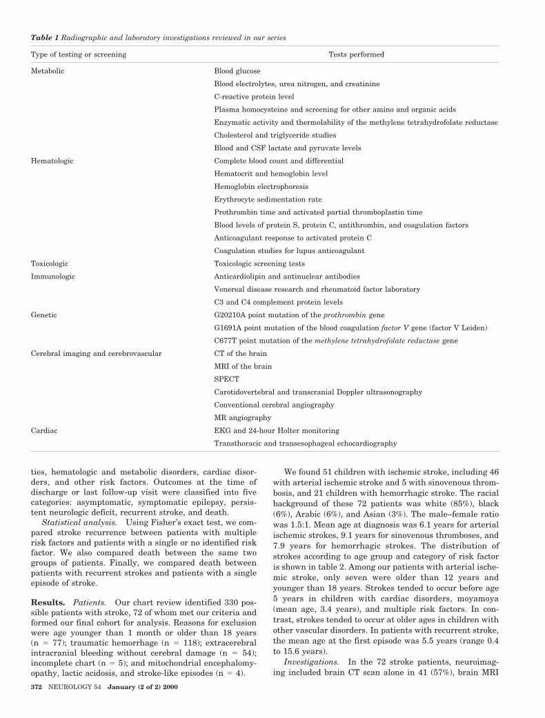
Data collection. The study neurologist (S.L.) reviewedthe charts of patients who met the inclusion criteria. Datacollected regarding stroke risk factors were ethnic origin,family history of thrombosis, medications, recreationaldrug use, infection or head trauma in the 4 weeks preced-ing stroke, headache, and associated systemic diseases.Table 1 lists the radiographic and laboratory investiga-tions reviewed for each case. The presence of anticardio-lipin antibody (aCLA) was defined as a significantlyelevated immunoglobulin M (IgM) or IgG titer. Risk factorswere classified into four categories: vascular abnormali-
From the Department of Pediatrics, Divisions of Neurology (Drs. Lanthier, Carmant, and Larbrisseau) and Hematology (Dr. David), Hopital Sainte-Justine,Universite de Montreal, Quebec; and the Department of Paediatrics, Division of Neurology (Drs. Lanthier and de Veber), The Hospital for Sick Children,University of Toronto, Ontario, Canada.Received November 10, 1998. Accepted in final form August 31, 1999.Address correspondence and reprint requests to Dr. L. Carmant, Department of Pediatrics, Division of Neurology, Hopital Sainte-Justine, 3175 Chemin de laCote Sainte-Catherine, Laboratoire d’Electrophysiologie Medicale (Room 2130), Montreal (Quebec) Canada, H3T 1C5.
Copyright © 2000 by the American Academy of Neurology 371
ties, hematologic and metabolic disorders, cardiac disor-ders, and other risk factors. Outcomes at the time ofdischarge or last follow-up visit were classified into fivecategories: asymptomatic, symptomatic epilepsy, persis-tent neurologic deficit, recurrent stroke, and death.
Statistical analysis. Using Fisher’s exact test, we com-pared stroke recurrence between patients with multiplerisk factors and patients with a single or no identified riskfactor. We also compared death between the same twogroups of patients. Finally, we compared death betweenpatients with recurrent strokes and patients with a singleepisode of stroke.
Results. Patients. Our chart review identified 330 pos-sible patients with stroke, 72 of whom met our criteria andformed our final cohort for analysis. Reasons for exclusionwere age younger than 1 month or older than 18 years(n 5 77); traumatic hemorrhage (n 5 118); extracerebralintracranial bleeding without cerebral damage (n 5 54);incomplete chart (n 5 5); and mitochondrial encephalomy-opathy, lactic acidosis, and stroke-like episodes (n 5 4).
We found 51 children with ischemic stroke, including 46with arterial ischemic stroke and 5 with sinovenous throm-bosis, and 21 children with hemorrhagic stroke. The racialbackground of these 72 patients was white (85%), black(6%), Arabic (6%), and Asian (3%). The male–female ratiowas 1.5:1. Mean age at diagnosis was 6.1 years for arterialischemic strokes, 9.1 years for sinovenous thromboses, and7.9 years for hemorrhagic strokes. The distribution ofstrokes according to age group and category of risk factoris shown in table 2. Among our patients with arterial ische-mic stroke, only seven were older than 12 years andyounger than 18 years. Strokes tended to occur before age5 years in children with cardiac disorders, moyamoya(mean age, 3.4 years), and multiple risk factors. In con-trast, strokes tended to occur at older ages in children withother vascular disorders. In patients with recurrent stroke,the mean age at the first episode was 5.5 years (range 0.4to 15.6 years).
Investigations. In the 72 stroke patients, neuroimag-ing included brain CT scan alone in 41 (57%), brain MRI
Table 1 Radiographic and laboratory investigations reviewed in our series
Type of testing or screening Tests performed
Metabolic Blood glucose
Blood electrolytes, urea nitrogen, and creatinine
C-reactive protein level
Plasma homocysteine and screening for other amino and organic acids
Enzymatic activity and thermolability of the methylene tetrahydrofolate reductase
Cholesterol and triglyceride studies
Blood and CSF lactate and pyruvate levels
Hematologic Complete blood count and differential
Hematocrit and hemoglobin level
Hemoglobin electrophoresis
Erythrocyte sedimentation rate
Prothrombin time and activated partial thromboplastin time
Blood levels of protein S, protein C, antithrombin, and coagulation factors
Anticoagulant response to activated protein C
Coagulation studies for lupus anticoagulant
Toxicologic Toxicologic screening tests
Immunologic Anticardiolipin and antinuclear antibodies
Venereal disease research and rheumatoid factor laboratory
C3 and C4 complement protein levels
Genetic G20210A point mutation of the prothrombin gene
G1691A point mutation of the blood coagulation factor V gene (factor V Leiden)
C677T point mutation of the methylene tetrahydrofolate reductase gene
Cerebral imaging and cerebrovascular CT of the brain
MRI of the brain
SPECT
Carotidovertebral and transcranial Doppler ultrasonography
Conventional cerebral angiography
MR angiography
Cardiac EKG and 24-hour Holter monitoring
Transthoracic and transesophageal echocardiography
372 NEUROLOGY 54 January (2 of 2) 2000
alone in 3 (4%), and both studies in 27 (38%). Only onepatient, who was diagnosed at autopsy, had neither studydone. Among the 51 patients with ischemic stroke, 34 pa-tients (67%) underwent conventional cerebral angiogra-phy, including 6 of 8 patients with no identified risk factor.Six patients (12%) underwent MR angiography, and 11patients (22%) had no vascular imaging studies. Amongthe 21 patients with hemorrhagic stroke, 15 patients (71%)underwent conventional cerebral angiography, including 2of 3 patients with no identified risk factor. None had MRangiography. Six patients (29%) had no vascular imagingstudies. Transthoracic or transesophageal echocardio-graphies were performed in 36 patients (71%) with ische-mic stroke and 7 (33%) with hemorrhagic stroke. Amongthe 51 children with ischemic stroke, prothrombotic testingincluded protein C, protein S, and antithrombin levels in38 patients (75%); the presence of an antiphospholipid(aPL) antibody (including aCLA and lupus anticoagulant)in 37 patients (73%); activated protein C resistance (aPCR)or factor V Leiden mutation in 15 patients (29%); plasmahomocysteine level in 9 patients (18%); the presence of themutant C677T methylene tetrahydrofolate reductase(MTHFR) gene in 3 patients (6%); and the presence of themutant G20210A prothrombin gene in 1 patient (2%).
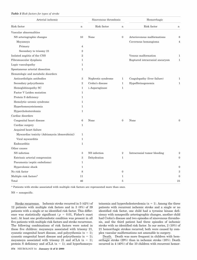
Stroke risk factors. The risk factors identified in eachof the three stroke types (arterial ischemic stroke, sino-venous thrombosis, and hemorrhagic stroke) are summa-rized in table 3.
Arterial ischemic stroke. In the 46 children with arte-rial ischemic stroke, hematologic or metabolic disorderswere identified in 9 (20%), and 7 of these had multiple riskfactors. The presence of aCLA (n 5 3) was the most fre-quent prothrombotic condition observed in our series. Hy-perhomocysteinemia was found in one child who washomozygous for C677T point mutation of the MTHFRgene. This child also had renal failure, hypercholesterol-emia, and folate deficiency.
A cardiac disorder was present in 9 (20%) of 46 patients.Six of these children had cyanotic congenital heart disease,including one child with trisomy 21 and another with pul-monary artery stenosis and a patent foramen ovale. Inthree children with cyanotic congenital heart disease, thestroke occurred in the context of polycythemia (n 5 2) orcardiac surgery (n 5 1).
Primary vascular disorders were present in 11 (31%) ofthe 36 patients with arterial ischemic stroke who under-went conventional or MR angiography. In 6 (17%), a moya-moya pattern was identified. All children with moyamoyawere white. Of the six moyamoya patients, one had tri-
somy 21 with congenital heart disease and polycythemia,another had trisomy 21 and aCLA, and a third had aCLA.
We found nonspecific arteriographic changes in a fur-ther 10 (24%) of the 36 patients with arterial ischemicstroke who underwent angiography. Clinical or laboratoryfindings did not suggest a specific cause in these patients.In particular, a history of varicella was absent. The arte-riographic abnormalities were localized to the proximalportion of the large intracranial arteries (n 5 7) or to thedistal portion (n 5 3). These abnormalities consisted ofsingle stenosis (n 5 3), multiple unilateral stenoses (n 55), and multiple bilateral stenoses (n 5 2). At the site ofthe stenoses, no specific signs of dissection were noted,including no intimal flap, double lumen, or string signs.MRI showed no sign of methemoglobin within the arterialwall. Nonspecific changes on arteriography were present inall three patients with arterial ischemic stroke who hadhad a nonspecific upper respiratory infection within thepreceding 4 weeks.
In ischemic stroke patients, three had extrinsic arterialcompression caused by underlying expanding processes,including subarachnoid hemorrhage with a large intracra-nial hematoma (n 5 1), subdural hematoma (n 5 1), anddiffuse brain edema secondary to acute liver failure withtranstentorial herniation and compression on posterior ce-rebral artery (n 5 1).
Sinovenous thrombosis. Risk factors were found in allfive patients with sinovenous thrombosis. Two patientshad a nonspecific infection. Three children had a hemato-logic or metabolic disorder, including one with nephroticsyndrome and dehydration.
Hemorrhagic stroke. Among the 21 patients with hem-orrhagic stroke, risk factors included vascular abnormali-ties in 14 patients (67%), hematologic disorder in 2patients (10%), and bleeding into an intracranial tumor in2 patients (10%). In 3 (14%) of the 21 patients, no riskfactor was identified. No patient with hemorrhagic strokehad multiple risk factors.
Outcome. The overall outcome in our cohort is pre-sented in tables 4 and 5. Fourteen patients (20%) died. Foranother 6 (8%) patients, the outcome was available only atthe time of discharge. The median length of the follow-upperiod for the remaining 52 patients (72%) was 1.9 years(range 0.25 to 7.2 years). Thirty-five percent of patientswith ischemic strokes and 38% of those with hemorrhagicstrokes were asymptomatic, whereas 49% of patients withischemic strokes and 33% of those with hemorrhagicstrokes had symptomatic epilepsy or persistent neurologicdeficit (see table 4).
Table 2 Distribution of strokes according to age group, type of stroke, and risk factor category
Age
Type of risk factor by type of stroke*
None Vasc only Hem only Cardi only Other Mult Total
1 mo–5 y 3/0/1 4/0/5 1/0/2 4/0/0 3/1/0 9/0/0 24/1/8
.5–12 y 3/0/1 8/0/4 1/2/0 1/0/0 0/0/1 2/1/0 15/3/6
.12–18 y 2/0/1 2/0/5 0/0/0 1/0/0 2/1/1 0/0/0 7/1/7
Total 8/0/3 14/0/14 2/2/2 6/0/0 5/2/2 11/1/0 46/5/21
* Values represent no. of arterial ischemic stroke/sinovenous thrombosis/hemorrhagic stroke, respectively.
Vasc 5 vascular abnormalities; Hem 5 hematologic or metabolic disorders; Cardi 5 cardiac disorders; Mult 5 multiple risk factors.
January (2 of 2) 2000 NEUROLOGY 54 373
Stroke recurrence. Ischemic stroke recurred in 5 (42%) of12 patients with multiple risk factors and in 3 (8%) of 39patients with a single or no identified risk factor. This differ-ence was statistically significant ( p 5 0.01, Fisher’s exacttest). At least one prothrombotic condition was present in allfive patients with multiple risk factors and stroke recurrence.The following combinations of risk factors were noted inthese five children: moyamoya associated with trisomy 21,cyanotic congenital heart disease, and polycythemia (n 5 1);cyanotic congenital heart disease and polycythemia (n 5 1);moyamoya associated with trisomy 21 and aCLA (n 5 1);protein S deficiency and aCLA (n 5 1); and hyperhomocys-
teinemia and hypercholesterolemia (n 5 1). Among the threepatients with recurrent ischemic stroke and a single or noidentified risk factor, one child had a tyrosine kinase defi-ciency with nonspecific arteriographic changes, another childhad Crohn’s disease and two episodes of sinovenous thrombo-sis, and the third patient had three episodes of ischemicstroke with no identified risk factor. In our series, 2 (10%) of21 hemorrhagic strokes recurred; both were caused by com-plex vascular malformations not amenable to surgery.
Death. Death was more frequent in children with hem-orrhagic stroke (29%) than in ischemic stroke (16%). Deathoccurred in 4 (40%) of the 10 children with recurrent hemor-
Table 3 Risk factors for types of stroke
Arterial ischemic Sinovenous thrombosis Hemorrhagic
Risk factor n Risk factor n Risk factor n
Vascular abnormalities
NS arteriographic changes 10 None 0 Arteriovenous malformations 8
Moyamoya Cavernous hemangioma 4
Primary 4
Secondary to trisomy 21 2
Isolated angiitis of the CNS 2 Venous malformation 1
Fibromuscular dysplasia 1 Ruptured intracranial aneurysm 1
Lupic vasculopathy 1
Spontaneous arterial dissection 1
Hematologic and metabolic disorders
Anticardiolipin antibodies 3 Nephrotic syndrome 1 Coagulopathy (liver failure) 1
Secondary polycythemia 2 Crohn’s disease 1 Hypofibrinogenemia 1
Hemoglobinopathy SC 1 L-Asparaginase 1
Factor V Leiden mutation 1
Protein S deficiency 1
Hemolytic–uremic syndrome 1
Hyperhomocysteinemia 1
Hypercholesterolemia 1
Cardiac disorders
Congenital heart disease 6 None 0 None 0
Cardiac surgery 1
Acquired heart failure
Myocardiac toxicity (Adriamycin [doxorubicin]) 1
Viral myocarditis 1
Endocarditis 1
Other causes
NS infection 3 NS infection 2 Intracranial tumor bleeding 2
Extrinsic arterial compression 3 Dehydration 1 0
Pneumonia (septic embolisms) 1
Hypovolemic shock 1 0
No risk factor 8 0 3
Multiple risk factors* 11 1 0
Total 46 5 21
* Patients with stroke associated with multiple risk factors are represented more than once.
NS 5 nonspecific.
374 NEUROLOGY 54 January (2 of 2) 2000
rhagic or ischemic stroke and in 10 (16%) of the 62 childrenwith a single episode of stroke. Deaths resulted from compli-cations of stroke (n 5 11), underlying cardiac disorders (n 52), and acute respiratory distress syndrome (n 5 1).
Discussion. Patients. Previous studies5,6 reportthat about 45% of strokes occur before the age of 5years. This agrees with our findings. The slight pre-dominance of male patients in our series also is con-sistent with previous studies,3,6,7 which report male–female ratios of 1:1 to 1.2:1. The frequency ofmoyamoya in our cohort is similar to that of thelarge non-Japanese series,7 which reports the disor-der in up to 10% of children with ischemic stroke.Our low proportion of black patients accounts for ourfinding only one case of hemoglobinopathy.
Investigations. During the study intervals, ourcenter followed the usual approach, in which investi-gation of children with stroke is selective and guidedby clinical suspicion. Therefore, investigations werenot uniform in our retrospective series. Hyperhomo-cysteinemia and mutant C677T MTHFR, aPCR andG1691A factor V gene, and G20210A prothrombingene, which have been identified recently, were inves-tigated in few of our patients. Because many children
in our series (76%) underwent angiographic evaluation,our study provides a realistic estimate of the preva-lence of vascular disorders in children with stroke.
Stroke risk factors. Few (15%) of our patientshad no identified stroke risk factor. Our results aresimilar to those of other recent studies, in which noetiology was found in 20% to 36%7,8 of patients withischemic stroke and in 11%1 of patients with hemor-rhagic stroke.
Hematologic and metabolic disorders. Despitethe absence of systematic screening in our cohort, wefound at least one hematologic or metabolic disorderin about 25% of ischemic stroke patients. Prothrom-botic disorders also were frequent in a recent series,3being associated with 35 (38%) of 92 pediatric ische-mic strokes. We have shown that hematologic andmetabolic disorders are frequently found in combina-tion with other risk factors in patients with ischemicstroke. The presence of multiple risk factors hasbeen previously reported to increase the risk ofthrombosis considerably.9 Based on these observa-tions, we believe that an extensive hematologic andmetabolic screening must be part of the workup ofpediatric ischemic strokes, even when a cause
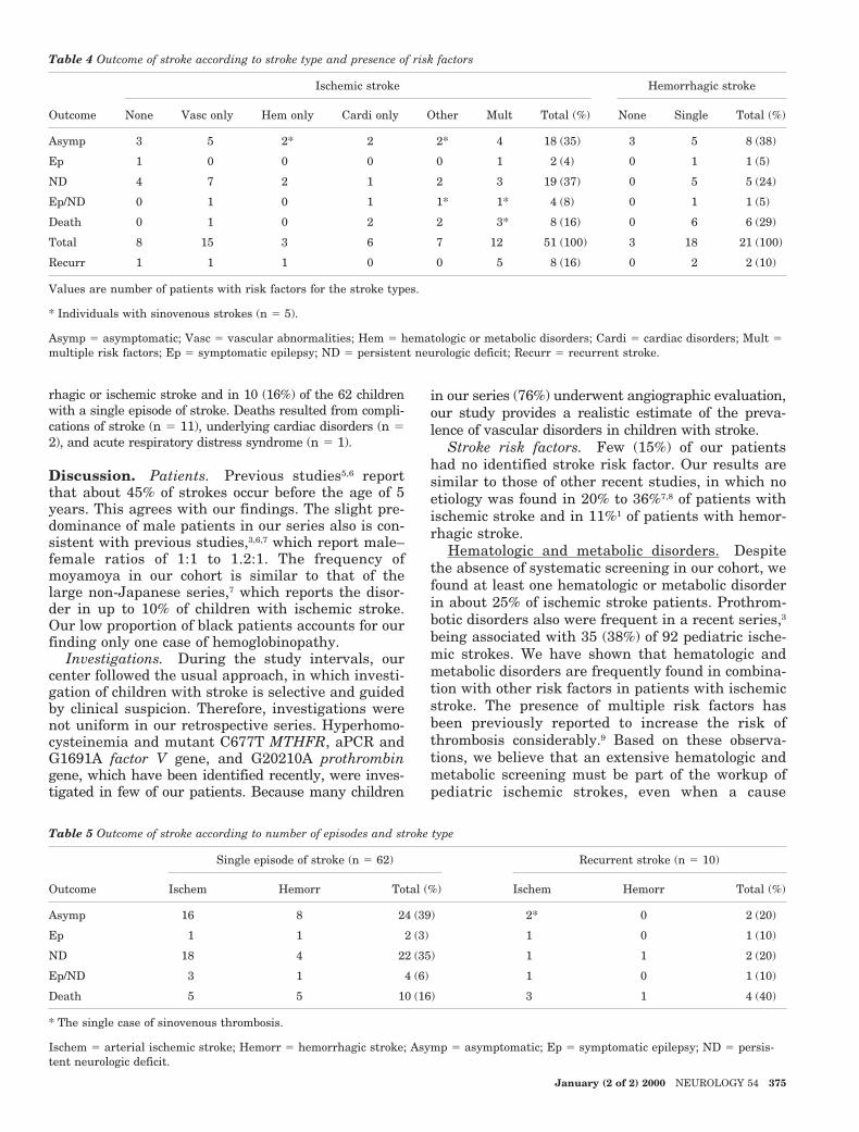
Table 4 Outcome of stroke according to stroke type and presence of risk factors
Outcome
Ischemic stroke Hemorrhagic stroke
None Vasc only Hem only Cardi only Other Mult Total (%) None Single Total (%)
Asymp 3 5 2* 2 2* 4 18 (35) 3 5 8 (38)
Ep 1 0 0 0 0 1 2 (4) 0 1 1 (5)
ND 4 7 2 1 2 3 19 (37) 0 5 5 (24)
Ep/ND 0 1 0 1 1* 1* 4 (8) 0 1 1 (5)
Death 0 1 0 2 2 3* 8 (16) 0 6 6 (29)
Total 8 15 3 6 7 12 51 (100) 3 18 21 (100)
Recurr 1 1 1 0 0 5 8 (16) 0 2 2 (10)
Values are number of patients with risk factors for the stroke types.
* Individuals with sinovenous strokes (n 5 5).
Asymp 5 asymptomatic; Vasc 5 vascular abnormalities; Hem 5 hematologic or metabolic disorders; Cardi 5 cardiac disorders; Mult 5multiple risk factors; Ep 5 symptomatic epilepsy; ND 5 persistent neurologic deficit; Recurr 5 recurrent stroke.
Table 5 Outcome of stroke according to number of episodes and stroke type
Outcome
Single episode of stroke (n 5 62) Recurrent stroke (n 5 10)
Ischem Hemorr Total (%) Ischem Hemorr Total (%)
Asymp 16 8 24 (39) 2* 0 2 (20)
Ep 1 1 2 (3) 1 0 1 (10)
ND 18 4 22 (35) 1 1 2 (20)
Ep/ND 3 1 4 (6) 1 0 1 (10)
Death 5 5 10 (16) 3 1 4 (40)
* The single case of sinovenous thrombosis.
Ischem 5 arterial ischemic stroke; Hemorr 5 hemorrhagic stroke; Asymp 5 asymptomatic; Ep 5 symptomatic epilepsy; ND 5 persis-tent neurologic deficit.
January (2 of 2) 2000 NEUROLOGY 54 375
is known. Arterial ischemic strokes should be inves-tigated as thoroughly as sinovenous thromboses, rul-ing out the presence of aPL antibodies and de-ficiencies in protein C, protein S, and antithrombin, aswell as aPCR (or factor V Leiden) and hyperhomocys-teinemia (or C677T MTHFR gene mutation). Althougha recent study reports that 20% of patients with sino-venous thrombosis were heterozygous carriers of theG20210A prothrombin gene point mutation,10 the rela-tive importance of this mutation in patients with arte-rial ischemic stroke remains to be defined.11 Sickle celldisease must be ruled out in black patients with strokebecause a transfusion program prevents stroke recur-rence.12 Mitochondrial disorders also are important toexclude in patients presenting with sudden neurologicdeficit.Cardiac disorders. Although echocardiogra-phy was not done for all patients in our series, wefound a cardiac risk factor in about 20% of patientswith ischemic stroke. In previous reports, up to 15%of ischemic strokes were attributed to cardiac disor-ders,7,13 most frequently to congenital heart disease.14
Therefore, cardiac evaluation remains essential inthe investigation of any ischemic stroke, especially inyounger children. Congenital heart disease tends tocause stroke during the first 4 years of life, often inthe setting of cardiac surgery or catheterization.14
Among our six patients with congenital heart dis-ease, only one had a cardioembolic stroke associatedwith cardiac surgery. As found in our series, cyanoticcongenital heart disease has been associated withmoyamoya disease and polycythemia.15 In childrenwith congenital heart disease and ischemic stroke,these two associated conditions must be ruled out be-cause such findings can modify therapeutic decisions.
Vascular abnormalities. A recent large series7
found specific vasculopathies in 18% of pediatric is-chemic strokes. We identified recognizable vascu-lopathies in almost 25% of our patients with arterialischemic stroke. This percentage may underestimatetheir prevalence because not all patients in our se-ries underwent vascular imaging. These findings un-derline the importance of vascular investigation inall arterial ischemic strokes in children. Moyamoyawas the most frequent recognizable vasculopathy inour cases of ischemic stroke. Although the exactpathophysiologic mechanism of moyamoya remainsunknown, it has been associated with many condi-tions, including trisomy 21 and congenital heart dis-ease.15,16 The association of moyamoya with lupusanticoagulant17 or with a significantly elevated bloodlevel of IgG aCLA18,19 also has been reported, albeitrarely. We report here two more cases of moyamoyaassociated with aCLA and arterial ischemic stroke.Surprisingly, in our series, aCLA was found in 2 of 6patients with moyamoya but in only 1 of 31 withoutmoyamoya ( p 5 0.06). Despite the few patients inour series, this finding may suggest an associationbetween aPL antibodies and moyamoya in childrenwith arterial ischemic stroke, rather than only a risein IgG aCLA as an epiphenomenon in response to anacute cerebral event. Although the link between aPL
antibodies and thrombosis also remains unknown,vascular endothelium is believed to be a major targetfor the antibodies.20 A direct cause–effect relation-ship cannot be established. However, a thrombogeniceffect of aPL antibodies may induce chronic progres-sive occlusion of cerebral vessels, characteristicallyof the supraclinoid portion of the internal carotidarteries with telangiectatic vessel proliferation, acombination that constitutes the moyamoya patternseen on angiography.21 Alternatively, moyamoyamay cause the remodeling of endothelial membranephospholipids into a more immunogenic target,thereby triggering the synthesis of aPL antibodies. Athird possibility is that both moyamoya and aPL an-tibodies result from a common underlying disorderand independently induce arterial ischemic stroke.Therefore, we believe that testing for aPL antibodiesshould be done in all children with ischemic stroke,and we agree with other investigators19 that it isespecially important in those with moyamoya.
We found nonspecific arteriographic changes in 10(22%) of 46 children with arterial ischemic stroke.These changes were classified as nonspecific changesbecause no evidence for specific causes of vasculopa-thies was found in these patients. Abnormalitieswere more often multiple, unilateral, and localizedproximally on the large intracranial arteries. Similarnonspecific changes have been reported in about 30%of a series6 of 48 Japanese children with ischemicstroke. In that series, mild head trauma with me-chanical vascular injury and vasospasm or vasculitissecondary to infection were proposed as causes. Inchildren with arterial ischemic stroke, unexplainedarterial stenosis at the circle of Willis (seen on angio-grams) has been reported to be frequently spontane-ously reversible.22 Among our 46 cases of arterialischemic stroke, a recent upper respiratory infectionwas found in 3 (30%) of 10 children with nonspecificarteriographic changes and in none of the 36 chil-dren without these changes. This finding suggeststhat, at least in some children, such changes aresecondary to a concurrent or recent infection. Non-specific infection of the head and neck that producesstimulation of the superior cervical ganglion couldpredispose cerebral vessels to inflammatory changesand thrombosis. This mechanism has been postulatedin postvaricella angiopathy23 and in moyamoya dis-ease.21 Nonspecific arteriographic changes could, insome cases, represent the earliest stages of moyamoya.
Vascular malformation and ruptured intracranialaneurysm were the principal causes of hemorrhagicstroke in our cohort. In one study1 that included bothintraparenchymal and extraparenchymal forms ofintracranial hemorrhage, these two causes repre-sented about two thirds of hemorrhagic strokes. Weagree with other researchers24 that angiographyshould be done in any spontaneous intracerebralhemorrhage in young patients.
Other risk factors. In our population, a recentnonspecific infection was found in about 10% of chil-dren with ischemic strokes. Another study13 found
376 NEUROLOGY 54 January (2 of 2) 2000
that a recent or concurrent infection, often affectingthe respiratory system, occurred more frequently inchildren with ischemic stroke than in control sub-jects (34% versus 11%). Dehydration, secondary hy-percoagulability, endothelial damage, altered lipidand prostaglandin metabolism, and secondary vascu-litis all can be involved.6,13 Infection is found lessoften in patients with hemorrhagic stroke.25 Wefound no ischemic stroke associated with trauma andonly one patient with spontaneous arterial dissec-tion. In one study7 of arterial ischemic stroke in theyoung, arterial dissection was present in none of 81children younger than 15 years of age but in 3 of 11children age 15 to 18 years. Our series included only7 patients with arterial ischemic stroke age 12 to 18years. This may explain our low frequency of arterialdissections. Recent or concurrent systemic or intra-cranial infection and mild head and neck traumashould not be overlooked as a stroke risk factor.
Outcome. Variability in length of follow-up andin inclusion/exclusion criteria may generate differentcohorts and outcomes. Because MRI was not fre-quently done for stroke at our center during thestudy period, it is possible that children with strokeundetectable by CT scan went undiagnosed. Thus,our cohort may underrepresent milder strokes. How-ever, in one large series5 of children with stroke, thereported outcomes were similar to ours, includingthe complete resolution of initial deficits in 23% anddeath in 23%. In this series, all deaths were attribut-able to neurologic complications of stroke.5 This alsowas the most frequent cause of death in our cohort.In our study, mortality was greater in hemorrhagicstroke, but neurologic outcomes were similar. Aworse outcome for hemorrhagic strokes is observed.5
Stroke recurrence and death. Our stroke recur-rence rate was similar to the 20% recurrence ratepreviously reported.2 Recurrence has been linked tomigraine,13 metabolic disease,15 moyamoya disease,6and sickle cell anemia.12 In a series14 of 50 strokesassociated with congenital heart disease, the prelim-inary outcome was death in 8%, no residual symp-toms in 40%, and recurrence in only 2%. We haveshown that the presence of multiple stroke risk fac-tors is frequent in children with ischemic stroke. Wealso have shown that recurrent strokes are signifi-cantly more likely in patients with multiple risk fac-tors than in those with a single or no identified riskfactor. We found a trend toward more frequent deathin patients with recurrent stroke, but this associa-tion was not statistically significant ( p 5 0.10). Fur-ther studies should re-evaluate this possibleassociation. Our findings provide a rationale for un-dertaking a vigorous search for every risk factor inevery pediatric stroke patient. In particular, risk fac-tors that are frequently found, including prothrom-botic conditions, cardiac disorders, and vascularabnormalities, must be ruled out in every ischemicstroke patient, even when a precise cause alreadyhas been identified. Some combinations of risk fac-tors may require a multifaceted therapeutic ap-
proach. In addition, less frequent risk factors alsoshould be excluded, if clinically suspected, when astroke remains unexplained and when stroke recurs.
Additional predictors of outcome were not ana-lyzed in our series. Such analyses should be carriedout in future studies because prediction of outcomeinfluences the aggressiveness of therapy and rehabil-itation and clarifies the prognosis. With our im-proved understanding of cerebrovascular diseasesand the wide availability of the new laboratory andimaging techniques, complete investigations shouldresult in fewer unexplained strokes and more chil-dren who can benefit from appropriate, specifictreatments designed to prevent stroke recurrence.AcknowledgmentThe authors thank Drs. Anne Lortie, Guy Geoffroy, and MichelVanasse, neurologists at Hopital Sainte-Justine, for providingclinical information on the patients. This report was preparedwith the assistance of Editorial Services, The Hospital for SickChildren, Toronto, Canada.
References1. Broderick J, Talbot GT, Prenger E, Leach A, Brott T. Stroke in
children within a major metropolitan area: the surprising impor-tance of intracerebral hemorrhage. J Child Neurol 1993;8:250–255.
2. Isler W. Stroke in childhood and adolescence. Eur Neurol1984;23:421–424.
3. de Veber G, Monagle P, Chan A. Prothrombotic disorders ininfants and children with cerebral thromboembolism. ArchNeurol 1998;55:1539–1543.
4. Ooiwa Y, Uematsu Y, Terada T, et al. Cerebral blood flow inmitochondrial myopathy, encephalopathy, lactic acidosis, andstrokelike episodes. Stroke 1993;24:304–309.
5. Higgins JJ, Kammerman LA, Fitz ChR. Predictors of survivaland characteristics of childhood stroke. Neuropediatrics 1991;22:190–193.
6. Satoh S, Shirane R, Yoshimoto T. Clinical survey of ischemiccerebrovascular disease in children in a district of Japan.Stroke 1991;22:586–589.
7. Williams LS, Garg BP, Cohen M, Fleck JD, Biller J. Subtypesof ischemic stroke in children and young adults. Neurology1997;49:1541–1545.
8. Mancini J, Girard N, Chabrol B, et al. Ischemic cerebrovascu-lar disease in children: retrospective study of 35 patients.J Child Neurol 1997;12:193–199.
9. Ridker PM, Hennekens CH, Selhub J, Miletich JP, MalinowMR, Stampfer MJ. Interrelation of hyperhomocyst(e)inemia,factor V Leiden, and risk of future venous thromboembolism.Circ J Am Heart Assoc 1997;95:1777–1782.
10. Martinelli I, Sacchi E, Landi G, Taioli E, Duca F, MannucciPM. High risk of cerebral-vein thrombosis in carriers of aprothrombin-gene mutation in users of oral contraceptives.N Engl J Med 1998;338:1793–1797.
11. Martinelli I, Franchi F, Akwan S, et al. The transition G to Aat position 20210 in the 39-untranslated region of the pro-thrombin gene is not associated with cerebral ischemia. Blood1997;90:3806.
12. Pegelow C, Adams RJ, McKie V, et al. Risk for recurrentstroke in sickle cell disease treated with erythrocyte transfu-sions. J Pediatr 1995;126:896–899.
13. Riikonen R, Santavuori P. Hereditary and acquired risk fac-tors for childhood stroke. Neuropediatrics 1994;25:227–233.
14. Cupido CM, de Veber G, Adams M. A prospective clinicalstudy of congenital heart disease in pediatric stroke. AnnNeurol 1996;40:338. Abstract.
15. Pavlakis SG, Gould, RJ, Zito, JL. Stroke in children. AdvPediatr 1991;38:151–179.
16. Riela AR, Roach ES. Etiology of stroke in children. J ChildNeurol 1993;8:201–220.
17. Fujiwara S, Miyazono M, Tsuda H, Fukui M. Intraventricularhemorrhage and cerebral ischemic attacks in the presence oflupus anticoagulant mimicking moyamoya disease. J Neuro-surg Sci 1993;37:161–164.
January (2 of 2) 2000 NEUROLOGY 54 377
18. Schoning M, Klein R, Krageloh-Mann I, et al. Antiphospho-lipid antibodies in cerebrovascular ischemia and stroke inchildhood. Neuropediatrics 1994;25:8–14.
19. Inoue R, Katamaya S, Kasai N, Hori S. Middle cerebral arteryocclusion with unilateral moyamoya like vessels and with rup-tured anterior cerebral artery aneurysm: its relation to theantiphospholipid antibody syndrome [in Japanese]. No ToShinkei 1994;46:995–998.
20. Bick RL, Kaplan H. Syndromes of thrombosis and hyperco-agulability: congenital and acquired causes of thrombosis.Med Clin North Am 1998;82:409–458.
21. Suzuki J, Kodama N. Moyamoya disease: a review. Stroke1983;14:104–109.
22. Chabrier S, Rodesch G, Lasjaunias P, Tardieu M, Landrieu P,Sevire G. Transient cerebral arteriopathy: a disorder recog-nized by serial angiograms in children with stroke. J ChildNeurol 1998;13:27–32.
23. Bodensteiner JB, Hille MR, Riggs JE. Clinical features ofvascular thrombosis following varicella. Am J Dis Child 1992;146:100–102.
24. Zhu XL, Chan MSY, Poon WS. Spontaneous intracranial hem-orrhage: which patients need diagnostic cerebral angiogra-phy? A prospective study of 206 cases and review of theliterature. Stroke 1997;28:1406–1409.
25. Eeg-Olofsson O, Ringheim Y. Stroke in children: clinical charac-teristics and prognosis. Acta Paediatr Scand 1983;72:391–395.
Neuro Images
Chinook arch over CalgaryW.J. Becker, MD, T. Feasby, MD,Calgary, Alberta, Canada
This phenomenon occurs when the warm dry chinook wind blows down from the Rocky Mountains,which can be seen in the background. Downtown Calgary is captured inside the chinook arch and theCalgary International Airport is in the foreground of the picture. The sky is clear over the mountainsand the arch is formed by the edge of the cloud formation over the prairies. Chinook winds are atrigger for migraines.
See also pages 280 and 302
Figure. Chinook arch over Calgary.
378 Copyright © 2000 by the American Academy of Neurology
DOI 10.1212/WNL.54.2.3782000;54;378 Neurology
Chinook arch over Calgary
This information is current as of January 25, 2000
ServicesUpdated Information &
http://n.neurology.org/content/54/2/378.fullincluding high resolution figures, can be found at:
Permissions & Licensing
http://www.neurology.org/about/about_the_journal#permissionsits entirety can be found online at:Information about reproducing this article in parts (figures,tables) or in
Reprints
http://n.neurology.org/subscribers/advertiseInformation about ordering reprints can be found online:
Online ISSN: 1526-632X.1951, it is now a weekly with 48 issues per year. Copyright . All rights reserved. Print ISSN: 0028-3878.
® is the official journal of the American Academy of Neurology. Published continuously sinceNeurology