Embed Size (px)
Citation preview
31T. Falcone and W.W. Hurd (eds.), Clinical Reproductive Medicine and Surgery: A Practical Guide,DOI 10.1007/978-1-4614-6837-0_2, © Springer Science+Business Media New York 2013
Introduction
The menstrual cycle is the result of an orchestra of hormones. It involves the interaction of many endocrine glands as well as a responsive uterus. The menstrual cycle remains a com-plex process where many aspects are still not well under-stood. In this chapter we will examine the control of the menstrual cycle through the interaction of the central ner-vous system, namely, the hypothalamus and pituitary, and the ovaries, resulting in the cyclic and ordered sloughing of the uterine endometrial lining. Key hormones that play a role in the control of the menstrual cycle include gonadotropin-releasing hormone (GnRH), follicle-stimulating hormone (FSH), luteinizing hormone (LH), estradiol, and progester-one (Table 2.1 ). In addition to these key hormones, there are other peptide and non-peptide hormones that play a role in the menstrual cycle that will also be discussed.
The Menstrual Cycle
The menstrual cycle can be divided into three phases: prolif-erative (follicular), ovulation, and secretory (luteal). The menstrual cycle is also described based on its length (number of days between onset of menstrual bleeding in one cycle and the onset of bleeding of the next cycle). The median duration of a menstrual cycle is 28 days [ 1– 3 ] . Most individuals will describe a cycle length of between 25 and 30 days [ 1– 3 ] . The variability in length of a menstrual cycle is based on the vari-able length of the follicular phase. The luteal phase is constant
in most women and is 14 days in length. Polymenorrhea is described as menstrual cycles that occur at intervals less than 21 days. Conversely, oligomenorrhea is described as men-strual cycles that occur at intervals more than 35 days. During menstruation, blood loss is typically 30 mL [ 4 ] , and amounts greater than 80 mL (menorrhagia) are considered abnormal [ 4 ] .
The proliferative phase begins at the onset of menses until ovulation takes place. Folliculogenesis takes place dur-ing this phase of the menstrual cycle. A dominant follicle is selected from a pool of growing follicles that will be destined to ovulate. The growth of follicles in this stage will depend on pituitary hormones such as FSH. The growth of the folli-cle also leads to production of estradiol from the layers of granulosa cells surrounding it. Estradiol is responsible for the proliferation of the endometrial lining of the uterus.
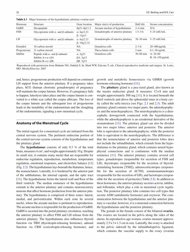
Ovulation happens at the peak of follicular growth in response to an LH surge [ 5 ] . Prior to ovulation, follicles grow to sizes greater than 20 mm in average diameter [ 6 ] . LH is then released in a positive-feedback mechanism from the anterior pituitary due to prolonged exposure to estradiol. For this positive feedback to take place, levels of estradiol above 200 pg/mL for approximately 50 h are necessary [ 7 ] (Fig. 2.1 ). Approximately 12 h after the LH pea, the oocyte is released [ 8, 9 ] . In order for the oocyte to release from the follicle, several proteolytic enzymes and prostaglandins are activated, leading to the digestion of the follicle wall collagen [ 10 ] . Once an oocyte is released, the fallopian tube is responsible for picking it up where it will await fertilization.
The secretory phase starts after ovulation. During this phase, the remaining granulosa cells that are not released with the oocyte during the ovulation process enlarge and acquire lutein (carotenoids), which is yellow in color. These granulosa cells are now called the corpus luteum and predominantly secrete progesterone. Peak progesterone production is noted 1 week after ovulation takes place (see Fig. 2.1 ). Progesterone is required to convert the endometrial lining of the uterus from a proliferative one into a secretory endometrium in preparation for embryo implantation. The life span of the corpus luteum
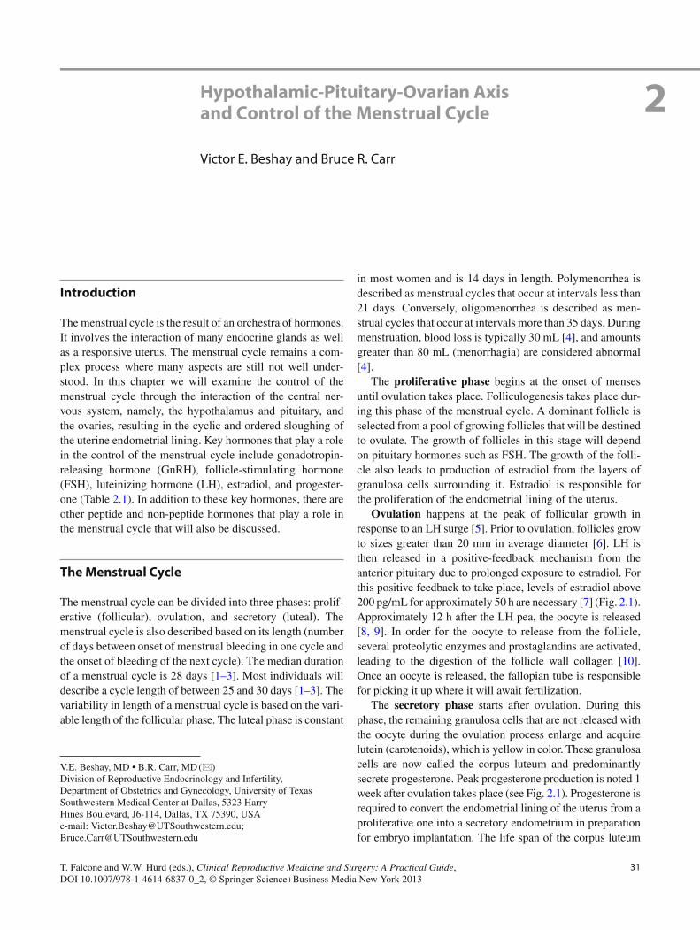
Hypothalamic-Pituitary-Ovarian Axis and Control of the Menstrual Cycle
Victor E. Beshay and Bruce R. Carr
2
V. E. Beshay , MD • B. R. Carr , MD (*) Division of Reproductive Endocrinology and Infertility, Department of Obstetrics and Gynecology , University of Texas Southwestern Medical Center at Dallas , 5323 Harry Hines Boulevard, J6-114 , Dallas , TX 75390 , USA e-mail: [email protected]; [email protected]
32 V.E. Beshay and B.R. Carr
and, hence, progesterone production will depend on continued LH support from the anterior pituitary. If a pregnancy takes place, hCG (human chorionic gonadotropin) of pregnancy will maintain the corpus luteum. However, if a pregnancy fails to happen, luteolysis takes place and the corpus luteum is con-verted to a white scar called the corpus albicans. The loss of the corpus luteum and the subsequent loss of progesterone leads to the instability of the endometrium and the sloughing of the endometrium, signaling a new menstrual cycle.
Anatomy of the Menstrual Cycle
The initial signals for a menstrual cycle are initiated from the central nervous system. The pertinent endocrine portion of the central nervous system consists of the hypothalamus and the pituitary gland.
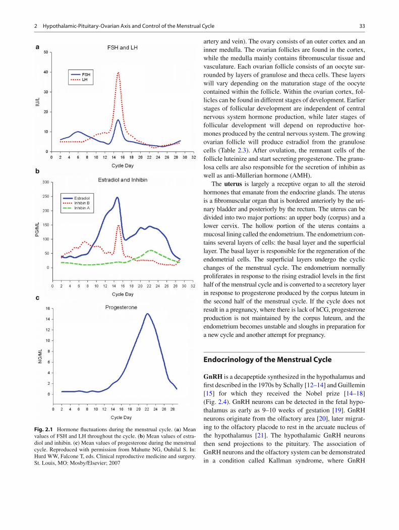
The hypothalamus consists of only 0.3 % of the total brain, measures 4 cm 3 , and weighs approximately 10 g. Despite its small size, it contains many nuclei that are responsible for endocrine regulation, reproduction, metabolism, temperature regulation, emotional responses, and electrolyte balance [ 11 ] (Fig. 2.2 ). The hypothalamus lays beneath the thalamus, hence, the nomenclature. Laterally, it is bordered by the anterior part of the subthalamus, the internal capsule, and the optic tract [ 11 ] . The hypothalamus forms the lateral wall and fl oor of the third ventricle. The median eminence of the hypothalamus extends to the anterior pituitary and contains neurosecretory neurons that affect hormone production from the anterior pitu-itary. The hypothalamus is comprised of three zones: lateral, medial, and periventricular. Within each zone lie several nuclei, where the arcuate nucleus is pertinent to reproduction. The arcuate nucleus is responsible for the production of GnRH. GnRH is secreted into the portal pituitary circulation, reaching the anterior pituitary to affect FSH and LH release from the anterior pituitary. The hypothalamus also in fl uences thyroid function via TRH (thyrotropin-releasing hormone), adrenal function via CRH (coricotropin-releasing hormone), and
growth and metabolic homeostasis via GHRH (growth hormone-releasing hormone) [ 11 ] .
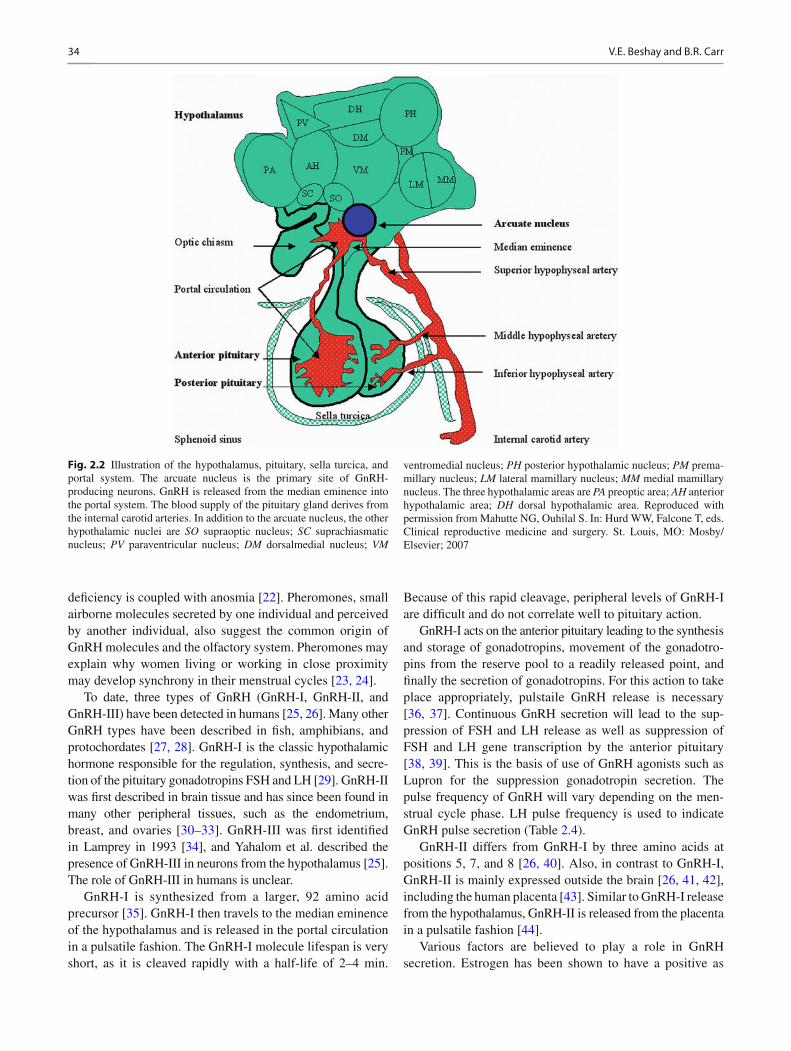
The pituitary gland is a pea-sized gland, also known as the master endocrine gland. It measures 12 × 8 mm and weight approximately 500 mg [ 11 ] . It is located beneath the third ventricle and above the sphenoidal sinus in a bony cav-ity called the sella turcica (see Figs. 2.1 and 2.3 ). The adult pituitary gland contains two major parts: the adenohypophy-sis and the neurohypophysis. The neurohypophysis is a dien-cephalic downgrowth connected with the hypothalamus, while the adenohypophysis is an ectodermal derivative of the stomatodeum [ 11 ] . The pituitary gland can also be divided into two major lobes: anterior and posterior. The anterior lobe is equivalent to the adenohypophysis, while the posterior lobe is equivalent to the neurohypophysis. The difference is that the nomenclature of anterior and posterior lobes does not include the infundibulum, which extends from the hypo-thalamus to the pituitary gland, which contains neural hypo-physial connections and is continuous with the median eminence [ 11 ] . The anterior pituitary contains several cell types: gonadotropes (responsible for secretion of FSH and LH), thyrotropes (responsible for the secretion of thyroid-stimulating hormone [TSH]), adrenocorticotropes (responsi-ble for the secretion of ACTH), somatomammotropes (responsible for the secretion of GH), and lactotropes (respon-sible for the secretion of prolactin) (Table 2.2 ). In addition to these hormones, the anterior pituitary secretes activin, inhibin, and follistatin, which play a role in menstrual cycle regula-tion. The posterior pituitary lobe contains two cell types that secrete ADH (antidiuretic hormone) and oxytocin. The com-munication between the hypothalamus and the anterior pitu-itary is vascular; however, it is a neuronal connection between the hypothalamus and the posterior pituitary.
The gonads in the female consist of the bilateral ovaries . The ovaries are located in the pelvis along the sides of the uterus. In reproductive-age women, ovaries measure approxi-mately 2.5 × 3 × 1.5 cm in size. Laterally, the ovary is attached to the pelvic sidewall by the infundibulopelvic ligament, which contains the vascular supply to the ovary (ovarian
Table 2.1 Major hormones of the hypothalamic-pituitary-ovarian axis a
Hormone Structure Gene location Major site(s) of production Half-life Serum concentration
GnRH Decapeptide 8p21–8p11.2 Arcuate nucleus of hypothalamus 2–4 min N/A FSH Glycoprotein with a - and b -subunits a : 6q12.21
b : 11p13 Gonadotrophs of anterior pituitary 1.5–4 h 5–25 mIU/mL
LH Glycoprotein with a - and b -subunits a : 6q12.21 b : 19q12.32
Gonadotrophs of anterior pituitary 20–30 min 5–25 mIU/mL
Estradiol 18 carbon steroid NA Granulosa cells 2–3 h 20–400 pg/mL Progesterone 21 carbon steroid NA Theca-lutein cells 5 min 0.1–30 ng/mL Inhibin Peptide with a - and b -subunits
Inhibin A = a + b A Inhibin B = a + b B
a : 2q33 b A: 2q13 b B: 7p15
Granulosa cells 30–60 min A: 10–60 B: 10–150 pg/mL
a Reproduced with permission from Mahutte NG, Ouhilal S. In: Hurd WW, Falcone T, eds. Clinical reproductive medicine and surgery. St. Louis, MO: Mosby/Elsevier; 2007
332 Hypothalamic-Pituitary-Ovarian Axis and Control of the Menstrual Cycle
artery and vein). The ovary consists of an outer cortex and an inner medulla. The ovarian follicles are found in the cortex, while the medulla mainly contains fi bromuscular tissue and vasculature. Each ovarian follicle consists of an oocyte sur-rounded by layers of granulose and theca cells. These layers will vary depending on the maturation stage of the oocyte contained within the follicle. Within the ovarian cortex, fol-licles can be found in different stages of development. Earlier stages of follicular development are independent of central nervous system hormone production, while later stages of follicular development will depend on reproductive hor-mones produced by the central nervous system. The growing ovarian follicle will produce estradiol from the granulose cells (Table 2.3 ). After ovulation, the remnant cells of the follicle luteinize and start secreting progesterone. The granu-losa cells are also responsible for the secretion of inhibin as well as anti-Müllerian hormone (AMH).
The uterus is largely a receptive organ to all the steroid hormones that emanate from the endocrine glands. The uterus is a fi bromuscular organ that is bordered anteriorly by the uri-nary bladder and posteriorly by the rectum. The uterus can be divided into two major portions: an upper body (corpus) and a lower cervix. The hollow portion of the uterus contains a mucosal lining called the endometrium. The endometrium con-tains several layers of cells: the basal layer and the super fi cial layer. The basal layer is responsible for the regeneration of the endometrial cells. The super fi cial layers undergo the cyclic changes of the menstrual cycle. The endometrium normally proliferates in response to the rising estradiol levels in the fi rst half of the menstrual cycle and is converted to a secretory layer in response to progesterone produced by the corpus luteum in the second half of the menstrual cycle. If the cycle does not result in a pregnancy, where there is lack of hCG, progesterone production is not maintained by the corpus luteum, and the endometrium becomes unstable and sloughs in preparation for a new cycle and another attempt for pregnancy.
Endocrinology of the Menstrual Cycle
GnRH is a decapeptide synthesized in the hypothalamus and fi rst described in the 1970s by Schally [ 12– 14 ] and Guillemin [ 15 ] for which they received the Nobel prize [ 14– 18 ] (Fig. 2.4 ). GnRH neurons can be detected in the fetal hypo-thalamus as early as 9–10 weeks of gestation [ 19 ] . GnRH neurons originate from the olfactory area [ 20 ] , later migrat-ing to the olfactory placode to rest in the arcuate nucleus of the hypothalamus [ 21 ] . The hypothalamic GnRH neurons then send projections to the pituitary. The association of GnRH neurons and the olfactory system can be demonstrated in a condition called Kallman syndrome, where GnRH
Fig. 2.1 Hormone fl uctuations during the menstrual cycle. ( a ) Mean values of FSH and LH throughout the cycle. ( b ) Mean values of estra-diol and inhibin. ( c ) Mean values of progesterone during the menstrual cycle. Reproduced with permission from Mahutte NG, Ouhilal S. In: Hurd WW, Falcone T, eds. Clinical reproductive medicine and surgery. St. Louis, MO: Mosby/Elsevier; 2007
34 V.E. Beshay and B.R. Carr
de fi ciency is coupled with anosmia [ 22 ] . Pheromones, small airborne molecules secreted by one individual and perceived by another individual, also suggest the common origin of GnRH molecules and the olfactory system. Pheromones may explain why women living or working in close proximity may develop synchrony in their menstrual cycles [ 23, 24 ] .
To date, three types of GnRH (GnRH-I, GnRH-II, and GnRH-III) have been detected in humans [ 25, 26 ] . Many other GnRH types have been described in fi sh, amphibians, and protochordates [ 27, 28 ] . GnRH-I is the classic hypothalamic hormone responsible for the regulation, synthesis, and secre-tion of the pituitary gonadotropins FSH and LH [ 29 ] . GnRH-II was fi rst described in brain tissue and has since been found in many other peripheral tissues, such as the endometrium, breast, and ovaries [ 30– 33 ] . GnRH-III was fi rst identi fi ed in Lamprey in 1993 [ 34 ] , and Yahalom et al. described the presence of GnRH-III in neurons from the hypothalamus [ 25 ] . The role of GnRH-III in humans is unclear.
GnRH-I is synthesized from a larger, 92 amino acid precursor [ 35 ] . GnRH-I then travels to the median eminence of the hypothalamus and is released in the portal circulation in a pulsatile fashion. The GnRH-I molecule lifespan is very short, as it is cleaved rapidly with a half-life of 2–4 min.
Because of this rapid cleavage, peripheral levels of GnRH-I are dif fi cult and do not correlate well to pituitary action.
GnRH-I acts on the anterior pituitary leading to the synthesis and storage of gonadotropins, movement of the gonadotro-pins from the reserve pool to a readily released point, and fi nally the secretion of gonadotropins. For this action to take place appropriately, pulstaile GnRH release is necessary [ 36, 37 ] . Continuous GnRH secretion will lead to the sup-pression of FSH and LH release as well as suppression of FSH and LH gene transcription by the anterior pituitary [ 38, 39 ] . This is the basis of use of GnRH agonists such as Lupron for the suppression gonadotropin secretion. The pulse frequency of GnRH will vary depending on the men-strual cycle phase. LH pulse frequency is used to indicate GnRH pulse secretion (Table 2.4 ).
GnRH-II differs from GnRH-I by three amino acids at positions 5, 7, and 8 [ 26, 40 ] . Also, in contrast to GnRH-I, GnRH-II is mainly expressed outside the brain [ 26, 41, 42 ] , including the human placenta [ 43 ] . Similar to GnRH-I release from the hypothalamus, GnRH-II is released from the placenta in a pulsatile fashion [ 44 ] .
Various factors are believed to play a role in GnRH secretion. Estrogen has been shown to have a positive as
Fig. 2.2 Illustration of the hypothalamus, pituitary, sella turcica, and portal system. The arcuate nucleus is the primary site of GnRH-producing neurons. GnRH is released from the median eminence into the portal system. The blood supply of the pituitary gland derives from the internal carotid arteries. In addition to the arcuate nucleus, the other hypothalamic nuclei are SO supraoptic nucleus; SC suprachiasmatic nucleus; PV paraventricular nucleus; DM dorsalmedial nucleus; VM
ventromedial nucleus; PH posterior hypothalamic nucleus; PM prema-millary nucleus; LM lateral mamillary nucleus; MM medial mamillary nucleus. The three hypothalamic areas are PA preoptic area; AH anterior hypothalamic area; DH dorsal hypothalamic area. Reproduced with permission from Mahutte NG, Ouhilal S. In: Hurd WW, Falcone T, eds. Clinical reproductive medicine and surgery. St. Louis, MO: Mosby/Elsevier; 2007
352 Hypothalamic-Pituitary-Ovarian Axis and Control of the Menstrual Cycle
well as a negative effect on GnRH-I secretion. Estrogen sup-presses GnRH-I secretion in a negative-feedback fashion [ 45 ] . In addition, estrogen has a differential regulation on GnRH-I
and GnRH-II mRNA levels. Estrogen increased GnRH-II mRNA levels while it decreased GnRH-I mRNA levels [ 46 ] . Progesterone is also noted to play a stimulatory role on GnRH-1 mRNA, which was decreased by the progesterone receptor antagonist RU48 [ 47 ] . However, no difference in the expression level of GnRH-II was seen with progester-one or the anti-progestin mifepristone [ 47 ] .
Fig. 2.3 X-ray and T1-weighted MRI images of the pituitary gland. ( a ) Lateral skull fi lm with the sphenoidal sinus and sella turcica. ( b ) Sagittal section demonstrating the relationship between the sphenoidal sinus and the pituitary gland. The normal posterior pituitary is brighter on MRI compared to the anterior pituitary. The sella turcica is not well seen on MRI. ( c ) Coronal section demon-
strating the relationship of the pituitary to the optic chiasm and the pituitary stalk. ( d ) Coronal section after gadolinium contrast, dem-onstrating the close proximity of the pituitary to the internal carotid arteries. Reproduced with permission from Mahutte NG, Ouhilal S. In: Hurd WW, Falcone T, eds. Clinical reproductive medicine and surgery. St. Louis, MO: Mosby/Elsevier; 2007
Table 2.2 Major cell types of the anterior pituitary gland a
Cell type
Appearance on light microscopy
Cellular frequency (%) Hormone products
Somatotrophs Acidophilic 50 Growth hormone Lactotrophs Acidophilic 20 Prolactin Corticotrophs Basophilic 20 Adrenocorticotrophic
hormone (ACTH) Thyrotrophs Basophilic 5 Thyroid-stimulating hormone
(TSH) and free a -subunit Gonadotrophs Basophilic 5 Follicle-stimulating hormone
(FSH), luteinizing hormone (LH) and free a -subunit
a Reproduced with permission from Mahutte NG, Ouhilal S. In: Hurd WW, Falcone T, eds. Clinical reproductive medicine and surgery. St. Louis, MO: Mosby/Elsevier; 2007
Table 2.3 Site of synthesis of major steroidogenic products of the ovary
Cell type Major steroid hormone products
Theca cells Androgens (androstenedione, DHEA, testosterone)
Granulosa cells Estrogens (estradiol, estrone, inhibin, AMH) Theca-lutein cells Progestogens (progesterone,
17-hydroxyprogesterone) Granulosa-lutein cells Estrogens (estradiol, estrone)
36 V.E. Beshay and B.R. Carr
Two types of GnRH receptors have been described in humans: GnRH-I receptor (GnRH-IR) and the GnRH-IIR (GnRH-IIR). The GnRH-IR is a G protein-coupled trans-membrane receptor (GPCR) [ 48, 49 ] . However, the mammalian GnRH-IR lacks the carboxyl-terminal tail [ 49, 50 ] . Activation of the GnRH-IR leads to the activation of phos-pholipase C, which in turn generated the second messengers inositol triphosphate and diacyl glycerol, stimulating protein kinase, cyclic adenosine monophosphate (cAMP), and release [ 51 ] of calcium ions. In addition to the brain, GnRH-IR can be found in the human placenta [ 52, 53 ] , ovarian follicles [ 33, 54 ] , in myometrium and leiomyomata [ 55, 56 ] , as well as human pancreas, liver, heart, skeletal muscle, kidney, placenta, and peripheral blood [ 57– 60 ] . GnRH-IIR is also a GPCR, but unlike the GnRH I-R, it has a C-terminal cytoplas-mic tail [ 61 ] . GnRH-IIR can be found the pituitary, placenta, ovary, uterus, prostate, mature sperm, pancreas, small and large intestines, kidney, and liver [ 26, 33, 62– 64 ] .
GnRH analogues have been developed by changes made to the amino acid sequence of the GnRH molecule. These changes result in the extension of the GnRH half-life as well as its biologic activity. There are two major groups of GnRH analogues: GnRH agonists and GnRH antagonists (Table 2.5 ) In the case of GnRH agonist use, the continuous activation of the GnRH receptor results in desensitization due to a confor-mational change of the receptor, uncoupling from G proteins, internalization of the receptor as well as reduced synthesis of the receptor [ 65, 66 ] . Prior to the desensitization by GnRH agonists, there is an initial fl are where there is increased gonadotropin secretion. Desensitization then takes place 7–14 days later. Unlike GnRH agonists, GnRH antagonists do not cause a fl are effect upon initial administration; instead, GnRH antagonists cause an immediate suppression of gonad-otropin secretion that is rapid and is reversible [ 67 ] . Currently,
GnRH analogues are available in injectable form in the treat-ment of many reproductive conditions, such as precocious puberty, endometriosis, and uterine leiomyomata; they are also being used in in vitro fertilization treatment cycles. Oral forms of GnRH analogues are under investigation. Elagolix is an orally active GnRH antagonist under investigation for use in reproductive conditions [ 68, 69 ] .
GnRH acts on the anterior pituitary to secrete gonadotro-pins: FSH and LH. FSH is a glycoprotein dimer consisting of two subunits: a (alpha)- and b (beta)-subunits. The a -subunit is common in FSH and LH as well as TSH and hCG. The b -subunit is distinct and hormone-speci fi c, which allows the differential function of each hormone. The a -subunit consists of 92 amino acids, while the FSH b -subunit consists of 118 amino acids and fi ve sialic acid residues. Sialic acid residues are responsible for the half-life of the hormone, where the higher the sialic acid content the longer the half-life of that molecule [ 70 ] . FSH has a half-life of several hours. The addition of sialic acid to urinary obtained or recombinant FSH products leads to their longer half-life. The rate-limiting step in gonadotropin production is the availability of b -subunits. In addition to GnRH stimulation of FSH b -subunit synthesis, FSH b -subunit synthesis is dependent on the presence of activin [ 71, 72 ] .
FSH starts to rise a few days prior to the onset of menses and is responsible for the recruitment of a cohort of ovarian follicles as well as a selection of the dominant follicle (see Fig. 2.1 ). FSH induces granulosa cell growth and activates aromatase activity, which converts androgens into estrogens.
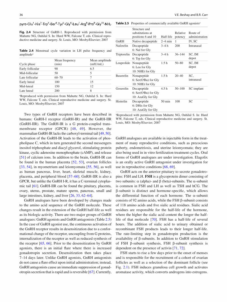
Fig. 2.4 Structure of GnRH-1. Reproduced with permission from Mahutte NG, Ouhilal S. In: Hurd WW, Falcone T, eds. Clinical repro-ductive medicine and surgery. St. Louis, MO: Mosby/Elsevier; 2007
Table 2.4 Menstrual cycle variation in LH pulse frequency and amplitude a
Cycle phase Mean frequency (min)
Mean amplitude (mIU/mL)
Early follicular 90 6.5 Mid-follicular 50 5 Late follicular 60–70 7 Early luteal 100 15 Mid-luteal 150 12 Late luteal 200 8
a Reproduced with permission from Mahutte NG, Ouhilal S. In: Hurd WW, Falcone T, eds. Clinical reproductive medicine and surgery. St. Louis, MO: Mosby/Elsevier; 2007
Table 2.5 Properties of commercially available GnRH agonists a
Structure and substitutions at positions 6 and 10 Half-life
Relative potency
Route of administration
GnRH Native decapeptide 2–4 min 1 IV, SC Naferelin Decapeptide
6: Nal for Gly 3–4 h 200 Intranasal
Triptorelin Decapeptide 6: Trp for Gly
3–4 h 36–144 SC, IM depot
Leuprolide Nonapeptide 6: Leu for Gly 10: NHEt for Gly
1.5 h 50–80 SC, IM depot
Buserelin Nonapeptide 6: Ser(O t Bu) for Gly 10: NHEt for Gly
1.5 h 20–40 SC, intranasal
Goserelin Decapeptide 6: Ser(O t Bu) for Gly 10: AzaGly for Gly
4.5 h 50–100 SC implant
Histrelin Decapeptide 6: DHis for Gly 10: AzaGly for Gly
50 min 100 SC
a Reproduced with permission from Mahutte NG, Ouhilal S. In: Hurd WW, Falcone T, eds. Clinical reproductive medicine and surgery. St. Louis, MO: Mosby/Elsevier; 2007
372 Hypothalamic-Pituitary-Ovarian Axis and Control of the Menstrual Cycle
FSH levels then start to decline owing to estrogen and inhibin B production by the growing follicular granulosa cells. Despite this drop in the FSH level, the dominant follicle continues to grow as it acquires the highest concentration of FSH receptors (secondary to increase in surrounding granu-losa cell number), making it more resistant to the drop in FSH level [ 73 ] . In addition, the drop in FSH level causes a higher androgenic microenvironment in the nondominant follicles. FSH then declines after ovulation of the dominant follicle.
LH is also a glycoprotein dimer consisting of two sub-units: a (alpha)- and b (beta)-subunits. The b -subunit of LH consists of 121 amino acids and one to two sialic acid resi-dues, giving it its shorter half-life of approximately 20 min. Because of this shorter half-life, LH needs to be rapidly syn-thesized and typically has pulses higher in amplitude than FSH. As with FSH, LH also starts to rise prior to the onset of menses. The LH increase throughout the follicular phase of the cycle is gradual. Immediately prior to ovulation, LH surges in response to estradiol production by the dominant follicle in a positive-feedback fashion. LH levels then decline in the secretory phase of the cycle (see Fig. 2.1 ).
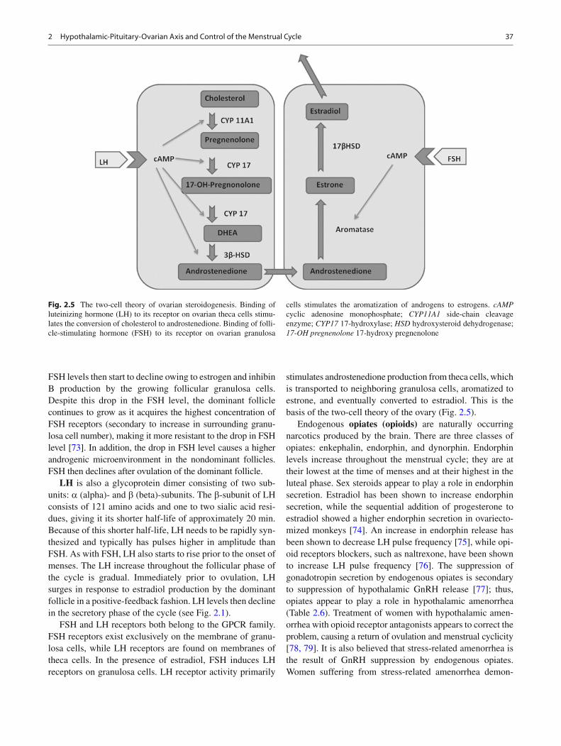
FSH and LH receptors both belong to the GPCR family. FSH receptors exist exclusively on the membrane of granu-losa cells, while LH receptors are found on membranes of theca cells. In the presence of estradiol, FSH induces LH receptors on granulosa cells. LH receptor activity primarily
stimulates androstenedione production from theca cells, which is transported to neighboring granulosa cells, aromatized to estrone, and eventually converted to estradiol. This is the basis of the two-cell theory of the ovary (Fig. 2.5 ).
Endogenous opiates (opioids) are naturally occurring narcotics produced by the brain. There are three classes of opiates: enkephalin, endorphin, and dynorphin. Endorphin levels increase throughout the menstrual cycle; they are at their lowest at the time of menses and at their highest in the luteal phase. Sex steroids appear to play a role in endorphin secretion. Estradiol has been shown to increase endorphin secretion, while the sequential addition of progesterone to estradiol showed a higher endorphin secretion in ovariecto-mized monkeys [ 74 ] . An increase in endorphin release has been shown to decrease LH pulse frequency [ 75 ] , while opi-oid receptors blockers, such as naltrexone, have been shown to increase LH pulse frequency [ 76 ] . The suppression of gonadotropin secretion by endogenous opiates is secondary to suppression of hypothalamic GnRH release [ 77 ] ; thus, opiates appear to play a role in hypothalamic amenorrhea (Table 2.6 ). Treatment of women with hypothalamic amen-orrhea with opioid receptor antagonists appears to correct the problem, causing a return of ovulation and menstrual cyclicity [ 78, 79 ] . It is also believed that stress-related amenorrhea is the result of GnRH suppression by endogenous opiates. Women suffering from stress-related amenorrhea demon-
Fig. 2.5 The two-cell theory of ovarian steroidogenesis. Binding of luteinizing hormone (LH) to its receptor on ovarian theca cells stimu-lates the conversion of cholesterol to androstenedione. Binding of folli-cle-stimulating hormone (FSH) to its receptor on ovarian granulosa
cells stimulates the aromatization of androgens to estrogens. cAMP cyclic adenosine monophosphate; CYP11A1 side-chain cleavage enzyme; CYP17 17-hydroxylase; HSD hydroxysteroid dehydrogenase; 17-OH pregnenolone 17-hydroxy pregnenolone

38 V.E. Beshay and B.R. Carr
strate higher hypothalamic corticotropin-releasing hormone. Proopiomelanocortin, the precursor to endorphins, is controlled mainly by corticotropin-releasing hormone [ 74 ] . In addition, hypothalamic amenorrhea that develops in athletes may also be secondary to opioid rise during exercise [ 80, 81 ] .
Ovarian peptide hormones such as inhibin, activin, and AMH also play a role in the menstrual cycle by modulating central nervous system hormone release. Inhibin, activin, and AMH all belong to the transforming growth factor- b (beta) superfamily (TGF- b ) of ligands.
Inhibin is a polypeptide mainly secreted by granulosa cells, but has also been found in pituitary gonadotropes [ 82, 83 ] . Inhibin is comprised of a a (alpha)- and b (beta)-sub-units. Two forms of inhibin have been identi fi ed: inhibin-A and inhibin-B, each containing an identical a -subunit but a unique b -subunit. Inhibin-A is predominantly secreted in the luteal phase of the menstrual cycle, while inhibin-B is pre-dominantly secreted in the follicular phase of the menstrual cycle [ 84 ] . Inhibin is released by granulosa cells in response to FSH [ 85 ] and selectively inhibits FSH secretion from the anterior pituitary [ 86 ] , thus creating a negative-feedback loop (see Fig. 2.1 ).
In contrast, activin , which is also secreted by the granu-losa cells, augments the secretion of FSH by enhancing GnRH receptor formation [ 87, 88 ] . The effects of activin are blocked by inhibin and follistatin [ 89 ] .
Follistatin is a peptide secreted by pituitary gonadotropes [ 90 ] . Follistatin inhibits FSH synthesis and secretion by sequestering activin [ 91, 92 ] . Inhibin inhibits follistatin pro-duction, while activin stimulates its production.
AMH is a product of the granulosa cells of small antral and pre-antral follicles and is re fl ective of their quantity [ 93 ] . It may be re fl ective of the ovarian reserve which is often a clinical term for the size of the primordial follicle pool. Although the role of AMH has been well described for caus-ing Müllerian duct regression in the male fetus, its role in females in the post-fetal life period has not been well de fi ned. It is believed that AMH, through a paracrine effect in the ovary, inhibits FSH-stimulated follicle growth, contributing to the emergence of the dominant follicle [ 74 ] . The relation-ship among AMH, the follicular pool, and recruitment throughout the reproductive life cycle is complex and is dependent on the stage of sexual development [ 94 ] . Clinically
AMH has been used in the prediction of ovarian reserve in women undergoing fertility evaluation and treatment [ 95 ] . However, the dichotomy of poor reserve vs. normal reserve is not evident [ 95 ] . AMH levels are elevated in patients with polycystic ovary syndrome and decreased in women exposed to antineoplastic drugs.
Leptin is a protein cytokine secreted by adipocytes. It con-sists of 167 amino acids and is secreted by adipose tissue, re fl ecting amounts of body fat [ 96 ] . Leptin’s most signi fi cant role is energy homeostasis. It is regulated by many factors, such as obesity, glucose, and insulin, which promote its secre-tion, whereas fasting, androgens, and thyroid hormone inhibit its secretion. Its role in reproduction is not well understood. As mentioned earlier, CRH is increased in stress-related amenorrhea and is also increased in weight-loss amenorrhea. It is not understood why CRH increases. The reduction in leptin level in these clinical scenarios may play a role in this CRH increase in the brain [ 97 ] . Leptin has also been shown to indirectly affect pituitary FSH and LH secretion in gonado-tropin-stimulated fertility treatment cycles [ 98 ] .
Estrogens are 18-carbon steroid hormones and include estrone (E1), estradiol (E2), and estriol (E3). The most potent estrogen is estradiol and is the product of the ovary. Estrone is mainly the product of peripheral androstenedione conversion. Estrone is also generated in the liver via 17 b (beta)-hydroxysteroid dehydrogenase conversion of estra-diol. Estriol is the principal estrogen formed by the pla-centa during pregnancy. Serum estradiol levels rise during the follicular phase of the menstrual cycle and are in paral-lel to the growth of the follicle. Estradiol is mainly found bound in the bloodstream to carrier proteins. Albumin car-ries approximately 60 % of estradiol, while sex hormone-binding globulin binds 38 % of estradiol, with 2 % remaining as free in the bloodstream. This free hormone is active and capable of entering target cells. In the early follicular phase, serum estradiol levels do not exceed 50 pg/mL. At peak follicular growth, the level rises to approximately 200–250 pg/mL. Estradiol levels drop with ovulation, but a sec-ond rise is seen in the mid-luteal phase, re fl ecting estrogen secretion from the corpus luteum (see Fig. 2.1 ). Circulating estrogens are conjugated in the liver to form sulfates and glucuronides; 80 % are excreted in the urine and the remaining 20 % in bile.
There are two known estrogen receptors: estrogen receptor-alpha (ER- a ) and estrogen receptor-beta (ER- b ) [ 99, 100 ] . Both receptors contain DNA-binding and hormone-binding domains, a hinge region, and a transcriptional activation function (TAF) domain. Estrogen will enter any cell, but only cells containing the estrogen receptor will respond. The receptor is typically nuclear in location, but can be shuttled to the cytoplasm via a process called nucleocytoplasmic shuttling [ 74 ] . Once estrogen binds to its receptor, activation of gene transcription then takes place.
Table 2.6 Neurotransmitter effects on GnRH release a
Neurotransmitter Effect
Dopamine Inhibits GnRH release Endorphin Inhibits GnRH release Serotonin Inhibits GnRH release Norepinephrine, epinephrine Stimulates GnRH release
a Reproduced with permission from Mahutte NG, Ouhilal S. In: Hurd WW, Falcone T, eds. Clinical reproductive medicine and surgery. St. Louis, MO: Mosby/Elsevier; 2007
392 Hypothalamic-Pituitary-Ovarian Axis and Control of the Menstrual Cycle
It is also known that estradiol has a negative-feedback effect on FSH secretion. This negative-feedback effect is the direct effect of estradiol coupled to its receptor, causing repression of FSH- b (beta) subunit transcription [ 101 ] .
Similar to estrogen, progesterone is a steroid hormone. Progesterone is a 21-carbon molecule and is the main steroid of the corpus luteum. In the follicular phase, progesterone levels are typically <2 ng/mL. Progesterone reaches its peak in the mid-luteal phase, with levels exceeding 5 ng/mL (see Fig. 2.1 ). The majority of progesterone in the bloodstream is bound to albumin (80 %) and corticosteroid-binding globu-lin (18 %). A very small amount of progesterone is bound to SHBG (0.5 %). The remaining progesterone is free in the circulation. The liver is responsible for clearing progesterone from the circulation by converting progesterone to pregnane-diol, which is conjugated to glucuronic acid and excreted in the urine.
Similar to estrogen, there are several progesterone recep-tors: progesterone receptor-A (PR-A), progesterone receptor-B (PR-B), and progesterone receptor-C (PR-C). PR-B is the positive regulator of progesterone effects, while PR-A and PR-C antagonize PR-B.
At high concentrations, progesterone inhibits FSH and LH secretion through effects on both the hypothalamus and pituitary [ 102 ] . The presence of progesterone in the luteal phase also causes the decline in GnRH pulse frequency in the hypothalamus. At low concentrations, progesterone can stimulate LH release only after exposure to estrogen and pro-gesterone [ 103 ] . Progesterone also causes a depletion of
estrogen receptors, which is the mechanism of protection against endometrial hyperplasia by progesterone.
Androgens are the major products of theca cells. Androgens are 19-carbon steroids and include: androstene-dione, testosterone, and dehydroepiandrosterone (DHEA). The principal secreted androgen by theca cells is androstene-dione. Most of the testosterone is the product of peripheral conversion of androstenedione through the actions of 17 b -hydroxysteroid dehydrogenase. Under the effect of FSH, androstenedione and testosterone are then further aromatized in granulosa cells and converted to estrogens (Fig. 2.6 ).
The androgen receptor exists in a full-length B form and a shorter A form [ 104 ] . Androgens and progestins can cross-react to their receptor but only when present in high concentration.
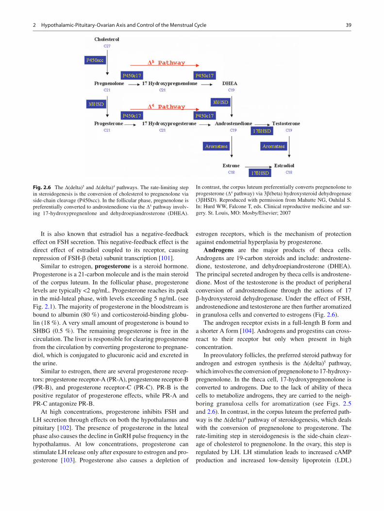
In preovulatory follicles, the preferred steroid pathway for androgen and estrogen synthesis is the D (delta) 5 pathway, which involves the conversion of pregnenolone to 17-hydroxy-pregnenolone. In the theca cell, 17-hydroxypregnonolone is converted to androgens. Due to the lack of ability of theca cells to metabolize androgens, they are carried to the neigh-boring granulosa cells for aromatization (see Figs. 2.5 and 2.6 ). In contrast, in the corpus luteum the preferred path-way is the D (delta) 4 pathway of steroidogenesis, which deals with the conversion of pregnenolone to progesterone. The rate-limiting step in steroidogenesis is the side-chain cleav-age of cholesterol to pregnenolone. In the ovary, this step is regulated by LH. LH stimulation leads to increased cAMP production and increased low-density lipoprotein (LDL)
Fig. 2.6 The D (delta) 5 and D (delta) 4 pathways. The rate-limiting step in steroidogenesis is the conversion of cholesterol to pregnenolone via side-chain cleavage (P450scc). In the follicular phase, pregnenolone is preferentially converted to androstenedione via the D 5 pathway involv-ing 17-hydroxypregnenlone and dehydroepiandrosterone (DHEA).
In contrast, the corpus luteum preferentially converts pregnenolone to progesterone ( D 4 pathway) via 3 b (beta) hydroxysteroid dehydrogenase (3 b HSD). Reproduced with permission from Mahutte NG, Ouhilal S. In: Hurd WW, Falcone T, eds. Clinical reproductive medicine and sur-gery. St. Louis, MO: Mosby/Elsevier; 2007
40 V.E. Beshay and B.R. Carr
receptor mRNA and subsequent increased LDL intake. LDL is the major form of cholesterol used for steroidogenesis. cAMP-activated steroidogenic acute regulatory protein (StAR) causes an increase in the transport of cholesterol across the mitochondrial membrane, where side-chain cleavage can take place [ 105 ] . From there, all the remaining ovarian hormones can be produced.
References
1. Treloar AE, Boynton RE, Behn BG, Brown BW. Variation of the human menstrual cycle through reproductive life. Int J Fertil. 1967;12(1 Pt 2):77–126.
2. Vollman RF. The menstrual cycle. Philadelphia: WB Saunders; 1977.
3. Presser HB. Temporal data relating to the human menstrual cycle. In: Ferin M, Halber F, Richart RM, et al., editors. Biorhythms and human reproduction. New York: Wiley; 1974. p. 145–60.
4. Hallberg L, Hogdahl AM, Nilsson L, Rybo G. Menstrual blood loss–a population study. Variation at different ages and attempts to de fi ne normality. Acta Obstet Gynecol Scand. 1966;45(3):320–51.
5. Cahill DJ, Wardle PG, Harlow CR, Hull MG. Onset of the preovu-latory luteinizing hormone surge: diurnal timing and critical fol-licular prerequisites. Fertil Steril. 1998;70(1):56–9.
6. Strauss J, Williams CJ. Neuroendocrine control of the menstrual cycle. 6th ed. Philadelphia: Saunders Elsevier; 2009.
7. Young JR, Jaffe RB. Strength-duration characteristics of estrogen effects on gonadotropin response to gonadotropin-releasing hor-mone in women. II. Effects of varying concentrations of estra-diol. J Clin Endocrinol Metab. 1976;42(3):432–42.
8. Pauerstein CJ, Eddy CA, Croxatto HD, Hess R, Siler-Khodr TM, Croxatto HB. Temporal relationships of estrogen, progesterone, and luteinizing hormone levels to ovulation in women and infra-human primates. Am J Obstet Gynecol. 1978;130(8):876–86.
9. Temporal relationships between ovulation and de fi ned changes in the concentration of plasma estradiol-17 beta, luteinizing hor-mone, follicle-stimulating hormone, and progesterone. I. Probit analysis. World Health Organization, Task Force on Methods for the Determination of the Fertile Period, Special Programme of Research, Development and Research Training in Human Reproduction. Am J Obstet Gynecol. 1980;138(4):383–90.
10. Espey LL, Lipner H. Ovulation. In: Knobil E, Neill JD, editors. The physiology of reproduction. New York: Raven; 1994.
11. Mancall EL, Brock DG, editors. Gray’s clinical neuroanatomy: the anatomic basis for clinical neuroscience. 1st ed. Philadelphia: Elsevier Saunders; 2011.
12. Baba Y, Matsuo H, Schally AV. Structure of the porcine LH- and FSH-releasing hormone. II. Con fi rmation of the proposed struc-ture by conventional sequential analyses. Biochem Biophys Res Commun. 1971;44(2):459–63.
13. Matsuo H, Baba Y, Nair RM, Arimura A, Schally AV. Structure of the porcine LH- and FSH-releasing hormone. I. The proposed amino acid sequence. Biochem Biophys Res Commun. 1971;43(6):1334–9.
14. Schally AV, Arimura A, Baba Y, Nair RM, Matsuo H, Redding TW, et al. Isolation and properties of the FSH and LH-releasing hormone. Biochem Biophys Res Commun. 1971;43(2):393–9.
15. Guillemin R. Chemistry and physiology of hypothalamic releas-ing factors for gonadotrophins. Int J Fertil. 1967;12(4):359–67.
16. Schally AV, Arimura A, Kastin AJ, Matsuo H, Baba Y, Redding TW, et al. Gonadotropin-releasing hormone: one polypeptide reg-ulates secretion of luteinizing and follicle-stimulating hormones. Science. 1971;173(4001):1036–8.
17. Arimura A, Matsuo H, Baba Y, Schally AV. Ovulation induced by synthetic luteinizing hormone-releasing hormone in the hamster. Science. 1971;174(4008):511–2.
18. Amoss M, Burgus R, Blackwell R, Vale W, Fellows R, Guillemin R. Puri fi cation, amino acid composition and N-terminus of the hypothalamic luteinizing hormone releasing factor (LRF) of ovine origin. Biochem Biophys Res Commun. 1971;44(1):205–10.
19. Grumbach M, Kaplan S. The neuroendocrinology of human puberty: an ontogenetic perspective. In: Grumbach M, Sizonenko P, Aubert M, editors. Control of the onset of puberty. Baltimore: Williams & Wilkins; 1990.
20. Schwanzel-Fukuda M, Pfaff DW. Origin of luteinizing hormone-releasing hormone neurons. Nature. 1989;338(6211):161–4.
21. Silverman AJ, Jhamandas J, Renaud LP. Localization of luteiniz-ing hormone-releasing hormone (LHRH) neurons that project to the median eminence. J Neurosci. 1987;7(8):2312–9.
22. Spratt DI, Carr DB, Merriam GR, Scully RE, Rao PN, Crowley Jr WF. The spectrum of abnormal patterns of gonadotropin-releasing hormone secretion in men with idiopathic hypogonadotropic hypogonadism: clinical and laboratory correlations. J Clin Endocrinol Metab. 1987;64(2):283–91.
23. McClintock MK. Menstrual synchorony and suppression. Nature. 1971;229(5282):244–5.
24. Stern K, McClintock MK. Regulation of ovulation by human pheromones. Nature. 1998;392(6672):177–9.
25. Yahalom D, Chen A, Ben-Aroya N, Rahimipour S, Kaganovsky E, Okon E, et al. The gonadotropin-releasing hormone family of neuropeptides in the brain of human, bovine and rat: identi fi cation of a third isoform. FEBS Lett. 1999;463(3):289–94.
26. White RB, Eisen JA, Kasten TL, Fernald RD. Second gene for gonadotropin-releasing hormone in humans. Proc Natl Acad Sci USA. 1998;95(1):305–9.
27. Morgan K, Millar RP. Evolution of GnRH ligand precursors and GnRH receptors in protochordate and vertebrate species. Gen Comp Endocrinol. 2004;139(3):191–7.
28. Millar RP, Lu ZL, Pawson AJ, Flanagan CA, Morgan K, Maudsley SR. Gonadotropin-releasing hormone receptors. Endocr Rev. 2004;25(2):235–75.
29. Yao B, Liu HY, Gu YC, Shi SS, Tao XQ, Li XJ, et al. Gonadotropin-releasing hormone positively regulates steroidogenesis via extra-cellular signal-regulated kinase in rat Leydig cells. Asian J Androl. 2011;13(3):438–45.
30. Cheon KW, Lee HS, Parhar IS, Kang IS. Expression of the second isoform of gonadotrophin-releasing hormone (GnRH-II) in human endometrium throughout the menstrual cycle. Mol Hum Reprod. 2001;7(5):447–52.
31. Fister S, Gunthert AR, Aicher B, Paulini KW, Emons G, Grundker C. GnRH-II antagonists induce apoptosis in human endometrial, ovarian, and breast cancer cells via activation of stress-induced MAPKs p38 and JNK and proapoptotic protein Bax. Cancer Res. 2009;69(16):6473–81.
32. Leung PC, Cheng CK, Zhu XM. Multi-factorial role of GnRH-I and GnRH-II in the human ovary. Mol Cell Endocrinol. 2003;202(1–2):145–53.
33. Choi JH, Gilks CB, Auersperg N, Leung PC. Immunolocalization of gonadotropin-releasing hormone (GnRH)-I, GnRH-II, and type I GnRH receptor during follicular development in the human ovary. J Clin Endocrinol Metab. 2006;91(11):4562–70.
34. Sower SA, Chiang YC, Lovas S, Conlon JM. Primary structure and biological activity of a third gonadotropin-releasing hormone from lamprey brain. Endocrinology. 1993;132(3):1125–31.
35. Nikolics K, Mason AJ, Szonyi E, Ramachandran J, Seeburg PH. A prolactin-inhibiting factor within the precursor for human gonadotropin-releasing hormone. Nature. 1985;316(6028):511–7.
36. Knobil E. The neuroendocrine control of the menstrual cycle. Recent Prog Horm Res. 1980;36:53–88.
412 Hypothalamic-Pituitary-Ovarian Axis and Control of the Menstrual Cycle
37. Mais V, Kazer RR, Cetel NS, Rivier J, Vale W, Yen SS. The dependency of folliculogenesis and corpus luteum function on pulsatile gonadotropin secretion in cycling women using a gonadotropin-releasing hormone antagonist as a probe. J Clin Endocrinol Metab. 1986;62(6):1250–5.
38. Belchetz PE, Plant TM, Nakai Y, Keogh EJ, Knobil E. Hypophysial responses to continuous and intermittent delivery of hypoptha-lamic gonadotropin-releasing hormone. Science. 1978;202(4368):631–3.
39. Haisenleder DJ, Dalkin AC, Ortolano GA, Marshall JC, Shupnik MA. A pulsatile gonadotropin-releasing hormone stimulus is required to increase transcription of the gonadotropin subunit genes: evidence for differential regulation of transcription by pulse frequency in vivo. Endocrinology. 1991;128(1):509–17.
40. Millar RP. GnRH II, and type II GnRH receptors. Trends Endocrinol Metab. 2003;14(1):35–43.
41. Serin IS, Tanriverdi F, Ata CD, Akalin H, Ozcelik B, Ozkul Y, et al. GnRH-II mRNA expression in tumor tissue and peripheral blood mononuclear cells (PBMCs) in patients with malignant and benign ovarian tumors. Eur J Obstet Gynecol Reprod Biol. 2010;149(1):92–6.
42. Poon SL, Klausen C, Hammond GL, Leung PC. 37-kDa laminin receptor precursor mediates GnRH-II-induced MMP-2 expression and invasiveness in ovarian cancer cells. Mol Endocrinol. 2011;25(2):327–38.
43. Chou CS, Beristain AG, MacCalman CD, Leung PC. Cellular localization of gonadotropin-releasing hormone (GnRH) I and GnRH II in fi rst-trimester human placenta and decidua. J Clin Endocrinol Metab. 2004;89(3):1459–66.
44. Siler-Khodr TM, Grayson M. Action of chicken II GnRH on the human placenta. J Clin Endocrinol Metab. 2001;86(2):804–10.
45. Messinis IE, Vanakara P, Zavos A, Verikouki C, Georgoulias P, Dafopoulos K. Failure of the GnRH antagonist ganirelix to block the positive feedback effect of exogenous estrogen in normal women. Fertil Steril. 2010;94(4):1554–6.
46. Chen A, Ganor Y, Rahimipour S, Ben-Aroya N, Koch Y, Levite M. The neuropeptides GnRH-II and GnRH-I are produced by human T cells and trigger laminin receptor gene expression, adhesion, chemot-axis and homing to speci fi c organs. Nat Med. 2002;8(12):1421–6.
47. An BS, Choi JH, Choi KC, Leung PC. Differential role of proges-terone receptor isoforms in the transcriptional regulation of human gonadotropin-releasing hormone I (GnRH I) receptor, GnRH I, and GnRH II. J Clin Endocrinol Metab. 2005;90(2):1106–13.
48. Cui J, Smith RG, Mount GR, Lo JL, Yu J, Walsh TF, et al. Identi fi cation of Phe313 of the gonadotropin-releasing hormone (GnRH) receptor as a site critical for the binding of nonpeptide GnRH antagonists. Mol Endocrinol. 2000;14(5):671–81.
49. Sealfon SC, Weinstein H, Millar RP. Molecular mechanisms of ligand interaction with the gonadotropin-releasing hormone recep-tor. Endocr Rev. 1997;18(2):180–205.
50. Stojilkovic SS, Reinhart J, Catt KJ. Gonadotropin-releasing hor-mone receptors: structure and signal transduction pathways. Endocr Rev. 1994;15(4):462–99.
51. Shacham S, Harris D, Ben-Shlomo H, Cohen I, Bon fi l D, Przedecki F, et al. Mechanism of GnRH receptor signaling on gonadotropin release and gene expression in pituitary gonadotrophs. Vitam Horm. 2001;63:63–90.
52. Lin LS, Roberts VJ, Yen SS. Expression of human gonadotropin-releasing hormone receptor gene in the placenta and its functional relationship to human chorionic gonadotropin secretion. J Clin Endocrinol Metab. 1995;80(2):580–5.
53. Wolfahrt S, Kleine B, Jarry H, Rossmanith WG. Endogenous reg-ulation of the GnRH receptor by GnRH in the human placenta. Mol Hum Reprod. 2001;7(1):89–95.
54. Bramley TA, Stirling D, Swanston IA, Menzies GS, McNeilly AS, Baird DT. Speci fi c binding sites for gonadotrophin-releasing hor-mone, LH/chorionic gonadotrophin, low-density lipoprotein, pro-
lactin and FSH in homogenates of human corpus luteum. II: concentrations throughout the luteal phase of the menstrual cycle and early pregnancy. J Endocrinol. 1987;113(2):317–27.
55. Reshkin S, Albarani V, Pezzetta A, Marinaccio M, Paradiso A. Gonadotrophin releasing hormone (GnRH) receptor and steroid receptors in human uterine leiomyoma, myometrium and endome-trium. Int J Oncol. 1997;11(3):603–7.
56. Kobayashi Y, Zhai YL, Iinuma M, Horiuchi A, Nikaido T, Fujii S. Effects of a GnRH analogue on human smooth muscle cells cultured from normal myometrial and from uterine leiomyomal tissues. Mol Hum Reprod. 1997;3(2):91–9.
57. Chen HF, Jeung EB, Stephenson M, Leung PC. Human peripheral blood mononuclear cells express gonadotropin-releasing hormone (GnRH), GnRH receptor, and interleukin-2 receptor gamma-chain messenger ribonucleic acids that are regulated by GnRH in vitro. J Clin Endocrinol Metab. 1999;84(2):743–50.
58. Kakar SS, Jennes L. Expression of gonadotropin-releasing hormone and gonadotropin-releasing hormone receptor mRNAs in various non-reproductive human tissues. Cancer Lett. 1995;98(1):57–62.
59. Cheung LW, Wong AS. Gonadotropin-releasing hormone: GnRH receptor signaling in extrapituitary tissues. FEBS J. 2008;275(22):5479–95.
60. Fekete M, Zalatnai A, Comaru-Schally AM, Schally AV. Membrane receptors for peptides in experimental and human pancreatic cancers. Pancreas. 1989;4(5):521–8.
61. Neill JD, Duck LW, Sellers JC, Musgrove LC. A gonadotropin-releasing hormone (GnRH) receptor speci fi c for GnRH II in pri-mates. Biochem Biophys Res Commun. 2001;282(4):1012–8.
62. Neill JD. GnRH and GnRH receptor genes in the human genome. Endocrinology. 2002;143(3):737–43.
63. Eicke N, Gunthert AR, Viereck V, Siebold D, Behe M, Becker T, et al. GnRH-II receptor-like antigenicity in human placenta and in cancers of the human reproductive organs. Eur J Endocrinol. 2005;153(4):605–12.
64. van Biljon W, Wykes S, Scherer S, Krawetz SA, Hapgood J. Type II gonadotropin-releasing hormone receptor transcripts in human sperm. Biol Reprod. 2002;67(6):1741–9.
65. Suarez-Quian CA, Wynn PC, Catt KJ. Receptor-mediated endocy-tosis of GnRH analogs: differential processing of gold-labeled ago-nist and antagonist derivatives. J Steroid Biochem. 1986;24(1):183–92.
66. Schvartz I, Hazum E. Internalization and recycling of receptor-bound gonadotropin-releasing hormone agonist in pituitary gonadotropes. J Biol Chem. 1987;262(35):17046–50.
67. Blockeel C, Sterrenburg MD, Broekmans FJ, Eijkemans MJ, Smitz J, Devroey P, et al. Follicular phase endocrine characteris-tics during ovarian stimulation and GnRH antagonist cotreatment for IVF: RCT comparing recFSH initiated on cycle day 2 or 5. J Clin Endocrinol Metab. 2011;96(4):1122–8.
68. Struthers RS, Nicholls AJ, Grundy J, Chen T, Jimenez R, Yen SS, et al. Suppression of gonadotropins and estradiol in premeno-pausal women by oral administration of the nonpeptide gonado-tropin-releasing hormone antagonist elagolix. J Clin Endocrinol Metab. 2009;94(2):545–51.
69. Chen C, Wu D, Guo Z, Xie Q, Reinhart GJ, Madan A, et al. Discovery of sodium R-(+)-4-{2-[5-(2- fl uoro-3-methoxyphenyl)-3-(2- fl uoro-6-[tri fl uoromethyl]be nzyl)-4-methyl-2,6-dioxo-3,6-dihydro-2H-pyrimidin-1-yl]-1-phenylethylamino}butyrate (elagolix), a potent and orally available nonpeptide antagonist of the human gonadotropin-releasing hormone receptor. J Med Chem. 2008;51(23):7478–85.
70. Morell AG, Gregoriadis G, Scheinberg IH, Hickman J, Ashwell G. The role of sialic acid in determining the survival of glycopro-teins in the circulation. J Biol Chem. 1971;246(5):1461–7.
71. Weiss J, Guendner MJ, Halvorson LM, Jameson JL. Transcriptional activation of the follicle-stimulating hormone beta-subunit gene by activin. Endocrinology. 1995;136(5):1885–91.
42 V.E. Beshay and B.R. Carr
72. Besecke LM, Guendner MJ, Schneyer AL, Bauer-Dantoin AC, Jameson JL, Weiss J. Gonadotropin-releasing hormone regulates follicle-stimulating hormone-beta gene expression through an activin/follistatin autocrine or paracrine loop. Endocrinology. 1996;137(9):3667–73.
73. Amsterdam A, Rotmensch S. Structure-function relationships during granulosa cell differentiation. Endocr Rev. 1987;8(3):309–37.
74. Fritz MA, Speroff L, editors. Clinical gynecologic endocrinology and infertility. Philadelphia: Lippincott Williams and Wilkins; 2005.
75. Rabinovici J, Rothman P, Monroe SE, Nerenberg C, Jaffe RB. Endocrine effects and pharmacokinetic characteristics of a potent new gonadotropin-releasing hormone antagonist (Ganirelix) with minimal histamine-releasing properties: studies in postmeno-pausal women. J Clin Endocrinol Metab. 1992;75(5):1220–5.
76. Evans WS, Weltman JY, Johnson ML, Weltman A, Veldhuis JD, Rogol AD. Effects of opioid receptor blockade on luteinizing hor-mone (LH) pulses and interpulse LH concentrations in normal women during the early phase of the menstrual cycle. J Endocrinol Invest. 1992;15(7):525–31.
77. Goodman RL, Par fi tt DB, Evans NP, Dahl GE, Karsch FJ. Endogenous opioid peptides control the amplitude and shape of gonadotropin-releasing hormone pulses in the ewe. Endocrinology. 1995;136(6):2412–20.
78. Wildt L, Sir-Petermann T, Leyendecker G, Waibel-Treber S, Rabenbauer B. Opiate antagonist treatment of ovarian failure. Hum Reprod. 1993;8 Suppl 2Suppl 2:168–74.
79. Wildt L, Leyendecker G, Sir-Petermann T, Waibel-Treber S. Treatment with naltrexone in hypothalamic ovarian failure: induc-tion of ovulation and pregnancy. Hum Reprod. 1993;8(3):350–8.
80. De Cree C. Endogenous opioid peptides in the control of the nor-mal menstrual cycle and their possible role in athletic menstrual irregularities. Obstet Gynecol Surv. 1989;44(10):720–32.
81. Harber VJ, Sutton JR. Endorphins and exercise. Sports Med. 1984;1(2):154–71.
82. Bauer-Dantoin AC, Weiss J, Jameson JL. Roles of estrogen, pro-gesterone, and gonadotropin-releasing hormone (GnRH) in the control of pituitary GnRH receptor gene expression at the time of the preovulatory gonadotropin surges. Endocrinology. 1995;136(3):1014–9.
83. Blumenfeld Z. Response of human fetal pituitary cells to activin, inhibin, hypophysiotropic and neuroregulatory factors in vitro. Early Pregnancy. 2001;5(1):41–2.
84. Groome NP, Illingworth PJ, O’Brien M, Pai R, Rodger FE, Mather JP, et al. Measurement of dimeric inhibin B throughout the human menstrual cycle. J Clin Endocrinol Metab. 1996;81(4):1401–5.
85. Bicsak TA, Tucker EM, Cappel S, Vaughan J, Rivier J, Vale W, et al. Hormonal regulation of granulosa cell inhibin biosynthesis. Endocrinology. 1986;119(6):2711–9.
86. Rivier C, Rivier J, Vale W. Inhibin-mediated feedback control of follicle-stimulating hormone secretion in the female rat. Science. 1986;234(4773):205–8.
87. Kaiser UB, Conn PM, Chin WW. Studies of gonadotropin-releas-ing hormone (GnRH) action using GnRH receptor-expressing pituitary cell lines. Endocr Rev. 1997;18(1):46–70.
88. Norwitz ER, Xu S, Jeong KH, Bedecarrats GY, Winebrenner LD, Chin WW, et al. Activin A augments GnRH-mediated transcrip-tional activation of the mouse GnRH receptor gene. Endocrinology. 2002;143(3):985–97.
89. Bilezikjian LM, Corrigan AZ, Blount AL, Vale WW. Pituitary follistatin and inhibin subunit messenger ribonucleic acid levels are differentially regulated by local and hormonal factors. Endocrinology. 1996;137(10):4277–84.
90. Kaiser UB, Lee BL, Carroll RS, Unabia G, Chin WW, Childs GV. Follistatin gene expression in the pituitary: localization in gonado-tropes and folliculostellate cells in diestrous rats. Endocrinology. 1992;130(5):3048–56.
91. Kogawa K, Nakamura T, Sugino K, Takio K, Titani K, Sugino H. Activin-binding protein is present in pituitary. Endocrinology. 1991;128(3):1434–40.
92. Besecke LM, Guendner MJ, Sluss PA, Polak AG, Woodruff TK, Jameson JL, et al. Pituitary follistatin regulates activin-mediated production of follicle-stimulating hormone during the rat estrous cycle. Endocrinology. 1997;138(7):2841–8.
93. Durlinger AL, Gruijters MJ, Kramer P, Karels B, Ingraham HA, Nachtigal MW, et al. Anti-Mullerian hormone inhibits initiation of primordial follicle growth in the mouse ovary. Endocrinology. 2002;143(3):1076–84.
94. Fleming R, Kelsey TW, Anderson RA, Wallace WH, Nelson SM. Interpreting human follicular recruitment and antimullerian hor-mone concencentraions throughout life. Fertil Steril. 2012;98:1097–102.
95. Anderson RA, Nelson SM, Wallace WH. Measuring anti-Mülle-rian hormone for the assessment of ovarian reserve: when and for whom is it indicated? Maturitas. 2012;71(1):28–33.
96. Considine RV, Sinha MK, Heiman ML, Kriauciunas A, Stephens TW, Nyce MR, et al. Serum immunoreactive-leptin concentra-tions in normal-weight and obese humans. N Engl J Med. 1996;334(5):292–5.
97. Kelesidis T, Kelesidis I, Chou S, Mantzoros CS. Narrative review: the role of leptin in human physiology: emerging clinical applica-tions. Ann Intern Med. 2010;152(2):93–100.
98. Brannian JD, Hansen KA. Leptin and ovarian folliculogenesis: implications for ovulation induction and ART outcomes. Semin Reprod Med. 2002;20(2):103–12.
99. Kuiper GG, Enmark E, Pelto-Huikko M, Nilsson S, Gustafsson JA. Cloning of a novel receptor expressed in rat prostate and ovary. Proc Natl Acad Sci USA. 1996;93(12):5925–30.
100. Mosselman S, Polman J, Dijkema R. ER beta: identi fi cation and characterization of a novel human estrogen receptor. FEBS Lett. 1996;392(1):49–53.
101. Miller CD, Miller WL. Transcriptional repression of the ovine follicle-stimulating hormone-beta gene by 17 beta-estradiol. Endocrinology. 1996;137(8):3437–46.
102. Wildt L, Hutchison JS, Marshall G, Pohl CR, Knobil E. On the site of action of progesterone in the blockade of the estradiol-induced gonadotropin discharge in the rhesus monkey. Endocrinology. 1981;109(4):1293–4.
103. Liu JH, Yen SS. Induction of midcycle gonadotropin surge by ovarian steroids in women: a critical evaluation. J Clin Endocrinol Metab. 1983;57(4):797–802.
104. Wilson CM, McPhaul MJ. A and B forms of the androgen receptor are present in human genital skin fi broblasts. Proc Natl Acad Sci USA. 1994;91(4):1234–8.
105. Clark BJ, Soo SC, Caron KM, Ikeda Y, Parker KL, Stocco DM. Hormonal and developmental regulation of the steroido-genic acute regulatory protein. Mol Endocrinol. 1995;9(10):1346–55.

http://www.springer.com/978-1-4614-6836-3