Embed Size (px)
Citation preview
Hypokalemia
CR, 51 y/o woman Na+ 139 K+ 2.7 Cl- 97 CO2 33 Creat1.0
SC, 33 y/o woman Na+ 138 K+ 3.1 Cl- 98 CO2 27 Creat0.8

Hypokalemia can only occur for four reasons: Decreased intake Shift into cells Extra-renal losses Renal losses

Causes of hypokalemia
Decreased intake: kidney can conserve to 5-25 mEq K+ daily; normal intake 40-120 daily.
Shift into cells: Alkalosis Insulin Beta adrenergic stimuli
Stress Beta agonists- e.g.: albuterol, ritodrine

Increased potassium entry into cells: Hypokalemic periodic paralysis- typically
oriental men with thyrotoxicosis; ? abnormal Ca++ channel; ? Increased Na/K atp ase activity.
Increased rbc uptake, e.g. after treatment with B12, folate.

Extra-renal losses of potassium:

Gastrointestinal losses of potassium Gastric juice contains 5 – 10 mEq K+/L. Intestinal fluids contain 20 – 50 mEq/L
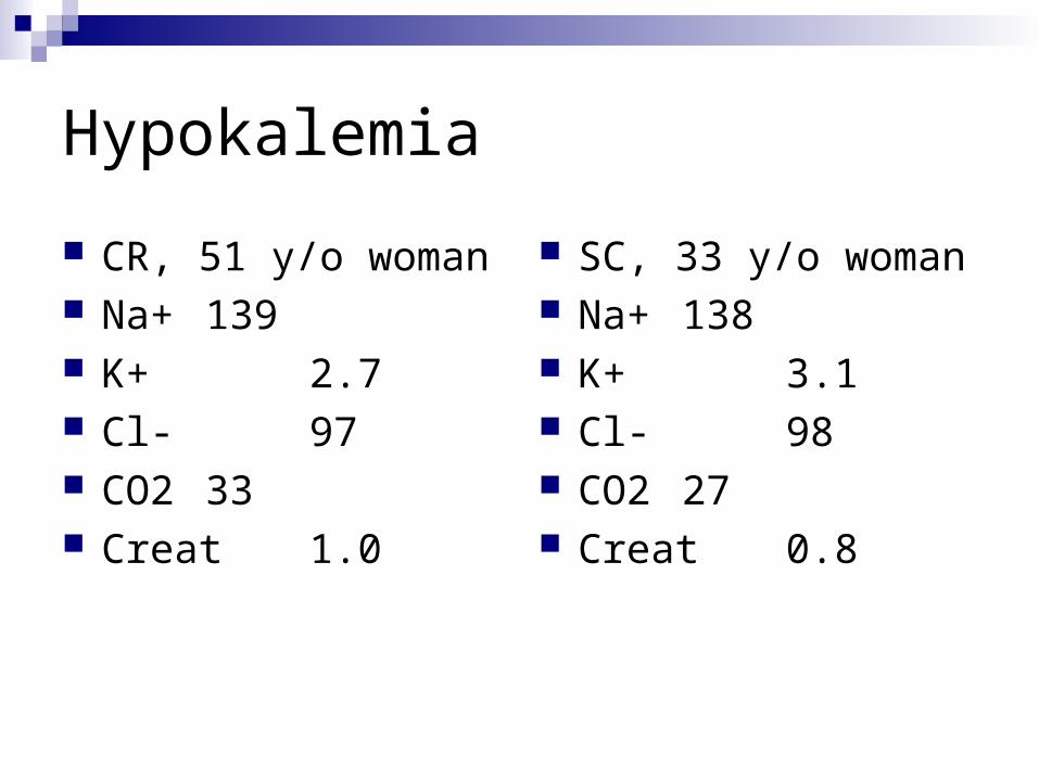
Hypokalemia from loss of gastric fluid. Loss of hydrogen ion increases plasma bicarbonate.
Coexisting volume depletion increases aldosterone
secretion. Increased delivery of bicarbonate to the distal
nephron obligates a cation. In the setting of increased aldosterone levels, sodium is retained and potassium excreted.
Potassium loss is most prominent early. Actual losses in gastric juice are relatively small.

Diarrheal losses are usually accompanied by metabolic acidosisVillous adenomaLaxative abuse

Sweat losses- 5 – 10 mEq/L

The kidney and potassium
Nearly all potassium filtered at the glomerulus is reabsorbed in the proximal nephron. Urinary potassium is the result of distal potassium secretion.
To excrete potassium, the kidney requires an adequate number of nephrons, aldosterone, and a circulation adequate to provide adequate distal delivery of sodium for sodium/potassium exchange.

Renal losses of potassium
Diuretics- activate the renin-angiotensin-aldosterone cascade.
Primary aldosteronism/increased steroids. Presentation of a non-resorbable anion distally,
obligating a cation, which will lead to increased potassium excretion in the presence of aldosterone. Bicarbonate Penicillin derivatives Betahydroxybutyrate
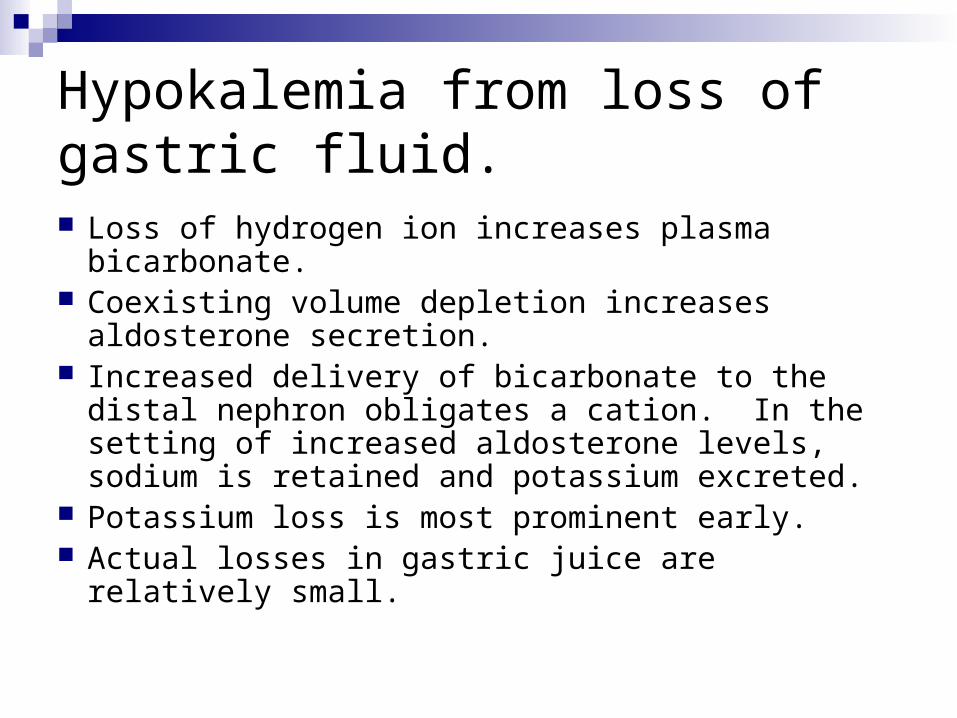
Renal losses of potassium
Renal tubular acidosis Proximal, especially with therapy Some distal types Type IV RTA patients are typically
hyperkalemic
Hypomagnesemia Polyuria
What data do we want to diagnose the cause of hypokalemia in these women? Urinary potassium: 24 hour values better
than spot specimens. Aldosterone and renin levels. Blood pressure measurements. A history.

Both women had urinary potassium levels that were elevated.
Both women had elevated renin and aldosterone levels.
Both women had low normal blood pressures
Therefore:
Potassium is being lost in the urine. Primary aldosteronism is r/o by normal
blood pressures. Electrolytes r/o renal tubular acidosis. Diuretic abuse?
C.R.
Admitted with repeated episodes of hypokalemia, many year history of chronic edema, treated periodically with lasix, zaroxolyn, and aldactone. Now vehemently denies diuretic use.
Subsequent urine positive for furosemide.
S.C.
Low potassium noted during initial lab drawn with pregnancy. Only medication is synthroid. No nausea, vomiting, diarrhea.
140 mEq KCl required daily to maintain K+ in the 3.5 range.
Other possibilities?
Bartter’s and Gitelman’s syndromes
Bartter’s syndrome is usually diagnosed in childhood, sometimes associated with growth and mental retardation. The defect is impaired NaCl reabsorption in the loop of Henle. Findings are similar to administration of a loop acting diuretic: Salt loss leading to volume depletion and activation of
the renin-angiotensin system Increased urinary calcium

Bartter’s and Gitelman’s Syndromes 3 or 4 types of Bartter’s have been identified:
• Defects in the luminal Na-K-Cl transporter• Defects in the luminal potassium channel• Defects in the basolateral chloride channel
I’ve never seen a patient with Bartter’s syndrome, although many have been referred.

Gitelman’s syndrome
Like Bartter’s an autosomal recessive disorder, but not usually diagnosed early in life.
Findings mimic administration of a thiazide diuretic: the defect is in the Na-Cl transporter.
Patients may complain of polyuria, cramps. They do not have hypercalciuria, but typically
have low serum magnesium levels.

Gitelman’s syndrome
Diagnosis is made by history as well as lab findings. Lab findings are indistinguishable from thiazide use:Hypokalemia, hypomagnesemia, increased
renin and aldosterone levels, decreased urinary calcium.
Genetic screening?

©2005 UpToDate® • www.uptodate.com • Contact Us

1: Clin Nephrol. 2001 Mar;55(3):233-7.Related Articles, Links
Mimicry of surreptitious diuretic ingestion and the ability to make a genetic diagnosis.
Schepkens H, Hoeben H, Vanholder R, Lameire N.
Department of Internal Medicine, University Hospital Gent, Belgium. [email protected]
Gitelman's syndrome, also known as "hypocalciuric variant of Bartter's syndrome", is a cause of chronic hypokalemia and hypomagnesemia in adults. A specific gene has been found responsible for this disorder, encoding the thiazide-sensitive NaCl coporter (TSC) in the distal convoluted tubule. We describe a psychiatric patient with chronic symptomatic hypokalemia and hypomagnesemia whose electrolyte disturbances were subsequently misdiagnosed as an acute alcohol and benzodiazepine withdrawal syndrome, as chronic diuretic abuse and as a classical Bartter's syndrome. Finally, genetic investigation revealed the presence of mutations in the SLC12A3 gene leading to the proper diagnosis of Gitelman's syndrome. We emphasize that Gitelman's syndrome should be suspected in every hypokalemic patient with biochemical resemblance of diuretic ingestion, especially when repeated toxic screens for diuretics are negative. The ability to make a molecular-genetic diagnosis can be of practical benefit in confusing clinical settings.

Gitelman’s syndrome: treatment
Potassium Magnesium Aldactone or amiloride ACEI’s NSAIDS of no benefit

S.C.
Continues to require very large doses of KCl, and is on amiloride.
Magnesium levels have consistently been low or low normal.

General comments about the treatment of hypokalemia Think about the cause of the hypokalemia
you are treating? A cellular shift, e.g. hypokalemic periodic paralysis, will require a lot less potassium to correct than hypokalemia from potassium loss.
Orally or i.v.? Orally is safer; limit i.v. repletion to 20 mEq./hour except in very unusual circumstances- then monitor.
Anticipate: has K+ loss stopped or will it be ongoing?
Are you giving other drugs that will influence K+ levels? E.g. NSAIDs, ACEIs, ARBs.
Generally, use KCl vs. other preparations. Followup with repeat levels- consider using the
replacement protocols.