Embed Size (px)
Citation preview

fphar-07-00288 September 2, 2016 Time: 17:32 # 1
ORIGINAL RESEARCHpublished: 05 September 2016doi: 10.3389/fphar.2016.00288
Edited by:Judith Maria Rollinger,
University of Vienna, Austria
Reviewed by:Mark Klitgaard Nøhr,
Aarhus University, DenmarkAdolfo Andrade-Cetto,
National Autonomous Universityof Mexico, Mexico
*Correspondence:Xinzhou Yang
†These authors have contributedequally to this work.
Specialty section:This article was submitted to
Ethnopharmacology,a section of the journal
Frontiers in Pharmacology
Received: 08 June 2016Accepted: 18 August 2016
Published: 05 September 2016
Citation:Huang M, Deng S, Han Q, Zhao P,
Zhou Q, Zheng S, Ma X, Xu C,Yang J and Yang X (2016)
Hypoglycemic Activityand the Potential Mechanism of the
Flavonoid Rich Extract from Sophoratonkinensis Gagnep. in KK-Ay Mice.
Front. Pharmacol. 7:288.doi: 10.3389/fphar.2016.00288
Hypoglycemic Activity and thePotential Mechanism of theFlavonoid Rich Extract from Sophoratonkinensis Gagnep. in KK-Ay MiceMi Huang1†, Shihao Deng1†, Qianqian Han1, Ping Zhao1, Qi Zhou1, Sijian Zheng1,Xinhua Ma1, Chan Xu1, Jing Yang1 and Xinzhou Yang1,2,3*
1 School of Pharmaceutical Sciences, South-Central University for Nationalities, Wuhan, China, 2 State Key Laboratory ofDrug Research, Shanghai Institute of Materia Medica, Chinese Academy of Sciences, Shanghai, China, 3 College ofBiological Engineering, Tianjin University of Science and Technology, Tianjin, China
This study investigated the active principles, hypoglycemic activity and potentialmechanisms of the flavonoid rich extract from Sophora tonkinensis Gagnep. (ST-EtOAc)in KK-Ay diabetic mice. An off-line semipreparative liquid chromatography-nuclearmagnetic resonance (LC-NMR) and liquid chromatography-ultraviolet-electrosprayionization mass spectrometry (LC-UV–ESIMS) protocol was performed to determine 13flavonoids from ST-EtOAc. ST-EtOAc administrated orally to the KK-Ay mice significantlyincreased their sensibility to insulin, reduced fasting blood-glucose levels and blood lipidindexes such as triglyceride and cholesterol. Moreover, ST-EtOAc exhibited a strongeffect of stimulation on glucose transporter 4 (GLUT4) translocation by 2.7-fold in L6cells. However, the selective AMP-activated protein kinase (AMPK) inhibitor compoundC can completely inhibit the activation of the AMPK pathway and prevent the GLUT4translocation caused by ST-EtOAc. In vivo, phosphorylation of the AMPK expressionin the liver and skeletal muscle was measured. The results showed phosphorylationof the AMPK had been improved and GLUT4 expression had been also enhanced. Inthis paper, we conclude that, ST-EtOAc seems to have potential beneficial effects onthe treatment of type 2 diabetes mellitus with the probable mechanism of stimulatingGLUT4 translocation modulated by the AMPK pathway.
Keywords: hypoglycemic agents, Sophora tonkinensis Gagnep., KK-Ay mice, GLUT4, p-AMPK
INTRODUCTION
Type 2 diabetes mellitus (T2DM), a metabolic disorder of the endocrine system characterizedby abnormal glucose and lipid metabolism, is caused by insulin resistance and relative insulindeficiency (Shulman, 2000). Because insulin resistance is the main metabolic abnormality ofT2DM, there has been considerable interest in insulin-sensitizing agents for the treatment of thisdisease (Moller, 2001; Ju et al., 2014). One of the most appealing targets for drug development is theinsulin-responsive glucose transporter 4 (GLUT4), which is vital to glucose homeostasis (Bryantet al., 2002). Increasing evidence suggests that enhanced translocation of GLUT4 can improveinsulin resistance of T2DM. Therefore, this protein may lead to the discovery of the next generationof anti-diabetic drugs (Zhang et al., 2007; Tsuchiya et al., 2015).
Frontiers in Pharmacology | www.frontiersin.org 1 September 2016 | Volume 7 | Article 288
brought to you by COREView metadata, citation and similar papers at core.ac.uk
provided by Frontiers - Publisher Connector

fphar-07-00288 September 2, 2016 Time: 17:32 # 2
Huang et al. An Oral Hypoglycemic Agent
Many traditional Chinese medicines (TCMs), which containmany active compounds targeting GLUT4 proteins, have revealedbeneficial hypoglycemic effects in vitro and in vivo, such as Coptischinensis (Turner et al., 2008), Momordica charantia (Tan et al.,2008), and Gardenia jasminoides (Zhang et al., 2006), etc. Inorder to identify potential hypoglycemic agents to fight T2DM,we developed a L6 cell-based GLUT4 translocation system withco-expressing recombinant GLUT4 and insulin regulation ofaminopeptidase (IRAP) using a confocal imaging technique toscreen the extracts or fractions from natural products (Wanget al., 2014; Yang X.Z. et al., 2014; Yang et al., 2015). Duringthe screening of a TCMs extract library (800 biotas) on GLUT4translocation, we have found that a flavonoid rich extractof Sophora tonkinensis Gagnep. (ST-EtOAc) had a potentiallybeneficial effect on GLUT4 translocation.
Sophora tonkinensis Gagnep. belongs to the leguminousfamily, and has been widely used as a traditional medicinein China (Tan et al., 2009). Clinically, it is mainly used totreat acute and chronic pharyngitis, tonsillitis, and chronichepatitis (Sheng et al., 2010). However, the hypoglycemic effect ofS. tonkinensis Gagnep. has not been reported until now. The aimof this investigation was to determine the active principles, andevaluate the potential hypoglycemic effects of ST-EtOAc in vitroand in vivo, and therefore elucidate its probable hypoglycemicmechanism.
MATERIALS AND METHODS
Instruments and ReagentsLiquid chromatography-photo-diode array-electrosprayionization mass spectrometry (LC-PDA-ESIMS) data wererecorded on a Waters ACQUITY SQD MS system (Waters,Milford, MA, USA) connected to a Waters 1525 highperformance liquid chromatography (HPLC) with a 2998Photodiode Array Detector (Waters, Milford, MA, USA) and aWaters SunfireTM C18 column (5 µm, 4.6 × 150 mm) (Waters,Ireland). The nuclear magnetic resonance (NMR) spectra wererecorded in dimethyl sulfoxide (DMSO)-d6 on an spectrometer(AVANCE III 500 MHz) equipped with 1.4 mm heavy wall MicroNMR tubes (NORELL, Landisville, NJ, USA). All the otheranalytical-reagent grade chemicals were used with no furtherpurification, and HPLC-grade solvents were obtained fromMerck Chemical Company (Darmstadt, Germany). The NMR(high-Resolution solution: MeOH-d4 or DMSO-d6) spectrawere kept a record in on an AVANCE III 500 MHz spectrometerequipped with Micro NMR tubes (1.4 mm). Typically, 1H spectraand 1H-1H COSY spectra were separated obtained at the scanrange of 32–128 and 4–32. The heteronuclear multiple bondcorrelation (HMBC) and the heteronuclear single quantumcoherence (HSQC) spectra were recorded at the scan range of16–96.
LC–PDA–ESIMS MethodAnalysis was performed on a C18 column (Waters Sunfire, 5 µm,4.6 × 150 mm). 0.1% formic acid added in Water (A) andacetonitrile (B) was used as mobile phases. The gradient elution
was used for analysis and the program was as follows: 0–20 min,10–100% B; 20–24 min, 100% B. The flow rate of the analysis was1.0 mL/min. UV-Vis spectra were obtained with the wavelengthrange of 200–500 nm with 10 scans per second. The splittedeluent was at a ratio of 1:5 before the mass spectrometer. Bothpositive and negative ion modes were used for ESIMS recording.The capillary voltage was 4000 V, the capillary exit voltage was140.0 V, and the skimmer voltage was 40 V. The nebulizer gaspressure was set to 40 psi, the dry gas flow to 10.0 L/min andthe dry temperature to 320◦C. Mass range was set from 120 to1500 m/z. Data acquisition and processing were achieved withMassLynxTM 4.0 software (Waters, Milford, MA, USA).
Plant Material and Preparation ofST-EtOAcThe roots of S. tonkinensis Gagnep. were collected fromJingxi county, Guangxi Zhuang Autonomous Region, China inJune 2013. And it was identified by Professor Jingquan Yuan(Guangxi Medicinal Botanical Garden, Nanning, China). Theplant voucher specimen was preserved as No. SC0060 anddeposited in College of Pharmacy, South-Central Universityfor Nationalities. Air-dried roots of S. tonkinensis Gagnep.(500 g) were smashed and then extracted sequentially at roomtemperature with n-hexane (4× 2.0 L, 3 h each), followed by ethylacetate (4 × 2.0 L, 3 h each) and methanol (4 × 2.0 L, 3 h each).The solvents were vacuum evaporated to yield n-hexane extract(4.9 g), ethyl acetate extract (ST-EtOAc, 36.8 g), and methanolextract (58.7 g), respectively.
Chemical CharacterizationSeparation of ST-EtOAc was carried by the procedures asfollows: 0.5 g of ST-EtOAc was dissolved in 2.0 mL of DMSOso that a concentration of ST-EtOAc was 250 mg/mL, andthen the solution was filtered. With the same solvent systemto that of LC–PDA–ESIMS, the optimized gradient programused as follows: 0–25 min, 15–100% B; 25–30 min, 100% B.The flow rate was set at 5.0 mL/min and the 200 µL injectedvolume was setup. Ten injections were carried out, and 13 peak-based fractions were collected manually. And combined thecorresponding fractions. Final purification were performed asfollows: peaks 1–6, 10, 12, and 13 were filtered by a SephadexLH-20 column (400 mm × 10 mm, MeOH containing 0.1%formic acid) to yield pure compounds 1 (2.9 mg), 2 (2.1 mg), 3(3.8 mg), 4 (3.5 mg), 5 (1.9 mg), 6 (23.3 mg), 10 (37.6 mg), 12(3.2 mg), and 13 (7.1 mg). Peak 7 was purified by preparativethin layer chromatography (TLC) (hexane:acetone:formic acid—100:10:0.5) to yield compound 7 (1.2 mg). Peak 8 was purifiedby preparative TLC (hexane:acetone:formic acid—100:10:0.4)to give compound 8 (1.8 mg). Peak 9 was purified bypreparative TLC (hexane:acetone:formic acid—100:8:0.4) to givecompound 9 (2.8 mg). Peak 11 was purified by preparativeTLC (hexane:acetone:formic acid—100:8:0.4) to give compound11 (1.4 mg). Compounds 1–13 with amounts 0.8–3.0 mg weredissolved in DMSO-d6 or MeOH-d4 for NMR tests on aspectrometer (AVANCE III 500 MHz) which was equipped with1.4 mm heavy wall Micro NMR tubes.
Frontiers in Pharmacology | www.frontiersin.org 2 September 2016 | Volume 7 | Article 288
fphar-07-00288 September 2, 2016 Time: 17:32 # 3
Huang et al. An Oral Hypoglycemic Agent
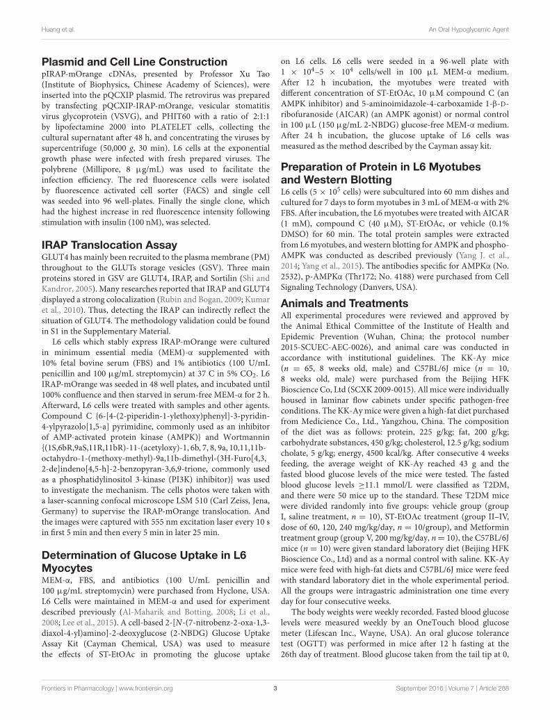
Plasmid and Cell Line ConstructionpIRAP-mOrange cDNAs, presented by Professor Xu Tao(Institute of Biophysics, Chinese Academy of Sciences), wereinserted into the pQCXIP plasmid. The retrovirus was preparedby transfecting pQCXIP-IRAP-mOrange, vesicular stomatitisvirus glycoprotein (VSVG), and PHIT60 with a ratio of 2:1:1by lipofectamine 2000 into PLATELET cells, collecting thecultural supernatant after 48 h, and concentrating the viruses bysupercentrifuge (50,000 g, 30 min). L6 cells at the exponentialgrowth phase were infected with fresh prepared viruses. Thepolybrene (Millipore, 8 µg/mL) was used to facilitate theinfection efficiency. The red fluorescence cells were isolatedby fluorescence activated cell sorter (FACS) and single cellwas seeded into 96 well-plates. Finally the single clone, whichhad the highest increase in red fluorescence intensity followingstimulation with insulin (100 nM), was selected.
IRAP Translocation AssayGLUT4 has mainly been recruited to the plasma membrane (PM)throughout to the GLUTs storage vesicles (GSV). Three mainproteins stored in GSV are GLUT4, IRAP, and Sortilin (Shi andKandror, 2005). Many researches reported that IRAP and GLUT4displayed a strong colocalization (Rubin and Bogan, 2009; Kumaret al., 2010). Thus, detecting the IRAP can indirectly reflect thesituation of GLUT4. The methodology validation could be foundin S1 in the Supplementary Material.
L6 cells which stably express IRAP-mOrange were culturedin minimum essential media (MEM)-α supplemented with10% fetal bovine serum (FBS) and 1% antibiotics (100 U/mLpenicillin and 100 µg/mL streptomycin) at 37 C in 5% CO2. L6IRAP-mOrange was seeded in 48 well plates, and incubated until100% confluence and then starved in serum-free MEM-α for 2 h.Afterward, L6 cells were treated with samples and other agents.Compound C {6-[4-(2-piperidin-1-ylethoxy)phenyl]-3-pyridin-4-ylpyrazolo[1,5-a] pyrimidine, commonly used as an inhibitorof AMP-activated protein kinase (AMPK)} and Wortmannin{(1S,6bR,9aS,11R,11bR)-11-(acetyloxy)-1, 6b, 7, 8, 9a, 10,11,11b-octahydro-1-(methoxy-methyl)-9a,11b-dimethyl-(3H-Furo[4,3,2-de]indeno[4,5-h]-2-benzopyran-3,6,9-trione, commonly usedas a phosphatidylinositol 3-kinase (PI3K) inhibitor)} was usedto investigate the mechanism. The cells photos were taken witha laser-scanning confocal microscope LSM 510 (Carl Zeiss, Jena,Germany) to supervise the IRAP-mOrange translocation. Andthe images were captured with 555 nm excitation laser every 10 sin first 5 min and then every 5 min in later 25 min.
Determination of Glucose Uptake in L6MyocytesMEM-α, FBS, and antibiotics (100 U/mL penicillin and100 µg/mL streptomycin) were purchased from Hyclone, USA.L6 Cells were maintained in MEM-α and used for experimentdescribed previously (Al-Maharik and Botting, 2008; Li et al.,2008; Lee et al., 2015). A cell-based 2-[N-(7-nitrobenz-2-oxa-1,3-diaxol-4-yl)amino]-2-deoxyglucose (2-NBDG) Glucose UptakeAssay Kit (Cayman Chemical, USA) was used to measurethe effects of ST-EtOAc in promoting the glucose uptake
on L6 cells. L6 cells were seeded in a 96-well plate with1 × 104–5 × 10⁴ cells/well in 100 µL MEM-α medium.After 12 h incubation, the myotubes were treated withdifferent concentration of ST-EtOAc, 10 µM compound C (anAMPK inhibitor) and 5-aminoimidazole-4-carboxamide 1-β-D-ribofuranoside (AICAR) (an AMPK agonist) or normal controlin 100 µL (150 µg/mL 2-NBDG) glucose-free MEM-α medium.After 24 h incubation, the glucose uptake of L6 cells wasmeasured as the method described by the Cayman assay kit.
Preparation of Protein in L6 Myotubesand Western BlottingL6 cells (5 × 10⁵ cells) were subcultured into 60 mm dishes andcultured for 7 days to form myotubes in 3 mL of MEM-α with 2%FBS. After incubation, the L6 myotubes were treated with AICAR(1 mM), compound C (40 µM), ST-EtOAc, or vehicle (0.1%DMSO) for 60 min. The total protein samples were extractedfrom L6 myotubes, and western blotting for AMPK and phospho-AMPK was conducted as described previously (Yang J. et al.,2014; Yang et al., 2015). The antibodies specific for AMPKα (No.2532), p-AMPKα (Thr172; No. 4188) were purchased from CellSignaling Technology (Danvers, USA).
Animals and TreatmentsAll experimental procedures were reviewed and approved bythe Animal Ethical Committee of the Institute of Health andEpidemic Prevention (Wuhan, China; the protocol number2015-SCUEC-AEC-0026), and animal care was conducted inaccordance with institutional guidelines. The KK-Ay mice(n = 65, 8 weeks old, male) and C57BL/6J mice (n = 10,8 weeks old, male) were purchased from the Beijing HFKBioscience Co, Ltd (SCXK 2009-0015). All mice were individuallyhoused in laminar flow cabinets under specific pathogen-freeconditions. The KK-Ay mice were given a high-fat diet purchasedfrom Medicience Co., Ltd., Yangzhou, China. The compositionof the diet was as follows: protein, 225 g/kg; fat, 200 g/kg;carbohydrate substances, 450 g/kg; cholesterol, 12.5 g/kg; sodiumcholate, 5 g/kg; energy, 4500 kcal/kg. After consecutive 4 weeksfeeding, the average weight of KK-Ay reached 43 g and thefasted blood glucose levels of the mice were tested. The fastedblood glucose levels ≥11.1 mmol/L were classified as T2DM,and there were 50 mice up to the standard. These T2DM micewere divided randomly into five groups: vehicle group (groupI, saline treatment, n = 10), ST-EtOAc treatment (group II–IV,dose of 60, 120, 240 mg/kg/day, n = 10/group), and Metformintreatment group (group V, 200 mg/kg/day, n= 10), the C57BL/6Jmice (n = 10) were given standard laboratory diet (Beijing HFKBioscience Co., Ltd) and as a normal control with saline. KK-Aymice were feed with high-fat diets and C57BL/6J mice were feedwith standard laboratory diet in the whole experimental period.All the groups were intragastric administration one time everyday for four consecutive weeks.
The body weights were weekly recorded. Fasted blood glucoselevels were measured weekly by an OneTouch blood glucosemeter (Lifescan Inc., Wayne, USA). An oral glucose tolerancetest (OGTT) was performed in mice after 12 h fasting at the26th day of treatment. Blood glucose taken from the tail tip at 0,
Frontiers in Pharmacology | www.frontiersin.org 3 September 2016 | Volume 7 | Article 288
fphar-07-00288 September 2, 2016 Time: 17:32 # 4
Huang et al. An Oral Hypoglycemic Agent
30, 60, and 120 min after glucose administration was measuredusing a blood glucose meter (OneTouch Ultra R©, Lifescan Inc.,Wayne, USA). The glucose load was 2.0 g/kg orally. At the endof the experiment, the mice were fasted for 12 h. Blood sampleswere collected by retro-orbital sinus puncture using capillarytubes under diethyl ether anesthesia. Then, the blood sampleswere centrifuging at 3000 rpm for continues 15 min so that theserum of each sample was obtained. At the same time, the micewere euthanized by cervical dislocation. And the livers, skeletalmuscles, and other tissues were harvested. Parts of livers andpancreas were immediately stored in liquid nitrogen tank, andthe rest parts were stored in 10% neutral buffered formalin to befixed.
Liver and Pancreas Histological AnalysisLivers and pancreas fixed in formalin were embedded inparaffin, and then they were cut into 5 µm-thick sections andstained with standard hematoxylin–eosin (HE). The stainedtissues were prepared for histopathological examinations, andpathological changes of the lesion and its vicinity in liverand pancreas were observed and photographed through anoptical microscope photographed (200×). The average isletsize (in µm2), was determined with software (program StereoInvestigator, Williston, VT, USA).
Biochemical Analyses of Serum andTissuesThe serum levels of insulin, total cholesterol (TC), triglycerides(TG), low density lipoprotein cholesterol (LDL-C), and highdensity lipoprotein cholesterol (HDL-C) were determined byautomatic biochemical analyzer (Hitachi 7180+ISE, Tokyo,Japan). Free fatty acid (FFA) was determined by correspondingassay kits (Jiancheng Bioengineering Institute, Nanjing, China).The tissue TC, TG, and FFA in mice were determinedaccording to the method described previously (Lee et al.,2006).
Tissue Extracts and Western BlottingThe total protein was extracted from skeletal muscle and liveraccording to the methods described previously (Yang et al., 2015).And the supernatant protein concentration was determined withbicinchoninic acid (BCA) assay kit (Abgent, San Diego, USA).An equivalent amount of samples were mounted on 10% sodiumdodecyl sulfate (SDS)-polyacrylamide gel electrophoresis. Theprotein transferred electrophoretically to polyvinylidene fluoridemembrane (Pall Corporation, Washington, USA) and incubatedovernight at 4◦C with antibodies specific for GLUT4 (No.2213), AMPKα (No. 2532), p-AMPKα (Thr172; No. 4188;Cell Signaling Technology, Danvers, USA). The membraneswere washed three times (10 min/wash) in tris buffered salinetween (TBST). Immune complexes were incubated with aperoxidase-conjugated antibody for 1 h. The blots were incubatedin enhanced chemiluminescence kits (Amersham-Pharmacia,Piscataway, NJ, USA). And the immunoreactive signals wereimaged and quantified with the Gel Image system (Aplegen Inc.,Pleasanton, USA).
Acute Toxicity StudyKung Ming (KM) mice were derived in 1944 from a pair ofSwiss mice that had been introduced from Hoffline Institutionof Hindustan into Kunming of China (Shang et al., 2009).Kunming mice are the most commonly used outbred mouseline in China. This type of mice shows strong disease resistanceand adaptability, high breeding coefficient and survival rate(Shang et al., 2009). So, KM mice have been widely utilizedin pharmacological, toxicological, medicinal, and biologicalresearch and testing.
KM mice [No. SCXK (E) 2008-0005] weighing between18 and 22 g were served as acute toxicity test. The animalswere housed environmentally controlled conditions at 25◦C,60% relative humidity, where 12 h dark–light cycles weremaintained with food and water. In the main study, the drug(ST-EtOAc dissolved in saline) was administered orally given to10 male mice and 10 female mice at doses of a single dose of(6500 mg/kg/day). KM mice were observed for clinical symptomsfor 15 days. At the end of the experiments, all the animals wereeuthanized followed and gross pathological examinations wereundertaken.
Statistical AnalysisData was shown as means ± standard error of the mean.One-way analysis of variance (ANOVA) was used for multiplegroup comparisons by Tukey’s post hoc test using GraphPadPrism 5.0 software package. P-values <0.05 were consideredsignificant.
RESULTS
Chemical Characterization of ST-EtOAcA LC–PDA–ESIMS experiment was carried out for chemicalprofiling of ST-EtOAc based on the standard operation procedureshown in Section “LC–PDA–ESIMS Method” (Figure 1A). Thesemipreparative HPLC chromatogram was used for large scalepreparative isolation of target compounds with the optimizedgradient conditions (Figure 1B). Thirteen peaks were collectedand continue to further purified with Sephadex LH-20 columnand preparative TLC to acquire 13 compounds for further 1D and2D NMR spectroscopic analysis. The two major principles weredetermined as maackiain (6; Lee et al., 2015) and sophoranone(10; Li et al., 2008). The remaining compounds (Figure 1C)were identified as genistin (1; Al-Maharik and Botting, 2008),trifolirhizin (2; Yang X.Z. et al., 2014), ononin (3; Yang X.Z.et al., 2014), trifolirhizin 6′-monoacetate (4; Yang X.Z. et al.,2014), quercetin (5; Yi et al., 2002), lespeflorin B4 (7; Mori-Hongo et al., 2009), dehydrolupinifolinol (8; Sutthivaiyakitet al., 2009), glabrol (9; Cho et al., 2012), euchrenone a2 (11;Mizuno et al., 1988), 6,8-diprenylkaempferol (12; Meragelmanet al., 2001), and sophoranochromene (13; Li et al., 2008) bycomparison with their mass spectrum (MS), UV, and NMRspectra with published reference data. Spectroscopic data ofcompounds 1–13 could be found in S2 in SupplementaryMaterial.
Frontiers in Pharmacology | www.frontiersin.org 4 September 2016 | Volume 7 | Article 288

fphar-07-00288 September 2, 2016 Time: 17:32 # 5
Huang et al. An Oral Hypoglycemic Agent
FIGURE 1 | (A) The liquid chromatography-photo-diode array-electrospray ionization mass spectrometry (LC-PDA-ESIMS) analysis of ST-EtOAc is shown at 280 nmwith peak labeling corresponding to compounds 1–13. (B) The optimized semipreparative high performance liquid chromatography (HPLC) separation of theST-EtOAc (50 mg in 200 µL dimethyl sulfoxide (DMSO)) is shown at 280 nm with peaks 1–13 collected for microprobe nuclear magnetic resonance (NMR) andfurther purified if required. (C) Structures of flavonoids from ST-EtOAc.
Frontiers in Pharmacology | www.frontiersin.org 5 September 2016 | Volume 7 | Article 288
fphar-07-00288 September 2, 2016 Time: 17:32 # 6
Huang et al. An Oral Hypoglycemic Agent
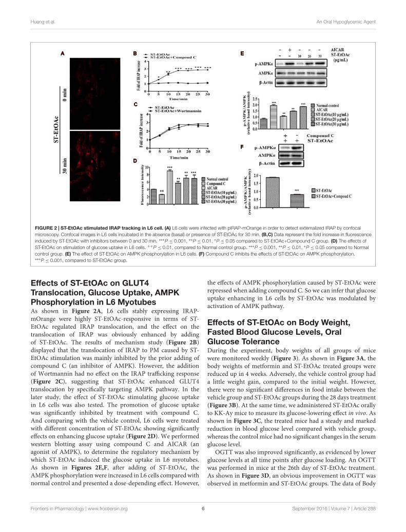
FIGURE 2 | ST-EtOAc stimulated IRAP tracking in L6 cell. (A) L6 cells were infected with pIRAP-mOrange in order to detect externalized IRAP by confocalmicroscopy. Confocal images in L6 cells incubated in the absence (basal) or presence of ST-EtOAc for 30 min. (B,C) Data represent the fold increase in fluorescenceinduced by ST-EtOAc with inhibitors between 0 and 30 min. ∗∗∗P ≤ 0.001, ∗∗P ≤ 0.01, ∗P ≤ 0.05 compared to ST-EtOAc+Compound C group. (D) The effects ofST-EtOAc on stimulation of glucose uptake in L6 cells. ++P ≤ 0.01, compared to Normal control group. ∗∗∗P ≤ 0.001, ∗∗P ≤ 0.01, ∗P ≤ 0.05 compared to Normalcontrol group. (E) The effect of ST-EtOAc on AMPK phosphorylation in L6 cells. (F) Compound C inhibits the effects of ST-EtOAc on AMPK phosphorylation.∗∗∗P ≤ 0.001, compared to ST-EtOAc group.
Effects of ST-EtOAc on GLUT4Translocation, Glucose Uptake, AMPKPhosphorylation in L6 MyotubesAs shown in Figure 2A, L6 cells stably expressing IRAP-mOrange were highly ST-EtOAc-responsive in terms of ST-EtOAc regulated IRAP translocation, and the effect on thetranslocation of IRAP was obviously enhanced by addingof ST-EtOAc. The results of mechanism study (Figure 2B)displayed that the translocation of IRAP to PM caused by ST-EtOAc stimulation was mainly inhibited by the prior adding ofcompound C (an inhibitor of AMPK). However, the additionof Wortmannin had no effect on the IRAP trafficking response(Figure 2C), suggesting that ST-EtOAc enhanced GLUT4translocation by specifically targeting AMPK pathway. In thelater study, the effect of ST-EtOAc stimulating glucose uptakein L6 cells was also tested. The promotion of glucose uptakewas significantly inhibited by treatment with compound C.And comparing with the vehicle control, L6 cells were treatedwith different concentration of ST-EtOAc showing significantlyeffects on enhancing glucose uptake (Figure 2D). We performedwestern blotting assay using compound C and AICAR (anagonist of AMPK), to determine the regulatory mechanism bywhich ST-EtOAc induced the glucose uptake in L6 myotubes.As shown in Figures 2E,F, after adding of ST-EtOAc, theAMPK phosphorylation were increased in L6 cells compared withnormal control and presented a dose-depending effect. However,
the effects of AMPK phosphorylation caused by ST-EtOAc wererepressed when adding compound C. So we can infer that glucoseuptake enhancing in L6 cells by ST-EtOAc was modulated byactivation of AMPK pathway.
Effects of ST-EtOAc on Body Weight,Fasted Blood Glucose Levels, OralGlucose ToleranceDuring the experiment, body weights of all groups of micewere monitored weekly (Figure 3). As shown in Figure 3A, thebody weights of metformin and ST-EtOAc treated groups werereduced up in 4 weeks. Adversely, the vehicle control group hada little weight gain, compared to the initial weight. However,there were no significant differences in food intake between thevehicle group and ST-EtOAc groups during the 28 days treatment(Figure 3B). At the same time, we administered ST-EtOAc orallyto KK-Ay mice to measure its glucose-lowering effect in vivo. Asshown in Figure 3C, the treated mice had a steady and markedreduction in blood glucose level compared with vehicle group,whereas the control mice had no significant changes in the serumglucose level.
OGTT was also improved significantly, as evidenced by lowerglucose levels at all time points after glucose loading. An OGTTwas performed in mice at the 26th day of ST-EtOAc treatment.As shown in Figure 3D, an obvious improvement in OGTT wasobserved in metformin and ST-EtOAc groups. The data of Body
Frontiers in Pharmacology | www.frontiersin.org 6 September 2016 | Volume 7 | Article 288

fphar-07-00288 September 2, 2016 Time: 17:32 # 7
Huang et al. An Oral Hypoglycemic Agent
FIGURE 3 | Effects of ST-EtOAc on body weight, serum glucose level, OGTT, and food intake. (A) The effects of ST-EtOAc on body weight. (B) The effectsof ST-EtOAc on food intake. (C) The effects of ST-EtOAc on OGTT. (D) The effects of ST-EtOAc on fasted blood glucose level. +++P ≤ 0.001, compared to normalcontrol; ∗∗∗P ≤ 0.001, ∗∗P ≤ 0.01, ∗P ≤ 0.05, compared to T2DM mice treated with vehicle.
FIGURE 4 | (A–E) Effects of ST-EtOAc on TC, TG, FFA, HDL-C, and LDL-C levels in the serum. (F) The effects of ST-EtOAc on serum insulin concentration.+++P ≤ 0.001, ++P ≤ 0.01, compared to normal control; ∗P ≤ 0.01, ∗∗P ≤ 0.05, ∗∗∗P ≤ 0.001, compared to T2DM mice treated with vehicle.
Frontiers in Pharmacology | www.frontiersin.org 7 September 2016 | Volume 7 | Article 288

fphar-07-00288 September 2, 2016 Time: 17:32 # 8
Huang et al. An Oral Hypoglycemic Agent
FIGURE 5 | Tissue lipid content after 4 weeks treatment. TC (A), TG (B), and FFA (C) levels in mice liver tissue; TC (D), TG (E), and FFA (F) levels in miceskeletal muscle tissue. +++P ≤ 0.001, ++P ≤ 0.01, compared to normal control; ∗∗∗P ≤ 0.001, ∗∗P ≤ 0.01, ∗P ≤ 0.05, compared to T2DM mice treated withvehicle.
Weight, Fasted Blood Glucose Levels and Oral Glucose Tolerancecould be found in S3 in Supplementary Material.
Effects of ST-EtOAc on GlucolipidMetabolismWe have examined the effects of ST-EtOAc on serum lipidparameters and adipose accumulation in liver and skeletal musclein all mice. It was shown that ST-EtOAc significantly reducedthe serum TC, TG, FFA, and LDL-C, and increased the serumHDL-C level in Figures 4A–E. Serum insulin was substantiallyreduced in the ST-EtOAc treated groups. The results showedthat after treating, the insulin levels in treated groups werereduced significantly, illustrating ST-EtOAc can relieve the IRin KK-Ay mice (Figure 4F). TC, TG, FFA in skeletal muscleand liver had been reduced significantly throughout 4 weekstreatment by ST-EtOAc (Figure 5). The data of related index ofGlucolipid Metabolism in KK-Ay mice could be found in S3 inSupplementary Material.
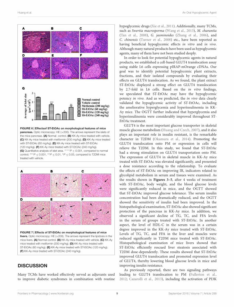
Mouse Pancreas and Liver HistologicalChangesIn the study, we performed histological examinations to observewhether ST-EtOAc have the capacity of protecting pancreasin KK-Ay mice. The results showed that ST-EtOAc couldalleviate the abnormity caused by diabetes in KK-Ay mice viaa dose-dependent manner (Figure 6). In the vehicle group, thehepatic steatosis and empty lipid vacuoles appeared obviouslyin liver, however, after the treatment of ST-EtOAc for 4weeks, the degree of hepatic steatosis and empty lipid vacuoles
was reversed, especially in the 240 mg/kg ST-EtOAc group(Figure 7).
Western Blot Analysis on TissuesIn this study, we have demonstrated the possible AMPK-GLUT4 pathway of the effects of ST-EtOAc in L6 cells. Invivo, additionally, we examined the expression of p-AMPK andGLUT4 in the skeletal muscles. As expected, the results showedthat the expression of GLUT4 and p-AMPK in skeletal muscles ofT2DM mice had been improved. Especially, the 120 mg/kg/daygroup revealed the similar effects to the 240 mg/kg/day group inenhancing the phosphorylation of AMPK after 28 days treatment(Figure 8A).
At the same time, we have examined the phosphorylation ofAMPK in liver by western blotting. As shown in Figure 8B, thelevels of phosphorylated AMPK significantly were increased inthe livers in KK-Ay mice treated with ST-EtOAc. These findingssuggested that ST-EtOAc may active the AMPK pathway toattenuate lipogenesis in hepatocytes.
Results of Acute Toxicity StudyDuring the acute toxicity study, 15 days observation periodperformed on ST-EtOAc treated KM mice have not producedmortality, abnormal clinical features, body weight disordersand food consumption, and toxicological changes in clinicalbiochemistry and hematology as well as other toxic signs (datahas not been shown). The acute oral toxicity results showed thatat the dose of 6500 mg/kg/day, ST-EtOAc concentrate is unlikelyto be poisonous in KM mice.
Frontiers in Pharmacology | www.frontiersin.org 8 September 2016 | Volume 7 | Article 288
fphar-07-00288 September 2, 2016 Time: 17:32 # 9
Huang et al. An Oral Hypoglycemic Agent
FIGURE 6 | Effectsof ST-EtOAc on morphological features of micepancreas. Optic microscopy: HE (×200). The arrows represent the islets ofthe mice pancreas. (A) Normal control. (B) KK-Ay mice treated with vehicle.(C) KK-Ay mice treated with metformin (200 mg/kg). (D) KK-Ay mice treatedwith ST-EtOAc (60 mg/kg). (E) KK-Ay mice treated with ST-EtOAc(120 mg/kg). (F) KK-Ay mice treated with ST-EtOAc (240 mg/kg).(G) Quantitative analysis of islet area. +++P ≤ 0.001, compared to normalcontrol; ∗∗∗P ≤ 0.001, ∗∗P ≤ 0.01, ∗P ≤ 0.05, compared to T2DM micetreated with vehicle.
FIGURE 7 | Effects of ST-EtOAc on morphological features of micelivers. Optic microscopy: HE (×200). The arrows represent the lipidosis in themice livers. (A) Normal control. (B) KK-Ay mice treated with vehicle. (C) KK-Aymice treated with metformin (200 mg/kg). (D) KK-Ay mice treated withST-EtOAc (60 mg/kg). (E) KK-Ay mice treated with ST-EtOAc (120 mg/kg).(F) KK-Ay mice treated with ST-EtOAc (240 mg/kg).
DISCUSSION
Many TCMs have worked effectively served as adjuvants usedto improve diabetic syndromes in combination with routine
hypoglycemic drugs (Xie et al., 2011). Additionally, many TCMs,such as Swertia macrosperma (Wang et al., 2013), M. charantia(Tan et al., 2008), G. jasminoides (Zhang et al., 2006), andC. chinensis (Turner et al., 2008) etc., have been reported ashaving beneficial hypoglycemic effects in vitro and in vivo.Although many natural products have been used as hypoglycemicagents, many of them have not been studied deeply.
In order to look for potential hypoglycemic agents in naturalproducts, we established a cell-based GLUT4 translocation assayusing stable L6 cells expressing pIRAP-mOrange cDNAs. Ourgoal was to identify potential hypoglycemic plant extracts,fractions, and their isolated compounds by evaluating theireffects on GLUT4 translocation. As we found, the plant extractST-EtOAc displayed a strong effect on GLUT4 translocationby 2.7-fold in L6 cells. Based on the in vitro findings,we speculated that ST-EtOAc may have the hypoglycemicpotency in vivo. And as we predicted, the in vivo data clearlyvalidated the hypoglycemic activity of ST-EtOAc, includingthe ameliorative hyperglycemia and hyperinsulinemia in KK-Ay mice. The OGTT further indicated that hyperglycemia andhyperinsulinemia were considerably improved throughout ST-EtOAc treatment.
GLUT4 is the most important glucose transporter in skeletalmuscle glucose metabolism (Huang and Czech, 2007), and it alsoplays an important role in insulin resistant, is the remarkablecharacter in T2DM (Okamura et al., 2014). Promoting theGLUT4 translocation onto PM or expression in cells willrelieve the T2DM. In this study, we found that ST-EtOAchad a strong stimulation on GLUT4 transportation onto PM.The expression of GLUT4 in skeletal muscle in KK-Ay micetreated with ST-EtOAc was elevated significantly, and presenteda dose resistance according to the relationship. To evaluatethe effects of ST-EtOAc on improving IR, indicators related toglycolipid metabolism in serum and tissues were examined. Asthe results shown in Figures 3–5, after 4 weeks of treatmentwith ST-EtOAc, body weight, and the blood glucose levelswere significantly reduced in mice, and the OGTT showedthat ST-EtOAc improved glucose tolerance. The serum insulinconcentration had been dramatically reduced, and the OGTTshowed the sensitivity of insulin had been improved. In thehistopathological examination, ST-EtOAc also showed significantprotection of the pancreas in KK-Ay mice. In addition, weobserved a significant decline of TG, TC, and FFA levelsin the serum of groups treated with ST-EtOAc. In anotheraspect, the level of HDL-C in the serum was in a certaindegree improved in the KK-Ay mice treated with ST-EtOAc.Levels of TG, TC, and FFA in the liver and muscles werereduced significantly in T2DM mice treated with ST-EtOAc.Histopathological examination of mice livers showed thatST-EtOAc efficiently rescued liver steatosis associated withT2DM dose-dependently. These results showed that ST-EtOAcimproved GLUT4 translocation and promoted expression levelof GLUT4, thereby lowering blood glucose levels in mice andimproving insulin resistance.
As previously reported, there are two signaling pathwaysleading to GLUT4 translocation to PM (Fullerton et al.,2012; Cazarolli et al., 2013), including the activation of PI3K
Frontiers in Pharmacology | www.frontiersin.org 9 September 2016 | Volume 7 | Article 288

fphar-07-00288 September 2, 2016 Time: 17:32 # 10
Huang et al. An Oral Hypoglycemic Agent
FIGURE 8 | (A) Effects of ST-EtOAc on AMPK phosphorylation and GLUT4 expression in skeletal muscle. (B) Effects of ST-EtOAc on AMPK phosphorylation in liver.+++P ≤ 0.001, compared to normal control; ∗∗∗P ≤ 0.001, ∗∗P ≤ 0.01, ∗P ≤ 0.05, compared to T2DM mice treated with vehicle.
and the activation of AMPK. In this study, when the L6cells were incubated with compound C for 30 min beforeaddition of ST-EtOAc, the translocation of GLUT4 to PMcaused by ST-EtOAc stimulation was mainly inhibited bythe addition of compound C (Figure 2B). However, theaddition of Wortmannin had no effect on the IRAP traffickingresponse (Figure 2C). These results suggested that ST-EtOAcenhanced GLUT4 translocation by specifically targeting theAMPK pathway.
AMPK, an enzyme that regulates the body’s balance of energy,has been investigated as a potential therapeutic target for thetreatment of T2DM (Fullerton et al., 2012). In glucose andlipid metabolism, the phosphorylation and activation of AMPKleads to GLUT4 translocation and eventually glucose uptake. Italso acts primarily by directly affecting the activity of enzymesinvolved in carbohydrate, lipid, and protein biosynthesesand secondarily by long-term transcriptional control of keycomponents (Rana et al., 2014). In our study, we detectedthe expression of p-AMPK in L6 cells and tissues usingwestern blotting after administration of ST-EtOAc. As thedata shown in Figures 8A,B, the content of p-AMPK in ST-EtOAc treated groups was significantly increased compared tothe non-treated group. The result means that in vitro, ST-EtOAc enhanced the GLUT4 translocation through upregulatingthe level of p-AMPK in L6 cells. In vivo, AMPK in mouseskeletal muscle and liver were also detected, and western
blotting data showed that the content of p-AMPK in liverand muscle in the ST-EtOAc-treated groups was significantlyincreased compared with the vehicle group. These in vitroand in vivo results coincided with each other, proving thatST-EtOAc upregulated contents of the p-AMPK and GLUT4,improved glucolipid metabolism, and relieved the IR in KK-Ay so that developing the effects on antidiabetes in vitro andin vivo.
This study showed ST-EtOAc had promising positiveactivity on GLUT4 translocation based on a cell-based GLUT4translocation system. Further investigation in vitro and in vivoshowed that ST-EtOAc indicated a strong stimulation on GLUT4translocation and enhanced the glucose uptake significantly.In vitro, ST-EtOAc improved glucose tolerance, reducedhyperglycemia and reduced insulin levels. ST-EtOAc has thepotential to treat diabetes by targeting the AMPK pathway.Furthermore, according to the oral acute toxicity study on ST-EtOAc, the single oral dose LD50 for KM mice was proposed to bemore than 6500 mg/kg/day. These results indicate that ST-EtOAc,at the dosages used in our study, did not result in any side effects.Therefore ST-EtOAc can be classified as a non-toxic substanceaccording to the common classification of the relative toxicityof chemicals. In order to identify the main components fromST-EtOAc effectively, 13 compounds had been isolated by theoff-line semipreparative HPLC–NMR. Some of these compoundshave been reported to be effective bioactive compounds, such as
Frontiers in Pharmacology | www.frontiersin.org 10 September 2016 | Volume 7 | Article 288

fphar-07-00288 September 2, 2016 Time: 17:32 # 11
Huang et al. An Oral Hypoglycemic Agent
genistin, which has shown an effect on weight gain in pregnantrats (Cao et al., 2015), and quercetin, which has shown tostimulate insulin secretion and reduce the viability of rat INS-1beta-cells (Kittl et al., 2016). The research suggests that ST-EtOAchas the potential to become an effective agent in the therapy ofdiabetes mellitus.
AUTHOR CONTRIBUTIONS
XY contributed to the conception of the study; MH andSD contributed significantly to analysis and manuscriptpreparation equally; QH, PZ, QZ, and SZ performed the dataanalyses and wrote the manuscript equally; XM, CX, andJY helped perform the analysis with constructive discussionsequally.
ACKNOWLEDGMENT
The work was financially supported by National NaturalScience Foundation of China grants (Nos. 81573561, 81102798,and 31070744), the State Key Laboratory of Drug ResearchFoundation (SIMM1403KF-07), and the Open Research Programfrom the Modernization Engineering Technology ResearchCenter of Ethnic Minority Medicine of Hubei Province (No.2015ZD004).
SUPPLEMENTARY MATERIAL
The Supplementary Material for this article can be foundonline at: http://journal.frontiersin.org/article/10.3389/fphar.2016.00288
REFERENCESAl-Maharik, N., and Botting, N. P. (2008). An efficient method for the
glycosylation of isoflavones. Eur. J. Org. Chem. 33, 5622–5629. doi: 10.1002/bab.1258
Bryant, N. J., Govers, R., and James, D. E. (2002). Regulated transport of the glucosetransporter GLUT4. Nat. Rev. Mol. Cell. Biol. 3, 267–277. doi: 10.1038/nrm782
Cao, J., Echelberger, R., Liu, M., Sluzas, E., McCaffrey, K., Buckley, B., et al.(2015). Soy but not bisphenol A (BPA) or the phytoestrogen genistinalters developmental weight gain and food intake in pregnant rats andtheir offspring. Reprod. Toxicol. 58, 282–294. doi: 10.1016/j.reprotox.2015.07.077
Cazarolli, L. H., Pereira, D. F., Kappel, V. D., Folador, P., Figueiredo, M. S.,Pizzolatti, M. G., et al. (2013). Insulin signaling: a potential signaling pathwayfor the stimulatory effect of kaempferitrin on glucose uptake in skeletal muscle.Eur. J. Pharmacol. 712, 1–7. doi: 10.1016/j.ejphar.2013.02.029
Cho, S., Park, J. H., Pae, A. N., Han, D., Kim, D., Cho, N. C., et al. (2012).Hypnotic effects and GABAergic mechanism of licorice (Glycyrrhiza glabra)ethanol extract and its major flavonoid constituent glabrol. Bioorg. Med. Chem.20, 3493–3501. doi: 10.1016/j.bmc.2012.04.011
Fullerton, M. D., Steinberg, G. R., and Schertzer, J. D. (2012). Immunometabolismof AMPK in insulin resistance and atherosclerosis. Mol. Cell. Endocrinol. 366,224–234. doi: 10.1016/j.mce.2012.02.004
Huang, S., and Czech, M. P. (2007). The GLUT4 glucose transporter. Cell. Metab.5, 237–252. doi: 10.1016/j.cmet.2007.03.006
Ju, T. J., Kwon, W. Y., Kim, Y. W., Kim, J. Y., Kim, Y. D., Lee, I. K., et al. (2014).Hemin improves insulin sensitivity in skeletal muscle in high fat–fed mice.J. Pharmacol. Sci. 126, 115–125. doi: 10.1254/jphs.14003FP
Kittl, M., Beyreis, M., Tumurkhuu, M., Fürst, J., Helm, K., Pitschmann, A., et al.(2016). Quercetin stimulates insulin secretion and reduces the viability ofrat INS-1 beta-Cells. Cell. Physiol. Biochem. 39, 278–293. doi: 10.1159/000445623
Kumar, A., Lawrence, J. C. Jr., Jung, D. Y., Ko, H. J., Keller, S. R., Kim, J. K., et al.(2010). Fat cellspecific ablation of rictor in mice impairs insulin-regulated fatcell and whole-body glucose and lipidmetabolism. Diabetes 59, 1397–1406. doi:10.2337/db09-1061
Lee, J. W., Lee, J. H., Lee, C., Jin, Q. H., Lee, D. H., Kim, Y. S., et al. (2015).Inhibitory constituents of Sophora tonkinensis on nitric oxide productionin RAW 264.7 macrophages. Bioorg. Med. Chem. Lett. 25, 960–962. doi:10.1016/j.bmcl.2014.12.012
Lee, Y. S., Kim, W. S., Kim, K. H., Yoon, M. J., Cho, H. J., Shen, Y., et al. (2006).Berberine, a natural plant product, activates AMP-activated protein kinase withbeneficial metabolic effects in diabetic and insulin-resistant state. Diabetes 55,2256–2264.
Li, X. N., Lu, Z. Q., Chen, G. T., Yan, H. X., Sha, N., Guan, S. H., et al. (2008). NMRspectral assignments of isoprenylated flavanones from Sophora tonkinensis.Magn. Reson. Chem. 46, 898–902. doi: 10.1002/mrc.2274
Meragelman, K. M., McKee, T. C., and Boyd, M. R. (2001). Anti-HIVprenylated flavonoids from Monotes africanus. J. Nat. Prod. 64, 546–548. doi:10.1021/np0005457
Mizuno, M., Tamura, K., Tanaka, T., and Iinuma, M. (1988). Threeprenylflavanones from Euchresta japonica. Phytochemistry 27, 1831–1834.doi: 10.1016/0031-9422(88)80454-0
Moller, D. E. (2001). New drug targets for type 2 diabetes and the metabolicsyndrome. Nature 414, 821–827. doi: 10.1038/414821a
Mori-Hongo, M., Takimoto, H., Katagiri, T., Kimura, M., Ikeda, Y., and Miyase, T.(2009). Melanin synthesis inhibitors from Lespedeza floribunda. J. Nat. Prod. 72,194–203. doi: 10.1021/np800395j
Okamura, T., Tawa, M., Geddawy, A., Shimosato, T., Iwasaki, H., Shintaku, H.,et al. (2014). Effects of atorvastatin, amlodipine, and their combination onvascular dysfunction in insulin-resistant rats. J. Pharmacol. Sci. 124, 76–85. doi:10.1254/jphs.13178FP
Rana, S., Blowers, E. C., and Natarajan, A. (2014). Small molecule adenosine5’-monophosphate activated protein kinase (AMPK) modulators and humandiseases. J. Med. Chem. 58, 2–29. doi: 10.1021/jm401994c
Rubin, B. R., and Bogan, J. S. (2009). Intracellular retention and insulinstimulatedmobilization of GLUT4 glucose transporters. Vitam. Horm. 80, 155–192. doi:10.1016/S0083-6729(08)00607-9
Shang, H. T., Wei, H., Yue, B. F., Xu, P., and Huang, H. G. (2009). Microsatelliteanalysis in two populations of Kunming mice. Lab. Anim. 43, 34–40. doi:10.1258/la.2008.008098
Sheng, Y. H., Li, F. J., Zhou, Q., and Jin, R. M. (2010). Study on the hepatotoxicityand pathological change induced by Radix et Rhizoma Sophorae tonkinensis inmice. Chin. J. Exp. Tradit. Med. Formulae 16:151.
Shi, J., and Kandror, K. V. (2005). Sortilin is essential and sufficient for theformation of glut4 storage vesicles in 3T3-L1 adipocytes. Dev. Cell 9, 99–108.doi: 10.1016/j.devcel.2005.04.004
Shulman, G. I. (2000). Cellular mechanisms of insulin resistance. J. Clin. Invest.106, 171–176. doi: 10.1172/JCI10583
Sutthivaiyakit, S., Thongnak, O., Lhinhatrakool, T., Yodchun, O., Srimark, R.,Dowtaisong, P., et al. (2009). Cytotoxic and antimycobacterial prenylatedflavonoids from the roots of Eriosema chinense. J. Nat. Prod. 72, 1092–1096.doi: 10.1021/np900021h
Tan, C. M., Fang, H. L., and Hu, T. J. (2009). Biological active ingredient andpharmacological effects of Radix Sophorae tonkinensis. Guangxi Agric. Sci. 40,1494–1497.
Tan, M. J., Ye, J. M., Turner, N., Hohnen-Behrens, C., Ke, C. Q., Tang, C. P.,et al. (2008). Antidiabetic activities of triterpenoids isolated from bitter melonassociated with activation of the AMPK pathway. Chem. Biol. 15, 263–273. doi:10.1016/j.chembiol.2008.01.013
Tsuchiya, A., Kanno, T., Shimizu, T., Tanaka, A., and Nishizaki, T. (2015). Rac1 andROCK are implicated in the cell surface delivery of GLUT4 under the control ofthe insulin signal mimetic diDCP-LA-PE. J. Pharmacol. Sci. 128, 179–184. doi:10.1016/j.jphs.2015.07.001
Frontiers in Pharmacology | www.frontiersin.org 11 September 2016 | Volume 7 | Article 288

fphar-07-00288 September 2, 2016 Time: 17:32 # 12
Huang et al. An Oral Hypoglycemic Agent
Turner, N., Li, J. Y., Gosby, A., To, S. W., Cheng, Z., Miyoshi, H., et al. (2008).Berberine and its more biologically available derivative, dihydroberberine,inhibit mitochondrial respiratory complex IA mechanism for the action ofberberine to activate AMP-activated protein kinase and improve insulin action.Diabetes 57, 1414–1418. doi: 10.2337/db07-1552
Wang, C., Yang, J., Zhao, P., Zhou, Q., Mei, Z. N., Yang, G. Z., et al. (2014).Chemical constituents from Eucalyptus citriodora Hook leaves and their glucosetransporter 4 translocation activities. Bioorg. Med. Chem. Lett. 24, 3096–3099.doi: 10.1016/j.bmcl.2014.05.014
Wang, Y. L., Xiao, Z. Q., Liu, S., Wan, L. S., Yue, Y. D., Zhang, Y. T., et al.(2013). Antidiabetic effects of Swertia macrosperma extracts in diabetic rats.J. Ethnopharmacol. 150, 536–544. doi: 10.1016/j.jep.2013.08.053
Xie, W., Zhao, Y., and Zhang, Y. (2011). Traditional Chinese medicinesin treatment of patients with type 2 diabetes mellitus. Evid.Based Complement. Alternat. Med. 2011:726723. doi: 10.1155/2011/726723
Yang, J., Zhao, P., Wan, D. R., Zhou, Q., Wang, C., Shu, G. W., et al. (2014).Antidiabetic effect of methanolic extract from Berberis julianae Schneid.via activation of AMP-activated protein kinase in type 2 diabetic mice.Evid. Based. Complement. Alternat. Med. 2014:106206. doi: 10.1155/2011/726723
Yang, X. Z., Yang, J., Wang, C., Wan, J. F., Yuan, J. Q., and Ren, Y. S.(2014). Sodium - dependent glucose cotransporter 2 (SGLT2) inhibitors fromSophora flavescens. J. Yunnan. Univ. (Nat. Sci.) 36, 267–272. doi: 10.7540/j.ynu.20130520
Yang, X. Z., Yang, J., Xu, C., Huang, M., Zhou, Q., Lv, J. N., et al. (2015). Antidiabeticeffects of flavonoids from Sophora flavescens EtOAc extract in type 2 diabeticKK-ay mice. J. Ethnopharmacol. 171, 161–170. doi: 10.1016/j.jep.2015.05.043
Yi, J. H., Zhang, G. L., and Li, B. G. (2002). Studies on the chemical constituents ofPseudotsuga sinensis. Acta Pharm. Sin. 37, 352–354.
Zhang, C. Y., Parton, L. E., Ye, C. P., Krauss, S., Shen, R., Lin, C. T., et al. (2006).Genipin inhibits UCP2-mediated proton leak and acutely reverses obesity-andhigh glucose-induced β cell dysfunction in isolated pancreatic islets. Cell Metab.3, 417–427. doi: 10.1016/j.cmet.2006.04.010
Zhang, H., Matsuda, H., Kumahara, A., Nakamura, S., and Yoshikawa, M. (2007).New type of anti-diabetic compounds from the processed leaves of Hydrangeamacrophylla var. thunbergii (Hydrangeae Dulcis Folium). Bioorg. Med. Chem.Lett. 17, 4972–4976. doi: 10.1016/j.bmcl.2007.06.027
Conflict of Interest Statement: The authors declare that the research wasconducted in the absence of any commercial or financial relationships that couldbe construed as a potential conflict of interest.
Copyright © 2016 Huang, Deng, Han, Zhao, Zhou, Zheng, Ma, Xu, Yang and Yang.This is an open-access article distributed under the terms of the Creative CommonsAttribution License (CC BY). The use, distribution or reproduction in other forumsis permitted, provided the original author(s) or licensor are credited and that theoriginal publication in this journal is cited, in accordance with accepted academicpractice. No use, distribution or reproduction is permitted which does not complywith these terms.
Frontiers in Pharmacology | www.frontiersin.org 12 September 2016 | Volume 7 | Article 288