Embed Size (px)
Citation preview

Neurobiology of Stress 15 (2021) 100357
Available online 22 June 20212352-2895/© 2021 The Authors. Published by Elsevier Inc. This is an open access article under the CC BY-NC-ND license(http://creativecommons.org/licenses/by-nc-nd/4.0/).
Hypoactivation of autonomtic nervous system-related orbitofrontal and motor cortex during acute stress in women with premenstrual syndrome
Yao Meng a,b, Dejian Huang c, Lulu Hou a,d, Renlai Zhou a,*
a Department of Psychology, Nanjing University, Nanjing, 210023, China b School of Nursing, Nanjing Medical University, Nanjing, 211166, China c Jiangsu Province Academy of Traditional Chinese Medicine, Nanjing, 210028, China d Department of Psychology, Shanghai Normal University, Shanghai, 200234, China
1. Introduction
Premenstrual syndrome (PMS) is a menstrual-related disorder, which refers to a series of cyclical and relapsing physical, emotional, and cognitive symptoms that regularly recur during the late luteal phase of each menstrual cycle (Ryu and Kim, 2015; Wu et al., 2016). Studies have shown that PMS is closely related to impaired stress reaction (Hamaideh et al., 2014; Klatzkin et al., 2014; Tamaki Matsumoto, Ushiroyama, Kimura, Hayashi and Moritani, 2007; Sadler et al., 2010). Therefore, numerous studies have explored the mechanism of PMS from the perspective of stress reactivity. Among them, the autonomic nervous system (ANS), one of the stress physiological responses (Kreibig, 2010; Dragomir, Gentile, Nolan, & D’Antono., 2014; Bruce S. Mcewen, 2007), has received much attention.
ANS comprises the sympathetic and parasympathetic systems, and it is closely related to emotion (Kreibig, 2010). When people’s ANS shows blunted activation or delayed recovery under pressure, their negative emotions increase (Conti et al., 2011; Gisela et al., 2003; Lupis et al., 2014; Bruce S. Mcewen, 2007; Shcheslavskaya et al., 2010; Sherman et al., 2009). Similarly, a low ANS activity has been observed in women with PMS, by either focusing on chronic stress (Baker et al., 2008) and acute stressors (Girdler et al., 1993; Klatzkin et al., 2014). Furthermore, the blunted response to stress predicts greater premenstrual symptoms and higher negative emotions (Klatzkin et al., 2014; Verkuil et al., 2009). However, it is not well understood how the neural functional dysregulation of ANS is involved in the etiology of PMS.
Actually, the ANS response at rest is related to the activation of relevant brain areas; for example, fluorodeoxyglucose positron emission tomography (FDG–PET) has been used to study glucose metabolism in the brain under different heart rates, heart rate variability, and blood pressure changes. The sympathetic activity of ANS shows a positive correlation with the motor cortex area (Schlindwein et al., 2008).
Moreover, a previous study showed that women with PMS involves the abnormal neural functional regulation of ANS-related regions during the resting state. Which were abnormal regional homogeneity and fractional amplitude of low-frequency fluctuation (fALFF), including the anterior cingulate cortex, prefrontal cortex (PFC), and precuneus (Liao et al., 2017). However, no neuroimaging studies have evaluated the neural functional regulation of ANS during acute stress in the late luteal phase of women with PMS.
It is acknowledged that ANS is easily triggered in the face of stress stimuli, therefore, it is better to introduce acute stress to detect the response of ANS-related brain areas. Under acute stressors, the ANS response is also related to the activation of certain brain areas, such as motor cortex (MC), PFC and orbitofrontal cortex (OFC) (Dum, 2016; Fechir et al., 2010; Holsen et al., 2012). Dum (2016) showed a positive correlation between the adrenal medulla that affects sympathetic func-tion and the activity of the motor and medial PFC based on rabies virus neural traceability. Furthermore, Holsen et al. (2012) found that the imbalanced ANS response is associated with dysfunction in neural re-gions, i.e., a dull parasympathetic response can result in the inactivation of the OFC among people with mood disorders. Presumably, in women with PMS, a blunted ANS response involved in acute stressful tasks may be accompanied by an inactivation reaction in the ANS-related regions.
Moreover, indicated that PMS women with a lower ANS response also have a higher subjective negative emotional experience (Matsu-moto et al., 2007). Neuroimaging studies showed that the ANS and emotional responses share a part of the neural mechanism. Specifically, the OFC, amygdala, and other areas not only control the body’s ANS response but also are inseparable from emotional reactions (Arnsten and Amy, 2015; Drevets, 2010; Protopopescu et al., 2008; Vitaly et al., 2008). Therefore, even if there is decreased activation in the ANS-related brain area of women with PMS during stressful tasks, it may have a close relationship with their subjective negative emotional
* Corresponding author. E-mail address: [email protected] (R. Zhou).
Contents lists available at ScienceDirect
Neurobiology of Stress
journal homepage: www.elsevier.com/locate/ynstr
https://doi.org/10.1016/j.ynstr.2021.100357 Received 11 October 2020; Received in revised form 8 June 2021; Accepted 15 June 2021

Neurobiology of Stress 15 (2021) 100357
2
experience. Further, women with PMS have more negative emotional experi-
ences during the stress, perhaps not only because of their slow ANS response, but also due to the prolonged recovery time afterwards. Studies have proposed that the longer the recovery time from a stressor, the greater is the individual’s subjective negative emotional experiences (Koolhaas et al., 2011; McEwen and Stellar, 1993; Shcheslavskaya et al., 2010; Sijtsema et al., 2015). The blunted ANS activity to stress has been observed in women with PMS, but it is not known whether an abnormal neural mechanism is operating in recovery time, and whether or not these related brain regions are relevant to their negative emotion.
Therefore, using the Montreal Imaging Stress Task (MIST), which can successfully activate ANS-related brain areas such as the MC, medial PFC, and OFC (Dedovic et al., 2005; Ming et al., 2017; Soliman et al., 2011), we investigated the neural mechanism of the stress process in women with PMS. We made two assumptions. First, we hypothesized that under the condition of an acute stressor, women with PMS experi-ence decreased activation of ANS-related brain areas, such as the MC and PFC, and their negative emotion would be correlated with the inactivation of the corresponding ANS-related brain region. Second, we further hypothesized that the ANS-related area in the PMS group has a slow recovery after the acute stressor, and this may be related to their negative emotion.
2. Methods
2.1. Participants
Participants were recruited from a local university and through an advertisement on a website. First, 193 women were asked to fill out the Premenstrual Syndrome Scales (PSS) (Bancroft, 1993; Gengli et al., 1998), which is the most common and widely used diagnostic criteria for PMS. Then, 62 participants (32 participants’ total score of PSS >11, 30 participants’ total score of PSS <6) were required to report their daily symptoms during the next three months. It was also evaluated according to the PSS scale, which has five affective symptoms (i.e., depression, nervous, irritability, anxiety, and neurotic), three behavioral symptoms (i.e., restless, absent-minded, and insomnia or hypersomnia), and three somatic symptoms (i.e., abdominal bloating, headache, and swelling of extremities). The inclusion criteria for PMS were met: (1) symptoms must occur during the five days before the menstrual bleeding for at least three consecutive menstrual cycles, and at least one affective and one somatic symptom must be present; (2) the symptoms are relieved within four days without recurrence until cycle day 13; (3) symptoms are present in the absence of medicine, hormone ingestion, or alcohol use.
Then, we organized a clinical diagnostic interview for each candi-date. During these interviews, we collected the demographic informa-tion and assessed the history of mental health problems. The exclusion criteria for participants were met: (1) being currently pregnant or lactating (n = 0); (2) having history of psychiatric disease (n = 1); (3) irregular menstrual cycles (n = 13) (4) alcohol and other drug use (i.e., hormonal compounds, oral contraceptives, or psychotropic drugs); (n =2); (5) being left-handed (n = 0).
Finally, 22 women with PMS and 24 women without PMS were invited to take part in the experiment in their late luteal phase (the five days before menstrual bleeding). According to G*Power instructions, the sample size has sufficient analytical power (1-β = 0.80, α = 0.05) for detecting medium-sized effects (r = 0.25) under the within-between interaction of repeated measures analysis of variance (ANOVA). Men-strual cycles of all participants were monitored for three months prior to fMRI scanning. Further, everyone was asked about the time of their next menstrual period after the experiment to reconfirm the accuracy of the test time. Five participants were excluded for their excessive head movement during the experiment. In total, 19 PMS (22.10 ± 2.15) and 22 non-PMS (23.24 ± 2.12) women were included in our analysis. The
PSS score differences between the two groups were significant (t(39) =16.81, p < 0.001).
The study was approved by the local ethics committee and performed in accordance with the Declaration of Helsinki. All participants provided written informed consent.
2.2. Materials
The PSS was compiled by Bancroft, and the Chinese version was revised by Gengli (Bancroft, 1993; Gengli et al., 1998). It consists of 12 items and covers both psychological and physical symptoms, such as follows: “In the last 3 months, how much anxiety did you feel 5 days before menstruation?” The questionnaire is scored on a 4-point Likert-type scale, with 0 = no symptom; 1 = mild symptoms; 2 =symptoms had some effect on daily work and life, but were endurable; and 3 = symptoms had a severe effect on daily work and life and required treatment. The higher the score, the more serious the symptom. Total scores of 6–10, 11–20, and >20 reflects mild, moderately severe, and severe PMS, respectively.
The Positive Affect and Negative Affect Scale (PANAS) comprises 20 items divided into two emotional dimensions (10 items in the positive dimension and 10 items in the negative dimension) (Tellegen, 1988). The participants responded to the questionnaire according to their current emotional state. It is scored on a 5-point Likert-type scale-r-anging from 1 (none at all) to 5 (very much). The Cronbach’s alpha of positive/negative dimensions for Chinese version is 0.85/0.84 (Qu et al., 2008).
The modified MIST is obtained from Matlab 3.0 and consists of a mental arithmetic task using up to four numbers ranging from 0 to 99 and up to four operands (add, subtract, multiply, and divide). The so-lution will be an integer between 0 and 9. The participants need to press a key corresponding to their answers. Feedback was provided on the submitted response (“correct,” “incorrect,” or “timeout”). A difficulty gradient for the mental arithmetic was built into the algorithm with three different categories. In the easiest categories, only tasks with two or three one-digit integers were created, and the operands are limited to + or − (example: 2 + 9 − 7). In the medium-difficulty categories, tasks with up to four integers are created, and the * operand is also allowed (example: 3 * 12–29). The most difficult category, tasks with four in-tegers, the * and/operands were used, and all the numbers may be in the two-digit range (example: 12 * 12/8–9). Three blocks are present in this task (Fig. 1), including (I) stress condition, (II) control condition, and (III) recovery condition. A block design was used with three runs con-sisting of stress, control, and recovery blocks. Each run had eight blocks (Fig. 1). To ensure the randomness of the experiment, both the control condition and stress condition are randomly presented. Each block takes 1 min, and each run takes 8 min with a 2-min break between the two runs. The total time was 28 min for each participant.
A. Control condition. The program tries to match for any cerebral activation caused by the mental arithmetic aspects of the task but without the stress components. For that purpose, mental arithmetic is presented with the same level of difficulty and at the same frequency as during the stress sessions, but individual performance and average users’ performance are not displayed.
B. Stress condition. The program tries to match for cerebral activa-tion, both caused by the stress and mental arithmetic task. Two per-formance indicators, one for the participant’s own performance and one for the average performance of all previous training participants, are shown on the upper performance bar. A time limit is enforced for each task; the elapsed time is displayed by a progress bar moving from the left to the right on the computer screen for each task; the elapsed time is displayed by a progress bar moving from the left to the right on the computer screen. Before the experiment, the subject is then reminded that there is a required minimum performance, and that her individual performance must be close to or equal to the average performance of all subjects. The participants were told that all the people in the scanner
Y. Meng et al.
Neurobiology of Stress 15 (2021) 100357
3
room (investigator, assistants, and MRI technicians) are following the subject’s performance on a second monitor in the control room of the scanning environment.
C. Recovery condition. It records a recovery state after stress/ calculation conditions. The interface of the computer program remains on the screen, but no tasks are shown.
Participants were asked to avoid high-fat and high-protein foods the day before sampling and to avoid alcohol consumption. No food or water was consumed half an hour before sampling. All the saliva samples were collected using a Cayman sampling device and stored at − 20 ◦C until being assayed. Estradiol and progesterone analysis were determined using a competitive enzyme-linked immunosorbent assay (c-ELISA). All the intra- and inter assay coefficients of variation were below 12%.
2.3. fMRI data acquisition
Images were acquired using a GE 32-channel head coil 3-T MRI (magnetic resonance imaging) scanner. A T1-weighted image used a magnetization-prepared rapid acquisition gradient echo (MPRAGE) with the following parameters: repetition time [TR] = 8.21 ms, echo time [TE] = 3.18 ms, flip angle = 15◦, slice thickness = 1.0 mm, matrix size = 256 × 256, effective voxel size = 1 × 1 × 1 mm3. Each run consisted of 148 vol.
Functional images were acquired with an EPI (gradient-echo planar imaging, EPI) sequence of 176 vol with the following parameters: TR/ TE = 2600 ms/3.02 ms, inversion time = 900 ms, flip angle = 8◦, matrix size = 256 × 256, slice thickness = 1.0 mm, and effective voxel size = 1 × 1 × 1 mm3.
2.4. Procedure
After arrival at the laboratory, participants received standardized instructions from a female researcher. Resting periods of 20 min pre-ceded the experiment, during which the participants were asked to sign an informed consent, complete PANAS, and do a training session to practice. Besides, their saliva samples were collected. Then, by taking off all metal objects and changing into lab clothes in advance, the partici-pants entered the lab to complete the fMRI paradigm. Finally, the par-ticipants were asked to fill out the PANAS again.
2.5. Statistical analysis
2.5.1. Demographic variables and behavioral data analysis The independent sample T test was used to compare the de-
mographic variables and behavioral data of the two groups. The changes in the PANAS results before and after the entire experiment were analyzed by 2 (condition: positive emotion, negative emotion) × 2 (time: pretest and posttest) × 2 (group: PMS/control group) repeated- measures ANOVA. The Greenhouse–Geisser freedom correction was
used. All the data were analyzed using SPSS 22.0.
2.5.2. Preprocessing of fMRI data The data were analyzed using DPARSF software (Yan and Zang,
2010). The first step of preprocessing is to convert the raw DICOM data to the NIFTI data format. Second, after realignment to the first image, the movement parameters for each participant were inspected. If a participant moved more than 3 mm in any direction (anterior posterior, right–left, inferior–superior), the data were excluded from further analysis. Third, for each participant, functional EPI scans were then coregistered to their corresponding high-resolution T1 anatomical image. Fourth, the unsegmented T1 anatomical images were then spatially normalized to the SPM12 MNI template using the default set-tings. To enhance the signal-to-noise ratios, these images were smoothed using a 4 mm FWHM (full-width-half-maximum) kernel.
2.5.3. Whole-brain analysis Significant hemodynamic changes for each condition were calcu-
lated using the general linear model (GLM). To explore the differences in the two groups under stress task and stress recovery condition, the task conditions (stress and control condition) and recovery conditions (re-covery after control condition and recovery after stress condition) were analyzed separately in the first level. Second-level effects were modeled by two full-factorial designs of 2 (group: PMS/control group) × 2 (conditions: stress and control; recovery after control condition and recovery after stress condition) ANOVA. As for the whole brain analysis, the voxel-level statistical threshold was set at p < 0.001, and corrected with a cluster-level of p ≤ 0.05 at the family-wise error (FWE).
3. Results
3.1. Demographic variables
The results of the independent sample T test indicate no significant differences in the age, BMI, menstrual duration, and menstrual cycle length [all t (39) <1.78, all p > 0.09] among the groups (Table 1).
3.2. fMRI results
3.2.1. Whole-brain activation The results (Table 2) of 2 (group: PMS/control group) × 2 (condi-
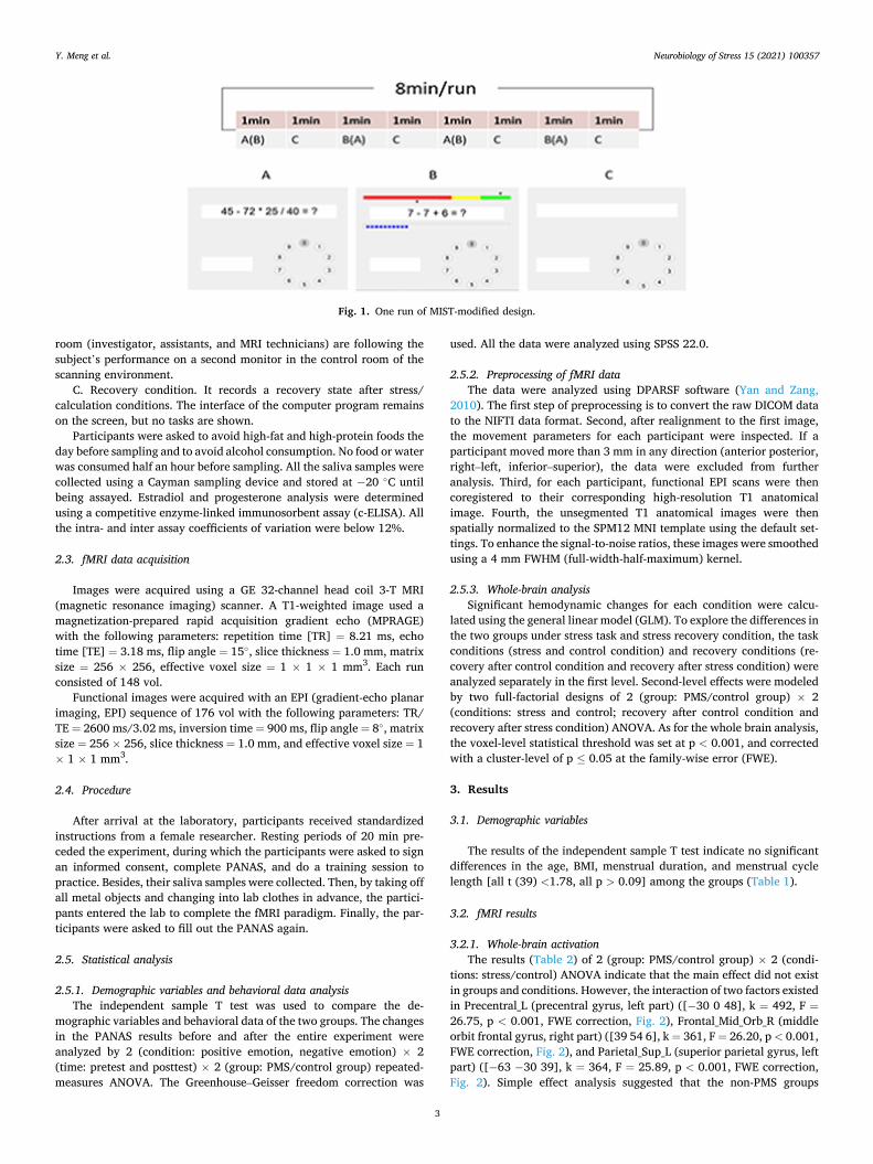
tions: stress/control) ANOVA indicate that the main effect did not exist in groups and conditions. However, the interaction of two factors existed in Precentral_L (precentral gyrus, left part) ([− 30 0 48], k = 492, F =26.75, p < 0.001, FWE correction, Fig. 2), Frontal_Mid_Orb_R (middle orbit frontal gyrus, right part) ([39 54 6], k = 361, F = 26.20, p < 0.001, FWE correction, Fig. 2), and Parietal_Sup_L (superior parietal gyrus, left part) ([− 63 − 30 39], k = 364, F = 25.89, p < 0.001, FWE correction, Fig. 2). Simple effect analysis suggested that the non-PMS groups
Fig. 1. One run of MIST-modified design.
Y. Meng et al.

Neurobiology of Stress 15 (2021) 100357
4
showed both stronger activation in the precentral_L (F = 52.49, p <0.001, η2 = 0.57), Frontal_Mid_Orb_R (F = 48.60, p = 0.017, η2 = 0.34), and Parietal_Sup_L (F = 51.60, p < 0.001, η2 = 0.51) compared to PMS groups under the stress condition, but no significant differences were observed between the two groups under the control condition (p =0.697/0.852/0.804).
The results (Table 2) of 2 (group: PMS/control group) × 2 (condi-tions: recovery after control condition/recovery after stress condition) ANOVA indicated that the main effect of groups existed in Frontal_Mid_R (middle frontal gyrus, right part) ([30 45 12], k = 429, F = 10.01, p =0.006, FWE correction, Fig. 3) and Insula_R (Insula, right part) ([24–72 − 57], k = 361, F = 21.26, p = 0.003, FWE correction). Specifically, the non-PMS groups showed stronger activation in the Frontal_Mid_R (F =17.89, p < 0.001, η2 = 0.32) compared to PMS groups. The interaction of two factors also existed in the Frontal_Mid_R (F = 5.76, p = 0.021, η2 =
0.13). Simple effect analysis showed that the MFG of PMS groups under recovery after stress condition showed stronger activation (F = 4.75, p = 0.035, η2 = 0.11) compared to recovery after the control condition.
3.2.2. Subjective emotion and its correlation with brain activation The change in PANAS results indicated that the interaction of con-
dition and group was significant (F = 7.15, p = 0.011, η2 = 0.16). The negative emotion of the PMS group both in the pretest (F = 6.06, p =0.018, η2 = 0.13) and posttest (F = 8.76, p = 0.005, η2 = 0.18) was higher than that in the non-PMS group. Meanwhile, the interaction be-tween condition, time, and group was marginally significant (F = 3.69, p = 0.06, η2 = 0.09). The negative emotion of the PMS group in the post- test was higher than that in the pretest (F = 6.35, p = 0.016, η2 = 0.14).
However, no significant change was observed in the positive emotion. The degree of pure stress activation in the MIST is represented by the
blood oxygen level dependent (BOLD) score of the stress condition minus the BOLD score of the control condition (Dedovic et al., 2005). Previous studies showed that individuals with a lower ANS response and delayed recovery have a higher negative emotional experience. How-ever, our neuroimaging study showed that the hypoactivation of ANS-related area just occurred during the stress. Thus, to study the correlation between ANS-related brain activation and the subjective negative emotional experience of women with PMS, we compared the changes in the emotional arousal (post-test minus pretest PANAS scores) of the two groups with the degree of stress activation (BOLD score of stress condition minus control condition) (Table 3). The results of the Pearson correlation test indicate that the change in negative emotion of the PMS group was negatively correlated with the degree of stress activation in the Frontal_Mid_Orb_R (r = − 0.499, p = 0.029) (Fig. 4).
4. Discussion
The results of our study showed that compared to the control group, women with PMS had a lower activation in the precentral gyrus, the right middle orbitofrontal gyrus under acute stress task, while they had a higher activation in the right middle frontal gyrus during the recovery period. In addition, the change in the negative emotion in women with PMS was negatively correlated with the degree of activation in their right middle orbitofrontal gyrus.
Specifically, the precentral gyrus is in the MC area of the brain. Studies showed that the activity of the MC area under an acute stressor is positively correlated with the sympathetic nervous system activity of the ANS (Fechir et al., 2010). Using the trans-neuronal transport of rabies virus, Dum (2016) found that the MC is the main connecting area for the adrenal medulla, which is responsible for the production of adrenal hormones that control the sympathetic activity (Dum, 2016). Studies have shown that women with PMS exhibit a dull response in the sym-pathetic system when they experience acute stressors (Girdler et al., 2001; Klatzkin et al., 2014; Roca et al., 2003). Therefore, this may ac-count for the inactivation of the precentral gyrus. Further, consistent with our results, Dubol et al. (2020a) reviewed the neuroimaging evi-dence for PMDD (the most severe form of PMS) and found that the hypoactivity of the PFC (i.e., MC) is associated with the severity of PMDD, suggesting a reduced cognitive control over the negative stim-ulation in women with premenstrual disorders. However, the reason for this deficiency is not clear. Our study can explain this from the perspective of a sympathetic system. The inactivation of the MC indi-cated a dull response in the sympathetic nervous system, which is indeed related to a worse selective attention performance (Giuliano et al., 2017). Therefore, the insensitive response of the nervous system may be regarded as one of the reasons for a reduced cognitive control in pre-menstrual disorders.
The OFC is a part of the PFC and plays an important role in both stress response and mood regulation (Arnsten and Amy, 2015; Drevets, 2010). It may have a potential impact on the initial pressure perception and persistence of stress response (Katarina et al., 2009). Our results also show the inactivation of the right middle orbitofrontal gyrus in women with PMS under an acute stressful task, which may be related to the hyporeactivity of their ANS response (sympathetic and parasympathetic system) in the late luteal phase (Girdler et al., 2001; Klatzkin et al., 2014; Roca et al., 2003). There are two reasons for this. First, the response of the sympathetic system can successfully lead to the activa-tion of the OFC, while a lower response results in its inactivation (Hagemann et al., 2003). The activation of the SNS can stimulate the adrenal medulla to release catecholamine. The transmission of cate-cholamine is necessary to maintain the activation of the PFC (Lupien et al., 2007; Sapolsky et al., 2000). Thus, a lower sympathetic reactivity can lead to insufficient secretion of catecholamine, which may affect the activation of OFC function (Brozoski et al., 1979; Drevets, 2010).
Table 1 Demographic variables (M±SD).
PMS (n = 19) Control (n = 22) p value
Age 22.10 ± 2.15 23.24 ± 2.12 0.112a
BMI 20.19 ± 2.11 20.65 ± 2.21 0.503a
Menstrual duration 5.47 ± 1.00 4.88 ± 0.93 0.091a
Menstrual length 29.47 ± 3.57 29.18 ± 4.11 0.615a
Progesterone (pg/mL) 638.03 ± 599.86 565.06 ± 550.63 0.432 a
Estradiol (pg/mL) 27.30 ± 20.04 18.22 ± 11.60 0.150 a
prepositive affect 22 ± 5.11 19.50 ± 4.34 0.102b
prenegative affect 22.53 ± 5.82 18.45 ± 4.76 0.018b
post-positive affect 22.21 ± 6.85 20.31 ± 5.01 0.323b
post-negative affect 25.84 ± 7.88 19.52 ± 5.54 0.005b
Note: BMI is the body mass index, which was calculated by dividing the weight (kg) with the height (m) squared (kg/m2). Menstrual duration refers to the number of menstrual days in a menstrual cycle. Cycle length refers to the time interval between two consecutive menstrual cycles.
a The p value was obtained from two sample t-tests. b The p value was obtained by ANOVA.
Table 2 Neuroticism effect and interactions.
Contrasts Cluster level sig.
Voxels X Y Z Region
Interaction: conditions × group
<0.001 492 − 30 0 48 Precentral_L 361 39 54 6 Frontal_Mid_Orb_R 364 − 63 − 30 39 Parietal_Sup_L
Main effect : recovery conditions (control recovery, stress recovery)
<0.05 429 30 45 12 Frontal_Mid_R 361 24 –72 − 57 Insula_R
Interaction: recovery conditions × group
<0.05 429 30 45 12 Frontal_Mid_R
Y. Meng et al.

Neurobiology of Stress 15 (2021) 100357
5
Fig. 2. The interaction of group and task conditions (all p < 0.001). Part A shows the volume in Precentral_L; Part B shows the volume in Frontal_Mid_Orb_R; Part C shows the volume in Parietal_Sup_L. During the stress condition, the activation of the PMS group was significantly lower than that of the control group. During the control condition, the difference between the two groups was not significant. Precentral_L = precentral gyrus, left part. Frontal_Mid_Orb_R = middle orbit frontal gyrus, right part. Parietal_Sup_L = superior parietal gyrus, left part.
Y. Meng et al.

Neurobiology of Stress 15 (2021) 100357
6
Second, the lower parasympathetic response was also significantly associated with the inactivation of OFC (Holsen et al., 2012). As mentioned above, the OFC is an important area responsible for emotional regulation. The lower parasympathetic response predicts poorer mood regulation under an acute stressor (Macatee et al., 2017), so that it may be one of the reasons why the right middle orbitofrontal gyrus in PMS shows a low activation. In addition, previous findings suggest that hormonal fluctuations during the menstrual cycle modulate the brain structure (i.e., OFC), thus influencing negative affect and cognition (Dubol et al., 2020b; Dreher et al., 2007). It has been found that the sex hormones secreted by the ovaries affect the secretion function of the hypothalamus and pituitary, thus causing the change in the related stress hormone release of epinephrine (i.e., adrenaline) from the adrenal medulla (Lustyk and Gerrish, 2010), i.e., sex hormones may serve as stressors with the potential of creating a negative feedback loop that further exacerbates the stress response, causing the hypoactive OFC in women with PMS. In sum, the activation of ANS is essential for adapting to stress and maintaining homeostasis (Fechir et al., 2010; Karatsoreos and Mcewen, 2011). However, the inactivation of ANS-related areas (precentral gyrus and right middle orbitofrontal gyrus) in women with PMS indicates that they may be characterized by reduced adaptation to an acute stressor.
The results of subjective emotions indicate that the negative emo-tions of women with PMS after the task increased significantly. Previous studies have shown a significant correlation between the abnormal ANS responses of women with PMS and negative emotional experience (T. Matsumoto, Hayashi, Kimura and Moritani, 2009; Tamaki Matsumoto et al., 2007). Therefore, we attempted to analyze whether there is also a correlation between the subjective changes of negative emotion (post task-pre task) and the activation of ANS-related brain regions during stress. Our results show that the activation of right middle orbitofrontal gyrus is significantly correlated with the change in negative emotions. The orbitofrontal gyrus is an important brain region involved in the cognitive emotional process (Deng et al., 2018; Hanson et al., 2010; Smoski et al., 2015; Wager et al., 2008). A study concerning cognitive reappraisal to negative emotion showed that the activation of right OFC in patients with depression was weaker than that of the healthy group. In the healthy group, reappraisal led to more positive emotions, triggering the response of the right OFC (Smoski et al., 2013). Rolls (2017) also found that the activation of OFC was associated with subjective emotional experience, while the impaired OFC can affect the normal emotional experience (Rolls, 2017), i.e., the negative emotional arousal of women with PMS under pressure is closely related to the inactivation of the right middle orbitofrontal gyrus. It is acknowledged that negative emotional experience is one of the most important symptoms of PMS, but no consistent reason is provided. Our results suggest that the inac-tivation of OFC in PMS caused by an imbalanced ANS response in acute pressure situations is probably one of the reasons for their increased negative emotions in the late luteal phase.
Furthermore, our research compared the recovery situation after the task conditions between two groups. The ANS-related areas (such as the MC, OFC, and mPFC) were not significantly different in the recovery phase between the two groups, so the second assumption was not satisfied. Nevertheless, the results show that irrespective of the stress or control recovery period, the activation of the right middle frontal gyrus in women with PMS was significantly higher than that in the control group. Furthermore, the activation of the right middle frontal gyrus in the PMS group in the stress recovery period was significantly higher than that during the control recovery. Thus, the right middle frontal gyrus plays an important role in executive control tasks such as inhibi-tory control and working memory tasks (Niendam et al., 2012; Satyshur et al., 2018). Consequently, the activation of the right middle frontal
Fig. 3. The interaction of group and recovery conditions in Frontal_Mid_R (p = 0.006). During the stress recovery, the activation of the PMS group was significantly higher than that of the control group. During the control recovery, the difference between the two groups was not significant. Frontal_Mid_R = middle frontal gyrus, right part.
Table 3 Correlation coefficient between the difference in PANAS before and after the experiment and main brain differences in stress and calculation conditions.
Precentral_L Frontal_Mid_Orb_R Parietal_Sup_L
ST-CT ST-CT ST-CT
PMS group
beforeNE- afterNE
.347 − .499* .313
beforePE- afterPE
− .131 − .140 − .101
Control group
beforeNE- afterNE
− .073 .003 − .112
beforePE- afterPE
.207 .215 .221
Note: *p < 0.05. CT = calculation task; ST = stress task; NE = negative emotion; PE = positive emotion; before NE-after NE = the negative emotional difference before and after the experiment; before PE-after PE = the positive emotional difference before and after the experiment.
Y. Meng et al.

Neurobiology of Stress 15 (2021) 100357
7
gyrus indicates normal cognitive processing in the task. It was still overactive in women with PMS during the stress recovery condition, indicating that it is difficult for them to recover from the task.
Some limitations must be noted. First, we invited participants to participate in our study during their late luteal phase. However, women had obvious hormonal fluctuations during the menstrual cycle, which is well known to play a role in stress processing (Lustyk and Gerrish, 2010). It remains to be further studied and confirmed whether the dysfunctions of stress processing indicated by the blood oxygen signal in PMS exist at other stages of the menstrual cycle. Second, the menstrual cycle was determined by subjective reporting, which may decrease the reliability of the results. Future studies should select more objective measurements to decide the cycle time. Third, it is better to explain the results if we collected both biological and fMRI data for stress tasks. Further research should achieve this. Last, we compared the data from subjective emotional changes and from the objective fMRI. Although the results were significant, repeatability should be further validated by studies with more participants. Future research should explore the correlation between subjective and objective data based on the number of subjects.
Funding
This study was supported by the Key Project of Philosophy and Social Science Research in Colleges and Universities in Jiangsu Province [2015JDXM001] and NJU National Demonstration Base for Innovation & Entrepreneurship [SCJD0406].
Data availability
Data will be made available on request.
CRediT authorship contribution statement
Yao Meng: Conceptualization, Methodology, Software. Dejian Huang: Data curation, Data collection. Lulu Hou: Investigation, Formal analysis. Renlai Zhou: Writing – review & editingWriting- Reviewing and Editing.
Declaration of competing interest
The authors declare no conflict of interest.
Acknowledgments
We gratefully acknowledge the help of PMS and healthy participants in our study. We also would like to thank all the instrument operators of MRI in our experiments.
References
Arnsten, Amy, F.T., 2015. Stress weakens prefrontal networks: molecular insults to higher cognition. Nat. Neurosci. 18 (10), 1376–1385. https://doi.org/10.1038/ nn.4087.
Baker, F.C., Colrain, I.M., Trinder, J., 2008. Reduced parasympathetic activity during sleep in the symptomatic phase of severe premenstrual syndrome. J. Psychosom. Res. 65 (1), 13–22. https://doi.org/10.1016/j.jpsychores.2008.04.008.
Bancroft, J., 1993. The premenstrual syndrome–a reappraisal of the concept and the evidence. Psychol. Med. Suppl 24 (Suppl. 24), 1–47. https://doi.org/10.1017/ S0264180100001272.
Brozoski, T.J., Brown, R.M., Rosvold, H.E., Goldman, P.S., 1979. Cognitive deficit caused by regional depletion of dopamine in prefrontal cortex of rhesus monkey. Science 205 (4409), 929–932. https://doi.org/10.1126/science.112679.
Conti, C.M., Maccauro, G., Fulcheri, M., 2011. Psychological stress and cancer. Int. J. Immunopathol. Pharmacol. 24 (1), 1. https://doi.org/10.1177/ 039463200101400101.
Dedovic, K., Renwick, R., Mahani, N.K., Engert, V., Lupien, S.J., Pruessner, J.C., 2005. The Montreal imaging stress task: using functional imaging to investigate the effects of perceiving and processing psychosocial stress in the human brain. J. Psychiatry Neurosci. 30 (5), 319–325. PMID: 16151536, PMCID: PMC1197276.
Deng, Y., Li, S., Zhou, R., Walter, M., 2018. Motivation but not valence modulates neuroticism-dependent cingulate cortex and insula activity. Hum. Brain Mapp. 39 (4), 1664–1672. https://doi.org/10.1002/hbm.23942.
Dragomir, A.I., Gentile, C., Nolan, R.P., D’Antono, 2014. Three-year stability of cardiovascular and autonomic nervous system responses to psychological stress. Psychophysiology 51 (9), 921–931. https://doi.org/10.1111/.12231.
Dreher, J.C., Schmidt, P.J., Kohn, P., Furman, D., Rubinow, D., Berman, K.F., 2007. Menstrual cycle phase modulates reward-related neural function in women. Proc. Natl. Acad. Sci. U.S.A. 104, 2465–2470. https://doi.org/10.1073/pnas.0605569104.
Drevets, W.C., 2010. Orbitofrontal cortex function and structure in depression. Ann. N. Y. Acad. Sci. 1121 (1), 499–527. https://doi.org/10.1196/annals.1401.029.
Dubol, M., Epperson, C.N., Lanzenberger, R., Sundstrom-Poromaa, I., Comasco, E., 2020a. Neuroimaging premenstrual dysphoric disorder: a systematic and critical review - sciencedirect. Front. Neuroendocrinol. 57 (139) https://doi.org/10.1016/j. yfrne.2020.100838.
Dubol, M., Comasco, E., Epperson, C.N., Pletzer, B., Sundstrm-Poromaa, I., 2020b. Neuroimaging the menstrual cycle: a multimodal systematic review. Front. Neuroendocrinol. 60 (3) https://doi.org/10.1016/j.yfrne.2020.100878.
Dum, R.P., 2016. Motor, cognitive, and affective areas of the cerebral cortex influence the adrenal medulla. Proc. Natl. Acad. Sci. Unit. States Am. 113 (35), 9922. https:// doi.org/10.1073/pnas.1605044113.
Fechir, M., Gamer, M., Blasius, I., Bauermann, T., Breimhorst, M., Schlindwein, P., Birklein, F., 2010. Functional imaging of sympathetic activation during mental stress. Neuroimage 50 (2), 847–854. https://doi.org/10.1016/j. neuroimage.2009.12.004.
Fig. 4. Associations between emotional arousal and brain volume of ORBmid.R in two groups (p = 0.029). There is a significant association between the emotional arousal in pms-NE and stress activation in ORBmid.R (p = 0.029). ORBmid.R is the middle orbit frontal gyrus, right part. pms-PE is the positive emotional difference before and after the experiment in the PMS group. pms-NE is the negative emotional difference before and after the experiment in the PMS group. control-PE is the positive emotional difference before and after the experiment in the control group. control-NE is the negative emotional difference before and after the experiment in the control group.
Y. Meng et al.

Neurobiology of Stress 15 (2021) 100357
8
Gengli, Z., Linhong, W., Q, C., 1998. Occurrence and influencing factors of premenstrual syndrome in women of childbearing age. Chin. J. Obstet. Gynecol. 4, 222–224.
Girdler, S.S., Pedersen, C.A., Stern, R.A., Light, K.C., 1993. Menstrual cycle and premenstrual syndrome: modifiers of cardiovascular reactivity in women. Health Psychol. 12 (3), 180–192. https://doi.org/10.1037/0278-6133.12.3.180.
Girdler, S.S., Straneva, P.A., Light, K.C., Pedersen, C.A., Morrow, A.L., 2001. Allopregnanolone levels and reactivity to mental stress in premenstrual dysphoric disorder. Biol. Psychiatr. 49 (9), 788–797. https://doi.org/10.1016/S0006-3223(00) 01044-1.
Gisela, L.V., Lumley, M.A., Elizabeth, J., Hillary, H., 2003. Age and gender differences in cardiac reactivity and subjective emotion responses to emotional autobiographical memories. Emotion 3 (2), 115–126. https://doi.org/10.1037/1528-3542.3.2.115.
Giuliano, R.J., Karns, C.M., Bell, T.A., Petersen, S., Skowron, E.A., Neville, H.J., 2017. Parasympathetic and sympathetic activity are associated with individual differences in neural indices of selective attention in adults. Psychophysiology. https://doi.org/ 10.1101/173377 e13079.
Hagemann, D., Waldstein, S.R., Thayer, J.F., 2003. Central and autonomic nervous system integration in emotion. Brain Cognit. 52 (1), 79–87. https://doi.org/ 10.1016/S0278-2626(03)00011-3.
Hamaideh, S.H., Al-Ashram, S.A., Al-Modallal, H., 2014. Premenstrual syndrome and premenstrual dysphoric disorder among Jordanian women. J. Psychiatr. Ment. Health Nurs. 21 (1), 60–68. https://doi.org/10.1111/jpm.12047.
Hanson, J.L., Chung, M.K., Avants, B.B., Shirtcliff, E.A., Gee, J.C., Davidson, R.J., Pollak, S.D., 2010. Early stress is associated with alterations in the orbitofrontal cortex: a tensor-based morphometry investigation of brain structure and behavioral risk. J. Neurosci. 30 (22), 7466–7472. https://doi.org/10.1523/JNEUROSCI.0859- 10.2010.
Holsen, L.M., Lee, J.H., Spaeth, S.B., Ogden, L.A., Klibanski, A., Whitfield-Gabrieli, S., Goldstein, J.M., 2012. Brain hypoactivation, autonomic nervous system dysregulation, and gonadal hormones in depression: a preliminary study. Neurosci. Lett. 514 (1), 57–61. https://doi.org/10.1016/j.neulet.2012.02.056.
Karatsoreos, I.N., Mcewen, B.S., 2011. Psychobiological allostasis: resistance, resilience and vulnerability. Trends Cognit. Sci. 15 (12), 576–584. https://doi.org/10.1016/j. tics.2011.10.005.
Katarina, D., Catherine, D.A., Pruessner, J.C., 2009. What stress does to your brain: a review of neuroimaging studies. Can. J. Psychiatr. 54 (1), 6–15. https://doi.org/ 10.1177/070674370905400104.
Klatzkin, R.R., Bunevicius, A., Forneris, C.A., Girdler, S., 2014. Menstrual mood disorders are associated with blunted sympathetic reactivity to stress. J. Psychosom. Res. 76 (1), 46–55. https://doi.org/10.1016/j.jpsychores.2013.11.002.
Koolhaas, J.M., Bartolomucci, A., Buwalda, B., Boer, S.F.D., Flügge, G., Korte, S.M., Palanza, P., 2011. Stress revisited: a critical evaluation of the stress concept. Neurosci. Biobehav. Rev. 35 (5), 1291–1301. https://doi.org/10.1016/j. neubiorev.2011.02.003.
Kreibig, S.D., 2010. Autonomic nervous system activity in emotion: a review. Biol. Psychol. 84 (3), 394–421. https://doi.org/10.1016/j.biopsycho.2010.03.010.
Liao, H., Duan, G., Liu, P., Liu, Y., Pang, Y., Liu, H., 2017. Altered fractional amplitude of low frequency fluctuation in premenstrual syndrome: a resting state fmri study. J. Affect. Disord. 218, 41–48. https://doi.org/10.1016/j.jad.2017.04.045.
Lupien, S.J., Maheu, F., Tu, M., Fiocco, A., Schramek, T.E., 2007. The effects of stress and stress hormones on human cognition: implications for the field of brain and cognition. Brain Cognit. 65 (3), 209–237. https://doi.org/10.1016/j. bandc.2007.02.007.
Lupis, S.B., Lerman, M., Wolf, J.M., 2014. Anger responses to psychosocial stress predict heart rate and cortisol stress responses in men but not women. Psychoneuroendocrinology 49 (1), 84–95. https://doi.org/10.1016/j. psyneuen.2014.07.004.
Lustyk, M.K.B., Gerrish, W.G., 2010. Premenstrual Syndrome and Premenstrual Dysphoric Disorder: Issues of Quality of Life, Stress and Exercise. Springer, New York, pp. 1952–1971. https://doi.org/10.1007/978-0-387-78665-0_115.
Macatee, R.J., Albanese, B.J., Schmidt, N.B., Cougle, J.R., 2017. The moderating influence of heart rate variability on stressor-elicited change in pupillary and attentional indices of emotional processing: an eye-Tracking study. Biol. Psychol. 123, 83–93. https://doi.org/10.1016/j.biopsycho.2016.11.013.
Matsumoto, T., Hayashi, T., Kimura, T., Moritani, T., 2009. Altered autonomic nervous system activity evaluated by heart rate variability among women with severe premenstrual emotional distress. Fertil. Steril. 92 (3) https://doi.org/10.1016/j. fertnstert.2009.07.1182. S131-S131.
Matsumoto, T., Ushiroyama, T., Kimura, T., Hayashi, T., Moritani, T., 2007. Altered autonomic nervous system activity as a potential etiological factor of premenstrual syndrome and premenstrual dysphoric disorder. Biopsychosoc. Med. 1 (1), 24. https://doi.org/10.1186/1751-0759-1-24.
Mcewen, B.S., 2007. Physiology and neurobiology of stress and adaptation: central role of the brain. Physiol. Rev. 87 (3), 873–904. https://doi.org/10.1152/ physrev.00041.2006.
McEwen, B.S., Stellar, 1993. Stress and the individual. Mechanisms leading to disease. Arch. Intern. Med. 153 (18), 2093–2101. https://doi.org/10.1001/ archinte.153.18.2093.
Ming, Q., Zhong, X., Zhang, X., Pu, W., Dong, D., Jiang, Y., Yao, S., 2017. State- independent and dependent neural responses to psychosocial stress in current and remitted depression. Am. J. Psychiatr. 174 (10), 2011. https://doi.org/10.1176/ appi.ajp.2017.16080974 appi.ajp.2017.
Niendam, T.A., Laird, A.R., Ray, K.L., Dean, Y.M., Glahn, D.C., Carter, C.S., 2012. Meta- analytic evidence for a superordinate cognitive control network subserving diverse executive functions. Cognit. Affect Behav. Neurosci. 12 (2), 241–268. https://doi. org/10.3758/s13415-011-0083-5.
Protopopescu, X., Tuescher, O., Pan, H., Epstein, J., Root, J., Chang, L., Stern, E., 2008. Toward a functional neuroanatomy of premenstrual dysphoric disorder. J. Affect. Disord. 108 (1–2), 87–94. https://doi.org/10.1016/j.jad.2007.09.015 (1), 87-94.
Qu, L., Xue, Z., W, Y., 2008. The Chinese revised positive and negative schedule (PANAS). Appl. Psychol. 3, 249–254.
Roca, C.A., Schmidt, P.J., Margaret, A., Patricia, D., Danaceau, M.A., Karen, P., Rubinow, D.R., 2003. Differential menstrual cycle regulation of hypothalamic- pituitary-adrenal axis in women with premenstrual syndrome and controls. J. Clin. Endocrinol. Metab. 88 (7), 3057–3063. https://doi.org/10.1210/jc.2002-021570.
Rolls, E.T., 2017. The orbitofrontal cortex and emotion in health and disease, including depression. Neuropsychologia 128, 14–43. https://doi.org/10.1016/j. neuropsychologia.2017.09.021.
Ryu, A., Kim, T.H., 2015. Premenstrual syndrome: a mini review. Maturitas 82 (4), 436–440. https://doi.org/10.1016/j.maturitas.2015.08.010.
Sadler, C., Smith, H., Hammond, J., Bayly, R., Borland, S., Panay, N., Inskip, H., 2010. Lifestyle factors, hormonal contraception, and premenstrual symptoms: the United Kingdom Southampton Women’s Survey. J. Wom. Health 19 (3), 391–396. https:// doi.org/10.1089/jwh.2008.1210.
Sapolsky, R.M., Romero, L.M., Munck, A.U., 2000. How do glucocorticoids influence stress responses? Integrating permissive, suppressive, stimulatory, and preparative actions. Endocr. Rev. 21 (1), 55–89. https://doi.org/10.1210/er.21.1.55.
Satyshur, M.D., Layden, E.A., Gowins, J.R., Buchanan, A., Gollan, J.K., 2018. Functional connectivity of reflective and brooding rumination in depressed and healthy women. Cognit. Affect Behav. Neurosci. 1–18 https://doi.org/10.3758/s13415-018-0611-7.
Schlindwein, P., Buchholz, H.G., Schreckenberger, M., Bartenstein, P., Dieterich, M., Birklein, F., 2008. Sympathetic activity at rest and motor brain areas: FDG–PET study. Auton. Neurosci. 143 (1), 27–32. https://doi.org/10.1016/j. autneu.2008.07.006.
Shcheslavskaya, O.V., Burg, M.M., Mckinley, P.S., Schwartz, J.E., William, G., Ryff, C.D., Sloan, R.P., 2010. Heart rate recovery after cognitive challenge is preserved with age. Psychosom. Med. 72 (2), 128–133. https://doi.org/10.1097/ PSY.0b013e3181c94ca0.
Sherman, D.K., Bunyan, D.P., J David, C., Jaremka, L.M., 2009. Psychological vulnerability and stress: the effects of self-affirmation on sympathetic nervous system responses to naturalistic stressors. Health Psychology Official Journal of the Division of Health Psychology American Psychological Association 28 (5), 554–562. https://doi.org/10.1037/a0014663.
Sijtsema, J.J., Roon, A.M.V., Groot, P.F.C., Riese, H., 2015. Early life adversities and adolescent antisocial behavior: the role of cardiac autonomic nervous system reactivity in the TRAILS study. Biol. Psychol. 110, 24–33. https://doi.org/10.1016/j. biopsycho.2015.06.012.
Smoski, M.J., Keng, S.L., Ji, J.L., Moore, T., Minkel, J., Dichter, G.S., 2015. Neural indicators of emotion regulation via acceptance vs reappraisal in remitted major depressive disorder. Soc. Cognit. Affect Neurosci. 10 (9), 1187. https://doi.org/ 10.1093/scan/nsv003.
Smoski, M.J., Keng, S.L., Schiller, C.E., Minkel, J., Dichter, G.S., 2013. Neural mechanisms of cognitive reappraisal in remitted major depressive disorder. J. Affect. Disord. 151 (1), 171–177. https://doi.org/10.1016/j.jad.2013.05.073.
Soliman, A., O’Driscoll, G.A., Pruessner, J., Joober, R., Ditto, B., Streicker, E., Dagher, A., 2011. Limbic response to psychosocial stress in schizotypy: a functional magnetic resonance imaging study. Schizophr. Res. 131 (1), 184–191. https://doi.org/ 10.1016/j.schres.2011.05.016.
Tellegen, A., 1988. Development and validation of brief measures of positive and negative affect: the PANAS scales. Pers Soc Psychol 54 (6), 1063–1070. https://doi. org/10.1037/0022-3514.54.6.1063.
Verkuil, B., Brosschot, J.F., Beurs, D.P.D., Thayer, J.F., 2009. Effects of explicit and implicit perseverative cognition on cardiac recovery after cognitive stress. Int. J. Psychophysiol. 74 (3), 220–228. https://doi.org/10.1016/j.ijpsycho.2009.09.003.
Vitaly, N., Rupali, D., Giulia, C., Nikos, M., Brown, E.N., Riccardo, B., 2008. Brain correlates of autonomic modulation: combining heart rate variability with fMRI. Neuroimage 42 (1), 169–177. https://doi.org/10.1016/j.neuroimage.2008.04.238.
Wager, T.D., Davidson, M.L., Hughes, B.L., Lindquist, M.A., Ochsner, K.N.n., 2008. Prefrontal-subcortical pathways mediating successful emotion regulation. Neuron 59 (6), 1037–1050. https://doi.org/10.1016/j.neuron.2008.09.006.
Wu, M., Liang, Y., Wang, Q., Zhao, Y., Zhou, R., 2016. Emotion dysregulation of women with premenstrual syndrome. Sci. Rep. 6, 38501. https://doi.org/10.1038/ srep38501.
Yan, C.G., Zang, Y.F., 2010. DPARSF: a matlab toolbox for “pipeline” data analysis of resting-state fMRI. Front. Syst. Neurosci. 4 (13), 13. https://doi.org/10.3389/ fnsys.2010.00013.
Y. Meng et al.