Embed Size (px)
Citation preview
Ann. rheum. Dis. (1956), 15, 46.
HYPERTROPHIC OSTEO-ARTHROPATHY INPOLYARTERITIS
BY
R. R. H. LOVELL AND G. B. D. SCOTTFrom the Medical Unit and Department of Morbid Anatomy, St. Mary's Hospital, Paddington
(RECEIVED FOR PUBLICATION AUGUST 16, 1955)
Hypertrophic osteo-arthropathy is most com-monly associated with bronchopulmonary or pleuraldiseases. It has also been described in severalapparently unrelated conditions and occasionally inpatients with no other recognizable disease. Theliterature is reviewed by Mendlowitz (1942). Thispaper reports hypertrophic osteo-arthropathy in apatient with polyarteritis nodosa and describes theeffect of cortisone.
Case ReportA male business executive, aged 51, had suffered noserious illness until 1947, when he started to get achingpains in the backs of the thighs; the pains were constant,and worse when he was tired, but unaffected by move-ment. Early in 1948 his right ankle became swollen andhe developed painful red papules, up to 3 mm. diameter,some of which ulcerated. About this time purplemottling was noticed on the legs and forearms. Laterin 1948 his knees became swollen and painful but he wasable to play golf. Early in 1949 the wrists, elbows, andmetacarpo-phalangeal joints were painful and swollenand he suffered increasingly severe limb pains unrelatedto joints. During 1950 he was disabled by pain, forwhich he took up to 24 tablets codeine co. B.P. daily.He now noticed small nodules under the skin; he sweatedprofusely and lost about 7 lb. weight in 6 months.Apart from symptoms of a sudden right subclavianvenous thrombosis while playing golf in 1949, his symp-toms from 1947 to 1950 related only to the skin andlocomotor system. He was seen in 1950 by Dr. W. S. C.Copeman and Dr. G. B. Mitchell-Heggs and was admit-ted to St. Mary's Hospital with a suggested diagnosis ofpolyarteritis nodosa.
Clinical Examination.-On admission in November,1950, his limbs were very painful. There was swellinground the elbows, wrists, metacarpo-phalangeal, knee,and ankle joints. The swelling was due partly to synovialeffusions, but there was also conspicuous peri-articularthickening extending 2-3 inches beyond the joints. Thewrists, knees, and ankles were warm, but movementswere free. There was slight clubbing of the fingers.The skin showed three types of lesion typical of poly-
arteritis:(a) purple mottling, most marked on the distal
parts of the arms and legs;(b) tender punched-out ulcers over the buttocks
and scars of similar lesions which had healed;
(c) subcutaneous nodules about 3 mm. diameteron the forearms.
Apart from dilated superficial collateral veins con-sistent with an occlusion of the right subclavian vein, theother systems appeared healthy. The blood pressurewas 105/65 mm. Hg.
Laboratory InvestigationsBlood: Erythrocyte sedimentation rate (Westergren)
75 mm./l hr.Haemoglobin 1I1 g./100 ml., 3,800,000 red cells/
c. mm., 15,000 white cells/c. mm. with 75 per cent.neutrophils, 19 per cent. lymphocytes, 3 per cent.eosinophils and 3 per cent. monocytes.
Urine: No abnormality.Radiology: Appearance of heart and lungs normal.
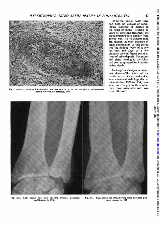
Hands, wrists, and elbows showed no joint changes.Knees showed some osteophytic lipping. Lower ends ofshafts of forearm bones showed minimal irregular forma-tion of new sub-periosteal bone (Fig. 2a, opposite).
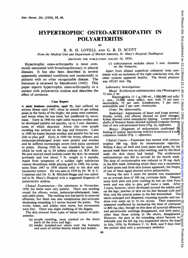
Biopsy: Diagnosis of polyarteritis confirmed byfinding of typical necrotizing arteritis in sections of a sub-cutaneous nodule (Fig. 1, opposite).
Clinical Course.-Treatment was started with cortico-trophin 100 mg. daily by intramuscular injection.Within 4 days all limb and joint pains had gone, by thesecond week there was no joint swelling, and by the thirdweek the skin ulcers had healed. The erythrocytesedimentation rate fell to normal by the fourth week.The dose of corticotrophin was reduced to 50 mg. dailyin the fifth week, following which there was a recurrenceof limb pains and fresh skin lesions appeared; the biopsyof one of these again showed active polyarteritis.During the next 3 years the patient was maintained
on an average dose of 100 mg. cortisone daily. Despitesome limb pain and joint swelling he lost no time fromwork and was able to play golf twice a week. After3 years, however, ulcers developed around the ankles andon the legs; patches of skin on his feet became cold andblue, and the arterial pulses in the feet diminished. Theulcers enlarged and deepened, exposing muscles and ten-dons over areas up to 16 cm. across. Their expansionappeared unaffected by increasing the dose of cortisoneto 400 mg./day, though on this dose all synovial effusionsand periarticular swellings disappeared, and limb pains,other than those arising in the ulcers, disappeared.However, the pain in the extending ulcers became in-tolerable and the left leg was amputated above the kneein July, 1954, by Professor C. G. Rob, and 9 days laterthe patient died with a pulmonary embolus.
46
copyright. on D
ecember 30, 2019 by guest. P
rotected byhttp://ard.bm
j.com/
Ann R
heum D
is: first published as 10.1136/ard.15.1.46 on 1 March 1956. D
ownloaded from
H YPERTROPHIC OSTEO-ARTHROPATHY IN POL YARTERITIS
Fig. 1.-Artery showing inflammation and necrosis in a section through a subcutaneousnodule removed in December, 1950.
Fig. 2(a).-Right radius and ulna, showing minimal periostealproliferation in 1950.
Up to the time of death therehad been no clinical or radio-logical evidence of disease inthe heart or lungs. During 3Jyears of cortisone treatment theblood pressure rose steadily from105/65 mm. Hg to 165/105 mm.Hg, though the only evidence ofrenal abnormality in this periodwas the finding twice of a fewred cells and once of a fewgranular casts in fifteen examina-tions of urine deposit. Symptomsand signs relating to the jointshad been suppressed for 3 monthsbefore death.
Radiological Changes in Jointsand Bones.-The joints of thehands, wrists, knees, and ankleswere examined radiologically atintervals from 1950 to 1954; therewere no changes in them otherthan those associated with syn-ovial effusions.
Fig. 2(b).-Right radius and ulna, showing more advanced pen-osteal changes in 1953.
47
ALI't
copyright. on D
ecember 30, 2019 by guest. P
rotected byhttp://ard.bm
j.com/
Ann R
heum D
is: first published as 10.1136/ard.15.1.46 on 1 March 1956. D
ownloaded from
ANNALS OF THE RHEUMATIC DISEASES
Iw' . I
441
is'~~~~~~~~~~~~~~~~~~~~~~~~'~ ~ ~ 0
tj.t4o
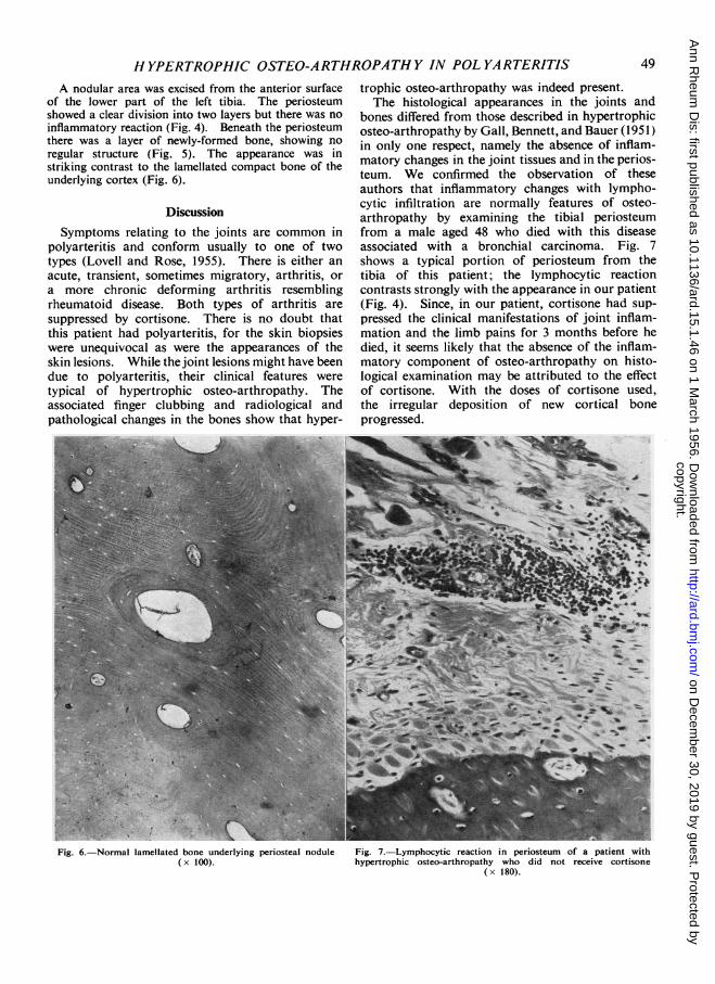
Fig. 4.-Thickened periosteum over a nodule excised at necropsy (x 180).
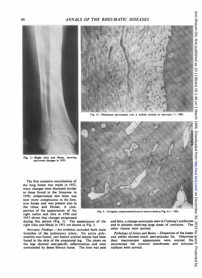
Fig. 3.-Right tibia and fibula, showingperiosteal changes in 1953.
The first extensive examination ofthe long bones was made in 1952,when changes were disclosed similarto those found in the forearms in1950; subperiosteal new bone wasnow more conspicuous in the fore-arm bones and was present also inthe tibiae and fibulae. A com-parison of the appearances of the Fig.5.-right radius and ulna in 1950 and1953 shows that changes progressedduring this period (Fig. 2). The appearances of theright tibia and fibula in 1953 are shown in Fig. 3.
Necropsy Findings.-An embolus occluded both mainbranches of the pulmonary artery. No active poly-arteritis was found; two healed arterial lesions had beenfound in the skin of the amputated leg. The ulcers onthe legs showed non-specific inflammation and weresurrounded by dense fibrous tissue. The liver was pale
-Irregular subperiosteal bone in same nodule as Fig. 4 ( x 100).
and fatty, a change commonly seen in Cushing's syndromeand in patients receiving large doses of cortisone. Theother viscera were normal.
Pathology ofJoints and Bones.-Dissection of the kneesand ankles showed much peri-articular fat. Otherwisetheir macroscopic appearances were normal. Onmicroscopy the synovial membranes and articularsurfaces were normal.
48
....._M w
............
LT: 'N
copyright. on D
ecember 30, 2019 by guest. P
rotected byhttp://ard.bm
j.com/
Ann R
heum D
is: first published as 10.1136/ard.15.1.46 on 1 March 1956. D
ownloaded from
H YPERTROPHIC OSTEO-ARTHROPATHY IN POL YARTERITIS
A nodular area was excised from the anterior surfaceof the lower part of the left tibia. The periosteumshowed a clear division into two layers but there was noinflammatory reaction (Fig. 4). Beneath the periosteumthere was a layer of newly-formed bone, showing noregular structure (Fig. 5). The appearance was instriking contrast to the lamellated compact bone of theunderlying cortex (Fig. 6).
DiscussionSymptoms relating to the joints are common in
polyarteritis and conform usually to one of twotypes (Lovell and Rose, 1955). There is either anacute, transient, sometimes migratory, arthritis, ora more chronic deforming arthritis resemblingrheumatoid disease. Both types of arthritis aresuppressed by cortisone. There is no doubt thatthis patient had polyarteritis, for the skin biopsieswere unequivocal as were the appearances of theskin lesions. While the joint lesions might have beendue to polyarteritis, their clinical features weretypical of hypertrophic osteo-arthropathy. Theassociated finger clubbing and radiological andpathological changes in the bones show that hyper-
trophic osteo-arthropathy was indeed present.The histological appearances in the joints and
bones differed from those described in hypertrophicosteo-arthropathy by Gall, Bennett, and Bauer (1951)in only one respect, namely the absence of inflam-matory changes in the joint tissues and in the perios-teum. We confirmed the observation of theseauthors that inflammatory changes with lympho-cytic infiltration are normally features of osteo-arthropathy by examining the tibial periosteumfrom a male aged 48 who died with this diseaseassociated with a bronchial carcinoma. Fig. 7shows a typical portion of periosteum from thetibia of this patient; the lymphocytic reactioncontrasts strongly with the appearance in our patient(Fig. 4). Since, in our patient, cortisone had sup-pressed the clinical manifestations of joint inflam-mation and the limb pains for 3 months before hedied, it seems likely that the absence of the inflam-matory component of osteo-arthropathy on histo-logical examination may be attributed to the effectof cortisone. With the doses of cortisone used,the irregular deposition of new cortical boneprogressed.
5- - a '!v, . *at
Fig. 6.-Normal lamellated bone underlying periosteal nodule(x 100).
Fig. 7.-Lymphocytic reaction in periosteum of a patient withhypertrophic osteo-arthropathy who did not receive cortisone
( x 180).
49
copyright. on D
ecember 30, 2019 by guest. P
rotected byhttp://ard.bm
j.com/
Ann R
heum D
is: first published as 10.1136/ard.15.1.46 on 1 March 1956. D
ownloaded from
ANNALS OF THE RHEUMATIC DISEASES
Summary
The occurrence of hypertrophic osteo-arthro-pathy is described in association with polyarteritis.Cortisone suppressed the arthritis and probablysuppressed the inflammatory component of theperiosteal reaction.
We thank Prof. G. W. Pickering and Dr. G. B.Mitchell-Heggs for permission to report this case.
REFERENCES
Gall, E. A., Bennett, G. A., and Bauer, W. (1951). Amer. J. Path.,27, 349.
Lovell, R. R. H., and Rose, G. A. (1955). Postgrad. med. J., 31, 382.Mendlowitz, M. (1942). Medicine (Baltimore), 21, 269.
Ostdoarthropathie hypertrophique dans la polyart6rite
RksuMiOn decrit l'incidence de l'osteoarthropathie hyper-
trophique en association avec la polyartdrite. Lacortisone supprima l'arthrite et, probablement, le corn-posant inflammatoire de la reaction periostique.
Osteoartropatia hipertr6fica de la poliarteritis
SUMARIO
Se describe la ocurrencia de la osteoartropatia hiper-tr6fica en asociaci6n con la poliarteritis. La cortisonesuprimi6 la artritis y, probablemente, el componentsinflamatorio de la reaccion peri6stica.
50
copyright. on D
ecember 30, 2019 by guest. P
rotected byhttp://ard.bm
j.com/
Ann R
heum D
is: first published as 10.1136/ard.15.1.46 on 1 March 1956. D
ownloaded from