Embed Size (px)
Citation preview
Henry Ford Hospital Medical Journal Henry Ford Hospital Medical Journal
Volume 11 Number 2 Article 5
6-1963
Hypertension, Muscle Weakness, And Polyuria: Medical Grand Hypertension, Muscle Weakness, And Polyuria: Medical Grand
Rounds Case Report Rounds Case Report
Fred W. Whitehouse
F. Robert Neher
Richmond W. Smith
John R. Caldwell
Follow this and additional works at: https://scholarlycommons.henryford.com/hfhmedjournal
Part of the Life Sciences Commons, Medical Specialties Commons, and the Public Health Commons
Recommended Citation Recommended Citation Whitehouse, Fred W.; Neher, F. Robert; Smith, Richmond W.; and Caldwell, John R. (1963) "Hypertension, Muscle Weakness, And Polyuria: Medical Grand Rounds Case Report," Henry Ford Hospital Medical Bulletin : Vol. 11 : No. 2 , 157-166. Available at: https://scholarlycommons.henryford.com/hfhmedjournal/vol11/iss2/5
This Article is brought to you for free and open access by Henry Ford Health System Scholarly Commons. It has been accepted for inclusion in Henry Ford Hospital Medical Journal by an authorized editor of Henry Ford Health System Scholarly Commons.

Henry Ford Hosp. Med. Bull. Vol. 11, June, 1963
HYPERTENSION, MUSCLE WEAKNESS, AND POLYURIA
MEDICAL GRAND ROUNDS CASE REPORT*
FRED W . WHITEHOUSE, M.D., DIVISION OF METABOLISM
F. ROBERT NEHER, M.D., MEDICAL RESIDENT
RICHMOND W . SMITH, M.D., DIVISION OF ENDOCRINOLOGY
JOHN R. CALDWELL, M.D., DIVISION OF HYPERTENSION
Dr. Fred W. Whitehouse: This morning we are going to discuss the mineral-ocorticoid, aldosterone, its effects on the body, and the problems raised by its excess secretion. Much of the groundwork in this area was prepared by steroid chemists and developed by clinical endocrinologists. For proper perspective, 4 historical points are of interest:
1) for many years an electrolyte-regulating principle in extracts of adrenal tissue was known; in 1953 this was identified as the 18-aldehyde derivative of corticosterone;
2) Reichstein synthesized 1 l-desoxycoriticosterone in 1937; investigative work suggested a striking similarity between this compound and the naturally-occurring, electrolyte-regulating substance;
3) Luetscher noted a sodium-retaining substance in urine, abundant in edematous states; this substance was known to be 18-aldehyde derivative of corticosterone, aldosterone.
4) In 1955, Conn described a patient with hypertension, hypokalemia, and systemic alkalosis, postulated an excess secretion of aldosterone, and corrected these abnormalities by removing an adrenal cortical adenoma.
Doctor Neher will present the first part of an illustrative case from our recent experience.
Dr. F. Robert Neher: The patient is a 61-year-old widowed schoolteacher. She was first seen at Henry Ford Hospital in September, 1961, because of intermittent diarrhea for two years with an exacerbation during the preceding three weeks. She had
"Edited by W. S. Haubrich, M.D., Grand Rounds Committee Chairman.
157
WHITEHOUSE, NEHER, SMITH, AND CALDWELL
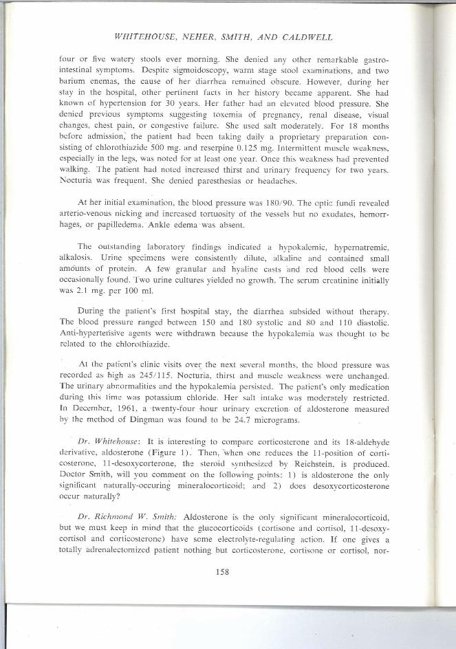
four or five watery stools ever morning. She denied any other remarkable gastrointestinal symptoms. Despite sigmoidoscopy, warm stage stool examinations, and two barium enemas, the cause of her diarrhea remained obscure. However, during her stay in the hospital, other pertinent facts in her history became apparent. She had known of hypertension for 30 years. Her father had an elevated blood pressure. She denied previous symptoms suggesting toxemia of pregnancy, renal disease, visual changes, chest pain, or congestive failure. She used salt moderately. For 18 months before admission^ the patient had been taking daily a proprietary preparation consisting of chlorothiazide 500 mg. and reserpine 0.125 mg. Intermittent muscle weakness, especially in the legs, was noted for at least one year. Once this weakness had prevented walking. The patient had noted increased thirst and urinary frequency for two years. Nocturia was frequent. She denied paresthesias or headaches.
At her initial examination, the blood pressure was 180/90. The optic fundi revealed arterio-venous nicking and increased tortuosity of the vessels but no exudates, hemorrhages, or papilledema. Ankle edema was absent.
The outstanding laboratory findings indicated a hypokalemic, hypernatremic, alkalosis. Urine specimens were consistently dilute, alkaline and contained small amounts of protein. A few granular and hyaline casts and red blood cells were occasionally found. Two urine cultures yielded no growth. The serum creatinine initially was 2.1 mg. per 100 ml.
During the patient's first hospital stay, the diarrhea subsided without therapy. The blood pressure ranged between 150 and 180 systolic and 80 and 110 diastolic. Anti-hyperterisive agents were withdrawn because the hypokalemia was thought to be related to the chlorothiazide.
At the patient's clinic visits over the next several months, the blood pressure was recorded as high as 245/115. Nocturia, thirst and muscle weakness were unchanged. The urinary abnormalities and the hypokalemia persisted. The patient's only medication during this time was potassium chloride. Her salt intake was moderately restricted. In December, 1961, a twenty-four -hour urinary excretion of aldosterone measured by the method of Dingman was found to be 24.7 micrograms.
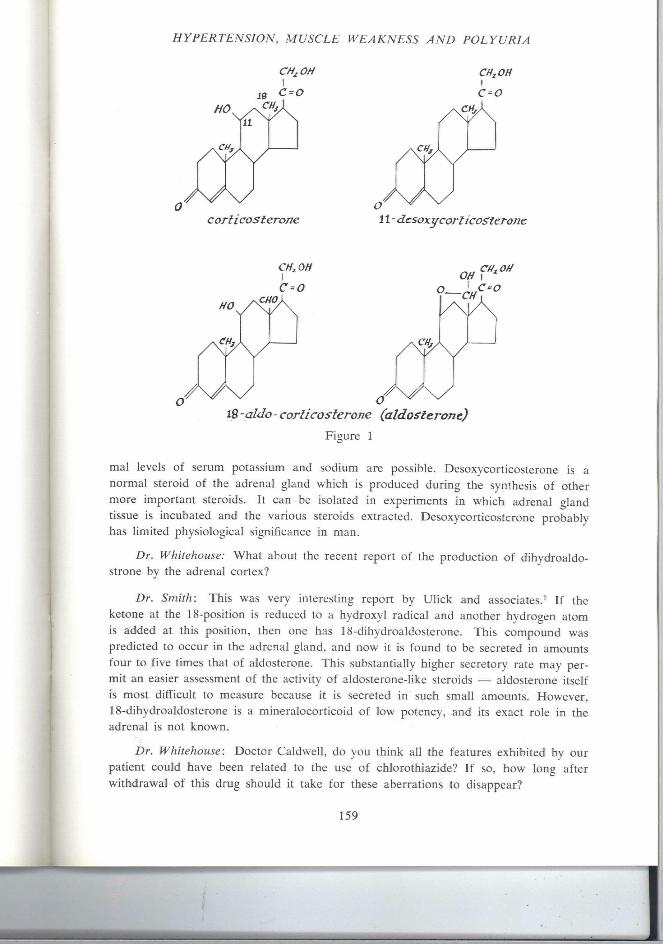
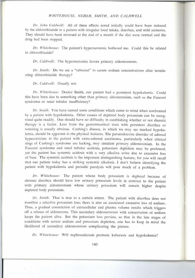
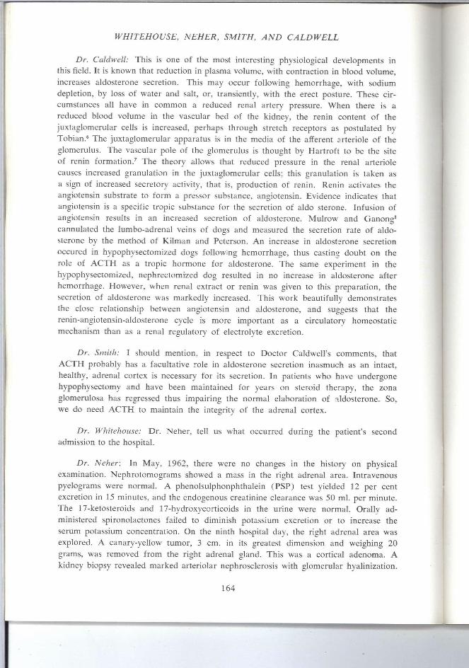
Dr. Whitehouse: It is interesting to com.pare corticosterone and its 18-aldehyde derivative, aldosterone (Figure 1). Then, when one reduces the 11-position of corticosterone, 11-desoxycorterone, the steroid synthesized by Reichstein, is produced. Doctor Smith, will you comment on the following, points: 1) is aldosterone the only significant naturally-occuring mineralocorticoid; and 2) does desoxycorticosterone occur naturally?
Dr. Richmond W. Smith: Aldosterone is the only significant mineralocorticoid, but we must keep in mind that the glucocorticoids (cortisone and Cortisol, 11-desoxy-cortisol and corticosterone) have seme electrolyte-regulating action. If one gives a totally adrenalectomized patient nothing but corticosterone, cortisone or Cortisol, nor-
158
HYPERTENSION, MUSCLE WEAKNESS AND POLYURIA
cor I icosterone 11- dcsoxycorticogleronc
CH^OM I C = 0
OH
18-aldo - corticosterone (aldosieronc) Figure 1
mal levels of serum potassium and sodium are possible. Desoxycorticosterone is a normal steroid of the adrenal gland which is produced during the synthesis of other more important steroids. It can be isolated in experiments in which adrenal gland tissue is incubated and the various steroids extracted. Desoxycorticosterone probably has limited physiological significance in man.
Dr. Whitehouse: What about the recent report of the production of dihydroaldo-strone by the adrenal cortex?
Dr. Smith: This was very interesting report by Uhck and associates.' I f the ketone at the 18-position is reduced to a hydroxyl radical and another hydrogen atom is added at this position, then one has 18-dihydroaldosterone. This compound was predicted to occur in the adrenal gland, and now it is found to be secreted in amounts four to five times that of aldosterone. This substantially higher secretory rate may permit an easier assessment of the activity of aldosterone-like steroids — aldosterone itself is most difficult to measure because it is secreted in such small amounts. However, 18-dihydroaldosterone is a mineralocorticoid of low potency, and its exact role in the adrenal is not known.
Dr. Whitehouse: Doctor Caldwell, do you think all the features exhibited by our patient could have been related to the use of chlorothiazide? I f so, how long after withdrawal of this drug should it take for these aberrations to disappear?
159
WHITEHOUSE, NEHER, SMITH, AND CALDWELL
Dr. John Caldwell: Al l of these effects noted initially could have been induced by the chlorothiazide in a patient with irregular food intake, diarrhea, and mild azotemia. They should have been reversed at the end of a month if the diet were normal and the drug had been stopped.
Dr. Whitehouse: The patient's hypernatremia bothered me. Could this be related to chlorothiazide?
Dr. Caldwell: The hypernatremia favors primary aldosteronism.
Dr. Smith: Do we see a "rebound" in serum sodium concentrations after terminating chlorothiazide therapy?
Dr. Caldwell: Usually not.
Dr. Whitehouse: Doctor Smith, our patient had a persistent hypokalemia. Could this have been due to something other than primary aldosteronism, such as the Fanconi syndrome or renal tubular insufficiency?
Dr. Smith: You have named some conditions which come to mind when confronted by a patient with hypokalemia. Other causes of depleted body potassium can be recognized quite readily. One should have no difficulty in establishing whether or not diuretic therapy is a factor. Loss from the gastrointestinal tract with persistent diarrhea or vomiting is usually obvious. Cushing's disease, in which we may see marked hypokalemia, should be apparent in the physical features. The paraendocrine disorder of adrenal hypecorticism in the patient with extra-adrenal carcinoma, particularly when clinical sings of Cushing's syndrome are lacking, may simulate primary aldosteronism. In the Fanconi syndrome and renal tubular acidosis, potassium depletion may be profound, yet the patient has systemic acidosis with a very alkaline urine due to excessive loss of base. The systemic acidosis is the important distinguishing feature, for you will recall that our patient today has a striking systemic alkalosis. I don't believe identifying the patient with hypokalemia and periodic paralysis will pose much of a problem.
Dr. Whitehouse: The patient whose body potassium is depleted because of chronic diarrhea should have low urinary potassium levels in contrast to the patient with primary aldosteronism whose urinary potassium will remain higher despite depleted body potassium.
Dr. Smith: That is true to a certain extent. The patient with diarrhea does not manifest a selective potassium loss; there is also an associated excessive loss of sodium. Thus, a gradual constriction of extracellular and plasma volume results which triggers off a release of aldosterone. This secondary aldosteronism with conservation of sodium keeps the patient alive. But the potassium loss persists, so that in the late stages of conditions with severe sodium and potassium depletion, one has to keep in mind the likelihood of secondary aldosteronism complicating the picture.
Dr. Whitehouse: Will nephrosclerosis promote kaliuresis and hypokalemia?
160
I
HYPERTENSION, MUSCLE WEAKNESS AND POLYURIA
Dr. Caldwell: No, we seldom see the serum potassium below 2 5 mEq/1. in uncomplicated nephrosclerosis. Of course, there is a functional disparity of the kidneys in many patients with hypertension, and there may be potassium wastage in some cases. This could occur as a consequence of occlusive renal arterial disease complicating longstanding hypertension. Aldosteronism with excessive potassium loss may result. In late stages of hypertensive disease, there may be secondary hyperplasia of the adrenal cortex; experimental evidence suggests that the hyperplasia is secondary to renal hypertension.
Dr. Whitehouse: Is it fair to say that most cases of "potassium-losing nephritis" have proved to be primary aldosteronism?
Dr. Smith: That is a fair statement. Have we sufficiently emphasized that chronic pyelonephritis itself can be a potassium-wasting disease? Precise diagnosis may be dif f i cult because of the high incidence of pyeloneprritis in patients with primary aldosteron-
Dr. Caldwell: Studies on patients with hypertensive disease and hyperaldosteron-uria also show that a large number my have pyelonephritis as well as renal artery stenosis.
Dr. Whitehouse: It's practically impossible to have a pure lesion, isn't it? Doctor Smith, will you comment on the 24.7 micrograms of aldosterone which our patient excreted in the twenty-four hour urine specimen?
Dr. Smith: This represents a three to four-fold increase over the normal aldosterone excretion as measured in our laboratory. Our results are quite comparable to those obtained elsewhere. Normal excretion is three to ten micrograms per day. We currently use a method which recovers "free" or acid extractable aldosterone as distinguished from the total aldosterone content. Much of the aldosterone is excreted in a conjugated state as reduced aldosterone. If this is measured, one may find values in the range of several hundred micrograms per day.
Dr. Whitehouse: We increased the dietary sodium before measuring the aldosterone excertion. Is this reasonable?
Dr. Smith: In a patient with primary aldosteronism, juggling of the sodium intake will not materially alter the level of aldosterone in urine, but in a normal subject or in patients with congestive heart failure or cirrhosis, restricting dietary sodium will have a profound effect on aldosterone excertion. This presumably occurs because constriction in plasma volume accelerates aldosterone release. In general restriction of dietary sodium should be avoided during measurement of aldosterone excretion.
Dr. Whitehouse: Do levels of aldosteronuria, as obtained in our patient, occur in patients with genetic hypertension?
Dr. Caldwell: When you say "genetic hypertension", you are postulating that there is a group of patients with hypertension entirely of genetic orgin. This is an unproved hypothesis, but it is an attractive one. No one denies the familial occurence of hyper-
161
WHITEHOUSE, NEHER, SMITH, AND CALDWELL
tension. Genest' analyzed over 200 determinations of urinary aldosterone in 110 patients and normal subjects; 46 patients had "essential" hypertension. Forty-three per cent of this latter group had hyperaldosteronuria, that is, an excretion of more than ten micrograms of aldosterone per twenty-four hours. Laragh,^ with the radioisotope dilution method, was not able to confirm this finding. He found hyperaldosteronuria of higher levels in patients with more severe hypertensive disease; the highest levels have occured in patients with malignant hypertension. Warier et al" report that 30 per cent of patients with so-called benign hypertension have excess aldosterone in the urine as measured by the physicochemical method of Nehr and Wettstein. Reproducibility of these findings was poor; perhaps some of the discrepancy in these studies may be related to the methods of measurements. With the physicochemical method, only 10 per cent of secreted aldosterone is recovered. Using the radio-isotope method, the rate of urinary excretion is said to be almost the same as the rate of secretion by the adrenal cortex itself.
Dr. Whitehouse: Ulick and co-workers' noted that all patients with primary aldosteronism failed to have high levels of aldosterone in the urine. They were attracted to the determination of the secretion rate of aldosterone by the adrenal cortex as a more accurate measure of hyperaldosteronism. Doctor Smith, will you comment on this point and assess its clinical applicability?
Dr. Smith: Most laboratories employ methods which measure "pH-1" aldosterone in urine. As with Cushing's disease, in which the urinary glucocorticoid excretion may be borderline and equivocal, so with primary aldosteronism due to a cortical tumor, aldosterone excretion in urine may be only slightiy increased. There is an overlap between the excretion levels by normal individuals and patients with definite organic disease. For this reason, isotope techniques were developed to refine the analysis of aldosterone in urine. In general, when measuring excretion rate, labelled aldosterone is added to the urine which permits a more accurate determination of the steroid without having to achieve quantitive recovery. When determining secretion rates, a known amount of labelled aldosterone is given to the patient and the specific .activity of the aldosterone metabolite in the urine is determined. From this information and with certain assumptions, the secretion rate can be estimated. The normal excretion of "pH-1" aldosterone is in the order or of 10 micrograms per day. The secretion rate may be 100 to 200 micrograms per day. Thus, we have a 10 to 20 fold margin in measuring secretion rates. Most of the aldosterone to be accounted for (the difference between 200 and 10 micrograms) can be found in the reduced fractions which are conjugated, and excreted as tetrahydroderivatives. As to a preference of approach, it is claimed that the secretion rate gives a more reliable, more meaningful figure. Since the introduction of Peterson's double isotope dilution technique, the advantages of determining secretion rates have been dissipated to some extent. I think it is a 50-50 choice now as to which procedure we should employ.
Dr. Whitehouse: Urinary aldosterone was measured only once in this patient. Would you say that sometimes the clinical picture can be so compelling that it is unnecessary to measure aldosterone in the urine before definitive therapy?
162
HYPERTENSION, MUSCLE WEAKNESS AND POLYURIA
Dr. Smith: This is one of the more important questions to be answered. We would be quite in error to operate on a patient merely with one quantitation of aldosterone in the absence of other supporting evidence. The important question is: how do we make a diagnosis of primary aldosteronism? Do we turn to the patient, look for the clinical features, or do we simply turn to the urine and find the level of aldosterone? May I cite a similar situation? Little would come of our efforts if we were to search for Cushing's syndrome by turning our backs on the patient and do nothing but send urine specimens to the laboratory for corticoid determinations. A great number of such unselected specimens would yield values above the normal range. The real key to the diagnosis of primary aldosteronism is in the clinical picture, especially the history. The patient described to us today brings out this point clearly. I f the physician is to ferret out the few patients with primary aldosteronism among the many suspects who may come to his attention, he must do so by a careful evaluation of the history. Then, if he strongly suspects primary aldosteronism on clinical grounds, he should leave a 24-hour urine specimen with the chemist. Several determinations are desirable.
Dr. Whitehouse: Aldosterone is said to act on the renal tubules whereby sodium is re-absorbed and potassium is excreted. Is this correct?
Dr. Smith: It is a clear, somewhat simplified statement, but I wouldn't limit the action to the renal tubules. Aldosterone, like glucocorticoids, probablv acts on cell membranes throughout the body, promoting sodium and hydrogen ion ingress and potassium ion egress. There is a mass exchange of sodium for potassium in cells generally.
Dr. Whitehouse: Doctor Caldwell, since hypertension is prominent in primary aldosteronism, does aldosterone produce vaso-constriction?
Dr. Caldwell: No, aldosterone does not have a direct vaso-eonstricting effect. A change in vascular tone is probably secondary to thesodium retention and potassium deficit in the artery wall. It is a paradox that aldosterone administered to rabbits, dogs, or even to human subjects does not raise the blood pressure. Perhaps if aldosterone is secreted in excess for many months or years, hypertension will ensue.
Dr. Whitehouse: Doctor Smith, how is the secretion of aldosterone controlled?
Dr. Smith: The influence on aldosterone secretion by corticotropin (ACTH) is inconsequential. Potassium loading is known to stimulate aldosterone secretion, but the prime regulator of aldosterone would appear to be the plasma volume, this being conditioned by the sodium intake. Constriction of the thoracic portion of the inferior veno cava increases aldosterone secretion while changes in carotid sinus pressures have a regulatory influence. Postulated baro-receptors therein respond to expansion and constriction of the plasma volume. Recent evidence has supported a more important humoral regulation of aldosterone secretion by way of the kidney. Through stimulatio of the juxtaglomerular appartus, renin and later angiotensin are formed; the latter stimulates aldosterone secretion.
Dr. Whitehouse: Doctor Caldwell, will you elaborate on the release of aldosterone through the action of angiotensin?
163
WHITEHOUSE, NEHER, SMITH, AND CALDWELL
Dr. Caldwell: This is one of the most interesting physiological developments in this field. It is known that reduction in plasma volume, with contraction in blood volume, increases aldosterone secretion. This may occur following hemorrhage, with sodium depletion, by loss of water and salt, or, transiently, with the erect posture. These circumstances all have in common a reduced renal artery pressure. When there is a reduced blood volume in the vascular bed of the kidney, the renin content of the juxtaglomerular cells is increased, perhaps through stretch receptors as postulated by Tobian.' The juxtaglomerular apparatus is in the media of the afferent arteriole of the glomerulus. The vascular pole of the glomerulus is thought by Hartroft to be the site of renin formation.' The theory allows that reduced pressure in the renal arteriole causes increased granulation in the juxtaglomerular cefls; this granulation is taken as a sign of increased secretory activity, that is, production of renin. Renin activates the angiotensin substrate to form a pressor substance, angiotensin. Evidence indicates that angiotensin is a specific tropic substance for the secretion of aldo sterone. Infusion of angiotensin results in an increased secretion of aldosterone. Mulrow and Ganong' cannulated the lumbo-adrenal veins of dogs and measured the secretion rate of aldosterone by the method of Kilman and Peterson. An increase in aldosterone secretion occured in hypophysectomized dogs following hemorrhage, thus castino doubt on the role of ACTH as a tropic hormone for aldosterone. The same experiment in the hypophysectomized, nephrectomized dog resulted in no increase in aldosterone after hemorrhage. However, when renal extract or renin was given to this preparation, the secretion of aldosterone was markedly increased. This work beautifully demonstrates the close relationship between angiotensin and aldosterone, and suggests that the renin-angiotensin-aldosterone cycle is more important as a circulatory homeostatic mechanism than as a renal regulatory of electrolyte excretion.
Dr. Smith: I should mention, in respect to Doctor Caldwell's comments, that ACTH probably has a facultative role in aldosterone secretion inasmuch as an intact, healthy, adrenal cortex is necessary for its secretion. In patients who have undergone hypophysectomy and have been maintained for years on steroid therapy, the zona glomerulosa has regressed thus impairing the normal elaboration of aldosterone. So, we do need ACTH to maintain the integrity of the adrenal cortex.
Dr. Whitehouse: Dr. Neher, tell us what occurred during the patient's second admission to the hospital.
Dr. Neher: In May, 1962, there were no changes in the history on physical examination. Nephrotomograms showed a mass in the right adrenal area. Intravenous pyelograms were normal. A phenolsulphonphthalein (PSP) test yielded 12 per cent excretion in 15 minutes, and the endogenous creatinine clearance was 50 ml. per minute. The 17-ketosteroids and 17-hydroxycorticoids in the urine were normal. Orally administered spironolactones failed to diminish potassium excretion or to increase the serum potassium concentration. On the ninth hospital day, the right adrenal area was explored. A canary-yellow tumor, 3 cm. in its greatest dimension and weighing 20 grams, was removed from the right adrenal gland. This was a cortical adenoma. A kidney biopsy revealed marked arteriolar nephrosclerosis with glomerular hyalinization.
164
HYPERTENSION, MUSCLE WEAKNESS AND POLYURIA
No changes characteristic of kaliopenic nephropathy were seen. The patient's postoperative course was uneventful.
Dr. Whitehouse: Her most recent blood pressure was 160/94. The serum potassium is now 4.6 mEq/1. The urine pH is 5, and the specific gravity is 1.018. The polyuria and polydipsia are gone, and the muscle strength seems improved. Although the clinical response is gratifying we don't expect a complete return to a normotensive state because of the considerable nephrosclerosis in the renal biopEV. In a coflected series' of patients with primary aldosteronism, 66 per cent experienced a return of blood pressure to normal; 20 percent noted improvement, and 14 per cent had no change. In these patients some renal biopsies were normal; others showed changes similar to those found in our patient. Many demonstrated vacuolization of the renal tubules characteristic of kaliopenic nephropathy. Are you surprised, Doctor Smith, that we found no renal tubular changes characteristic of kaliopenic nephropathy?
Dr. Sinith: Very much so. I doubt that she received sufficient supplemental potassium to have reversed the expected changes.
Dr. Caldwell: Perhaps the lack of vacuolization in the renal tubules means that the adrenal adenoma was a recent development. The patient had hypertension for 30 years, but she had polyria for only two years. Perhaps there was a chronic stimulation of the adrenal cortex resulting in hyperaldosteronuria. This tropic activity may have eventually induced a cortical adenoma. Of course, a de novo adenoma may arise in a younger person resulting in truly primary aldosteronism.
Dr. Smith: That's nice thinking, but I can't accept a 20 gram, solitary cortical adenoma as being the result of "tropic hormone" stimulation.
Dr. Caldwell: Well, about that we could speculate and debate. However, I think the very presence of hypertension is an indication for a screening test for primary aldosteronism. A careful history to detect muscle weakness, headache, polyuria, and polydipsia will be most helpful. Every patient should have the serum potassium measured and an electrocardiogram taken for evidence of hypokalem.ia. It is also important in any patient with hyperaldosteronuria and long standing hypertension to do a thorough evaluation of the severity of cardiovascular renal disease, for pyelonephritis and, in particular, renal artery stenosis.
Dr. Thomas N . James: I want to object to the concept of stretch receptors being present in the juxtaglomerular apparatus. Stretch receptors have a distinctive anatomic structure and no such morphology exists in the area of the juxtaglomerular apparatus. Stretch receptors are well recognized in the right atrium, great veins, aortic arch and carotid sinus. The term, "stretch mechanism" has been suggested by others to clarify the misconception introduced by this unfortunate choice of terms.
Dr. John W. Keyes: With regard to the use of thiazide diuretics, I do not believe that these, in themselves, can produce hypokalemia of a severe degree. It is
165

WHITEHOUSE, NEHER, SMITH, AND CALDWELL
true that they can lower potassium in the presence of severe heart failure, but in
your patient, the profound hypokalemia itself is a good screening test for primary
aldosteronism. In fact, another case of primary aldosteronism was discovered at
this hospital by the occurrence of a profound hypokalemia following the use of
chlorothiazide.
REFERENCES
1. Ulick, S., and Vetter, K. K. The secretion of 18-dihydroaldesterone by the human adrenal J.C.L 41:1406; 1962.
2. Genest, J., Nowaczynski, W., Koiw, E., Sander T., and Biron, P.: Adrenocortical function in essential hypertension, In, International Symposium on Hypertension, ed. by K. D. Bock, and P. T. Cottier, Beriin, Springer-Verlag, 1960, pp. 126-47.
3. Laragh, J. H., Ulick, S., Januszewicz, V., Deming, Q.B., Kelly, W.G. and Lieberman, S.: Aldosterone secretion and primary and malignant hypertension. J. Clin. Inves 39-1091 1960. & .Jf
4. Warter J., Schwartz, J., and Block, R.: Significance of hyperaldosterounria in hypertension, In, International Symposium on Hypertension, ed. by K. D. Bock, and P. T. Cottier Berlin, Springer, 1960, pp. 147-58.
5. Ulick, S., Laragh, J.H., and Lieberman, S. The isolation of a urinary metabolite of aldesterone and its use to measure the rate or secretion of aldosterone by the adrenal cortex of man. Tr. Assn. Am. Phys. 71:225, 1958.
6. Tobian, L. Relationship of the juxtaglomerular apparatus to renin and angiotensin. Circ. 25:189, 1962. J f t - 6
7. Hartcroft, W.S. and Hartcroft, P.M. New approaches in the study of cardiovascular disease: aldosterone, renin, hypertension and juxtaglomerular cells. Fed. Proc. 20:845, 1961.
8. Mulrow, P.J. and Ganong, W.F. Role of the kidney and the reninangiotensin system in the response of aldosterone secretion to hemorrhage. Circulation 25:213, 1962.
9. Conn, J.W. and Conn, E.S. Primary aldosteronism versus hypertensive disease with secondary aldosteronism. Recent Progr. in Hormone Res. 17:389, 1961.
166
![Medical Treatment of Nocturia in Men with Lower …...balance [2], leading to excessive production of urine at all times (global polyuria) or primarily at night (nocturnal polyuria),](https://img.pdfslide.us/doc/110x75/5fa935277a549e105b2545fb/medical-treatment-of-nocturia-in-men-with-lower-balance-2-leading-to-excessive.jpg)


![[Critica] Apple's Weakness](https://img.pdfslide.us/doc/110x75/54b2dc494a7959d10e8b456b/critica-apples-weakness.jpg)