Embed Size (px)
Citation preview
Research ArticleHypertension and CardiovascularRemodelling in Rats Exposed to Continuous Light: Protection byACE-Inhibition and Melatonin
Fedor Simko,1,2,3,4 Olga Pechanova,4,5 Kristina Repova Bednarova,1
Kristina Krajcirovicova,1 Peter Celec,1,6 Natalia Kamodyova,6 Stefan Zorad,3,4
Jarmila Kucharska,2 Anna Gvozdjakova,2 Michaela Adamcova,7 and Ludovit Paulis1,4
1 Department of Pathophysiology, School of Medicine, Comenius University, Sasinkova 4, 81372 Bratislava, Slovakia2 3rd Clinic of Medicine, School of Medicine, Comenius University, 83305 Bratislava, Slovakia3 Institute of Experimental Endocrinology, Slovak Academy of Sciences, 83305 Bratislava, Slovakia4Center of Excellence NOREG, 81372 Bratislava, Slovakia5 Institute of Normal and Pathological Physiology, Slovak Academy of Sciences, 81371 Bratislava, Slovakia6 Institute of Molecular Biomedicine, School of Medicine, Comenius University, 81372 Bratislava, Slovakia7 Department of Physiology, School of Medicine, Charles University, 50038 Hradec Kralove 1, Czech Republic
Correspondence should be addressed to Fedor Simko; [email protected]
Received 10 January 2014; Revised 27 May 2014; Accepted 29 May 2014; Published 6 July 2014
Academic Editor: Russel J. Reiter
Copyright © 2014 Fedor Simko et al. This is an open access article distributed under the Creative Commons Attribution License,which permits unrestricted use, distribution, and reproduction in any medium, provided the original work is properly cited.
Exposure of rats to continuous light attenuates melatonin production and results in hypertension development. This studyinvestigated whether hypertension induced by continuous light (24 hours/day) exposure induces heart and aorta remodelling andif these alterations are prevented by melatonin or angiotensin converting enzyme inhibitor captopril. Four groups of 3-month-oldmaleWistar rats (10 per group) were treated as follows for six weeks: untreated controls, exposed to continuous light, light-exposed,and treated with either captopril (100mg/kg/day) or melatonin (10mg/kg/day). Exposure to continuous light led to hypertension,left ventricular (LV) hypertrophy and fibrosis, and enhancement of the oxidative load in the LV and aorta. Increase in systolicblood pressure by continuous light exposure was prevented completely by captopril and partially by melatonin. Both captopril andmelatonin reduced the wall thickness and cross-sectional area of the aorta and reduced the level of oxidative stress. However, onlycaptopril reducedLVhypertrophy development andonlymelatonin reducedLVhydroxyproline concentration in insoluble and totalcollagen in rats exposed to continuous light. In conclusion, captopril prevented LV hypertrophy development in the continuouslight-induced hypertension model, while only melatonin significantly reduced fibrosis. This antifibrotic action of melatonin maybe protective in hypertensive heart disease.
1. Introduction
Left ventricular (LV) hypertrophy, although representing anadaptation to hemodynamic overload, is associated withincreased cardiovascular risk [1, 2]. The search for newapproaches for the prevention or regression of LV hypertro-phy in different models of pathological myocardial growthcontinues [3–7].
Melatonin (N-acetyl-5-methoxytryptamine), the mostabundant secretory product of the vertebrate pineal glandthat controls biological rhythms [8], has numerous beneficial
actions in the heart [9–12] and plays an important rolein the pathogenesis of hypertension [13–16]. Experimentalpinealectomy, a procedure that is easily performed in rats[17], reduced the level of both day-time and nocturnalmelatonin [8] and resulted in an enhancement of vascularresponsiveness to vasoconstriction stimuli [18], increasedblood pressure, and myocardial fibrosis [19]. An alternativeapproach to attenuate melatonin production, associated witha blood pressure rise, is the exposure of rats to continuous24 hours/day chronic lighting [20]. This model of melatonin-deficient hypertension seems to be more physiological than
Hindawi Publishing CorporationMediators of InflammationVolume 2014, Article ID 703175, 10 pageshttp://dx.doi.org/10.1155/2014/703175

2 Mediators of Inflammation
pinealectomy because it reduces only nocturnal melatoninsecretion and no surgery is involved [20]. Since melatoninexerts cardiovascular protection on several levels includ-ing antioxidant and scavenging actions [21–23], endothelialprotection, sympatholytic effect [24–28], and antiarrhythmiceffects [29], it is comprehensible that a deficiency of mela-tonin could result in pathological alterations in circulationand target organ damage [2, 8]. Indeed, several studies withpatients suffering from acute myocardial infarction with STsegment elevation indicated that deficit of melatonin couldbe a negative prognostic factor of the severity of infarction,subsequent remodelling, and prognosis [30–33] and thatmelatonin supplementation may exert beneficial effects as aradical scavenger in a human model of myocardial ischemia-reperfusion damage [34, 35].
This study investigatedwhether continuous light-inducedhypertension results in the pathological growth of the heartand the aorta and whether melatonin can modify thesepotential alterations. Moreover, the effect of melatonin wascompared with the angiotensin converting enzyme (ACE)inhibitor, captopril. Since ACE-inhibitors are antihyperten-sives with well-established antihypertrophic effect, which isalso based on inhibition of angiotensin II production andinterference with aldosterone, catecholamines, or endothelinproduction [37, 38], ACE-inhibitionmay be protective also incontinuous light-induced hypertension.
2. Material and Methods
2.1. Animals and Treatment. Male adult three-month-oldWistar rats (Dobra Voda, Slovak Republic) were randomlydivided into four groups (𝑛 = 10 in each group): control(Wistar) rats (c), rats exposed to 24 hours/day continuouslight (24), and continuous light-exposed rats treated witheither captopril (100mg/kg/day; Egis Pharmaceuticals Ltd,Budapest; 24C) ormelatonin (10mg/kg/day; Sigma ChemicalCo., Deisenhofen, Germany; 24M). Captopril and melatoninwere dissolved in drinking water and their concentrationwas adjusted to daily water consumption to ensure correctdosage. The natural water consumption of rats was about 12-13mL/100 g of body weight. In order to ensure that all theamount of water with dissoluted melatonin was really drankby a particular rat, only 10mL/100mg of water-melatoninsolution was offered. The solution was prepared as follows:10mg of melatonin was dissoluted in 100mL water, while noadditional substance was added to dissolute the substance.Melatonin containing solutions were protected from lightexposure by using black bottles. All rats were housed inindividual cages at 22–24∘C and fed a regular pellet diet adlibitum in accordance with the Guide for the Care and Use ofLaboratory Animals published by the US National Institutesof Health (NIH Publication number 8523, revised 1985).
Systolic blood pressure (SBP) was measured each weekby noninvasive tail-cuff plethysmography (Hugo-Sachs Elek-tronic, Freiburg, Germany). After six weeks, rats were decap-itated and body weight (BW), heart weight (HW), and LVand right ventricle weights (LVW, RVW) were determinedand their relative weights (LVW/BW and RVW/BW ratio)were calculated. Left ventricle samples were frozen at −80∘C
and later used for the determination of hydroxyprolineconcentrations and oxidative stress parameters.
2.2. Morphometry of Aorta. Thoracic aorta samples werefixed for 24 hours in 4% formaldehyde, embedded in paraffin,cut in serial 5𝜇m thick sections, and stained with haema-toxylin and eosin. Wall thickness (WT) in 𝜇m and the innercircumference in mm were measured using light microscopyand a two-dimensional image analyzer (Impor Pro; Kvants.r.o., Bratislava, Slovak Republic). Inner diameter (ID)expressed in mm and cross-sectional area (CSA) expressedin mm2 were then calculated [28]. During morphometricmeasurements the observer was blinded (only the numbersof animals were accessible for the observer).
2.3. Determination of Hydroxyproline. Collagenous proteinswere isolated according to Pelouch et al. [39, 40]. Hydrox-yproline concentrations were analyzed spectrophotometri-cally at 550 nm [41].
2.4. Oxidative Load Measurement. Tissue samples werehomogenized in 1mL phosphate buffered saline and cen-trifuged at 19320 g (4∘C) for 5 minutes and the supernatantwas used for further biochemical analyses.
Advanced glycation end-products (AGEs) as markersof carbonyl stress were measured using the characteristicfluorescence (𝜆ex. = 370 nm, 𝜆em. = 440 nm) [42]. Briefly,tissue homogenates were diluted 10-fold with phosphatebuffered saline and the AGEs concentration was calculatedbased on the calibration curve prepared with AGE-BSAcalibrator [43].
Advanced oxidation protein products (AOPP) weredetermined spectrophotometrically [44]. Briefly, 200𝜇L ofappropriately diluted tissue homogenates were incubatedwith glacial acetic acid for 2 minutes and the absorbance wasread at 340 nm using Sapphire II instrument (Tecan, Grodig,Austria). AOPP concentration was calculated on the basis ofthe calibration curve of chloramin T with potassium iodide.Proteins were quantified using BCA protein assay kit (SigmaAldrich, Steinheim, Germany) [42].
2.5. CoQ and Tocopherol Measurement. Concentrations ofCoQ9ox, CoQ10ox, and gamma- and alpha-tocopherol weredetermined by HPLC method according to Lang et al. [45]with some modifications [46]. Concentrations of CoQ andtocopherols were detected spectrophotometrically at 275 nmand 295 nm, respectively.
2.6. Statistical Analysis. Results are expressed as mean ±S.E.M. One-way, two-tailed analysis of variance (ANOVA)and the Bonferroni test were used for statistical analysis.Differences were considered significant if the 𝑃 value <0.05.
3. Results
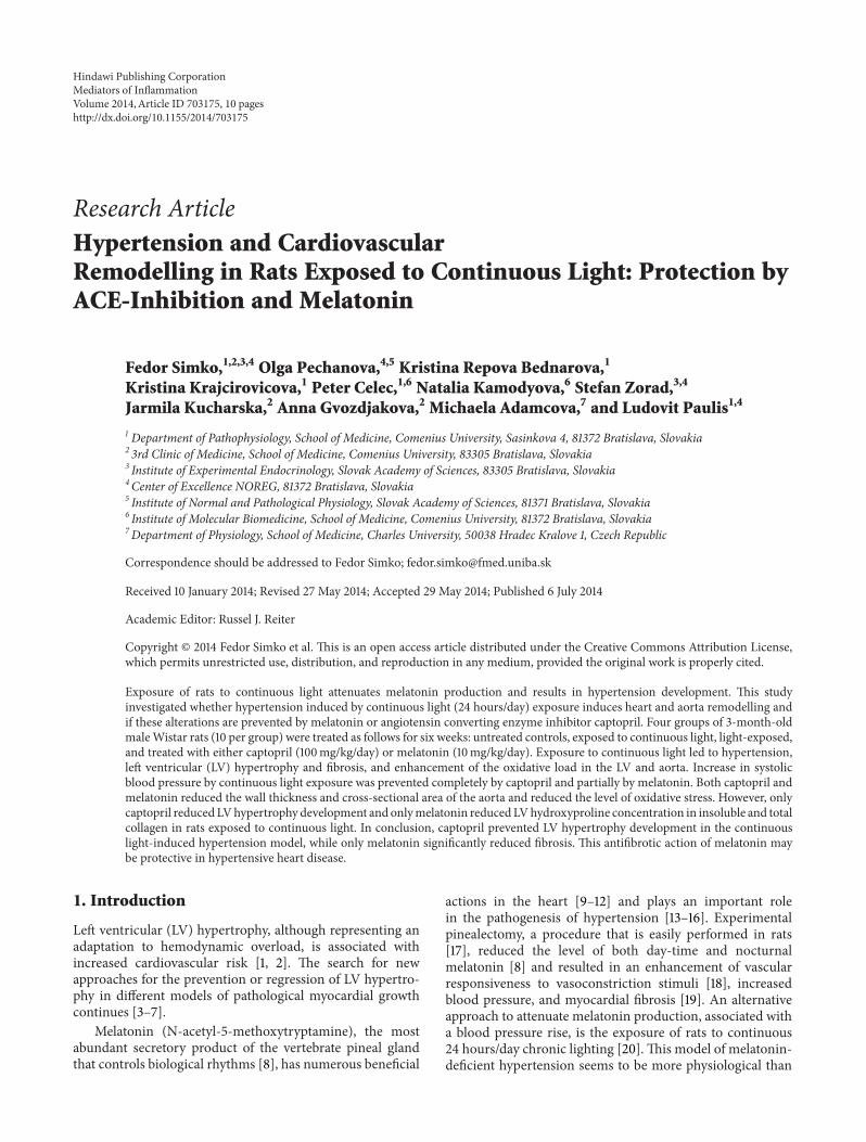
3.1. Cardiovascular Parameters. SBP was 125 ± 0.79mmHgin control rats and 163 ± 0.48mmHg in continuous light-group. SBP decreased significantly (𝑃 < 0.05) by bothcaptopril (21%) and melatonin (15%) treatment compared
Mediators of Inflammation 3
c
24
24C
24M
100
110
120
130
140
150
160
170
0 1 2 3 4 5 6
Systo
lic b
lood
pre
ssur
e (m
mH
g)
Weeks
c24
24C24M
∗∗
∗
∗
∗
∗#∗#
∗# ∗#∗# ∗#
∗#
∗#∗#
∗#
∗#
(a)
0
0.2
0.4
0.6
0.8
1
1.2
1.4
c 24 24C 24M
Relat
ive L
V w
eigh
t (m
g/g)
∗#
∗
(b)
Figure 1: The influence of captopril (24C) and melatonin (24M) on blood pressure (a) and relative left ventricle (LV) weight (LVW/BW) (b)in 24 hours/day continuous light exposure-induced hypertension (24). c: Wistar controls. ∗𝑃 < 0.05 versus c; #
𝑃 < 0.05 versus 24.
0.00
0.02
0.04
0.06
0.08
0.10
0.12
0.14
0.16
c 24 24C 24M
Aort
ic w
all t
hick
ness
(mm
)
∗#∗#
(a)
#
0.00
0.10
0.20
0.30
0.40
0.50
0.60
0.70
0.80
0.90
c 24 24C 24M
Cros
s-se
ctio
nal a
rea o
f the
aort
a (m
m2)
∗#
(b)
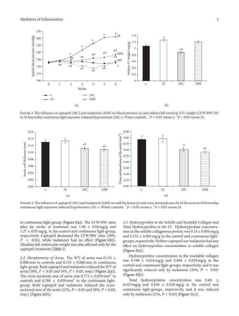
Figure 2:The influence of captopril (24C) andmelatonin (24M) on wall thickness (a) and cross-sectional area (b) of the aorta in 24 hours/daycontinuous light exposure-induced hypertension (24). c: Wistar controls. ∗𝑃 < 0.05 versus c; #
𝑃 < 0.05 versus 24.
to continuous light-group (Figure 1(a)). The LVW/BW ratioafter six weeks of treatment was 1.08 ± 0.04mg/g and1.27 ± 0.05mg/g, in the control and continuous light-group,respectively. Captopril decreased the LVW/BW ratio (30%,𝑃 < 0.05), while melatonin had no effect (Figure 1(b)).Absolute left ventricular weight was also affected only by thecaptopril treatment (Table 1).
3.2. Morphometry of Aorta. The WT of aorta was 0.131 ±0.006mm in controls and 0.135 ± 0.006mm in continuouslight-group. Both captopril andmelatonin reduced theWTofaorta (18%, 𝑃 < 0.05 and 19%, 𝑃 < 0.05, resp.) (Figure 2(a)).The cross-sectional area of aorta was 0.772 ± 0.059mm2 incontrols and 0.780 ± 0.059mm2 in the continuous light-group. Both captopril and melatonin reduced the cross-sectional area of the aorta (22%, 𝑃 < 0.05 and 28%, 𝑃 < 0.05,resp.) (Figure 2(b)).
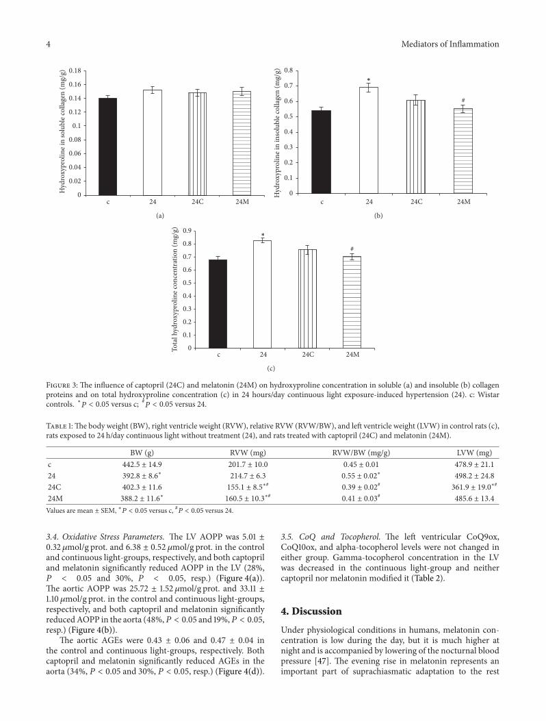
3.3. Hydroxyproline in the Soluble and Insoluble Collagen andTotal Hydroxyproline in the LV. Hydroxyproline concentra-tion in the soluble collagenous protein was 0.14 ± 0.004mg/gand 0.152 ± 0.005mg/g in the control and continuous light-groups, respectively. Neither captopril normelatonin had anyeffect on hydroxyproline concentration in soluble collagen(Figure 3(a)).
Hydroxyproline concentration in the insoluble collagenwas 0.540 ± 0.024mg/g and 0.690 ± 0.029mg/g in thecontrol and continuous light-groups, respectively, and it wassignificantly reduced only by melatonin (20%, 𝑃 < 0.05)(Figure 3(b)).
Total hydroxyproline concentration was 0.68 ±0.025mg/g and 0.830 ± 0.020mg/g in the control andcontinuous light-groups, respectively, and it was reducedonly by melatonin (15%, 𝑃 < 0.05) (Figure 3(c)).
4 Mediators of Inflammation
0
0.02
0.04
0.06
0.08
0.1
0.12
0.14
0.16
0.18
c 24 24C 24M
Hyd
roxy
prol
ine i
n so
lubl
e col
lage
n (m
g/g)
(a)
#
0
0.1
0.2
0.3
0.4
0.5
0.6
0.7
0.8
c 24 24C 24M
Hyd
roxy
prol
ine i
n in
solu
ble c
olla
gen
(mg/
g)
∗
(b)
#
0
0.1
0.2
0.3
0.4
0.5
0.6
0.7
0.8
0.9
c 24 24C 24MTota
l hyd
roxy
prol
ine c
once
ntra
tion
(mg/
g)
∗
(c)
Figure 3: The influence of captopril (24C) and melatonin (24M) on hydroxyproline concentration in soluble (a) and insoluble (b) collagenproteins and on total hydroxyproline concentration (c) in 24 hours/day continuous light exposure-induced hypertension (24). c: Wistarcontrols. ∗𝑃 < 0.05 versus c; #
𝑃 < 0.05 versus 24.
Table 1:The body weight (BW), right ventricle weight (RVW), relative RVW (RVW/BW), and left ventricle weight (LVW) in control rats (c),rats exposed to 24 h/day continuous light without treatment (24), and rats treated with captopril (24C) and melatonin (24M).
BW (g) RVW (mg) RVW/BW (mg/g) LVW (mg)c 442.5 ± 14.9 201.7 ± 10.0 0.45 ± 0.01 478.9 ± 21.1
24 392.8 ± 8.6∗
214.7 ± 6.3 0.55 ± 0.02∗
498.2 ± 24.8
24C 402.3 ± 11.6 155.1 ± 8.5∗#
0.39 ± 0.02#
361.9 ± 19.0∗#
24M 388.2 ± 11.6∗
160.5 ± 10.3∗#
0.41 ± 0.03#
485.6 ± 13.4
Values are mean ± SEM, ∗𝑃 < 0.05 versus c, #𝑃 < 0.05 versus 24.
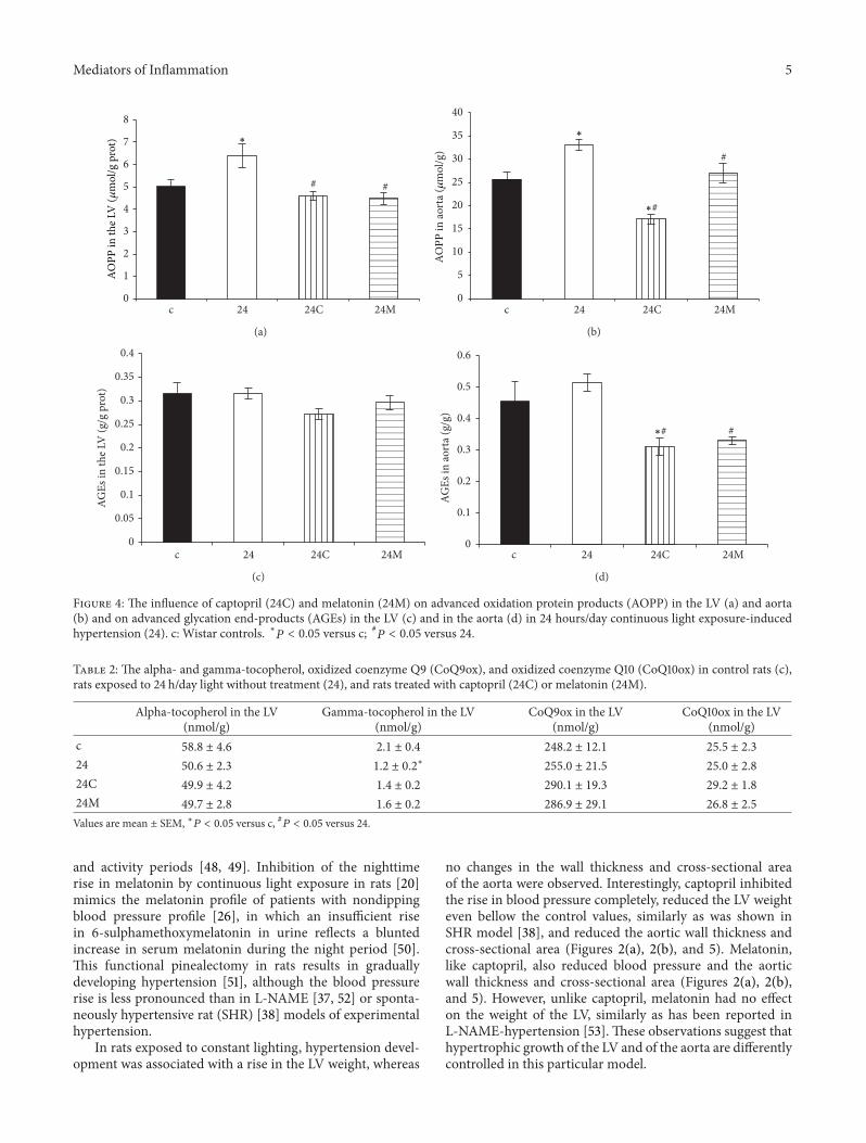
3.4. Oxidative Stress Parameters. The LV AOPP was 5.01 ±0.32 𝜇mol/g prot. and 6.38 ± 0.52 𝜇mol/g prot. in the controland continuous light-groups, respectively, and both captopriland melatonin significantly reduced AOPP in the LV (28%,𝑃 < 0.05 and 30%, 𝑃 < 0.05, resp.) (Figure 4(a)).The aortic AOPP was 25.72 ± 1.52 𝜇mol/g prot. and 33.11 ±1.10 𝜇mol/g prot. in the control and continuous light-groups,respectively, and both captopril and melatonin significantlyreducedAOPP in the aorta (48%,𝑃 < 0.05 and 19%,𝑃 < 0.05,resp.) (Figure 4(b)).
The aortic AGEs were 0.43 ± 0.06 and 0.47 ± 0.04 inthe control and continuous light-groups, respectively. Bothcaptopril and melatonin significantly reduced AGEs in theaorta (34%, 𝑃 < 0.05 and 30%, 𝑃 < 0.05, resp.) (Figure 4(d)).
3.5. CoQ and Tocopherol. The left ventricular CoQ9ox,CoQ10ox, and alpha-tocopherol levels were not changed ineither group. Gamma-tocopherol concentration in the LVwas decreased in the continuous light-group and neithercaptopril nor melatonin modified it (Table 2).
4. Discussion
Under physiological conditions in humans, melatonin con-centration is low during the day, but it is much higher atnight and is accompanied by lowering of the nocturnal bloodpressure [47]. The evening rise in melatonin represents animportant part of suprachiasmatic adaptation to the rest
Mediators of Inflammation 5
# #
0
1
2
3
4
5
6
7
8
c 24 24C 24M
∗
AOPP
in th
e LV
(𝜇m
ol/g
pro
t)
(a)
#
0
5
10
15
20
25
30
35
40
c 24 24C 24M
AOPP
in ao
rta (
𝜇m
ol/g
)
∗#
∗
(b)
0
0.05
0.1
0.15
0.2
0.25
0.3
0.35
0.4
c 24 24C 24M
AGEs
in th
e LV
(g/g
pro
t)
(c)
#
0
0.1
0.2
0.3
0.4
0.5
0.6
c 24 24C 24M
AGEs
in ao
rta (
g/g)
∗#
(d)
Figure 4: The influence of captopril (24C) and melatonin (24M) on advanced oxidation protein products (AOPP) in the LV (a) and aorta(b) and on advanced glycation end-products (AGEs) in the LV (c) and in the aorta (d) in 24 hours/day continuous light exposure-inducedhypertension (24). c: Wistar controls. ∗𝑃 < 0.05 versus c; #
𝑃 < 0.05 versus 24.
Table 2: The alpha- and gamma-tocopherol, oxidized coenzyme Q9 (CoQ9ox), and oxidized coenzyme Q10 (CoQ10ox) in control rats (c),rats exposed to 24 h/day light without treatment (24), and rats treated with captopril (24C) or melatonin (24M).
Alpha-tocopherol in the LV(nmol/g)
Gamma-tocopherol in the LV(nmol/g)
CoQ9ox in the LV(nmol/g)
CoQ10ox in the LV(nmol/g)
c 58.8 ± 4.6 2.1 ± 0.4 248.2 ± 12.1 25.5 ± 2.3
24 50.6 ± 2.3 1.2 ± 0.2∗
255.0 ± 21.5 25.0 ± 2.8
24C 49.9 ± 4.2 1.4 ± 0.2 290.1 ± 19.3 29.2 ± 1.8
24M 49.7 ± 2.8 1.6 ± 0.2 286.9 ± 29.1 26.8 ± 2.5
Values are mean ± SEM, ∗𝑃 < 0.05 versus c, #𝑃 < 0.05 versus 24.
and activity periods [48, 49]. Inhibition of the nighttimerise in melatonin by continuous light exposure in rats [20]mimics the melatonin profile of patients with nondippingblood pressure profile [26], in which an insufficient risein 6-sulphamethoxymelatonin in urine reflects a bluntedincrease in serum melatonin during the night period [50].This functional pinealectomy in rats results in graduallydeveloping hypertension [51], although the blood pressurerise is less pronounced than in L-NAME [37, 52] or sponta-neously hypertensive rat (SHR) [38] models of experimentalhypertension.
In rats exposed to constant lighting, hypertension devel-opment was associated with a rise in the LV weight, whereas
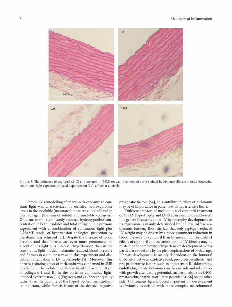
no changes in the wall thickness and cross-sectional areaof the aorta were observed. Interestingly, captopril inhibitedthe rise in blood pressure completely, reduced the LV weighteven bellow the control values, similarly as was shown inSHR model [38], and reduced the aortic wall thickness andcross-sectional area (Figures 2(a), 2(b), and 5). Melatonin,like captopril, also reduced blood pressure and the aorticwall thickness and cross-sectional area (Figures 2(a), 2(b),and 5). However, unlike captopril, melatonin had no effecton the weight of the LV, similarly as has been reported inL-NAME-hypertension [53]. These observations suggest thathypertrophic growth of the LV and of the aorta are differentlycontrolled in this particular model.
6 Mediators of Inflammation
c 24
100𝜇m
24C 24M
Figure 5: The influence of captopril (24C) and melatonin (24M) on wall thickness of aorta stained by hematoxylin-eosin in 24 hours/daycontinuous light exposure-induced hypertension (24). c: Wistar controls.
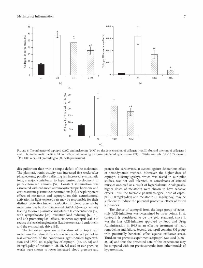
Fibrotic LV remodelling after six-week exposure to con-stant light was characterized by elevated hydroxyprolinelevels in the insoluble (maturated, more cross-linked) and intotal collagen (the sum of soluble and insoluble collagens).Only melatonin significantly reduced hydroxyproline con-centration in both insoluble and total collagen. In a previousexperiment with a combination of continuous light plusL-NAME model of hypertension analogical protection bymelatonin was achieved [51]. Despite the increase of bloodpressure and that fibrosis was even more pronounced ina continuous light plus L-NAME hypertension than in thecontinuous light model, melatonin reduced blood pressureand fibrosis in a similar way as in this experiment and alsowithout attenuation of LV hypertrophy [51]. Moreover, thisfibrosis-reducing effect of melatonin was confirmed in SHRmodel [38]. The indolamine also reduced the accumulationof collagens I and III in the aorta in continuous light-induced hypertension [36] (Figures 6 and 7). Since the qualityrather than the quantity of the hypertrophied myocardiumis important, while fibrosis is one of the decisive negative
prognostic factors [54], this antifibrotic effect of melatoninmay be of importance in patients with hypertensive heart.
Different impacts of melatonin and captopril treatmenton the LV hypertrophy and LV fibrosis need to be addressed.It is generally accepted that LV hypertrophy development orits regression is mainly determined by the level of haemo-dynamic burden. Thus, the fact that only captopril reducedLV weight may be driven by a more prominent reduction inblood pressure by captopril than by melatonin. The distincteffects of captopril and melatonin on the LV fibrosis may berelated to the complexity of hypertension development in thisparticularmodel and by the pleiotropic actions of both drugs.Fibrosis development is mainly dependent on the humoraldisbalance between oxidative load, pro-proteosynthetic, andpro-proliferative factors such as angiotensin II, aldosterone,endothelin, or catecholamines on the one side and substanceswith growth attenuating potential, such as nitric oxide (NO),prostacyclin, or atrial natriuretic peptide [54–56] on the otherside. Continuous light-induced hypertension developmentis obviously associated with more complex neurohumoral
Mediators of Inflammation 7
0
5
10
15
20
25
30
35
c 24 24C 24M
Col
lage
n I i
n ao
rtic
med
ia (%
)
∗#
∗
∗
(a)
#
0
0.01
0.02
0.03
0.04
c 24 24C 24M
Col
lage
n II
I in
aort
ic m
edia
(%)
∗#
∗
(b)
0
5
10
15
20
25
30
35
c 24 24C 24M
Col
lage
n I +
III i
n ao
rtic
med
ia (%
)
∗#
∗
∗
(c)
Figure 6: The influence of captopril (24C) and melatonin (24M) on the concentration of collagen I (a), III (b), and the sum of collagens Iand III (c) in the aortic media in 24 hours/day continuous light exposure-induced hypertension (24). c: Wistar controls. ∗𝑃 < 0.05 versus c;#𝑃 < 0.05 versus 24 (according to [36] with permission).
disequilibrium than with a simple deficit of the melatonin.The plasmatic renin activity was increased five weeks afterpinealectomy, possibly reflecting an increased sympathetictone, a major contributor to hypertension development inpinealectomized animals [57]. Constant illumination wasassociated with enhanced adrenocorticotropic hormone andcorticosterone plasmatic concentrations [58].Thepluripotenteffects of melatonin and captopril on this neurohumoralactivation in light-exposed rats may be responsible for theirdistinct protective impact. Reduction in blood pressure bymelatonin may be due to increased GABA(A)—ergic activityleading to lower plasmatic angiotensin II concentration [59]with sympatholytic [28], oxidative load reducing [60, 61],and NO-promoting [25] effects. However, captopril is able toreduce the level of angiotensin II, aldosterone, and endothelinand the sympathetic drive [62].
The important question is the dose of captopril andmelatonin that should be chosen to counteract patholog-ical alterations of the continuous light-induced hyperten-sion and LVH. 100mg/kg/day of captopril [36, 38, 51] and10mg/kg/day of melatonin [38, 51, 53] used in our previousworks were shown to lower increased blood pressure and
protect the cardiovascular system against deleterious effectof hemodynamic overload. Moreover, the higher dose ofcaptopril (150mg/kg/day), which was tested in our pilotstudies, was not well tolerated, as convulsions of striatedmuscles occurred as a result of hyperkalemia. Analogically,higher doses of melatonin were shown to have sedativeeffects. Thus, the tolerable pharmacological dose of capto-pril (100mg/kg/day) and melatonin (10mg/kg/day) may besufficient to induce the potential protective effects of testedsubstances.
The choice of captopril from the large group of acces-sible ACE-inhibitors was determined by three points. First,captopril is considered to be the gold standard, since itwas the first ACE-inhibitor approved by Food and DrugAdministration in 1993 as an effective treatment of heartremodeling and failure. Second, captopril contains SH groupwith potentially beneficial effect against oxidative stress.Third, in our previous experiments captopril was used [4, 36–38, 51] and thus the presented data of this experiment maybe compared with our previous results from other models ofhypertension.
8 Mediators of Inflammation
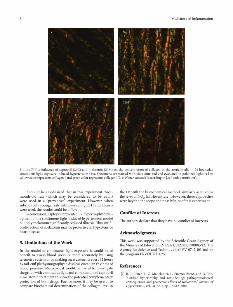
c 24
24C 24M
Figure 7: The influence of captopril (24C) and melatonin (24M) on the concentration of collagen in the aortic media in 24 hours/daycontinuous light exposure-induced hypertension (24). Specimens are stained with picrosirius red and evaluated in polarized light: red toyellow color represents collagen I and green color represents collagen III. c: Wistar controls (according to [36] with permission).
It should be emphasized that in this experiment three-month-old rats (which may be considered to be adult)were used in a “preventive” experiment. However, whensubstantially younger rats with developing LVH and fibrosiswere used, the results could be different.
In conclusion, captopril prevented LV hypertrophy devel-opment in the continuous light-induced hypertension modelbut only melatonin significantly reduced fibrosis. This antifi-brotic action of melatonin may be protective in hypertensiveheart disease.
5. Limitations of the Work
In the model of continuous light exposure it would be ofbenefit to assess blood pressure more accurately by usingtelemetry system or by making measurements every 12 hoursby tail-cuff plethysmography to disclose circadian rhythms ofblood pressure. Moreover, it would be useful to investigatethe groupwith continuous light and combination of captopril+ melatonin treatment to show the potential complementaryprotection of both drugs. Furthermore, it may be useful tocompare biochemical determination of the collagen level in
the LV with the histochemical method, similarly as to knowthe level of NO
𝑥(nitrite-nitrate). However, these approaches
were beyond the scope and possibilities of this experiment.
Conflict of Interests
The authors declare that they have no conflict of interests.
Acknowledgments
This work was supported by the Scientific Grant Agency ofthe Ministry of Education (VEGA 1/0227/12, 2/00183/12), theAgency for Science and Technique (APVV-0742-10) and bythe program PRVOUK P37/5.
References
[1] R. J. Reiter, L. C. Manchester, L. Fuentes-Broto, and D. Tan,“Cardiac hypertrophy and remodelling: pathophysiologicalconsequences and protective effects of melatonin,” Journal ofHypertension, vol. 28, no. 1, pp. S7–S12, 2010.
Mediators of Inflammation 9
[2] F. Simko andO. Pechanova, “Remodelling of the heart and vess-els in experimental hypertension: advances in protection,” Jour-nal of Hypertension, vol. 28, supplement 1, pp. S1–S6, 2010.
[3] F. Simko, J. Matuskova, I. Luptak et al., “Effect of simvastatin onremodeling of the left ventricle and aorta in L-NAME-inducedhypertension,” Life Sciences, vol. 74, no. 10, pp. 1211–1224, 2004.
[4] F. Simko, V. Pelouch, J. Torok et al., “Protein remodeling of theheart ventricles in hereditary hypertriglyceridemic rat: effect oface-inhibition,” Journal of Biomedical Science, vol. 12, no. 1, pp.103–111, 2005.
[5] C. A. Mandarim-de-Lacerda and L. M. M. Pereira, “Effect oftelmisartan on preexistent cardiac and renal lesions in sponta-neously hypertensive mature rats,” Histology and Histopathol-ogy, vol. 19, no. 3, pp. 727–733, 2004.
[6] F. Simko, “Statins: a perspective for left ventricular hypertrophytreatment,”European Journal of Clinical Investigation, vol. 37, no.9, pp. 681–691, 2007.
[7] J. Kyselovic, P. Krenek, M. Wibo, and T. Godfraind, “Effects ofamlodipine and lacidipine on cardiac remodelling and reninproduction in salt-loaded stroke-prone hypertensive rats,” TheBritish Journal of Pharmacology, vol. 134, no. 7, pp. 1516–1522,2001.
[8] F. Simko, R. J. Reiter,O. Pechanova, andL. Paulis, “Experimentalmodels of melatonin-deficient hypertension,” Frontiers in Bio-science, vol. 18, no. 2, pp. 616–625, 2013.
[9] M. Ozturk, M. Ozler, Y. G. Kurt et al., “Efficacy of melatonin,mercaptoethylguanidine and 1400W in doxorubicin- andtrastuzumab-induced cardiotoxicity,” Journal of Pineal Res-earch, vol. 50, no. 1, pp. 89–96, 2011.
[10] E. Grossini, C. Molinari, F. Uberti, D. A. S. G. Mary, G. Vacca,and P. P. Caimmi, “Intracoronary melatonin increases coronaryblood flow and cardiac function through 𝛽-adrenoreceptors,MT1/MT2 receptors, and nitric oxide in anesthetized pigs,”Journal of Pineal Research, vol. 51, no. 2, pp. 246–257, 2011.
[11] S. Samimi-Fard, P. Abreu-Gonzalez, A. Dominguez-Rodriguez,and A. Jimenez-Sosa, “A case-control study of melatonin recep-tor type 1A polymorphism and acute myocardial infarction ina Spanish population,” Journal of Pineal Research, vol. 51, no. 4,pp. 400–404, 2011.
[12] M. Zeman and I. Herichova, “Melatonin and clock genesexpression in the cardiovascular system,” Frontiers in Bioscience,vol. 1, no. 5, pp. 743–753, 2013.
[13] F. Simko and L. Paulis, “Melatonin as a potential antihyperten-sive treatment,” Journal of Pineal Research, vol. 42, no. 4, pp. 319–322, 2007.
[14] S. Tengattini, R. J. Reiter, D. Tan, M. P. Terron, L. F. Rodella, andR. Rezzani, “Cardiovascular diseases: protective effects of mela-tonin,” Journal of Pineal Research, vol. 44, no. 1, pp. 16–25, 2008.
[15] A. Dominguez-Rodriguez, P. Abreu-Gonzalez, J. J. Sanchez-Sanchez, J. C. Kaski, and R. J. Reiter, “Melatonin and circadianbiology in human cardiovascular disease,” Journal of PinealResearch, vol. 49, no. 1, pp. 14–22, 2010.
[16] L. Paulis and F. Simko, “Blood pressure modulation andcardiovascular protection by melatonin: potential mechanismsbehind,” Physiological Research, vol. 56, no. 6, pp. 671–684, 2007.
[17] R. A.Hoffman andR. J. Reiter, “Rapid pinealectomy in hamstersand other small rodents,”The Anatomical Record, vol. 153, no. 1,pp. 19–21, 1965.
[18] S. C. Cunnane,M. S.Manku,M.Oka, andD. F. Horrobin, “Enh-anced vascular reactivity to various vasoconstrictor agentsfollowing pinealectomy in the rat: role of melatonin,” Canadian
Journal of Physiology and Pharmacology, vol. 58, no. 3, pp. 287–293, 1980.
[19] A. Zanoboni and W. Zanoboni-Muciaccia, “Experimental hyp-ertension in pinealectomized rats,” Life Sciences, vol. 6, no. 21,pp. 2327–2331, 1967.
[20] G.M. Brown, A. Bar-Or, D. Grossi, S. Kashur, and E. Johannson,“Urinary 6-sulphatoxymelatonin, an index of pineal function inthe rat,” Journal of Pineal Research, vol. 10, no. 3, pp. 141–147, 1991.
[21] D. X. Tan, L. D. Chen, B. Poeggeler et al., “Melatonin: a potent,endogenous hydroxyl radical scavenger,” Endocrine Journal, vol.1, pp. 157–160, 1993.
[22] R. J. Reiter,D. X. Tan,M. P. Terron, L. J. Flores, andZ.Czarnocki,“Melatonin and its metabolites: new findings regarding theirproduction and their radical scavenging actions,”Acta Biochim-ica Polonica, vol. 54, no. 1, pp. 1–9, 2007.
[23] A. Galano, D. X. Tan, and R. J. Reiter, “Melatonin as a naturalally against oxidative stress: a physicochemical examination,”Journal of Pineal Research, vol. 51, no. 1, pp. 1–16, 2011.
[24] R. J. Reiter and D. X. Tan, “Melatonin: a multitaskingmolecule,”Progress in Brain Research, vol. 181, pp. 127–151, 2010.
[25] R. Rezzani, E. Porteri, C. de Ciuceis et al., “Effects of melatoninand pycnogenol on small artery structure and function inspontaneously hypertensive rats,” Hypertension, vol. 55, no. 6,pp. 1373–1380, 2010.
[26] R. J. Reiter, D. Tan, and A. Korkmaz, “The circadian melatoninrhythm and its modulation: possible impact on hypertension,”Journal of Hypertension, vol. 27, supplement 6, pp. S17–S20,2009.
[27] A. Dominguez-Rodriguez, P. Abreu-Gonzalez, and R. J. Reiter,“Melatonin and cardiovascular disease: myth or reality?”Revista Espanola de Cardiologia, vol. 65, no. 3, pp. 215–218, 2012.
[28] L. Paulis, J. Matuskova, M. Adamcova et al., “Regression of leftventricular hypertrophy and aortic remodelling in NO-deficient hypertensive rats: effect of L-arginine and spiro-nolactone,” Acta Physiologica, vol. 194, no. 1, pp. 45–55, 2008.
[29] T. Benova, C. Viczenczova, J. Radosinska et al., “Melatoninattenuates hypertension-related proarrhythmic myocardialmaladaptation of connexin-43 and propensity of the heart tolethalarrhythmias,” Canadian Journal of Physiology and Pha-rmacology, vol. 91, no. 8, pp. 633–639, 2013.
[30] A. Dominguez-Rodriguez, P. Abreu-Gonzalez, M. Garcia-Gon-zalez, and R. J. Reiter, “Prognostic value of nocturnal melatoninlevels as a novel marker in patients with ST-segment elevationmyocardial infarction,”TheAmerican Journal of Cardiology, vol.97, no. 8, pp. 1162–1164, 2006.
[31] A. Dominguez-Rodriguez, P. Abreu-Gonzalez, and R. J. Reiter,“Clinical aspects of melatonin in the acute coronary syndrome,”Current Vascular Pharmacology, vol. 7, no. 3, pp. 367–373, 2009.
[32] A. Dominguez-Rodriguez, P. Abreu-Gonzalez, E. Arroyo-Ucar,and R. J. Reiter, “Decreased level of melatonin in serum predictsleft ventricular remodelling after acute myocardial infarction,”Journal of Pineal Research, vol. 53, no. 3, pp. 319–323, 2012.
[33] A. Dominguez-Rodriguez, P. Abreu-Gonzalez, E. Arroyo-Ucar,P. Avanzas, and R. J. Reiter, “Global left ventricular longitudinalstrain is associated with decreased melatonin levels in patientswith acute myocardial infarction: a two-dimensional speckletracking study,” Biomarkers, vol. 18, no. 4, pp. 310–313, 2013.
[34] A. Dominguez-Rodriguez, P. Abreu-Gonzalez, M. J. Garcia-Gonzalez, J. C. Kaski, R. J. Reiter, and A. Jimenez-Sosa, “A uni-center, randomized, double-blind, parallel-group, placebo-con-trolled study of Melatonin as an Adjunct in patients with acute
10 Mediators of Inflammation
myocaRdial Infarction undergoing primary Angioplasty. TheMelatonin Adjunct in the acute myocaRdial Infarction treatedwith Angioplasty (MARIA) trial: study design and rationale,”Contemporary Clinical Trials, vol. 28, no. 4, pp. 532–539, 2007.
[35] A. Dominguez-Rodriguez, P. Abreu-Gonzalez, and R. J. Reiter,“Melatonin and cardioprotection in the acute myocardialinfarction: a promising cardioprotective agent,” InternationalJournal of Cardiology, vol. 158, no. 2, pp. 309–310, 2012.
[36] K. Repova-Bednarova, S. Aziriova, and J.Hrenak, “Effect of cap-topril and melatonin on fibrotic rebuilding of the aorta in 24hour light-induced hypertension,” Physiological Research, vol.62, supplement 1, pp. S135–S141, 2013.
[37] I. Bernatova, O. Pechanova, V. Pelouch, and F. Simko, “Regres-sion of chronic L-NAME-treatment-induced left ventricularhypertrophy: effect of captopril,” Journal of Molecular andCellular Cardiology, vol. 32, no. 2, pp. 177–185, 2000.
[38] F. Simko, O. Pechanova, V. Pelouch et al., “Effect of melatonin,captopril, spironolactone and simvastatin on blood pressureand left ventricular remodelling in spontaneously hypertensiverats,” Journal of Hypertension, vol. 27, supplement 6, pp. S5–S10,2009.
[39] V. Pelouch,M.Milerova, B. Ostadal, M. Samanek, and B. Hucın,“Protein profiling of human atrial and ventricular musculature:the effect of normoxaemia and hypoxaemia in congenital heartdiseases,” Physiological Research, vol. 42, no. 4, pp. 235–242,1993.
[40] V. Pelouch,M.Milerova, B. Ostadal, B. Hucin, andM. Samanek,“Differences between atrial and ventricular protein profiling inchildren with congenital heart disease,” Molecular and CellularBiochemistry, vol. 147, no. 1–4, pp. 43–49, 1995.
[41] G. K. Reddy and C. S. Enwemeka, “A simplified method for theanalysis of hydroxyproline in biological tissues,” Clinical Bio-chemistry, vol. 29, no. 3, pp. 225–229, 1996.
[42] G. Munch, R. Keis, A. Wessels et al., “Determination of adva-nced glycation end products in serum by fluorescence spec-troscopy and competitive ELISA1,” European Journal of ClinicalChemistry and Clinical Biochemistry, vol. 35, no. 9, pp. 669–677,1997.
[43] A.D. Bhatwadekar andV. S. Ghole, “Rapidmethod for the prep-aration of an AGE-BSA standard calibrator using thermal glyc-ation,” Journal of Clinical Laboratory Analysis, vol. 19, no. 1, pp.11–15, 2005.
[44] V. Witko-Sarsat, M. Friedlander, C. Capeillere-Blandin et al.,“Advanced oxidation protein products as a novel marker ofoxidative stress in uremia,” Kidney International, vol. 49, no. 5,pp. 1304–1313, 1996.
[45] J. K. Lang, K. Gohil, and L. Packer, “Simultaneous determina-tion of tocopherols, ubiquinols, and ubiquinones in blood, pla-sma, tissue homogenates, and subcellular fractions,” AnalyticalBiochemistry, vol. 157, no. 1, pp. 106–116, 1986.
[46] J. Kucharska, A. Gvozdjakova, and F. Simko, “Simvastatin dec-reased coenzyme Q in the left ventricle and skeletal muscle butnot in the brain and liver in L-NAME-induced hypertension,”Physiological Research, vol. 56, supplement 2, pp. S49–S54, 2007.
[47] M. Zeman, K. Dulkova, V. Bada, and I. Herichova, “Plasmamel-atonin concentrations in hypertensive patients with the dippingand non-dipping blood pressure profile,” Life Sciences, vol. 76,no. 16, pp. 1795–1803, 2005.
[48] R. M. Buijs and A. Kalsbeek, “Hypothalamic integration ofcentral and peripheral clocks,” Nature Reviews Neuroscience,vol. 2, no. 7, pp. 521–526, 2001.
[49] F. A. J. L. Scheer, G. A. van Montfrans, E. J. W. van Someren, G.Mairuhu, and R. M. Buijs, “Daily nighttime melatonin reducesblood pressure in male patients with essential hypertension,”Hypertension, vol. 43, no. 2, pp. 192–197, 2004.
[50] M. Jonas, D. Garfinkel, N. Zisapel, M. Laudon, and E. Gross-man, “Impaired nocturnal melatonin secretion in non-dipperhypertensive patients,” Blood Pressure, vol. 12, no. 1, pp. 19–24,2003.
[51] F. Simko, O. Pechanova, V. Pelouch et al., “Continuous light andL-NAME-induced left ventricular remodelling: different pro-tection with melatonin and captopril,” Journal of Hypertension,vol. 28, supplement 1, pp. S13–S18, 2010.
[52] O. Pechanova, J. Zicha, L. Paulis et al., “The effect of N-acet-ylcysteine and melatonin in adult spontaneously hypertensiverats with established hypertension,” European Journal of Phar-macology, vol. 561, no. 1–3, pp. 129–136, 2007.
[53] L. Paulis, O. Pechanova, J. Zicha et al., “Melatonin preventsfibrosis but not hypertrophy development in the left ventricle ofNG-nitro-L-arginine-methyl ester hypertensive rats,” Journal ofHypertension, vol. 27, supplement 6, pp. S11–S16, 2009.
[54] K. T. Weber, “From inflammation to fibrosis: a stiff stretch ofhighway,” Hypertension, vol. 43, no. 4, pp. 716–719, 2004.
[55] F. Simko and J. Simko, “The potential role of nitric oxide in thehypertrophic growth of the left ventricle,” Physiological Res-earch, vol. 49, no. 1, pp. 37–46, 2000.
[56] F. Simko, “Is NO the king? Pathophysiological benefit with unc-ertain clinical impact [editorial],”Physiological Research, vol. 56,supplement 2, pp. S1–S6, 2007.
[57] H. Karppanen, S. Lahovaara, P. Mannisto, and H. Vapaatalo,“Plasma renin activity and in vitro synthesis of aldosterone bythe adrenal glands of rats with spontaneous, renal, or pinealec-tomy induced hypertension,” Acta Physiologica Scandinavica,vol. 94, no. 2, pp. 184–188, 1975.
[58] V.Milosievic, S. Trifunovic,M. Sekulic et al., “Chronic exposureto constant light affects morphology and secretion of adrenalzona fascikulata cells in female rats,” General Physiology andBiophysics, vol. 24, no. 3, pp. 299–309, 2005.
[59] H. Li, Y. Kang, L. Yu, H. Xu, and H. Zhao, “Melatoninreduces blood pressure in rats with stress-induced hypertensionviaGABAA receptors,”Clinical and Experimental Pharmacologyand Physiology, vol. 36, no. 4, pp. 436–440, 2009.
[60] R. J. Reiter, D. X. Tan, L. C. Manchester, M. Pilar Terron, L. J.Flores, and S. Koppisepi, “Medical implications of melatonin:receptor-mediated and receptor-independent actions,” Adva-nces in Medical Sciences, vol. 52, pp. 11–28, 2007.
[61] M. Kozirog, A. R. Poliwczak, P. Duchnowicz, M. Koter-Micha-lak, J. Sikora, and M. Broncel, “Melatonin treatment improvesblood pressure, lipid profile, and parameters of oxidativestress in patients with metabolic syndrome,” Journal of PinealResearch, vol. 50, no. 3, pp. 261–266, 2011.
[62] F. Simko, J. Simko, and M. Fabryova, “ACE-inhibition and ang-iotensin II receptor blockers in chronic heart failure: pathophys-iological consideration of the unresolved battle,”CardiovascularDrugs andTherapy, vol. 17, no. 3, pp. 287–290, 2003.


![Tail suspension is useful as a sarcopenia model in rats...muscle weakness [9, 10]. Spontaneously hypertensive rats (SHR) are widely used in hypertension and insulin-resistance models](https://img.pdfslide.us/doc/110x75/60fd29b9db06e05a8002d7af/tail-suspension-is-useful-as-a-sarcopenia-model-in-rats-muscle-weakness-9.jpg)