Embed Size (px)
Citation preview
CASE REPORT
Journal of the College of Physicians and Surgeons Pakistan 2021, Vol. 31(06): 735-736 735
Hypersensitivity Pneumonitis in a Pediatric PatientMadiha Naz, Shazia Parveen, Afsheen Asghar Khan and Fauzia Zafar
Department of Pediatric Medicine, Nishtar Medical University, Multan, Pakistan
ABSTRACTHypersensitivity pneumonitis (HP) is a rarely diagnosed interstitial lung disease with variable manifestations. It results fromrepeated inhalation of certain antigens, e.g. mold, avian antigen, etc. in susceptible patients. The diagnosis is made by expo-sure history, relevant clinical presentation, and specific radiologic features. It is treated by avoidance of triggers and use of corti-costeroids. We report a 7.5-year girl with HP. She was admitted for cough and severe respiratory distress.
Key Words: Hypersensitivity pneumonitis, Antigen, Allergy, Child.
How to cite this article: Naz M, Parveen S, Khan AA, Zafar F. Hypersensitivity Pneumonitis in a Pediatric Patient. J Coll Physicians SurgPak 2021; 31(06):735-736.
INTRODUCTION
Hypersensitivity pneumonitis (HP), or extrinsic allergic alve-olitis, is described as a complex syndrome with variableseverity, diverse presentation, and disease course.1 It is an inter-stitial lung disease that results from repeated inhalation ofcertain organic or inorganic antigens, in susceptible patients. Inmost reported pediatric cases, common provocation agentsinclude bird antigens, fungal aerosols and certain drugs.2 HP hasbeen classified into acute, sub-acute, and chronic forms;3 butthese are ill defined and there are no definite criteria. The diag-nosis is established by documented exposure, relevant clinicalsigns and symptoms, and specific radiologic features. If high-re-solution CT (HRCT) is inconclusive, then broncho-alveolarlavage (BAL) or lung biopsy is indicated.4 Treatment optionsinclude avoidance of triggering agent in all cases, and corticos-teroids in some patients.5 The prognosis is excellent when if diag-nosed and treated early. However, deaths from HP have beenreported in children as well as adults.
CASE REPORT
A 7.5-year girl, presented with complaints of fever and severerespiratory distress. There was history of fever, gradual inonset, high grade, continuous, and associated with cough andrespiratory distress. Cough was productive with white sputum,and there was history of similar complaints for the last 9 months.She also took anti-tuberculous treatment (ATT) for 7 monthswithout being properly investigated. History of exposure topigeon’s droppings for long time was positive.
Correspondence to: Dr. Madiha Naz, Department of Pedia-tric Medicine, Nishtar Medical University, MultanE-mail: madddnaz@gmail.com.....................................................Received: January 18, 2020; Revised: February 28, 2020;Accepted: March 09, 2020DOI: https://doi.org/10.29271/jcpsp.2021.06.735
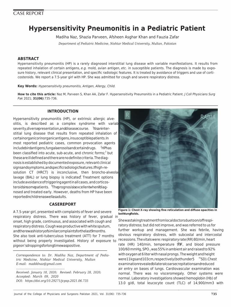
Figure 1: Chest X-ray showing fine reticulation and diffuse opacities inboth lung fields.
She was taking treatment from local doctors due to on/off respi-ratory distress; but did not improve, and was referred to us forfurther workup and management. She was febrile, havingobvious respiratory distress, with subcostal and intercostalrecessions. The vitals were: respiratory rate (RR) 80/min, heartrate (HR) 140/min, temperature 990 F, and blood pressure100/60 mmHg, SPO2 was 55% in ambient air and raised to 92%with oxygen at 6 liter with nasal prongs. The weight and heightwere 11 kgs and 103 cm, respectively (both under 5th SD). Chestexamination revealed bilateral coarse crepitations and reducedair entry on bases of lungs. Cardiovascular examination wasnormal. There was no visceromegaly. Other systems werenormal. Laboratory investigations showed hemoglobin (Hb) of13.0 g/dl, total leucocyte count (TLC) of 14,900/mm3 with
Madiha Naz, Shazia Parveen, Afsheen Asghar Khan and Fauzia Zafar
Journal of the College of Physicians and Surgeons Pakistan 2021, Vol. 31(06): 735-736736
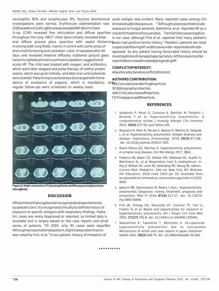
neutrophils 90% and lymphocytes 9%. Routine biochemicalinvestigations were normal. Erythrocyte sedimentation rate(ESR) was 6 mm/1st hr. IgE level was raised at 499.9 IU/ml. ChestX-ray (CXR) revealed fine reticulation and diffuse opacitiesthroughout the lung. HRCT chest done initially revealed bilat-eral diffuse ground glass opacities with septal thickeninginvolving both lung fields, mainly in centre with some areas ofbronchial thickening and cavitation. Later, it repeated after 40days and revealed bilateral diffusely scattered ground glasshaze in lung fields almost in symmetrical pattern, suggestive ofacute HP. The child was treated with oxygen, and antibiotics,which were later stopped and pulse therapy of methyl predni-solone, which was given initially, and later oral corticosteroidswere started. Patient improved and was discharged with homeadvice of avoidance of pigeons, which is mandatory; andregular follow-ups were scheduled on weekly basis.
Figure 2: High-resolution CT showing bilateral diffuse ground glass hazein lung fields.
DISCUSSION
HP is an interstitial lung disorder recognised and reported rarelyby pediatricians. It is recognised clinically by definite history ofexposure to specific antigens with respiratory findings. Pedia-tric cases are rarely diagnosed or reported, so limited data isavailable and is largely based on few case reports and smallseries of patients. Till 2005, only 95 cases were reported.6
Although we report a female patient, slight male predominancewas noted by Fink et al.6 In our patient, history of inhalation of
avian antigen was evident. Many reported cases among chil-dren also had birds exposure.2,5Although cases reported includeexposure to fungal aerosols. Nakshima et al. reported HP as aresult of inhalation of isocyanates.7 Family history was negativein our case; although Fink et al. reported that many pediatriccases had positive family history.6 Pediatric patients are leastsuspected of having HP, and thus are under-reported and misdi-agnosed. So any patient having favourable history should beinvestigated and managed appropriately. All the cases must bereported to increase knowledge regarding HP.
CONFLICT OF INTEREST:All authors declared no conflict of interest.
AUTHORS’ CONTRIBUTION:MN: Conception and writing the article.SP: Bibliography collection.AAK: Critical revision of the article.FZ: Final approval of the article.
REFERENCES
Spagnolo P, Rossi G, Cavazza A, Bonifazi M, Paladini I,1.Bonella F, et al. Hypersensitivity pneumonitis: Acomprehensive review. J Investig Allergol Clin Immunol2015; 25(4):237-50; quiz follow 250.Nogueira R, Melo N, Novais E, Bastos H, Martins N, Delgado2.L, et al. Hypersensitivity pneumonitis: Antigen diversity anddisease implications. Pulmonology 2019; 25(2):97-108. doi: 10.1016/j.pulmoe.2018.07.003.Riario Sforza GG, Marinou A. Hypersensitivity pneumonitis:3.A complex lung disease. Clin Mol Allergy 2017; 15:6.Federico MJ, Baker CD, Deboer EM, Halbower AC, Kupfer O,4.Martiniano SL, et al. Respiratory tract & mediastinum. In:Hay Jr William W, Levin MJ, Deterding RR, Abzug MJ, editors.Current Pent: Pediatrics. 24th ed. New York, NY: McGraw-Hill Education; 2018 cited 2020 Jan 10. Available from:accessmedicine.mhmedical.com/content.aspx?aid=1153308493Jędrych ME, Szturmowicz M, Bestry I, Kuś J. Hypersensitivity5.pneumonitis: Diagnostic criteria, treatment, prognosis andprevention. Med Pr 2016; 67(4):517-27. doi: 10.13075/mp.5893.00406.Fink JN, Ortega HG, Reynolds HY, Cormier YF, Fan LL,6.Franks TJ, et al. Needs and opportunities for research inhypersensitivity pneumonitis. Am J Respir Crit Care Med2005; 171(7):792-8. doi: 10.1164/rccm.200409-1205WS. Nakashima K, Takeshita T, Morimoto K. Occupational7.hypersensitivity pneumonitis due to isocyanates:Mechanisms of action and case reports in Japan. IndustrialHealth 2001; 39(3):269-79. doi: 10.2486/indhealth.39.269.
••••••••••