Embed Size (px)
Citation preview

Journal of Molecular and Cellular Cardiology 47 (2009) 275–287
Contents lists available at ScienceDirect
Journal of Molecular and Cellular Cardiology
j ourna l homepage: www.e lsev ie r.com/ locate /y jmcc
Original article
Hyperbaric oxygenation enhances transplanted cell graft and functional recovery inthe infarct heart
Mahmood Khan a,⁎, Sarath Meduru a, Iyyapu K. Mohan a, M. Lakshmi Kuppusamy a, Sheik Wisel a,Aditi Kulkarni a, Brian K. Rivera a, Robert L. Hamlin b, Periannan Kuppusamy a
a Davis Heart and Lung Research Institute, Division of Cardiovascular Medicine, Department of Internal Medicine, The Ohio State University, Columbus, OH 43210, USAb Department of Veterinary Biosciences, The Ohio State University, Columbus, OH 43210, USA
Abbreviations: DAPI, 4′,6-diamidino-2-phenylindoleEjection fraction; EKG, Electrocardiography; EPR, ElecHBO, Hyperbaric oxygenation; LAD, Left-anterior-desMyocardial infarction; MRI, Magnetic resonance imagingSPIO, Superparamagnetic iron oxide; VCG, Vectorcendothelial growth factor.⁎ Corresponding author. Davis Heart and Lung Rese
University, 420 W. 12th Ave., Room 116A, Columbus, OH8383; fax: +1 614 292 8454.
E-mail address: [email protected] (M. Kha
0022-2828/$ – see front matter © 2009 Elsevier Inc. Adoi:10.1016/j.yjmcc.2009.04.005
a b s t r a c t
a r t i c l e i n f oArticle history:Received 9 February 2009Received in revised form 2 April 2009Accepted 6 April 2009Available online 17 April 2009
Keywords:Mesenchymal stem cellHyperbaric oxygenMyocardial infarctionStem-cell therapyAngiogenesisElectrocardiographyEchocardiographyMyocardial oxygenation
A major limitation to the application of stem-cell therapy to repair ischemic heart damage is the low survivalof transplanted cells in the heart, possibly due to poor oxygenation. We hypothesized that hyperbaricoxygenation (HBO) can be used as an adjuvant treatment to augment stem-cell therapy. Therefore, the goalof this study was to evaluate the effect of HBO on the engraftment of rat bone marrow-derived mesenchymalstem cells (MSCs) transplanted in infarct rat hearts. Myocardial infarction (MI) was induced in Fisher-344rats by permanently ligating the left-anterior-descending coronary artery. MSCs, labeled with fluorescentsuperparamagnetic iron oxide (SPIO) particles, were transplanted in the infarct and peri-infarct regions ofthe MI hearts. HBO (100% oxygen at 2 ATA for 90 min) was administered daily for 2 weeks. Four MI groupswere used: untreated (MI); HBO; MSC; MSC+HBO. Echocardiography, electro-vectorcardiography, andmagnetic resonance imaging were used for functional evaluations. The engraftment of transplanted MSCs inthe heart was confirmed by SPIO fluorescence and Prussian-blue staining. Immunohistochemical stainingwas used to identify key cellular and molecular markers including CD29, troponin-T, connexin-43, VEGF,α-smooth-muscle actin, and von Willebrand factor in the tissue. Compared to MI and MSC groups, theMSC+HBO group showed a significantly increased recovery of cardiac function including left-ventricular(LV) ejection fraction, fraction shortening, LV wall thickness, and QRS vector. Further, HBO treatmentsignificantly increased the engraftment of CD29-positive cells, expression of connexin-43, troponin-T andVEGF, and angiogenesis in the infarct tissue. Thus, HBO appears to be a potential and clinically-viableadjuvant treatment for myocardial stem-cell therapy.
© 2009 Elsevier Inc. All rights reserved.
1. Introduction
Cell transplantation shows great promise for repair and restorationof heart function following myocardial infarction [1,2]. However, thepotential of cell-based therapy for myocardial tissue repair is limitedby the death of transplanted cells, mostly within the first few daysafter transplantation in the infarct tissue, likely from a combination ofischemia (deprivation of nutrients and oxygen), inflammation, andapoptosis [3]. Several interventions, including the use of cells over-expressing pro-survival/anti-apoptotic proteins [4,5], and pharmaco-
; ECHO, Echocardiography; EF,tron paramagnetic resonance;cending coronary artery; MI,; MSC, Mesenchymal stem cell;ardiography; VEGF, Vascular
arch Institute, The Ohio State43210, USA. Tel.: +1 614 292
n).
ll rights reserved.
logical agents [6], have been reported to augment survival, function,and homing of the transplanted cells in the hostile ischemicenvironment [7]. Laflamme et al. used a “cocktail” of pro-survivalfactors to inhibit death-promoting pathways and upregulate survival-signaling. They reported a higher proportion of engraftment whencompared to simpler interventions [8]. Recently, we have shown thebeneficial effect of preconditioning cells with trimetazidine, an anti-ischemic drug, prior to cardiac cell therapy [9]. Most of the abovementioned approaches involved manipulation of the cells prior to, orduring, transplantation in the infarct heart. However, to date, no post-transplantation strategies have been considered for promotingcellular engraftment.
Hypoxia is a formidable factor in the ischemic tissue that can leadto the production of oxygen free radicals. The hostile environmentwith persistent oxidative stress ultimately leads to apoptosis of themajority of the transplanted cells. Therefore, we considered astraightforward physiological approach to reduce the severity ofhypoxia by hyperoxygenating the infarct tissue following celltransplantation. Hyperbaric oxygenation (HBO) is a safe, clinically-viable treatment that has been used as a primary therapy in patients
276 M. Khan et al. / Journal of Molecular and Cellular Cardiology 47 (2009) 275–287
with decompression sickness, arterial gas embolism and carbon-monoxide poisoning [10]. It is also used as an adjuvant therapy topromote wound healing [11], and for the treatment of variousconditions, including ischemic injury [12]. HBO involves inhalationof 100% oxygen under greater-than-one atmospheric absolute (ATA)pressure. Such doses of oxygen have a number of beneficialbiochemical, cellular, and physiologic effects [13]. HBO, administeredat the onset of reperfusion in an open-chest rabbit model ofmyocardial ischemia–reperfusion injury, showed a significant reduc-tion in infarct size [14]. More recent studies have shown that HBOattenuates myocardial injury via nitric oxide signaling [13], improvescardiac function in patients with acute myocardial infarction [15], andhelps mobilization of stem cells by enhancing CXCR4 and VEGFR-2 inhumans [16]. However, the efficacy of HBO as an adjuvant to celltherapy has not yet been studied.
We therefore hypothesized that HBO, when applied in conjunctionwith stem-cell therapy, would improve oxygenation in the infarctheart, leading to increased cell engraftment and cardiac function.Using a rat model of myocardial infarction, induced by permanentligation of the left-anterior-descending (LAD) coronary artery,followed by transplantation of rat bone marrow-derived mesenchy-mal stem cells (MSCs), we have demonstrated a substantial increase incell engraftment, reduction in infarct size, recovery of myocardialfunction, restoration of electrophysiological normalcy, and angioge-nesis in cell-transplanted hearts subjected to HBO treatment.
2. Materials and methods
2.1. Reagents
Dulbecco's Modified Eagle medium (DMEM), fetal bovine serum,penicillin, streptomycin, trypsin, sodium pyruvate, and phosphate-
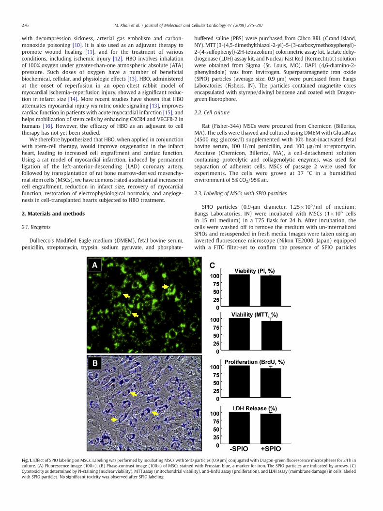
Fig. 1. Effect of SPIO labeling on MSCs. Labeling was performed by incubating MSCs with SPIOculture. (A) Fluorescence image (100×). (B) Phase-contrast image (100×) of MSCs stainedCytotoxicity as determined by PI-staining (nuclear viability), MTTassay (mitochondrial viabilwith SPIO particles. No significant toxicity was observed after SPIO labeling.
buffered saline (PBS) were purchased from Gibco BRL (Grand Island,NY).MTT (3-(4,5-dimethylthiazol-2-yl)-5-(3-carboxymethoxyphenyl)-2-(4-sulfophenyl)-2H-tetrazolium) colorimetric assay kit, lactate dehy-drogenase (LDH) assay kit, and Nuclear Fast Red (Kernechtrot) solutionwere obtained from Sigma (St. Louis, MO). DAPI (4,6-diamino-2-phenylindole) was from Invitrogen. Superparamagnetic iron oxide(SPIO) particles (average size, 0.9 μm) were purchased from BangsLaboratories (Fishers, IN). The particles contained magnetite coresencapsulated with styrene/divinyl benzene and coated with Dragon-green fluorophore.
2.2. Cell culture
Rat (Fisher-344) MSCs were procured from Chemicon (Billerica,MA). The cells were thawed and cultured using DMEMwith GlutaMax(4500 mg glucose/l) supplemented with 10% heat-inactivated fetalbovine serum, 100 U/ml penicillin, and 100 μg/ml streptomycin.Accutase (Chemicon, Billerica, MA), a cell-detachment solutioncontaining proteolytic and collagenolytic enzymes, was used forseparation of adherent cells. MSCs of passage 2 were used forexperiments. The cells were grown at 37 °C in a humidifiedenvironment of 5% CO2/95% air.
2.3. Labeling of MSCs with SPIO particles
SPIO particles (0.9-μm diameter, 1.25×10⁵/ml of medium;Bangs Laboratories, IN) were incubated with MSCs (1×10⁶ cellsin 15 ml medium) in a T75 flask for 24 h. After incubation, thecells were washed off to remove the medium with un-internalizedSPIOs and resuspended in fresh media. Images were taken using aninverted fluorescence microscope (Nikon TE2000, Japan) equippedwith a FITC filter-set to confirm the presence of SPIO particles
particles (0.9 μm) conjugated with Dragon-green fluorescence microspheres for 24 h inwith Prussian blue, a marker for iron. The SPIO particles are indicated by arrows. (C)ity), anti-BrdU assay (proliferation), and LDH assay (membrane damage) in cells labeled
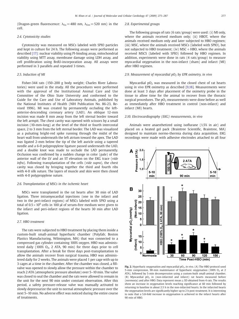
Fig. 2. Hyperbaric oxygenation and myocardial pO2, in vivo. (A) The HBO protocol used5-min compression, 90-min maintenance of hyperbaric oxygenation (100% O2 at 2ATA), followed by 5-min decompression using a custom-built small-animal chamber.(B) Myocardial pO2 in (non-infarcted and infarct) rat hearts measured before(normoxia) and after HBO. Data represent mean±SD obtained from 4 rats. The resultsshow an increase in oxygenation levels reaching significance at 60 min followed byreturning to baseline in about 2.5 h in the non-infarcted hearts. In the infarcted heartsthe oxygenation levels are significantly higher up to 2 h post-treatment. It is interestingto note that a 5.8-fold increase in oxygenation is achieved in the infarct hearts after90 min of HBO.
277M. Khan et al. / Journal of Molecular and Cellular Cardiology 47 (2009) 275–287
(Dragon-green fluorescence: λEx=480 nm, λEm=520 nm) in thecell.
2.4. Cytotoxicity studies
Cytotoxicity was measured on MSCs labeled with SPIO particlesand kept in culture for 24 h. The following assays were performed asdescribed [17]: nuclear viability using PI-binding assay, mitochondrialviability using MTT assay, membrane damage using LDH assay, andcell proliferation using BrdU-incorporation assay. All assays wereperformed in 3 parallels and repeated 3 times.
2.5. Induction of MI
Fisher-344 rats (150–200 g body weight; Charles River Labora-tories) were used in the study. All the procedures were performedwith the approval of the Institutional Animal Care and UseCommittee of the Ohio State University and conformed to theGuide for the Care and Use of Laboratory Animals, published bythe National Institutes of Health (NIH Publication No. 86-23, Re-vised 1996). MI was created by permanently occluding the left-anterior-descending coronary artery (LAD). An oblique 12-mmincision was made 8 mm away from the left sternal border towardthe left armpit. The chest cavity was opened with scissors by a smallincision (10-mm-long) at the level of the third or fourth intercostalspace, 2 to 3 mm from the left sternal border. The LAD was visualizedas a pulsating bright-red spike running through the midst of theheart wall from underneath the left atrium toward the apex. The LADwas ligated 2-mm below the tip of the left auricle using a taperedneedle and a 6-0 polypropylene ligature passed underneath the LAD,and a double knot was made to occlude the LAD permanently.Occlusion was confirmed by a sudden change in color (pale) of theanterior wall of the LV and an ST elevation on the EKG trace (videinfra). Following transplantation of the cells (vide supra), the chestcavity was closed by bringing together the third and fourth ribswith 4-0 silk suture. The layers of muscle and skin were then closedwith 4-0 polypropylene suture.
2.6. Transplantation of MSCs in the ischemic heart
MSCs were transplanted in the rat hearts after 30 min of LADligation. Three intramyocardial injections (one in the infarct andtwo in the peri-infarct regions) of MSCs labeled with SPIO using atotal of 0.5×10⁶ cells in 100 μl of serum-free medium were given tothe infarct and peri-infarct regions of the hearts 30 min after LADligation.
2.7. HBO treatment
The rats were subjected to HBO treatment by placing them inside acustom-built small-animal hyperbaric chamber (Polyfab; BostonPlastics Manufacturing, Wilmington, MA) that was connected to acompressed gas cylinder containing 100% oxygen. HBO was adminis-tered daily (100% O2, 2 ATA, 90 min) for three days prior to celltransplantation. After a break for three days post-transplantation toallow the animals recover from surgical trauma, HBO was adminis-tered daily for 2 weeks. The animals were placed 1 per cagewith up to3 cages at a time in the chamber. After the chamber was closed, a fill-valve was opened to slowly allow the pressure within the chamber toreach 2 ATA (atmospheric pressure absolute) over 5–10min. The valvewas closed to seal the chamber and the rats were allowed to remain inthe unit for the next 90 min under constant observation. After thisperiod, a safety pressure-release valve was manually activated toslowly depressurize the unit to normal atmospheric pressure over thenext 5–10 min. No adverse effect was noticed during the entire courseof treatments.
2.8. Experimental groups
The following groups of rats (6 rats/group) were used: (i) MI only,where the animals received medium only; (ii) HBOT, where theanimals received medium only and later subjected to HBO regimen;(iii) MSC, where the animals received MSCs (labeled with SPIO), butnot subjected to HBO treatment; (iv) MSC+HBO, where the animalsreceived MSCS (labeled with SPIO) followed by HBO regimen. Inaddition, experiments were done in rats (4 rats/group) to measuremyocardial oxygenation in the non-infarct (sham) and infarct (MI)after HBO regimen.
2.9. Measurement of myocardial pO2 by EPR oximetry, in vivo
Myocardial pO2 was measured in the closed chest of rat heartsusing in vivo EPR oximetry as described [9,18]. Measurements weredone at least 3 days after placement of the oximetry probe in thetissue to allow time for the animal to recover from the thoracicsurgical procedures. The pO2 measurements were done before as wellas immediately after HBO treatment in control (non-infarct) andinfarct (MI) hearts.
2.10. Electrocardiography (EKG) measurements, in vivo
Animals were anaesthetized using isoflurane (1.5% in air) andplaced on a heated gel pack (Braintree Scientific, Braintree, MA)designed to maintain normo-thermia during data acquisition. EKGrecordings were made with adhesive electrodes attached to all four
278 M. Khan et al. / Journal of Molecular and Cellular Cardiology 47 (2009) 275–287
paws and chest with the rat in a supine position. Electrocardiogramswere digitally recorded using a physiologic data acquisition system(PowerLab, AD Instruments, CO), with a sampling rate of 1 kHz. TheEKGs were signal-averaged (from lead II) over 150–200 beats peracquisition for determination of the P wave duration, P–R interval, andQT interval.
Evaluation of the orientation of the mean QRS vector in thefrontal plane was done by the average precession of the depolariza-tion wave through the ventricles. For most mammals the orientationof the mean spatial QRS vector in the frontal plane is tail-ward,leftward and slightly dorsal. However, with occlusion of the LAD andsubsequent death of cardiac tissue within the region, the leftanterior region of the ventricular mass is electrophysiologicallyremoved from the activation sequence, and the resulting processionis dominated by the left-ventricular free-wall, activated in a cranio-caudal (tail-ward) orientation. Thus, the orientation of the meanQRS in the frontal plane would be oriented tail-ward. Theorientation of the QRS vector in all hearts was tracked over timeto assess changes in the left-ventricular depolarization wave in theinfarcted region.
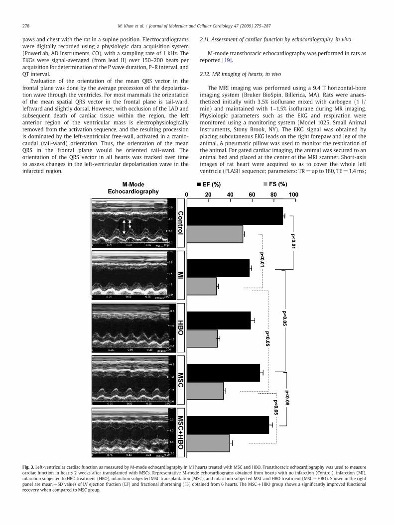
Fig. 3. Left-ventricular cardiac function as measured by M-mode echocardiography in MI hecardiac function in hearts 2 weeks after transplanted with MSCs. Representative M-modeinfarction subjected to HBO treatment (HBO), infarction subjected MSC transplantation (MSpanel are mean±SD values of LV ejection fraction (EF) and fractional shortening (FS) obtrecovery when compared to MSC group.
2.11. Assessment of cardiac function by echocardiography, in vivo
M-mode transthoracic echocardiography was performed in rats asreported [19].
2.12. MR imaging of hearts, in vivo
The MRI imaging was performed using a 9.4 T horizontal-boreimaging system (Bruker BioSpin, Billerica, MA). Rats were anaes-thetized initially with 3.5% isoflurane mixed with carbogen (1 l/min) and maintained with 1–1.5% isoflurane during MR imaging.Physiologic parameters such as the EKG and respiration weremonitored using a monitoring system (Model 1025, Small AnimalInstruments, Stony Brook, NY). The EKG signal was obtained byplacing subcutaneous EKG leads on the right forepaw and leg of theanimal. A pneumatic pillow was used to monitor the respiration ofthe animal. For gated cardiac imaging, the animal was secured to ananimal bed and placed at the center of the MRI scanner. Short-axisimages of rat heart were acquired so as to cover the whole leftventricle (FLASH sequence; parameters: TR=up to 180, TE=1.4 ms;
arts treated with MSC and HBO. Transthoracic echocardiography was used to measureechocardiograms obtained from hearts with no infarction (Control), infarction (MI),C), and infarction subjected MSC and HBO treatment (MSC+HBO). Shown in the rightained from 6 hearts. The MSC+HBO group shows a significantly improved functional
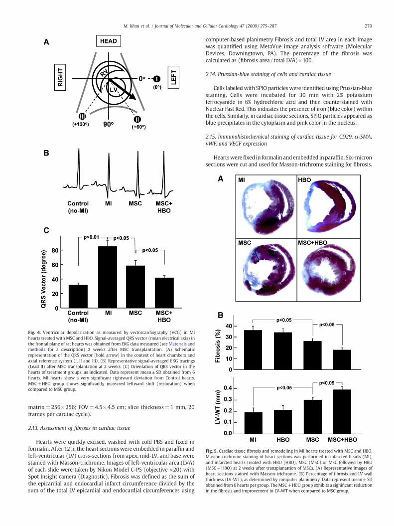
Fig. 4. Ventricular depolarization as measured by vectorcardiography (VCG) in MIhearts treated with MSC and HBO. Signal-averaged QRS vector (mean electrical axis) inthe frontal plane of rat hearts was obtained from EKG data measured (see Materials andmethods for a description) 2 weeks after MSC transplantation. (A) Schematicrepresentation of the QRS vector (bold arrow) in the context of heart chambers andaxial reference system (I, II and III). (B) Representative signal-averaged EKG tracings(Lead II) after MSC transplantation at 2 weeks. (C) Orientation of QRS vector in thehearts of treatment groups, as indicated. Data represent mean±SD obtained from 6hearts. MI hearts show a very significant rightward deviation from Control hearts.MSC+HBO group shows significantly increased leftward shift (restoration) whencompared to MSC group.
Fig. 5. Cardiac tissue fibrosis and remodeling in MI hearts treated with MSC and HBO.Masson-trichrome staining of heart sections was performed in infarcted hearts (MI),and infarcted hearts treated with HBO (HBO), MSC (MSC) or MSC followed by HBO(MSC+HBO) at 2 weeks after transplantation of MSCs. (A) Representative images ofheart sections stained with Masson-trichrome. (B) Percentage of fibrosis and LV wallthickness (LV-WT), as determined by computer planimetry. Data represent mean±SDobtained from 6 hearts per group. TheMSC+HBO group exhibits a significant reductionin the fibrosis and improvement in LV-WT when compared to MSC group.
279M. Khan et al. / Journal of Molecular and Cellular Cardiology 47 (2009) 275–287
matrix=256×256; FOV=4.5×4.5 cm; slice thickness=1 mm, 20frames per cardiac cycle).
2.13. Assessment of fibrosis in cardiac tissue
Hearts were quickly excised, washed with cold PBS and fixed informalin. After 12 h, the heart sections were embedded in paraffin andleft-ventricular (LV) cross-sections from apex, mid-LV, and base werestained with Masson-trichrome. Images of left-ventricular area (LVA)of each slide were taken by Nikon Model C-PS (objective ×20) withSpot Insight camera (Diagnostic). Fibrosis was defined as the sum ofthe epicardial and endocardial infarct circumference divided by thesum of the total LV epicardial and endocardial circumferences using
computer-based planimetry Fibrosis and total LV area in each imagewas quantified using MetaVue image analysis software (MolecularDevices, Downingtown, PA). The percentage of the fibrosis wascalculated as (fibrosis area/total LVA)×100.
2.14. Prussian-blue staining of cells and cardiac tissue
Cells labeled with SPIO particles were identified using Prussian-bluestaining. Cells were incubated for 30 min with 2% potassiumferrocyanide in 6% hydrochloric acid and then counterstained withNuclear Fast Red. This indicates the presence of iron (blue color) withinthe cells. Similarly, in cardiac tissue sections, SPIO particles appeared asblue precipitates in the cytoplasm and pink color in the nucleus.
2.15. Immunohistochemical staining of cardiac tissue for CD29, α-SMA,vWF, and VEGF expression
Heartswerefixed in formalin and embedded inparaffin. Six-micronsections were cut and used for Masson-trichrome staining for fibrosis.
280 M. Khan et al. / Journal of Molecular and Cellular Cardiology 47 (2009) 275–287
For immunofluorescence staining, the fixed tissue sections wereserially rehydrated in 100%, 95%, and 80% ethanol after deparaffiniza-tionwith xylene. Slideswere kept in steam for 30min and thenwashedin PBS (pH 7.4) three times for 5 min each. The tissue sections werethen incubated with 2% goat serum and 5% bovine-serum albumin inPBS to reduce nonspecific binding. The sections were then incubatedfor 4 h with mouse anti-CD29, anti-α-smooth-muscle actin (α-SMA)or anti-von Willebrand factor (vWF) monoclonal antibody or anti-VEGF. The sections were then incubated with appropriate anti-mousesecondary antibodies (1:1000 dilution) conjugated toTexas red (CD29,α-SMA), FITC (vWF and VEGF). Nuclei were counterstained withHardSet DAPI (Vector Labs). The tissue slides were visualized using aninverted Nikon fluorescence microscope. Separate sections were alsostained without primary antibodies to examine nonspecific binding.Blood vessels staining positive for α-SMA and vWF were counted inboth infarct and peri-infarct regions of the heart.
2.16. Immunohistochemistry for connexin-43 and troponin-T
Heart tissues were fixed in Histochoice fixative (Amresco, Solon,OH) and paraffin-embedded. Immunohistochemistry was performedas follows. Paraffin-embedded tissue sections were deparaffinized
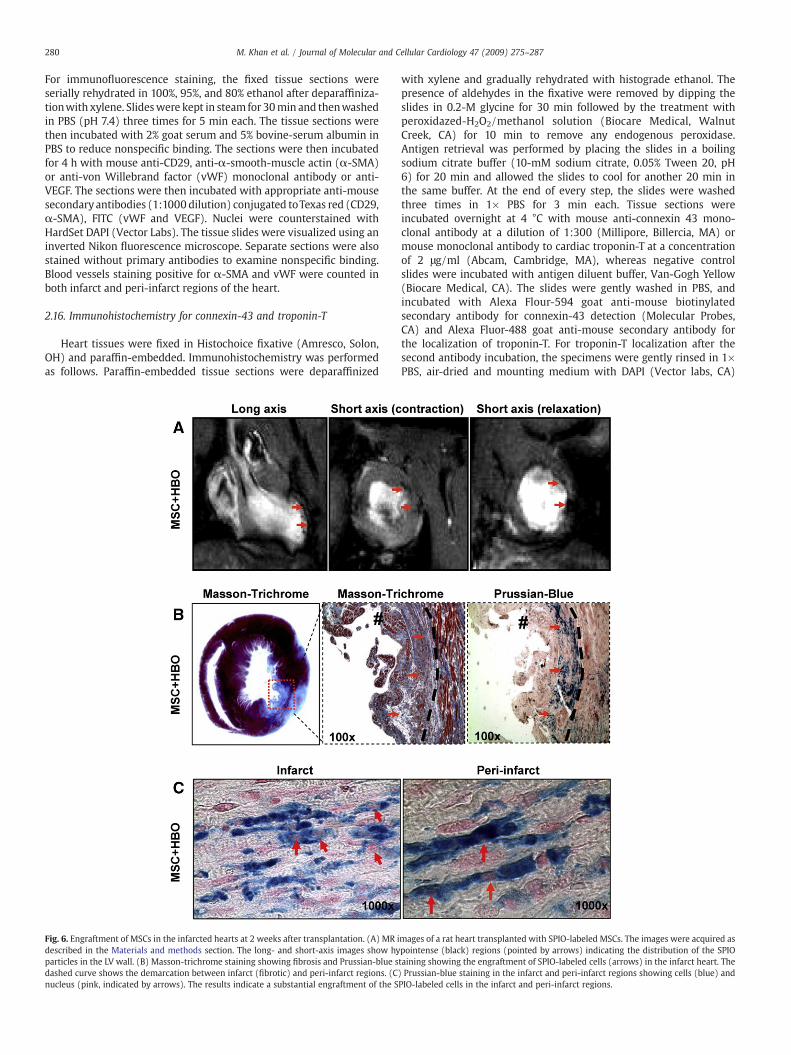
Fig. 6. Engraftment of MSCs in the infarcted hearts at 2 weeks after transplantation. (A) MR idescribed in the Materials and methods section. The long- and short-axis images show hyparticles in the LV wall. (B) Masson-trichrome staining showing fibrosis and Prussian-blue sdashed curve shows the demarcation between infarct (fibrotic) and peri-infarct regions. (Cnucleus (pink, indicated by arrows). The results indicate a substantial engraftment of the S
with xylene and gradually rehydrated with histograde ethanol. Thepresence of aldehydes in the fixative were removed by dipping theslides in 0.2-M glycine for 30 min followed by the treatment withperoxidazed-H2O2/methanol solution (Biocare Medical, WalnutCreek, CA) for 10 min to remove any endogenous peroxidase.Antigen retrieval was performed by placing the slides in a boilingsodium citrate buffer (10-mM sodium citrate, 0.05% Tween 20, pH6) for 20 min and allowed the slides to cool for another 20 min inthe same buffer. At the end of every step, the slides were washedthree times in 1× PBS for 3 min each. Tissue sections wereincubated overnight at 4 °C with mouse anti-connexin 43 mono-clonal antibody at a dilution of 1:300 (Millipore, Billercia, MA) ormouse monoclonal antibody to cardiac troponin-T at a concentrationof 2 μg/ml (Abcam, Cambridge, MA), whereas negative controlslides were incubated with antigen diluent buffer, Van-Gogh Yellow(Biocare Medical, CA). The slides were gently washed in PBS, andincubated with Alexa Flour-594 goat anti-mouse biotinylatedsecondary antibody for connexin-43 detection (Molecular Probes,CA) and Alexa Fluor-488 goat anti-mouse secondary antibody forthe localization of troponin-T. For troponin-T localization after thesecond antibody incubation, the specimens were gently rinsed in 1×PBS, air-dried and mounting medium with DAPI (Vector labs, CA)
mages of a rat heart transplanted with SPIO-labeled MSCs. The images were acquired aspointense (black) regions (pointed by arrows) indicating the distribution of the SPIOtaining showing the engraftment of SPIO-labeled cells (arrows) in the infarct heart. The) Prussian-blue staining in the infarct and peri-infarct regions showing cells (blue) andPIO-labeled cells in the infarct and peri-infarct regions.
281M. Khan et al. / Journal of Molecular and Cellular Cardiology 47 (2009) 275–287
was applied topically and slides were viewed under fluorescencemicroscope (Nikon TE 2000, Japan).
2.17. Data analysis
The statistical significance of the results was evaluated usingANOVA and a Student's t-test. Repeated-measures ANOVA andBonferroni t-test were used for multiple comparisons of pO2 data.The values were expressed as mean±SD. A p value of b0.05 wasconsidered to be significant.
3. Results
3.1. Effect of SPIO labeling on MSCs
MSCs were labeled with Dragon-green fluorescence-conjugatedSPIO particles by co-incubation in culture for 24 h. Fluorescencemicroscopy and phase-contrast optical images confirmed uptake ofSPIO particles by MSCs (Fig. 1). The cells were then examined forpossible cytotoxicity induced by the uptake of SPIO particles. Thelabeled cells were kept in culture for 24 h, and subjected to standardcytotoxicity tests including PI-binding assay for nuclear viability, MTTassay for mitochondrial function, BrdU-incorporation assay forproliferation, and LDH-release assay for membrane integrity. Theresults (Fig. 1C) showed that labeling of MSCs with SPIO had nosignificant effect upon cellular viability and proliferation.
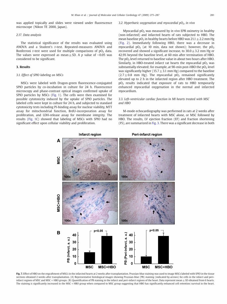
Fig. 7. Effect of HBO on the engraftment of MSCs in the infarcted hearts at 2weeks after transpsections obtained 2 weeks after transplantation. (A) Representative histological images shoinfarct regions of MSC and MSC+HBO groups. (B) Quantification of PB staining in the infarctThe staining is significantly increased in the MSC+HBO group when compared to MSC gro
3.2. Hyperbaric oxygenation and myocardial pO2, in vivo
Myocardial pO2 was measured by in vivo EPR oximetry in healthy(non-infarcted) and infarcted hearts of rats subjected to HBO. Themean baseline pO2 in healthy hearts before HBOwas 21.1±2.2mmHg(Fig. 2). Immediately following HBO, there was a decrease inmyocardial pO2 (at 10 min, data not shown); however, the pO2
recovered and showed a significant increase, to 30.0±3.2 mm Hg or42.9% beyond the baseline level, at 60 min after termination of HBO.The pO2 level returned to baseline value in about two hours after HBO.Similarly, in HBO-treated infarct rat hearts the myocardial pO2 wassubstantially elevated; for example, at 90-min post-HBO the pO2 levelwas significantly higher (15.7±3.1 mm Hg) compared to the baseline(2.7±0.8 mm Hg). The myocardial pO2 remained significantlyelevated up to 2 h in the infarcted region after HBO treatment. ThepO2 results indicated that exposure of rats to HBO temporarilyenhanced myocardial oxygenation in the normal and infarctedmyocardium.
3.3. Left-ventricular cardiac function in MI hearts treated with MSCand HBO
M-mode echocardiography was performed in rats at 2 weeks aftertreatment of infarcted hearts with MSC alone, or MSC followed byHBO. The results, LV ejection fraction (EF) and fraction shortening(FS), are summarized in Fig. 3. Therewas a significant decrease in both
lantation. Prussian-blue staining was used to imageMSCs labeled with SPIO in the tissuewing Prussian-blue (PB) staining (indicated by arrows) for cells in the infarct and peri-and peri-infarct regions of the heart. Data represent mean±SD obtained from 6 hearts.up suggesting that HBO has significantly enhanced cell retention/survival in the heart.
282 M. Khan et al. / Journal of Molecular and Cellular Cardiology 47 (2009) 275–287
EF and FS in the MI group when compared to control group (pb0.01).MSC treatment significantly restored the cardiac functions. However,HBO by itself had no significant effect on the recovery. In contrast, HBOin combination with MSC treatment significantly improved both thefunctions when compared to MSC alone treatment. The echocardio-graphy results indicated that HBO augmented the recovery of cardiacfunction by MSC treatment.
3.4. Ventricular depolarization in MI hearts treated with MSC and HBO
The QRS vector is used as a measure of abnormalities in ventriculardepolarization that may occur as result of loss of functional cardio-myocytes in MI heart [20]. It represents the net orientation of electricalimpulse propagation during ventricular depolarization. The mean QRSvector is obtained from signal-averaged QRS complex EKG data. Aschematic illustration of the QRS vector with reference to cardiacanatomy and traditional reference axis system is shown in Fig. 4A. The
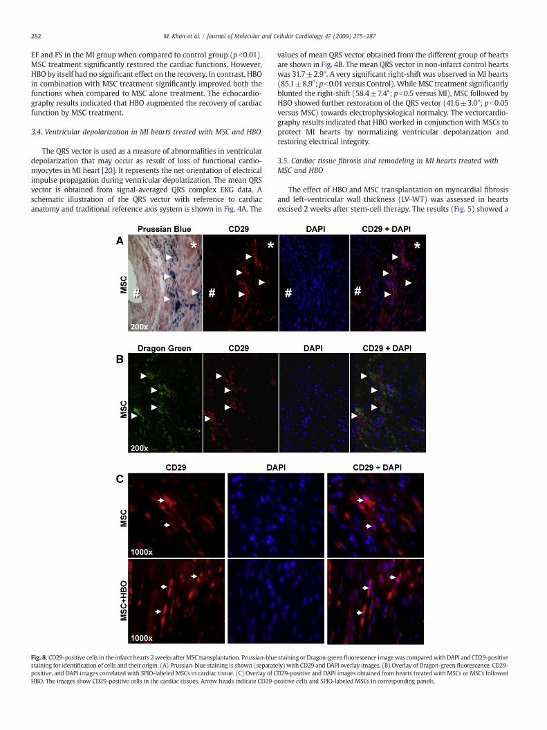
Fig. 8. CD29-positive cells in the infarct hearts 2weeks afterMSC transplantation. Prussian-bluestaining for identification of cells and their origin. (A) Prussian-blue staining is shown (separatpositive, and DAPI images correlated with SPIO-labeled MSCs in cardiac tissue. (C) Overlay of CHBO. The images show CD29-positive cells in the cardiac tissues. Arrow heads indicate CD29-p
values of mean QRS vector obtained from the different group of heartsare shown in Fig. 4B. The mean QRS vector in non-infarct control heartswas 31.7±2.9°. A very significant right-shift was observed in MI hearts(85.1±8.9°; pb0.01 versus Control).WhileMSC treatment significantlyblunted the right-shift (58.4±7.4°; pb0.5 versus MI), MSC followed byHBO showed further restoration of the QRS vector (41.6±3.0°; pb0.05versus MSC) towards electrophysiological normalcy. The vectorcardio-graphy results indicated that HBO worked in conjunction with MSCs toprotect MI hearts by normalizing ventricular depolarization andrestoring electrical integrity.
3.5. Cardiac tissue fibrosis and remodeling in MI hearts treated withMSC and HBO
The effect of HBO and MSC transplantation on myocardial fibrosisand left-ventricular wall thickness (LV-WT) was assessed in heartsexcised 2 weeks after stem-cell therapy. The results (Fig. 5) showed a
staining or Dragon-green fluorescence imagewas comparedwith DAPI and CD29-positiveely) with CD29 and DAPI overlay images. (B) Overlay of Dragon-green fluorescence, CD29-D29-positive and DAPI images obtained from hearts treated with MSCs or MSCs followedositive cells and SPIO-labeled MSCs in corresponding panels.

Fig. 9. Expression of connexin-43 in the engrafted cells 2 weeks after transplantation. Overlay images of Dragon-green fluorescence (green), connexin-43 immunofluorescence (red),and DAPI staining (blue) in the infarct and peri-infarct sections of a heart from MSC+HBO group. White broken circles indicate positive staining for connexin-43 expression.
Fig. 10. Cardiac troponin-T expression in the MI heart at 2 weeks after transplantation of MSC. (A) Overlay images of anti-troponin-T immunofluorescence (green) and DAPI staining(blue) in heart sections in the experimental groups including a non-MI (Control) group. (B) Quantification of troponin-T levels in the cardiac tissues. Data represent mean±SD from4 hearts per group. The troponin-T level is significantly higher in the MSC+HBO group when compared to the MSC group.
283M. Khan et al. / Journal of Molecular and Cellular Cardiology 47 (2009) 275–287
284 M. Khan et al. / Journal of Molecular and Cellular Cardiology 47 (2009) 275–287
significant reduction in tissue fibrosis and recovery of LV-WT in boththe MSC and MSC+HBO groups when compared to MI group.Combined treatment of MSC and HBO resulted in a significantattenuation of fibrosis and restoration of LV-WT when compared toMSC alone treatment.
3.6. Engraftment of MSCs in the infarcted hearts
The retention and distribution of SPIO-labeled cells in the infarctheart at 2 weeks post-transplantation were verified by in vivo MRimaging as well as by histochemical staining using Prussian blue. Twoweeks after intramyocardial injection of MSCs labeled with SPIO, MRimaging was performed using a 9.4 T MRI system. Large, hypointenseareas were detected at the site where SPIO-labeled MSCs were injected(Fig. 6A). Masson-trichrome staining (for fibrosis) and Prussian-bluestaining (for SPIO) in the heart tissue harvested 2 weeks post-transplantation showed positive staining for engrafted cells (Fig. 6B).The images further revealed a substantial distribution of the Prussian-blue-stained cells in the peri-infarct region as well (Fig. 6C). Weestimated the relative amounts of engrafted cells in the infarct and peri-infarct regions in the MSC and MSC+HBO group of hearts. Fig. 7Ashows representative histological slides stained with Prussian blue. Asubstantial increase in the stainingwas observed in both the infarct andperi-infarct regions of the MSC+HBO group. Quantitative analysis ofthe Prussian-blue staining revealed significantly higher levels of cellengraftment up on HBO treatment (Fig. 7B). Taken together, the in vivoMRI and histology results clearly suggested engraftment of thetransplanted cells in the infarcted heart and further that HBO enhancedcell engraftment both in the infarct as well peri-infarct regions.However, the above results do not identify the type of cell being stained.
3.7. Identification of CD29-positive cells in the infarct heartstransplanted with MSCs
To identify whether the cells stained by Prussian blue (vide supra)are of MSC-type, we used immunohistochemical staining for CD29
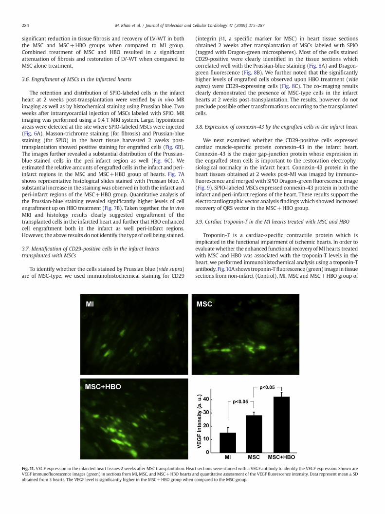
Fig. 11. VEGF expression in the infarcted heart tissues 2 weeks after MSC transplantation. HearVEGF immunofluorescence images (green) in sections from MI, MSC, and MSC+HBO hearts aobtained from 3 hearts. The VEGF level is significantly higher in the MSC+HBO group when
(integrin β1, a specific marker for MSC) in heart tissue sectionsobtained 2 weeks after transplantation of MSCs labeled with SPIO(tagged with Dragon-green microspheres). Most of the cells stainedCD29-positive were clearly identified in the tissue sections whichcorrelated well with the Prussian-blue staining (Fig. 8A) and Dragon-green fluorescence (Fig. 8B). We further noted that the significantlyhigher levels of engrafted cells observed upon HBO treatment (videsupra) were CD29-expressing cells (Fig. 8C). The co-imaging resultsclearly demonstrated the presence of MSC-type cells in the infarcthearts at 2 weeks post-transplantation. The results, however, do notpreclude possible other transformations occurring to the transplantedcells.
3.8. Expression of connexin-43 by the engrafted cells in the infarct heart
We next examined whether the CD29-positive cells expressedcardiac muscle-specific protein connexin-43 in the infarct heart.Connexin-43 is the major gap-junction protein whose expression inthe engrafted stem cells is important to the restoration electrophy-siological normalcy in the infarct heart. Connexin-43 protein in theheart tissues obtained at 2 weeks post-MI was imaged by immuno-fluorescence and merged with SPIO Dragon-green fluorescence image(Fig. 9). SPIO-labeledMSCs expressed connexin-43 protein in both theinfarct and peri-infarct regions of the heart. These results support theelectrocardiographic vector analysis findings which showed increasedrecovery of QRS vector in the MSC+HBO group.
3.9. Cardiac troponin-T in the MI hearts treated with MSC and HBO
Troponin-T is a cardiac-specific contractile protein which isimplicated in the functional impairment of ischemic hearts. In order toevaluatewhether the enhanced functional recovery ofMI hearts treatedwith MSC and HBO was associated with the troponin-T levels in theheart, we performed immunohistochemical analysis using a troponin-Tantibody. Fig.10A shows troponin-Tfluorescence (green) image in tissuesections from non-infarct (Control), MI, MSC and MSC+HBO group of
t sections were stained with a VEGF antibody to identify the VEGF expression. Shown arend quantitative assessment of the VEGF fluorescence intensity. Data represent mean±SDcompared to the MSC group.
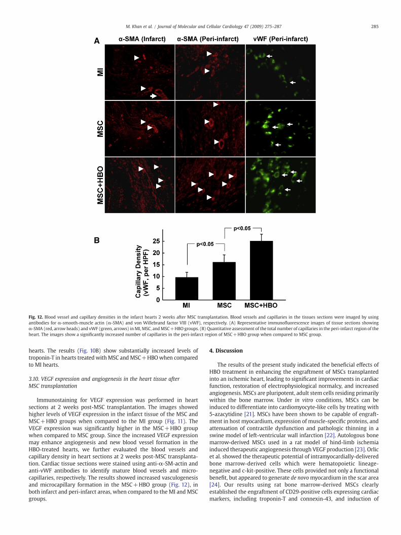
Fig. 12. Blood vessel and capillary densities in the infarct hearts 2 weeks after MSC transplantation. Blood vessels and capillaries in the tissues sections were imaged by usingantibodies for α-smooth-muscle actin (α-SMA) and von Willebrand factor VIII (vWF), respectively. (A) Representative immunofluorescence images of tissue sections showingα-SMA (red, arrow heads) and vWF (green, arrows) inMI, MSC, andMSC+HBO groups. (B) Quantitative assessment of the total number of capillaries in the peri-infarct region of theheart. The images show a significantly increased number of capillaries in the peri-infarct region of MSC+HBO group when compared to MSC group.
285M. Khan et al. / Journal of Molecular and Cellular Cardiology 47 (2009) 275–287
hearts. The results (Fig. 10B) show substantially increased levels oftroponin-T in hearts treatedwithMSC andMSC+HBOwhen comparedto MI hearts.
3.10. VEGF expression and angiogenesis in the heart tissue afterMSC transplantation
Immunostaining for VEGF expression was performed in heartsections at 2 weeks post-MSC transplantation. The images showedhigher levels of VEGF expression in the infarct tissue of the MSC andMSC+HBO groups when compared to the MI group (Fig. 11). TheVEGF expression was significantly higher in the MSC+HBO groupwhen compared to MSC group. Since the increased VEGF expressionmay enhance angiogenesis and new blood vessel formation in theHBO-treated hearts, we further evaluated the blood vessels andcapillary density in heart sections at 2 weeks post-MSC transplanta-tion. Cardiac tissue sections were stained using anti-α-SM-actin andanti-vWF antibodies to identify mature blood vessels and micro-capillaries, respectively. The results showed increased vasculogenesisand microcapillary formation in the MSC+HBO group (Fig. 12), inboth infarct and peri-infarct areas, when compared to the MI and MSCgroups.
4. Discussion
The results of the present study indicated the beneficial effects ofHBO treatment in enhancing the engraftment of MSCs transplantedinto an ischemic heart, leading to significant improvements in cardiacfunction, restoration of electrophysiological normalcy, and increasedangiogenesis. MSCs are pluripotent, adult stem cells residing primarilywithin the bone marrow. Under in vitro conditions, MSCs can beinduced to differentiate into cardiomyocyte-like cells by treating with5-azacytidine [21]. MSCs have been shown to be capable of engraft-ment in host myocardium, expression of muscle-specific proteins, andattenuation of contractile dysfunction and pathologic thinning in aswine model of left-ventricular wall infarction [22]. Autologous bonemarrow-derived MSCs used in a rat model of hind-limb ischemiainduced therapeutic angiogenesis through VEGF production [23]. Orlicet al. showed the therapeutic potential of intramyocardially-deliveredbone marrow-derived cells which were hematopoietic lineage-negative and c-kit-positive. These cells provided not only a functionalbenefit, but appeared to generate de novomyocardium in the scar area[24]. Our results using rat bone marrow-derived MSCs clearlyestablished the engraftment of CD29-positive cells expressing cardiacmarkers, including troponin-T and connexin-43, and induction of
286 M. Khan et al. / Journal of Molecular and Cellular Cardiology 47 (2009) 275–287
angiogenesis through VEGF production. The engraftment and differen-tiation of MSCs were markedly augmented by adjuvant HBO.
HBO is emerging as a potential therapy for cardiovascular diseases[12]. In a randomized multicenter study using patients with acute MI,adjunctive HBOwith thrombolytic agents (recombinant tissue plasmi-nogen activator or streptokinase) resulted in a decrease in creatinephosphokinase levels, rapid resolution of pain and an improvement inejection fraction [15,25]. Preconditioning of rats with HBO (2.5 ATA,60 min, twice daily for two days) has been shown to alleviate theinjury to ischemic myocardium induced by LAD coronary arteryligation [26]. The results of the present study did not show anysignificant effect of HBO on cardiac function (Fig. 3) or infarct size(Fig. 5) measured 2 weeks after induction of infarction. Tritto et al.used a rat model of permanent coronary artery occlusion to show thatHBO could only delay the progression of ischemic cell death, withoutaffecting the ultimate extent of necrosis measured after 3 weeks [27].Our results also seemed to indicate that HBO alone could not provideany beneficial effect in the long term, although HBO was administeredthroughout the two-week period.
HBO significantly increased the recovery of cardiac function andreduced infarct size in hearts transplanted with MSCs. The beneficialeffects of adjuvant HBO were significantly greater than that providedby MSCs alone. The synergistic effect could stem from a HBO-mediated increase in CD29-positive graft survival (Fig. 8), leading toan increase in VEGF production (Fig. 11) and angiogenesis (Fig. 12).Hyperbaria, as well as hyperoxia, has been shown to limit infarct sizeand attenuate ischemia–reperfusion injury [14,28]. Pre-exposure ofrats to one-time, 60-min hyperbaric oxygenation, which includes bothhyperoxia and hyperbaria, was shown to be protective of heartsisolated and then subjected to ischemia–reperfusion injury [29]. Thestudy concluded that the protective effect was mediated by nitricoxide synthase and dependent on the oxygen availability (partialpressure of oxygen) rather than hyperbaria alone. In contrast, in ourpresent study using in vivo hearts we did not observe any significantreduction in infarct size in the HBO group. The difference could beattributed to the long period (2 weeks) after the induction of MI in ourstudy. However, when HBO was used as an adjuvant with stem-celltherapy, we observed a significant reduction in myocardial fibrosisand wall thinning.
The HBO-mediated recovery of the QRS vector in the MSC+HBOgroup of hearts (Fig. 4) is reflective of connexin-43 expression in thegrafted cells (Fig. 9). Taken together, the results seem to suggest thatHBO works in conjunction with transplanted MSCs, although by itselfmay not be beneficial in the long term. It has been reported that HBOmobilized bonemarrow-derived stem/progenitor cells in humans andanimals by stimulating nitric oxide synthase and that this mobiliza-tion of cells remained elevated in human blood over the course of 20HBO treatments [16]. While the contribution of a similar effect tomobilize endogenous stem cells by HBO cannot be ruled out in ourmodel, the observed results do not suggest any significance in theircontribution.
We measured myocardial tissue oxygenation by implanting anoxygen-sensitive EPR probe in the myocardium as reported previously[18,19]. Immediately after HBO administration, there was a substantialincrease in myocardial oxygenation in the infarct hearts, and itremained significantly elevated up to 2.5 h following termination ofthe HBO session. Although the magnitude (absolute value) of increasewas similar to that in non-infarct hearts, the hyperoxygenated infarcthearts showed a 5.8-fold increase when compared to the normoxicbaseline. The in vivo oximetry measurements clearly established thatHBO increases myocardial tissue oxygenation in the infarct tissue. Theobserved improvements in cell engraftment and cardiac functionindicated by HBO suggest that the hyperoxic post-conditioning couldplay a major role in enhancing graft survival and retention.
There is an increasing body of evidence that MSCs transfected withVEGF improves cardiac function and increases angiogenesis in the
infarct heart [30]. In addition to its potent angiogenic functions, VEGFcan stimulate proliferation, delay senescence, suppress apoptosis andpromote survival of various cells [31]. Different oxygen tensions appearto activate different signaling pathways to stimulate VEGF expressionfor angiogenesis [32]. Hypoxia enhances reactive oxygen species (ROS)production, which potently induces VEGF expression via a hypoxia-inducible factor-1 (HIF-1)-independent pathway [32]. A recent studyshowed that HBO reduces liver injury in regenerating rat liver after apartial hepatectomy through enhanced VEGF expression [33]. Simi-larly, the results from our study show that there was a significantincrease in VEGF expression in the MSC+HBO hearts at 2 weeks post-transplantation. A possible mechanism responsible for the increasedangiogenesis observed in the MSC+HBO group in our study could bethis increase in VEGF expression. Thus, there is strong evidence tosupport our contention that HBO, used in conjunction with MSCcardiomyoplasty, enhances the engraftment and survival of trans-planted cells through increased VEGF expression and angiogenesis.
Electrocardiography remains one of the most important and non-invasive means of identifying and quantifying the severity ofmyocardial infarction. A typical frontal plane QRS loop for carnivoresandprimates is oriented inferiorly and leftward, because the prevailingportion of the QRS loop is produced by waves of depolarizationtraversing the normally-dominant left-ventricular free-wall in anendocardial to epicardial direction. When a left-anterior-descendingcoronary artery is ligated, an anteroseptal infarct is produced, and thatregion of myocardium does not contribute to the QRS loop. In the ratviewed from the front (from the ventral surface of the torso of aquadruped) surface, the QRS loop would be displaced from the left,caudal (tail-ward) surface of the torso. Thus, instead of the QRS loopbeing inscribed leftward and caudal in a usually clockwise manner, itsdirection of inscription becomes counter-clockwise having beendeviated from the region of infarction. If the regenerated tissue iscapable of conducting cardiac electrical signals it will becomedepolarized and the orientation of the QRS loop tends to returntowards electrophysiologic normalcy. The significant restoration of themeanQRS vector orientation in theMSC+HBO group of hearts (Fig. 4)indicates not only the regenerative capability of transplanted stemcells, but also their capability to express connexin-43 to restoreelectrical integrity in the infarct myocardium.
The differentiation of MSCs into fully-functional cardiomyocytes isstill controversial. A recent study by Haider et al. showed that MSCshave the ability to engraft in the ischemic heart and differentiate toadopt a cardiac phenotype [34]. The differentiating MSCs showed aspectrum of morphological changes that were characteristic ofdeveloping myofibers. Immunostaining for connexin-43, along withcardiac actin showed that the neofibers also developed intercellularcommunication junctions [34]. The tendency of MSCs to displayplasticity towards a cardiac lineage has been reported [35]. Bonemarrow-derived MSCs, co-cultured with rat embryonic cardiomyo-cytes, exhibited expression of troponin-T, troponin-I, α-actin, andconnexin-43 [35]. The induction of MSCs towards the cardiomyocytelineage was attributed to local cell-to-cell contacts and/or thepresence of soluble factors in the culture medium. Similarly, ourstudy demonstrated that the engrafted MSCs expressed bothtroponin-T and connexin-43. Connexin-43 expression was mostlyobserved in MSCs present in the peri-infarct regions of the heart. Thetendency towards connexin-43 expressionwas relatively higher in theMSC+HBO group.
Overall, the present study demonstrated that HBO could serve asan effective adjuvant to augment MSC-based cardiomyoplasty. HBOtreatment significantly enhanced the survival of transplanted MSCs inthe infarct heart, leading to improvements in cardiac function. Inaddition, MSC transplantation, along with HBO, resulted in increasedVEGF and connexin-43 expressions, as well as angiogenesis in theischemic tissue. Thus, HBO appears to be an effective and clinically-applicable method to improve the survival and retention of stem cells
287M. Khan et al. / Journal of Molecular and Cellular Cardiology 47 (2009) 275–287
used in the treatment of myocardial infarction and/or heart failure,thereby improving therapeutic efficacy and overall clinical outcome.
Acknowledgments
This study was supported by grants from AHA (SDG 0930181N toMK) and NIH (R01 EB006153 to PK). We thank Dr. Chandan K. Sen andMs. Lynn Lambert for advice and technical support for using the HBOchamber. We also thank Dr. Adriana Pedraza-Toscano for help withEKG data analysis.
References
[1] Murry CE, Field LJ, Menasche P. Cell-based cardiac repair: reflections at the 10-yearpoint. Circulation 2005;112:3174–83.
[2] Dimmeler S, Burchfield J, Zeiher AM. Cell-based therapy of myocardial infarction.Arterioscler Thromb Vasc Biol 2008;28:208–16.
[3] Robey TE, Saiget MK, Reinecke H, Murry CE. Systems approaches to preventingtransplanted cell death in cardiac repair. J Mol Cell Cardiol 2008;45:567–81.
[4] LiW,MaN, Ong LL, NesselmannC, Klopsch C, Ladilov Y, et al. Bcl-2 engineeredMSCsinhibited apoptosis and improved heart function. Stem Cells 2007;25:2118–27.
[5] Mangi AA, Noiseux N, Kong D, He H, Rezvani M, Ingwall JS, et al. Mesenchymalstem cells modified with Akt prevent remodeling and restore performance ofinfarcted hearts. Nat Med 2003;9:1195–201.
[6] Pasha Z, Wang Y, Sheikh R, Zhang D, Zhao T, Ashraf M. Preconditioning enhancescell survival and differentiation of stem cells during transplantation in infarctedmyocardium. Cardiovasc Res 2008;77:134–42.
[7] Seeger FH, Zeiher AM, Dimmeler S. Cell-enhancement strategies for the treatmentof ischemic heart disease. Nat Clin Pract Cardiovasc Med 2007;4(Suppl 1):S110–3.
[8] Laflamme MA, Chen KY, Naumova AV, Muskheli V, Fugate JA, Dupras SK, et al.Cardiomyocytes derived from human embryonic stem cells in pro-survival factorsenhance function of infarcted rat hearts. Nat Biotechnol 2007;25:1015–24.
[9] Wisel S, Khan M, Kuppusamy ML, Mohan IK, Chacko SM, Rivera BK, et al.Pharmacological preconditioning of mesenchymal stem cells with Trimetazidineprotects hypoxic cells against oxidative stress and enhances recovery ofmyocardial function in infarcted heart through Bcl-2 expression. J PharmacolExp Therap 2009;329:543–50.
[10] Tibbles PM, Edelsberg JS. Hyperbaric-oxygen therapy. N Engl JMed1996;334:1642–8.[11] Thackham JA, McElwain DL, Long RJ. The use of hyperbaric oxygen therapy to treat
chronic wounds. A Rev Wound Repair Regen 2008;16:321–30.[12] Yogaratnam JZ, Laden G, Madden LA, Seymour AM, Guvendik L, Cowen M, et al.
Hyperbaric oxygen: a new drug in myocardial revascularization and protection?Cardiovasc Revasc Med 2006;7:146–54.
[13] Yogaratnam JZ, Laden G, Guvendik L, Cowen M, Cale A, Griffin S. Pharmacologicalpreconditioning with hyperbaric oxygen: can this therapy attenuate myocardialischemic reperfusion injury and induce myocardial protection via nitric oxide?J Surg Res 2007.
[14] Sterling DL, Thornton JD, Swafford A, Gottlieb SF, Bishop SP, Stanley AW, et al.Hyperbaric oxygen limits infarct size in ischemic rabbit myocardium in vivo.Circulation 1993;88:1931–6.
[15] Dekleva M, Neskovic A, Vlahovic A, Putnikovic B, Beleslin B, Ostojic M. Adjunctiveeffect of hyperbaric oxygen treatment after thrombolysis on left ventricularfunction in patients with acute myocardial infarction. Am Heart J 2004;148:E14.
[16] Thom SR, Bhopale VM, Velazquez OC, Goldstein LJ, Thom LH, Buerk DG. Stem cellmobilization by hyperbaric oxygen. Am J Physiol Heart Circ Physiol 2006;290:H1378–86.
[17] Wisel S, Chacko SM, KuppusamyML, Pandian RP, KhanM, Kutala VK, et al. Labelingof skeletal myoblasts with a novel oxygen-sensing spin probe for noninvasivemonitoring of in situ oxygenation and cell therapy in heart. Am J Physiol Heart CircPhysiol 2007;292:H1254–61.
[18] Khan M, Kutala VK, Wisel S, Chacko SM, Kuppusamy ML, Kwiatkowski P, et al.Measurement of oxygenation at the site of stem cell therapy in a murine model ofmyocardial infarction. Adv Exp Med Biol 2008;614:45–52.
[19] Khan M, Kutala VK, Vikram DS, Wisel S, Chacko SM, Kuppusamy ML, et al. Skeletalmyoblasts transplanted in the ischemic myocardium enhance in situ oxygenationand recovery of contractile function. Am J Physiol Heart Circ Physiol 2007;293:H2129–39.
[20] Hamlin RL. The QRS electrocardiogram, epicardiogram, vectorcardiogram andventricular excitation of swine. Am J Physiol 1960;198:537–42.
[21] Makino S, Fukuda K, Miyoshi S, Konishi F, Kodama H, Pan J, et al. Cardiomyo-cytes can be generated from marrow stromal cells in vitro. J Clin Invest 1999;103:697–705.
[22] Shake JG, Gruber PJ, Baumgartner WA, Senechal G, Meyers J, Redmond JM, et al.Mesenchymal stem cell implantation in a swine myocardial infarct model: engraft-ment and functional effects. Ann Thorac Surg 2002;73:1919–25 [discussion 26].
[23] Al-Khaldi A, Al-Sabti H, Galipeau J, Lachapelle K. Therapeutic angiogenesis usingautologous bone marrow stromal cells: improved blood flow in a chronic limbischemia model. Ann Thorac Surg 2003;75:204–9.
[24] Orlic D, Kajstura J, Chimenti S, Jakoniuk I, Anderson SM, Li B, et al. Bone marrowcells regenerate infarcted myocardium. Nature 2001;410:701–5.
[25] Stavitsky Y, Shandling AH, Ellestad MH, Hart GB, Van Natta B, Messenger JC, et al.Hyperbaric oxygen and thrombolysis in myocardial infarction: the ‘HOT MI’randomized multicenter study. Cardiology 1998;90:131–6.
[26] Han C, Lin L, ZhangW, Zhang L, Lv S, Sun Q, et al. Hyperbaric oxygen preconditioningalleviates myocardial ischemic injury in rats. Exp Biol Med (Maywood) 2008;233:1448–53.
[27] Tritto I, Ambrosio G, Capelli-Bigazzi M, Perrone-Filardi P, Lepore S, Tifano R, et al.Effects of hyperbaric oxygen therapy on experimental infarct size: Salvage versusdelay of myocardial necrosis. J Appl Cardiol 1991;6:359–66.
[28] Tahep Id P, Valen G, Starkopf J, Kairane C, Zilmer M, Vaage J. Pretreating rats withhyperoxia attenuates ischemia–reperfusion injury of the heart. Life Sci 2001;68:1629–40.
[29] Cabigas BP, Su J, Hutchins W, Shi Y, Schaefer RB, Recinos RF, et al. Hyperoxic andhyperbaric-induced cardioprotection: role of nitric oxide synthase 3. CardiovascRes 2006;72:143–51.
[30] Yang J, Zhou W, Zheng W, Ma Y, Lin L, Tang T, et al. Effects of myocardialtransplantation of marrow mesenchymal stem cells transfected with vascularendothelial growth factor for the improvement of heart function and angiogenesisafter myocardial infarction. Cardiology 2007;107:17–29.
[31] Ferrara N, Gerber HP, LeCouter J. The biology of VEGF and its receptors. Nat Med2003;9:669–76.
[32] Sen CK, Khanna S, Babior BM, Hunt TK, Ellison EC, Roy S. Oxidant-induced vascularendothelial growth factor expression in human keratinocytes and cutaneouswound healing. J Biol Chem 2002;277:33284–90.
[33] Ijichi H, Taketomi A, Yoshizumi T, Uchiyama H, Yonemura Y, Soejima Y, et al.Hyperbaric oxygen induces vascular endothelial growth factor and reduces liverinjury in regenerating rat liver after partial hepatectomy. J Hepatol 2006;45:28–34.
[34] Haider H, Jiang S, Idris NM, Ashraf M. IGF-1-overexpressing mesenchymal stemcells accelerate bone marrow stem cell mobilization via paracrine activation ofSDF-1{alpha}/CXCR4 signaling to promote myocardial repair. Circ Res 2008;103:1300–8.
[35] Rose RA, Jiang H,Wang X, Helke S, Tsoporis JN, Gong N, et al. Bonemarrow-derivedmesenchymal stromal cells express cardiac-specific markers, retain the stromalphenotype, and do not become functional cardiomyocytes in vitro. Stem Cells2008;26:2884–92.