Embed Size (px)
Citation preview
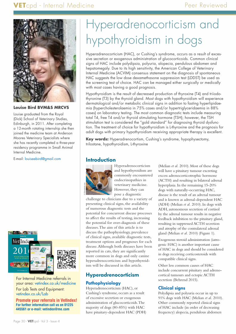
Page 30 - VETcpd - Vol 3 - Issue 4
Hyperadrenocorticism and hypothyroidism in dogs
VETcpd - Internal Medicine Peer Reviewed
Hyperadrenocorticism (HAC), or Cushing’s syndrome, occurs as a result of exces-sive secretion or exogenous administration of glucocorticoids. Common clinical signs of HAC include polydipsia, polyuria, alopecia, pendulous abdomen and hepatomegaly. Due to its high sensitivity, the American College of Veterinary Internal Medicine (ACVIM) consensus statement on the diagnosis of spontaneous HAC suggests the low dose dexamethasone suppression test (LDDST) be used as the screening test of choice. HAC can be managed either surgically or medically with most cases having a good prognosis.
Hypothyroidism is the result of decreased production of thyroxine (T4) and triiodo-thyronine (T3) by the thyroid gland. Most dogs with hypothyroidism will experience dermatological and/or metabolic clinical signs in addition to fasting hyperlipidae-mia (hypercholesterolaemia in 75% cases and/or hypertriglyceridaemia in 88% cases) on laboratory testing. The most common diagnostic tests include measuring total T4, free T4 and/or thyroid stimulating hormone (TSH); however, the TSH stimulation test is considered the “gold standard” for diagnosing thyroid dysfunc-tion. The treatment of choice for hypothyroidism is L-thyroxine and the prognosis for adult dogs with primary hypothyroidism receiving appropriate therapy is excellent.
Key words: Hyperadrenocortism, Cushing’s syndrome, hypophysectomy, trilostane, hypothyroidism, L-thyroxine
IntroductionHyperadrenocorticism and hypothyroidism are commonly encountered endocrinopathies in veterinary medicine. However, they can pose a diagnostic
challenge to clinicians due to a variety of presenting clinical signs, the availability of numerous diagnostic tests and the potential for concurrent disease processes to affect the results of testing, increasing the potential for over-diagnosis of these diseases. The aim of this article is to discuss the pathophysiology, prevalence of clinical signs, available diagnostic tests, treatment options and prognoses for each disease. Although both diseases have been reported in cats, they are significantly more common in dogs and only canine hyperadrenocorticism and hypothyroid-ism will be discussed in this article.
HyperadrenocorticismPathophysiologyHyperadrenocorticism (HAC), or Cushing’s syndrome, occurs as a result of excessive secretion or exogenous administration of glucocorticoids. The majority of dogs (80-85%) with HAC have pituitary-dependent HAC (PDH)
Louise Bird BVM&S MRCVSLouise graduated from the Royal (Dick) School of Veterinary Studies, Edinburgh, in 2011. After completing a 12-month rotating internship she then joined the medicine team at Anderson Moores Veterinary Specialists where she has recently completed a three-year residency programme in Small Animal Internal Medicine.
E-mail: [email protected]
For Internal Medicine referrals in your area: vetindex.co.uk/medicineFor Lab Tests and Equipment:vetindex.co.uk/Lab
Promote your referrals in VetIndex! For further information call us on 01225 445561 or e-mail: [email protected]
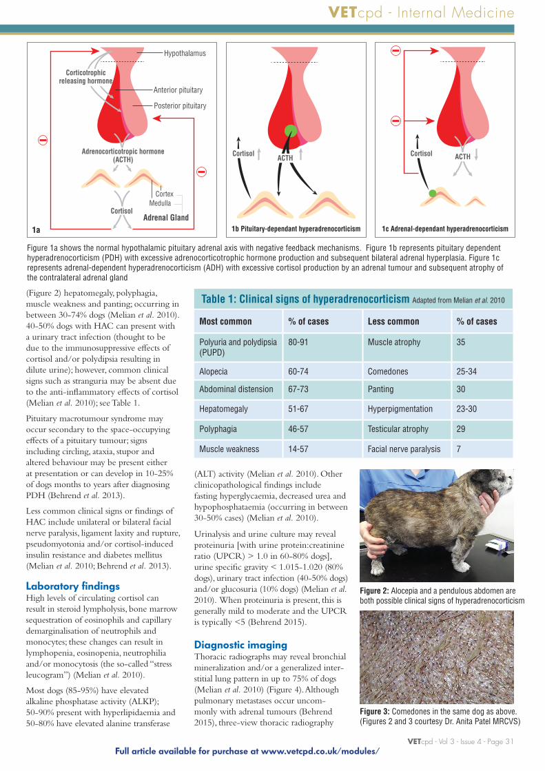
(Melian et al. 2010). Most of these dogs will have a pituitary tumour excreting excess adrenocorticotrophic hormone (ACTH) and resulting in bilateral adrenal hyperplasia. In the remaining 15-20% dogs with naturally-occurring HAC, disease is the result of an adrenal tumour and is known as adrenal-dependent HAC (ADH) (Melian et al. 2010). In dogs with ADH, autonomous secretion of cortisol by the adrenal tumour results in negative feedback inhibition to the pituitary gland, resulting in suppressed ACTH secretion and atrophy of the contralateral adrenal gland (Melian et al. 2010) (Figure 1).
Exogenous steroid administration (iatro-genic HAC) is another important cause of HAC in dogs and should be considered in dogs receiving corticosteroids with compatible clinical signs.
Other less common causes of HAC include concurrent pituitary and adreno-cortical tumours and ectopic ACTH secretion (Behrend 2015).
Clinical signsPolydipsia and polyuria occur in up to 91% dogs with HAC (Melian et al. 2010). Other commonly reported clinical signs of HAC include (in order of decreasing frequency) alopecia, pendulous abdomen
© iS
tock
phot
o.co
m
Full article available for purchase at www.vetcpd.co.uk/modules/ VETcpd - Vol 3 - Issue 4 - Page 31
1a
Hypothalamus
Posterior pituitary
Anterior pituitary
CortexMedulla
Adrenocorticotropic hormone (ACTH)
Cortisol
Corticotrophicreleasing hormone
1b Pituitary-dependant hyperadrenocorticism 1c Adrenal-dependant hyperadrenocorticism
CortisolACTH
Cortisol ACTH
Adrenal Gland
VETcpd - Internal Medicine
(Figure 2) hepatomegaly, polyphagia, muscle weakness and panting; occurring in between 30-74% dogs (Melian et al. 2010). 40-50% dogs with HAC can present with a urinary tract infection (thought to be due to the immunosuppressive effects of cortisol and/or polydipsia resulting in dilute urine); however, common clinical signs such as stranguria may be absent due to the anti-inflammatory effects of cortisol (Melian et al. 2010); see Table 1.
Pituitary macrotumour syndrome may occur secondary to the space-occupying effects of a pituitary tumour; signs including circling, ataxia, stupor and altered behaviour may be present either at presentation or can develop in 10-25% of dogs months to years after diagnosing PDH (Behrend et al. 2013).
Less common clinical signs or findings of HAC include unilateral or bilateral facial nerve paralysis, ligament laxity and rupture, pseudomyotonia and/or cortisol-induced insulin resistance and diabetes mellitus (Melian et al. 2010; Behrend et al. 2013).
Laboratory findingsHigh levels of circulating cortisol can result in steroid lympholysis, bone marrow sequestration of eosinophils and capillary demarginalisation of neutrophils and monocytes; these changes can result in lymphopenia, eosinopenia, neutrophilia and/or monocytosis (the so-called “stress leucogram”) (Melian et al. 2010).
Most dogs (85-95%) have elevated alkaline phosphatase activity (ALKP); 50-90% present with hyperlipidaemia and 50-80% have elevated alanine transferase
Most common % of cases Less common % of cases
Polyuria and polydipsia (PUPD)
80-91 Muscle atrophy 35
Alopecia 60-74 Comedones 25-34
Abdominal distension 67-73 Panting 30
Hepatomegaly 51-67 Hyperpigmentation 23-30
Polyphagia 46-57 Testicular atrophy 29
Muscle weakness 14-57 Facial nerve paralysis 7
Table 1: Clinical signs of hyperadrenocorticism Adapted from Melian et al. 2010
Figure 1a shows the normal hypothalamic pituitary adrenal axis with negative feedback mechanisms. Figure 1b represents pituitary dependent hyperadrenocorticism (PDH) with excessive adrenocorticotrophic hormone production and subsequent bilateral adrenal hyperplasia. Figure 1c represents adrenal-dependent hyperadrenocorticism (ADH) with excessive cortisol production by an adrenal tumour and subsequent atrophy of the contralateral adrenal gland
Figure 3: Comedones in the same dog as above. (Figures 2 and 3 courtesy Dr. Anita Patel MRCVS)
Figure 2: Alocepia and a pendulous abdomen are both possible clinical signs of hyperadrenocorticism
(ALT) activity (Melian et al. 2010). Other clinicopathological findings include fasting hyperglycaemia, decreased urea and hypophosphataemia (occurring in between 30-50% cases) (Melian et al. 2010).
Urinalysis and urine culture may reveal proteinuria [with urine protein:creatinine ratio (UPCR) > 1.0 in 60-80% dogs], urine specific gravity < 1.015-1.020 (80% dogs), urinary tract infection (40-50% dogs) and/or glucosuria (10% dogs) (Melian et al. 2010). When proteinuria is present, this is generally mild to moderate and the UPCR is typically <5 (Behrend 2015).
Diagnostic imagingThoracic radiographs may reveal bronchial mineralization and/or a generalized inter-stitial lung pattern in up to 75% of dogs (Melian et al. 2010) (Figure 4). Although pulmonary metastases occur uncom-monly with adrenal tumours (Behrend 2015), three-view thoracic radiography