Embed Size (px)
Citation preview
HYDROQUINONE
Data were last reviewed in IARC (1977) and the compound was classified in IARCMonographs Supplement 7 (1987).
1. Exposure Data
1.1 Chemical and physical data1.1.1 Nomenclature
Chem. Abstr. Serv. Reg. No.: 123-31-9Chem. Abstr. Name: 1,4-Benzenediol IUPAC Systematic Name: HydroquinoneSynonym: Benzoquinol
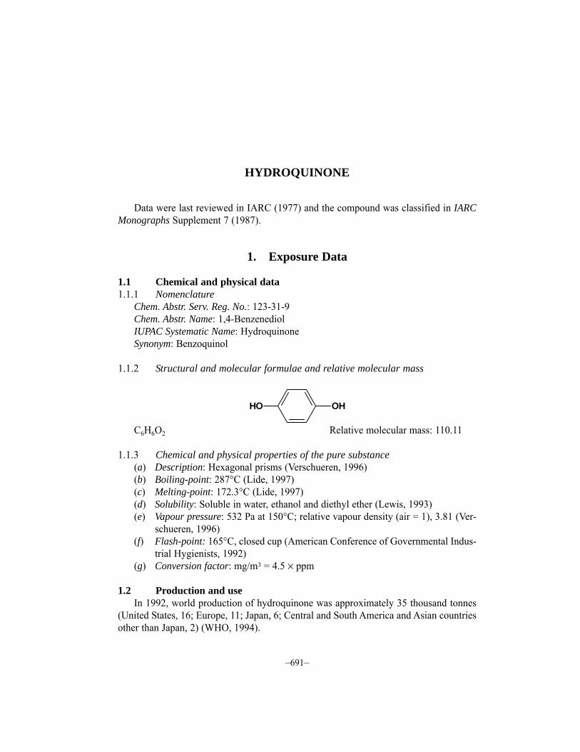
1.1.2 Structural and molecular formulae and relative molecular mass
C6H6O2 Relative molecular mass: 110.11
1.1.3 Chemical and physical properties of the pure substance(a) Description: Hexagonal prisms (Verschueren, 1996)(b) Boiling-point: 287°C (Lide, 1997)(c) Melting-point: 172.3°C (Lide, 1997)(d) Solubility: Soluble in water, ethanol and diethyl ether (Lewis, 1993)(e) Vapour pressure: 532 Pa at 150°C; relative vapour density (air = 1), 3.81 (Ver-
schueren, 1996)(f) Flash-point: 165°C, closed cup (American Conference of Governmental Indus-
trial Hygienists, 1992)(g) Conversion factor: mg/m3 = 4.5 × ppm
1.2 Production and useIn 1992, world production of hydroquinone was approximately 35 thousand tonnes
(United States, 16; Europe, 11; Japan, 6; Central and South America and Asian countriesother than Japan, 2) (WHO, 1994).
–691–
OHHO

Hydroquinone is used as a photographic developer (with black-and-white film), adye intermediate, a stabilizer in paints, varnishes, motor fuels and oils, an antioxidant forfats and oils, an inhibitor of polymerization and in the treatment of skin hyperpigmen-tation (Lewis, 1993).
1.3 Occurrence1.3.1 Occupational exposure
According to the 1981–83 National Occupational Exposure Survey (NOES, 1997),approximately 100 000 workers in the United States were potentially exposed to hydro-quinone (see General Remarks). Occupational exposures to hydroquinone may occur inits production and use in the production of dyes, paints, motor fuels and oils, and somepolymers. Dermal contact with hydroquinone may occur in the development of black-and-white photographs.
1.3.2 Environmental occurrenceHydroquinone is both a natural and an anthropogenic compound. It occurs naturally
as a conjugate with β-D-glucopyranoside in the leaves, bark and fruit of a number ofplants, especially the ericaceous shrubs such as cranberry, cowberry, bearberry and blue-berry. It may be released to the environment as a fugitive emission during its production,formulation and use as a chemical intermediate, photographic chemical and stabilizer(United States National Library of Medicine, 1997). Users of skin-bleaching formu-lations may be exposed to hydroquinone.
1.4 Regulations and guidelinesThe American Conference of Governmental Industrial Hygienists (ACGIH) (1997)
has recommended 2 mg/m3 as the 8-h time-weighted average threshold limit value foroccupational exposures to hydroquinone in workplace air. Similar values have been usedas standards or guidelines in many countries (International Labour Office, 1991).
No international guideline for hydroquinone in drinking-water has been established(WHO, 1993).
2. Studies of Cancer in Humans
One of the most prominent uses of hydroquinone is in photographic development andit is possible that work as a photographic processor often involved hydroquinone expo-sure in the past. Several studies have examined cancer risks among photographic pro-cessors. However, the Working Group did not use these except where the report providedsome information indicating that the workers concerned had indeed been exposed tohydroquinone.
IARC MONOGRAPHS VOLUME 71692
2.1 Cohort studiesPifer et al. (1995) reported a cohort mortality study of 879 workers (22 895 person–
years of follow-up) at a Tennessee (United States) plant in which hydroquinone was manu-factured and used over several decades. Job history records were linked to extensive indus-trial hygiene data and expertise to estimate cumulative exposure to hydroquinone. Averagehydroquinone dust levels ranged from 0.1 to 6.0 mg/m3, with levels over 2 mg/m3 for mostof the period of operation of the plant. Mean employment duration was 13.7 years andmean follow-up from first exposure was 26.8 years. Relative risk estimates (standardizedmortality ratios (SMRs)) for this cohort were derived by comparison with the general popu-lation of Tennessee as well as with an occupational cohort not exposed to hydroquinone (aplant of the same company, located in New York State). The SMR for all causes of deathcombined (n = 168) was significantly below 1.0, as was the SMR for all cancers combined(n = 33). Only two sites, colon (n = 5) and lung (n = 14) had more than three observedcases. Most site-specific SMRs were well below 1.0. The results were similar with bothcomparison populations. The dose–response analyses of selected cancer sites did not revealany meaningful trend or heterogeneity. [The numbers for individual cancer sites were smalland the power to detect effects was weak. The Working Group noted that this cohort hadsystematically lower SMRs than the comparison industrial cohort.]
Nielsen et al. (1996) carried out a cohort incidence study among 837 Danish litho-graphers born between 1933 and 1942 and registered with the Danish Union of Litho-graphers in 1974 or later. Questionnaires were sent to cohort members in 1989 to obtaininformation on job exposures; usable responses were received from 620 workers. Aboutone-quarter of the cohort members reported working regularly with hydroquinone forphotographic development. The entire cohort was traced in the Danish Cancer Registryfrom 1974 to 1989. Relative risk estimates (standardized incidence ratios (SIRs)) for thiscohort were derived by comparison with the general population of Denmark. There werea total of 24 cancers registered, giving an SIR of 0.9. For no site except skin were theremore than three cases. Five cases of malignant melanoma occurred, with 1.5 expected(SIR, 3.4; 95% confidence interval, 1.2–7.5). Among these five, two had reportedly beenexposed to hydroquinone.
3. Studies of Cancer in Experimental Animals
In skin painting studies in mice, hydroquinone was inactive as an initiator of skincarcinogenesis. In bladder implantation studies, hydroquinone in cholesterol pelletsincreased the incidence of bladder carcinomas in mice (IARC, 1977).
3.1 Oral administration3.1.1 Mouse
Groups of 55 male and 55 female B6C3F1 mice, eight to 10 weeks of age, were admi-nistered 0, 50 or 100 mg/kg bw hydroquinone (purity, > 99%) by gavage on five days per
HYDROQUINONE 693
week for 103 weeks. Mean body weights of high-dose mice at the end of the study werelower than those of vehicle controls, and the relative liver weights were increased forexposed males and high-dose females. Survival in treated mice was similar to that in con-trols. No increase in tumours was found in exposed males. In females, hepatocellularadenomas were found in 2/55 controls, 15/55 low-dose group (p = 0.001) and 12/55 high-dose group (p = 0.005) (United States National Toxicology Program, 1989).
Groups of 28–30 male and 28–30 female B6C3F1 mice, six weeks of age, were givenhydroquinone (purity, > 99%) in the diet at concentrations of 0 or 0.8% for 96 weeks. Thefinal body weight was reduced in hydroquinone-treated females. The incidence of hepa-tocellular adenoma was increased to 14/30 in exposed males (p < 0.05) compared with6/28 in controls. Incidence of no other tumour type was significantly increased by expo-sure in males, although three renal adenomas occurred. No increase in tumour incidencewas found in females (Shibata et al., 1991).
3.1.2 RatGroups of 55 male and 55 female Fischer 344/N rats, seven to nine weeks of age,
were administered 0, 25 or 50 mg/kg bw hydroquinone (purity, > 99%) by gavage on fivedays per week for 103 weeks. Mean body weights of exposed males were reduced andthe relative kidney weights for high-dose males were greater than those for vehiclecontrols. Survival was reduced in exposed animals. In exposed males, renal tubule celladenomas developed in 4/55 low-dose group (p = 0.069) and 8/55 high-dose group(p = 0.003) compared with 0/55 controls. In exposed females, mononuclear cell leu-kaemia developed in 15/55 low-dose group (p = 0.048) and 22/55 high-dose group(p = 0.003) compared with 9/55 controls. The historical incidence of leukaemia forwater/vehicle control female rats was 25 ± 15% (United States National Toxicology Pro-gram, 1989). [The Working Group noted that the incidences of leukaemia in the exposedgroup were within the historical control range.]
Groups of 30 male and 30 female Fischer 344 rats, six weeks of age, were givenhydroquinone (purity, > 99%) in the diet at concentrations of 0 or 0.8% for 104 weeks.Body weight gain was decreased in both exposed males and females. Chronic nephro-pathy was more severe in males given hydroquinone. In the kidneys of exposed male rats,the incidence of tubule hyperplasia was 30/30 (100%) and that of adenomas was 14/30(47%; p < 0.01), compared with 1/30 (3%) and 0/30 (0%), respectively in unexposedcontrols. Incidence of no other tumour type was increased by exposure (Shibata et al.,1991).
3.2 Administration with known carcinogens3.2.1 Rat
Groups of 15 male Fischer 344 rats, six weeks of age, were administered 0 or 0.05%N-nitrosobutyl-N-(4-hydroxybutyl)amine in the drinking-water for two weeks followedby ureteric ligation one week later to initiate bladder carcinogenesis. Hydroquinone[purity unspecified] was administered at concentrations of 0 or 0.2% in the diet for 22
IARC MONOGRAPHS VOLUME 71694
weeks and all animals were killed at week 24. Hydroquinone alone induced no bladderlesions. When hydroquinone was given after initiation, no increase in bladder lesions wasobserved (Miyata et al., 1985).
Groups of 10 or 15 male Fischer 344 rats, six weeks of age, were given hydro-quinone (purity > 99%) at concentrations of 0 or 0.8% in the diet for 51 weeks, whileother groups of 15 or 16 animals were fed 0 or 0.8% hydroquinone for 51 weeks startingone week after exposure to 150 mg/kg bw N-methyl-N′-nitro-N-nitrosoguanidine by oralgavage to initiate stomach carcinogenesis. The body weights of rats given hydroquinoneafter initiator were lower than those given only initiator. Hydroquinone alone did notinduce forestomach lesions, nor did it enhance the incidence of forestomach or glandularstomach lesions induced by the initiator (Hirose et al., 1989).
Groups of 7–10 male Sprague-Dawley rats, weighing 200 g, were given hydro-quinone (purity, > 99%) in the diet at concentrations of 0, 100 or 200 mg/kg for sixweeks beginning one week after partial hepatectomy and intraperitoneal injection of300 mg/kg bw N-nitrosodiethylamine to initiate liver carcinogenesis. One group under-went only partial hepatectomy and was fed the high dose of hydroquinone. In the hepa-tectomized group exposed only to hydroquinone, no liver enzyme-altered (γ-glutamyl-transpeptidase) foci were induced. Hydroquinone after initiation increased the multi-plicity of foci from 0.08 per cm2 to 0.68 in the low-dose group and to 0.34 in the high-dose group [statistical analysis not given]. In a second experiment, groups of 10 ratsunderwent the regimen to initiate liver carcinogenesis and received 0 or 1 mg/kg bwhydroquinone by oral gavage on five days per week for seven weeks. Hydroquinone didnot increase the multiplicity of enzyme-altered foci, but their area was increased from amean of 1.00 × 10⁻⁴ cm2 to 1.30 × 10⁻⁴ cm2 (p < 0.05) and their volume from 1.49 ×10⁻⁴ cm3 to 3.12 × 10⁻⁴ cm3 (p < 0.01) (Stenius et al., 1989).
Groups of 15 or 12 male Fischer 344 rats, seven to eight weeks of age, were givenhydroquinone (purity, > 99%) at concentrations of 0 or 0.8% in the diet for 49 weeksalone or starting one week after six intraperitoneal injections of 25 mg/kg bw N-nitroso-methyl-n-amylamine to initiate upper digestive tract carcinogenesis. Hydroquinonealone reduced weight gain. In animals given hydroquinone after carcinogen, the inci-dence of oesophageal carcinoma was 4/12 rats (not significant) compared with 0/11 inthe group given initiator only, and the multiplicity was increased to 0.33 tumours per rat(p < 0.05) compared with 0 in the controls (Yamaguchi et al., 1989).
Groups of 10 or 20 male Fischer 344/Du Crj rats [age unspecified] were given hydro-quinone [purity unspecified] at concentrations of 0 or 0.8% in the diet for 30 weeks eitheralone or after exposure to 0.1% N-nitroso-bis(2-hydroxypropyl)amine in the drinking-water for two weeks to initiate carcinogenesis in several organs. No unexposed controlswere included. Body weight was reduced by hydroquinone given after the initiator andliver weight was increased compared with the group given initiator only. Hydroquinonealone induced no lung or thyroid tumours. Rats given initiator developed low incidencesof tumours in the thyroid, lung, urinary bladder and kidney. None of these incidences wasincreased by hydroquinone (Hasegawa et al., 1990).
HYDROQUINONE 695
IARC MONOGRAPHS VOLUME 71696
Groups of 10 or 20 male Fischer 344 rats, six weeks of age, were given hydroquinone(purity, > 99%) in the diet at a concentration of 0.8% for 36 weeks alone or after exposureto 0.05% N-nitrosobutyl-N-(4-hydroxybutyl)amine in the drinking-water for four weeks toinitiate bladder carcinogenesis. Hydroquinone alone did not affect body weight or bladderweight. Hydroquinone exposure alone did not induce bladder tumours and feeding ofhydroquinone after initiator did not increase the incidence or multiplicity of bladder neo-plasms induced by the initiatior alone (Kurata et al., 1990).
Groups of 15 or 20 male Wistar/Crj rats, six weeks of age, were given hydroquinone[purity unspecified] at concentrations of 0 or 0.8% in the diet for 36 weeks starting oneweek after exposure to 0.1% N-nitrosoethyl-N-hydroxyethylamine in the drinking-waterfor three weeks to initiate liver and kidney carcinogenesis. The final body weights of ratsgiven hydroquinone were lower than those of animals given only basal diet or initiation.The relative liver and kidney weights of rats receiving hydroquinone were higher thanthose of the basal diet group. Hydroquinone alone did not induce preneoplastic or neo-plastic liver or kidney lesions. In the kidney, hydroquinone exposure after initiationincreased the multiplicity of renal cell tumours to 5.22 per rat (p < 0.01) compared with2.58 after initiation only and increased the multiplicity of microadenomas to 2.77(p < 0.05) compared with 0.94 after initiation only (Okazaki et al., 1993).
3.2.2 HamsterGroups of female Syrian golden hamsters, six weeks of age, were given hydro-
quinone (purity, > 99%) at concentrations of 0 or 1.5% in the diet for 16 weeks eitheralone (10 and 15 hamsters) or after two subcutaneous injections of 70 mg/kg bw N-nitro-sobis(2-oxopropyl)amine (20 hamsters) to initiate pancreatic carcinogenesis. Hydro-quinone alone did not affect body weights or liver or pancreas weights compared withuntreated controls. Given after the initiator, hydroquinone did not affect body weight orliver weight, but reduced pancreas weight compared with hamsters given only initiator.Hydroquinone alone did not induce neoplastic lesions in the pancreas or liver. Inhamsters given hydroquinone after initiator, the multiplicity of pancreatic lesions wasreduced (Maruyama et al., 1991).
4. Other Data Relevant to an Evaluation of Carcinogenicityand its Mechanisms
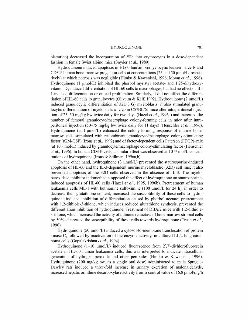
4.1 Absorption, distribution, metabolism and excretionThe major metabolism of hydroquinone is to the sulfate and, at higher exposure,
glucuronide conjugates. Oxidation to 1,4-benzoquinone results in a reactive metabolite,that may form mono- or polyglutathione conjugates (see Figure 1).
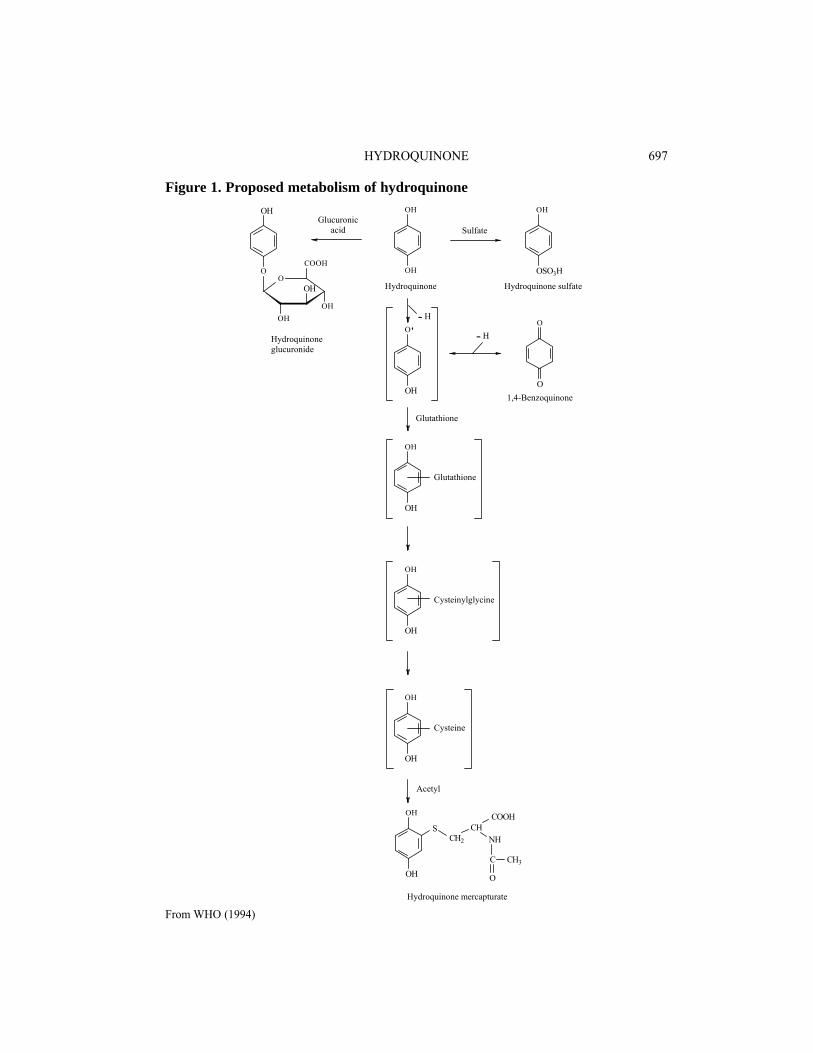
HYDROQUINONE 697
From WHO (1994)
Figure 1. Proposed metabolism of hydroquinone
Hydroquinone mercapturate
Acetyl
SulfateGlucuronic
acid
Hydroquinoneglucuronide
OH
OH
SCH2
CHNH
COOH
C
O
CH3
Glutathione
OH
OH
OH
OSO3H
Hydroquinone Hydroquinone sulfate
O
O
O
OH1,4-Benzoquinone
Glutathione
OH
OH
Cysteine
OH
OH
Cysteinylglycine
OH
OH
OH
OO
OH
OH
OH
COOH
- H
- H
4.1.1 HumansRates of percutaneous absorption of hydroquinone in 5% aqueous solution through
human stratum corneum in vitro were approximately half those through full-thickness ratskin; the human skin penetration rate was classified as ‘slow’ (Barber et al., 1995). Thedata allowed calculation of skin absorbance in workers in photographic development.
Rates of hydroquinone glucuronidation in human liver microsomes showed a two- tothree-fold variation between individual liver samples; they were somewhat higher thanin the rat, and lower than in the mouse liver (Seaton et al., 1995). A compartmental phar-macokinetic model was derived to describe the pharmacokinetics of hydroquinonein vivo in humans, rats and mice, incorporating hydroquinone glucuronidation rates;sulfation of hydroquinone was not included in this model. NAD(P)H:quinone acceptoroxidoreductases protect against reactive quinones by reducing them to the hydroquinone;this enzyme seems to be absent in some individuals, which will lead to loss of such pro-tection and make them more sensitive to hydroquinone toxicity (Ross, 1996).
4.1.2 Experimental systemsPercutaneous absorption of hydroquinone from an aqueous solution was studied in
full-thickness rat skin in vitro; the permeability constant was 2.3 × 10⁻⁵ cm/h, which wasapproximately two-fold faster than that of human skin (Barber et al., 1995).
The disposition of [14C]hydroquinone after oral administration to Sprague-Dawleyrats was studied by Divincenzo et al. (1984). Whether mixed with the diet or administeredas a single dose, the compound was almost completely excreted in urine, with up to 4%in the faeces. By far the major metabolites were the sulfate and glucuronide conjugates,with a small amount of unconjugated hydroquinone. Apparently no analysis for mercap-turates was performed. These results were confirmed by Saito and Takeichi (1995), whoalso demonstrated a wide tissue distribution of hydroquinone. Hill et al. (1993) foundappreciable amounts of hydroquinone–glutathione conjugates in bile after intraperitonealadministration of hydroquinone to rats that had been pretreated with AT-125, an inhibitorof γ-glutamyltranspeptidase: both mono-, di- and triglutathione conjugates were found, aswell as a mercapturate in urine. More than 4% of the dose was recovered as glutathioneconjugates, indicating considerable formation of the highly toxic 1,4-benzoquinone (seethis volume) metabolite. Nerland and Pierce (1990) identified the hydroquinone mercap-turate (N-acetyl-S-(2,5-dihydroxyphenol)-L-cysteine) in untreated rats after administrationof hydroquinone.
A simple compartmental pharmacokinetic model was proposed by Seaton et al.(1995) to describe the pharmacokinetics of hydroquinone in mice, rats and humans. Themodel did not include hydroquinone sulfation, which does occur in rats and possibly inmice, although glucuronidation is the major reaction. Phenol and hydroquinone maymutually inhibit their sulfation if both are present simultaneously in the rat (Legatheet al., 1994).
Hydroquinone can be converted to the very reactive 1,4-benzoquinone by severalenzymes. A major activity is myeloperoxidase (Subrahmanyam et al., 1991), which is
IARC MONOGRAPHS VOLUME 71698
stimulated by phenol and some other phenols. Microsomal cytochrome P450 may alsoplay a role (Hill et al., 1993). Macrophage peroxidase activity converting hydroxyqui-none to 1,4-benzoquinone may be important in the myelotoxicity of benzene (Schlosser& Kalf, 1989; Smith et al., 1989; Snyder & Hedli, 1996). Copper(II) ions stronglyenhance this process, in which hydrogen peroxide and other reactive oxygen species maybe involved (Eyer, 1991; Rao, 1991; Li & Trush, 1993a,b).
Hydroquinone forms DNA adducts in the peroxidase-containing promyelocytic HL-60 cell line; this process is enhanced by addition of hydrogen peroxide or cumene hydro-peroxide (Lévay & Bodell, 1996), presumably because the hydroquinone is oxidized bya cellular peroxidase to a reactive, DNA-binding metabolite.
4.1.3 Comparison of human and rodent dataThe metabolism of hydroquinone seems very similar in man and rodents: sulfate and
glucuronide conjugates are the major metabolites. Through the 1,4-benzoquinone meta-bolite, a reactive intermediate can be formed, in particular in macrophages by peroxidases,that may be trapped by conjugation with glutathione. The reactive intermediate may formDNA adducts, and may also be responsible for kidney toxicity.
4.2 Toxic effectsThe toxicity of hydroquinone has been reviewed (WHO, 1994).
4.2.1 HumansNo data were available to the Working Group.
4.2.2 Experimental systemsLong-term feeding of hydroquinone to rats led to aplastic anaemia, liver cord-cell
atrophy and ulceration of the gastric mucosa. A single high dose was reported to inducerenal tubule necrosis in rats (IARC, 1977).
In a carcinogenicity study (United States National Toxicology Program, 1989; Kariet al., 1992), nephropathy was observed in nearly all male and most female rats of alldosed groups and vehicle controls. The nephropathy was characterized by degenerationand regeneration of tubule epithelium, atrophy and dilatation of some tubules, hyalinecasts in the tubule lamina, glomerulosclerosis, interstitial fibrosis, and chronic inflam-mation. In males, the nephropathy was more severe in the high-dose (50 mg/kg bw perday) group, while in females no dose-dependence was observed. Nephropathy wasobserved in males also in 13-week studies. Presence of hyaline droplets was not reported.In another carcinogenicity study (Shibata et al., 1991), the prevalence and severity ofchronic nephropathy was more marked in dosed males than in females. It was stated thatthe nephropathy observed was not of the α2u-globulin nephropathy type. In a reanalysis ofthe histology of the United States National Toxicology Program study, it was observedthat the atypical tubule hyperplasias and adenomas were located in areas of severe chronicprogressive nephropathy (Hard et al., 1997).
HYDROQUINONE 699
After six weeks of oral administration (50 mg/kg bw per day) of hydroquinone tomale Fischer 344 rats, modestly elevated urinary excretion of alanine aminopeptidase,alkaline phosphatase, γ-glutamyltranspeptidase and N-acetylglucosaminidase wasobserved (English et al., 1994a). No such indication of renal toxicity was observed infemale Fischer 344 rats or male Sprague-Dawley rats. Interstitial inflammation and dege-nerative/regenerative tubule foci were more frequent in high-dose (25 or 50 mg/kg bw/day) male Fischer 344 rats. Similarly, the proportion of proliferating cells, measured bybromodeoxyuridine (BrdU) labelling, was elevated in the proximal tubules in maleFischer 344 rats given the highest dose (50 mg/kg bw per day), while no consistentchange in the labelling was observed in the renal tubules from male Sprague-Dawley orfemale Fischer 344 rats. On the other hand, after a single dose of hydroquinone (Boatmanet al., 1996), female Fischer 344 rats were more sensitive to hydroquinone-inducednephrotoxicity, as measured by urinary excretion of alanine aminopeptidase, N-acetyl-glucosaminidase, alkaline phosphatase, γ-glutamyltranspeptidase, glucose and creati-nine, by urinary osmolality or by blood levels of urea nitrogen. In these acute experi-ments, no nephrotoxicity was observed in Sprague-Dawley rats.
In 14-day studies (United States National Toxicology Program, 1989), tremors,convulsions and death following gavage were observed at doses ≥ 500 mg/kg bw per day.In 13-week studies, lethargy, tremor and convulsions leading to death were also observedat doses ≥ 200 mg/kg bw per day.
In a two-year study (United States National Toxicology Program, 1989; Kari et al.,1992), dose-dependent hepatic morphological changes (anisokaryosis, elevated fre-quency of multinucleated cells) were observed in male mice. In a long-term feedingstudy (0.8% in the diet) (Shibata et al., 1991), hepatic centrilobular hypertrophy wasobserved in males and forestomach hyperplasia in both males and females, while no non-neoplastic changes in the kidney were reported.
Administration of hydroquinone (0.5% in the diet) for 20 weeks did not induce hyper-plasia or papillomatous lesions in the forestomach in Syrian golden hamsters (Hiroseet al., 1986). In male Fischer 344 rats, oral administration of hydroquinone for eightweeks (0.8% in the diet) did not induce hyperplasia or DNA synthesis, as measured byBrdU-labelling index in the forestomach epithelium. No cell proliferation, increased DNAsynthesis or increase in pepsinogen-isoenzyme-1-altered neoplastic foci was observed inthe pyloric mucosa (Shibata et al., 1990).
A large number of studies have been performed on the effects of hydroquinone onbone marrow, in order to elucidate the mechanisms of the myelodepressive and leukae-mogenic activity of benzene.
Hydroquinone decreased interleukin (IL)-1 secretion and protein and RNA synthesisof isolated human peripheral blood monocytes induced by Escherichia coli lipopoly-saccharide at micromolar concentrations (Carbonnelle et al., 1995). Hydroquinone(4 μmol/L) inhibited the growth of bone marrow cells from female C57BL/6 × DBA/2mice (Seidel et al., 1991) and from male Swiss Webster and C57BL/6J mice (10 μmol/L)(Neun et al., 1992). Hydroquinone (50, 75 or 100 mg/kg bw, single intraperitoneal admi-
IARC MONOGRAPHS VOLUME 71700
nistration) decreased the incorporation of 59Fe into erythrocytes in a dose-dependentfashion in female Swiss albino mice (Snyder et al., 1989).
Hydroquinone induced apoptosis in HL60 human promyelocytic leukaemia cells andCD34+ human bone-marrow progenitor cells at concentrations (25 and 50 μmol/L, respec-tively) at which necrosis was negligible (Hiraku & Kawanishi, 1996; Moran et al., 1996).Hydroquinone (1 μmol/L) inhibited the phorbol myristyl acetate- and 1,25-dihydroxy-vitamin D3-induced differentiation of HL-60 cells to macrophages, but had no effect on IL-1-induced differentiation or on cell proliferation. Similarly, it did not affect the differen-tiation of HL-60 cells to granulocytes (Oliveira & Kalf, 1992). Hydroquinone (2 μmol/L)induced granulocytic differentiation of 32D.3(G) myeloblasts; it also stimulated granu-locytic differentiation of myeloblasts in vivo in C57BL/6J mice after intraperitoneal injec-tion of 25–50 mg/kg bw twice daily for two days (Hazel et al., 1996a) and increased thenumber of femoral granulocyte/macrophage colony-forming cells in mice after intra-peritoneal injection (50–75 mg/kg bw twice daily for 11 days) (Henschler et al., 1996).Hydroquinone (at 1 μmol/L) enhanced the colony-forming response of murine bone-marrow cells stimulated with recombinant granulocyte/macrophage colony-stimulatingfactor (rGM-CSF) (Irons et al., 1992) and of factor-dependent cells Paterson (FDCP)–mix(at 10–9 mol/L) induced by granulocyte/macrophage colony-stimulating factor (Henschleret al., 1996). In human CD34+ cells, a similar effect was observed at 10–21 mol/L concen-trations of hydroquinone (Irons & Stillman, 1996a,b).
On the other hand, hydroquinone (3 μmol/L) prevented the staurosporine-inducedapoptosis of HL-60 and the IL-3-dependent murine myeloblastic (32D) cell line; it alsoprevented apoptosis of the 32D cells observed in the absence of IL-3. The myelo-peroxidase inhibitor indomethacin opposed the effect of hydroquinone on staurosporine-induced apoptosis of HL-60 cells (Hazel et al., 1995, 1996b). Pretreatment of humanleukaemia cells ML-1 with buthionine sulfoximine (100 μmol/L for 24 h), in order todecrease their glutathione content, increased the susceptibility of these cells to hydro-quinone-induced inhibition of differentiation caused by phorbol acetate; pretreatmentwith 1,2-dithiole-3-thione, which induces reduced glutathione synthesis, prevented thedifferentiation inhibition of hydroquinone. Treatment of DBA/2 mice with 1,2-dithiole-3-thione, which increased the activity of quinone reductase of bone-marrow stromal cellsby 50%, decreased the susceptibility of these cells towards hydroquinone (Trush et al.,1996).
Hydroquinone (50 μmol/L) induced a cytosol-to-membrane translocation of proteinkinase C, followed by inactivation of the enzyme activity, in cultured LL/2 lung carci-noma cells (Gopalakrishna et al., 1994).
Hydroquinone (1–10 μmol/L) induced fluorescence from 2′,7′-dichlorofluorescinacetate in HL-60 human leukaemia cells; this was interpreted to indicate intracellulargeneration of hydrogen peroxide and other peroxides (Hiraku & Kawanishi, 1996).Hydroquinone (200 mg/kg bw, as a single oral dose) administered to male Sprague-Dawley rats induced a three-fold increase in urinary excretion of malonaldehyde,increased hepatic ornithine decarboxylase activity from a control value of 16.8 pmol/mg/h
HYDROQUINONE 701
to 86.5 pmol/mg/h and, in vitro, 0.3 mmol/L induced a rapid depletion (30%) of the gluta-thione content of isolated hepatocytes (Stenius et al., 1989). Hydroquinone (10 μmol/L)induced formation of 8-hydroxydeoxyguanosine in the DNA of HL-60 cells in vitro, butnot in bone-marrow cells of B6C3F1 mice in vivo after a single intraperitoneal dose of75 mg/kg bw (Kolachana et al., 1993). An increase in urinary excretion of 8-hydroxy-guanine was observed in rats given a single intraperitoneal dose of 11 mg/kg bw hydro-quinone (Suzuki et al., 1995).
Hydroquinone (≥ 0.25 μmol/L) prevented the elimination by apoptosis of G418-resistant, transformed Swiss 3T3 MxCl1 cells by co-cultured TGF-β-treated C3H 10T½Cl8 cells (Schaeffer et al., 1995).
In a study on the immunotoxic effects of cigarette tar components, hydroquinone, at aconcentration that did not affect the viability of the cells (50 μmol/L), decreased IL-2-dependent DNA synthesis and cell proliferation by > 90% in cultured human T lympho-blasts (Li et al., 1997). Hydroquinone inhibited Fc-receptor-mediated phagocytosis inmouse peritoneal macrophages only at rather high concentrations (100 μmol/L) (Manninget al., 1994).
4.3 Reproductive and developmental effects4.3.1 Humans
No data were available to the Working Group.
4.3.2 Experimental systemsIn a developmental toxicity study in COBS-CD-BR rats dosed by gavage, hydro-
quinone (30, 100 or 300 mg/kg bw per day on days 6 through 15 of gestation) did notinduce malformation, gross variations or skeletal variations, with the exception of anincrease in the incidence of total common vertebral variations at the highest dose. At thehighest dose, slight reductions of mean fetal body weight and of maternal body weightgain were also observed (Krasavagne et al., 1992).
In New Zealand white rabbits administered 25–150 mg/kg bw per day hydroquinoneby gavage on days 6 through 18 of gestation, the only treatment-related changes observedwere nonsignificant increases in minor skeletal malformations (vertebral/rib defects,angulated hyoid arch) and microphthalmia at the highest dose level, at which maternalweight gain was also decreased (Murphy et al., 1992).
In a two-generation reproductive toxicity study in rats, no adverse effect wasobserved on feed consumption, survival, reproductive parameters, pup weight, sex distri-bution, survival, gross lesions or microscopic anatomy after oral doses of 15–150 mg/kgbw per day (Blacker et al., 1993).
Hydroquinone had no adverse effect upon cultured whole rat conceptuses at a con-centration of 50 μmol/L, but killed all embryos at 100 μmol/L (Chapman et al., 1994).
IARC MONOGRAPHS VOLUME 71702
4.4 Genetic and related effects4.4.1 Humans
No data were available to the Working Group.
4.4.2 Experimental systems (see Table 1 for references)Hydroquinone did not induce SOS repair and did not increase the numbers of
mutants when tested against commonly used strains of Salmonella typhimurium.However, it was shown to be mutagenic to S. typhimurium TA104 and TA102, which aresensitive to oxidative mutagens. The activity demonstrated with TA104 was almost com-pletely inhibited by co-incubation with superoxide dismutase and catalase and isconsistent with superoxide and hydrogen peroxide being the mutagen(s). Hydroquinoneinduced gene conversion and mutations in Saccharomyces cerevisiae. It did not inducesex-linked recessive lethal mutations in Drosophila melanogaster.
In cultured mammalian cells, hydroquinone induced DNA single-strand breaks in rathepatocytes, gene mutations, chromosomal aberrations and sister chromatid exchanges.Positive results were obtained in a cell transformation assay using Syrian hamsterembryo cells. Increased frequencies of CREST-positive micronuclei (indicating chromo-some loss) and CREST-negative micronuclei (indicating chromosome breakage) wereobserved following exposure of Chinese hamster lung cells to hydroquinone in oneextensive study; only kinetochore-negative micronuclei were found in another study. Theformation of micronuclei was dependent on arachidonic acid supplementation. Themicronuclei induced in the presence of a superoxide-generation system (hypoxanthineand xanthine oxidase) consisted exclusively of CREST-negative micronuclei and theirformation was completely inhibited by pre-treatment with catalase. In addition,glutathione treatment inhibited both CREST-positive and negative micronuclei (Dobo &Eastmond, 1994).
In vitro in human cells, induction of DNA strand breaks was shown to be dependenton the presence of Cu(II). Hydroquinone induced sister chromatid exchanges and chro-mosomal aberrations without an exogenous metabolic system. The metabolic activationsystem was not required for the induction of micronuclei in human lymphocytes wherekinetochore-positive micronuclei were found.
In vivo in mouse bone marrow, hydroquinone induced micronuclei and chromosomalaberrations in several studies but not sister chromatid exchanges in a single study. Hyper-ploidy and chromosome loss (as demonstrated by centromere-positive micronuclei) butnot polyploidy were also found in mouse bone marrow. In mouse spermatocytes, chro-mosomal aberrations and hyperploidy were observed.
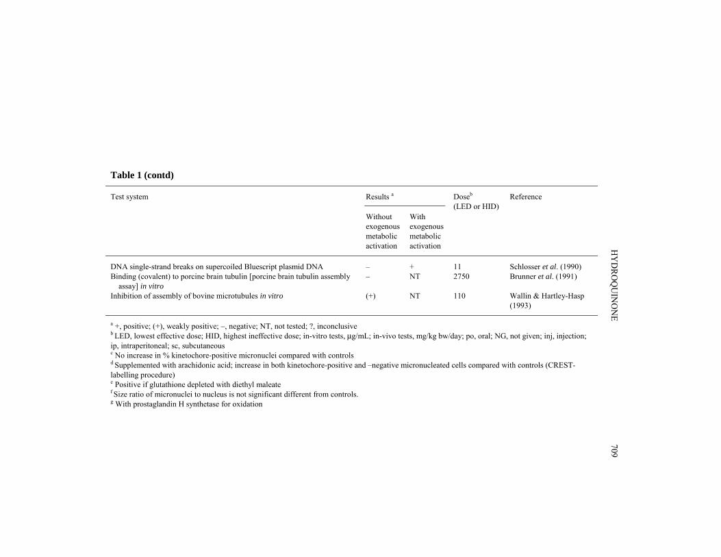
Hydroquinone inhibited intercellular communication in Chinese hamster cellsin vitro. Topoisomerase II (Frantz et al., 1996; Hutt & Kalf, 1996) but not topoiso-merase I (Chen & Eastmond, 1995b) activity was inhibited in vitro by hydroquinonetreatment.
Hydroquinone binding to calf thymus DNA and cysteine is enhanced by oxidation(prostaglandin H synthetase or cumene hydroperoxide) and inhibited by indomethacin
HYDROQUINONE 703
IARC M
ON
OG
RAPH
S VO
LUM
E 71704Table 1. Genetic and related effects of hydroquinone
Test system Results a Reference
Withoutexogenousmetabolicactivation
Withexogenousmetabolicactivation
Doseb
(LED or HID)
PRB, SOS repair activity, Salmonella typhimurium TA1535/pSK1002, umu test
– – 3300 Nakamura et al. (1987)
SA0, Salmonella typhimurium TA100, reverse mutation – – 333 Haworth et al. (1983)SA0, Salmonella typhimurium TA100, reverse mutation – – 125 Sakai et al. (1985)SA2, Salmonella typhimurium TA102, reverse mutation + NT NG Hakura et al. (1996)SA4, Salmonella typhimurium TA104, reverse mutation + NT 25 Hakura et al. (1996)SA5, Salmonella typhimurium TA1535, reverse mutation – – 333 Haworth et al. (1983)SA7, Salmonella typhimurium TA1537, reverse mutation – – 333 Haworth et al. (1983)SA9, Salmonella typhimurium TA98, reverse mutation – – 333 Haworth et al. (1983)SA9, Salmonella typhimurium TA98, reverse mutation – – 125 Sakai et al. (1985)SAS, Salmonella typhimurium TA97, reverse mutation – – 125 Sakai et al. (1985)SCG, Saccharomyces cerevisiae MP1, gene conversion + NT 1320 Fahrig (1984)SCH, Saccharomyces cerevisiae MP1, homozygosis by mitotic recombination or gene conversion
– NT 1320 Fahrig (1984)
SCF, Saccharomyces cerevisiae MP1, forward mutation + NT 1320 Fahrig (1984)DMX, Drosophila melanogaster, sex-linked recessive lethal mutation ? 28 000 ppm
feedFoureman et al. (1994)
DMX, Drosophila melanogaster, sex-linked recessive lethal mutation – 1500 ppminj × 1
Foureman et al. (1994)
DIA, DNA single strand breaks, cross-links or related damage, LYS mouse lymphoma cells, alkaline elution in vitro
– NT 11 Pellack-Walker & Blumer(1986)
DIA, DNA single strand breaks, isolated rat hepatocytes, alkaline elution in vitro
+ NT 33 Walles (1992)
G5T, Gene mutation, mouse lymphoma L5178Y cells, tk locus in vitgro + NT 2.5 McGregor et al. (1988a,b)GIA, Gene mutation, Syrian hamster embryo cells, hprt locus in vitro + NT 1.1 Tsutsui et al. (1997)
HY
DRO
QU
INO
NE
705
Table 1 (contd)
Test system Results a Reference
Withoutexogenousmetabolicactivation
Withexogenousmetabolicactivation
Doseb
(LED or HID)
GIA, Gene mutation, Syrian hamster embryo cells, ouabain resistance in vitro
+ NT 1.1 Tsutsui et al. (1997)
SIC, Sister chromatid exchange, Chinese hamster ovary CHO cells in vitro + + 0.5 Galloway et al. (1987)SIS, Sister chromatid exchange, Syrian hamster embryo cells in vitro + NT 0.11 Tsutsui et al. (1997)MIA, Micronucleus test, Chinese hamster embryonic lung CL-1 cells in vitro +c NT 1 Antoccia et al. (1991)MIA, Micronucleus test, Chinese hamster lung V79 cells in vitro (+) NT 31.6 Seelbach et al. (1993)MIA, Micronucleus test, Chinese hamster lung V79 cells in vitro + NT 2.8 Ellard & Parry (1993)MIA, Micronucleus test, Chinese hamster XEM2 (V79 exp CYP1A1) cells in vitro
+ NT 2.8 Ellard & Parry (1993)
MIA, Micronucleus test, Chinese hamster SD1 (V79 exp CYP2B1) cells in vitro
+ NT 2.8 Ellard & Parry (1993)
MIA, Micronucleus test, Chinese hamster lung V79 cells in vitro NT +d 11.5 Dobo & Eastmond (1994)CIC, Chromosomal aberrations, Chinese hamster ovary CHO cells in vitro – + 450 Galloway et al. (1987)CIS, Chromosomal aberrations, Syrian hamster embryo cells in vitro + NT 3.3 Tsutsui et al. (1997)AIA, Aneuploidy, DON:Wg3h Chinese hamster cells, dislocating metaphase chromosomes in vitro
+ NT 10 Warr et al. (1993)
AIA, Aneuploidy, LUC2 Chinese hamster cells, in vitro – NT 5 Warr et al. (1993)AIA, Aneuploidy, Syrian hamster embryo cells in vitro – NT 3.3 Tsutsui et al. (1997)TCS, Cell transformation, Syrian hamster embryo cells, clonal assay + NT 0.33 Tsutsui et al. (1997)DIH, DNA strand breaks, cross-links or related damage, human lymphocytes, comet assay in vitro
? + 11 Anderson et al. (1995)
DIH, DNA strand breaks, human promyelocytic HL60 cells, pulse field electrophoresis in vitro
+ NT 1.1 Hiraku & Kawanishi(1996)
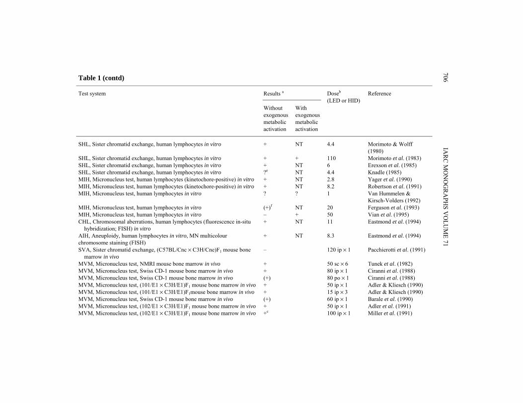
IARC M
ON
OG
RAPH
S VO
LUM
E 71706Table 1 (contd)
Test system Results a Reference
Withoutexogenousmetabolicactivation
Withexogenousmetabolicactivation
Doseb
(LED or HID)
SHL, Sister chromatid exchange, human lymphocytes in vitro + NT 4.4 Morimoto & Wolff(1980)
SHL, Sister chromatid exchange, human lymphocytes in vitro + + 110 Morimoto et al. (1983)SHL, Sister chromatid exchange, human lymphocytes in vitro + NT 6 Erexson et al. (1985)SHL, Sister chromatid exchange, human lymphocytes in vitro ?e NT 4.4 Knadle (1985)MIH, Micronucleus test, human lymphocytes (kinetochore-positive) in vitro + NT 2.8 Yager et al. (1990)MIH, Micronucleus test, human lymphocytes (kinetochore-positive) in vitro + NT 8.2 Robertson et al. (1991)MIH, Micronucleus test, human lymphocytes in vitro ? ? 1 Van Hummelen &
Kirsch-Volders (1992)MIH, Micronucleus test, human lymphocytes in vitro (+)f NT 20 Ferguson et al. (1993)MIH, Micronucleus test, human lymphocytes in vitro – + 50 Vian et al. (1995)CHL, Chromosomal aberrations, human lymphocytes (fluorescence in-situ hybridization; FISH) in vitro
+ NT 11 Eastmond et al. (1994)
AIH, Aneuploidy, human lymphocytes in vitro, MN multicolourchromosome staining (FISH)
+ NT 8.3 Eastmond et al. (1994)
SVA, Sister chromatid exchange, (C57BL/Cnc × C3H/Cne)F1 mouse bone marrow in vivo
– 120 ip × 1 Pacchierotti et al. (1991)
MVM, Micronucleus test, NMRI mouse bone marrow in vivo + 50 sc × 6 Tunek et al. (1982)MVM, Micronucleus test, Swiss CD-1 mouse bone marrow in vivo + 80 ip × 1 Ciranni et al. (1988)MVM, Micronucleus test, Swiss CD-1 mouse bone marrow in vivo (+) 80 po × 1 Ciranni et al. (1988)MVM, Micronucleus test, (101/E1 × C3H/E1)F1 mouse bone marrow in vivo + 50 ip × 1 Adler & Kliesch (1990)MVM, Micronucleus test, (101/E1 × C3H/E1)F1mouse bone marrow in vivo + 15 ip × 3 Adler & Kliesch (1990)MVM, Micronucleus test, Swiss CD-1 mouse bone marrow in vivo (+) 60 ip × 1 Barale et al. (1990)MVM, Micronucleus test, (102/E1 × C3H/E1)F1 mouse bone marrow in vivo + 50 ip × 1 Adler et al. (1991)MVM, Micronucleus test, (102/E1 × C3H/E1)F1 mouse bone marrow in vivo +c 100 ip × 1 Miller et al. (1991)
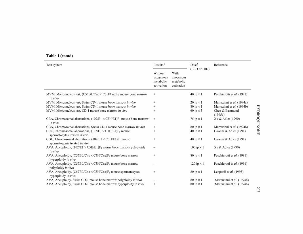
HY
DRO
QU
INO
NE
707
Table 1 (contd)
Test system Results a Reference
Withoutexogenousmetabolicactivation
Withexogenousmetabolicactivation
Doseb
(LED or HID)
MVM, Micronucleus test, (C57BL/Cnc × C3H/Cne)F1 mouse bone marrow in vivo
+ 40 ip × 1 Pacchierotti et al. (1991)
MVM, Micronucleus test, Swiss CD-1 mouse bone marrow in vivo + 20 ip × 1 Marrazinni et al. (1994a)MVM, Micronucleus test, Swiss CD-1 mouse bone marrow in vivo + 80 ip × 1 Marrazinni et al. (1994b)MVM, Micronucleus test, CD-1 mouse bone marrow in vivo + 60 ip × 3 Chen & Eastmond
(1995a)CBA, Chromosomal aberrations, (102/E1 × C3H/E1)F1 mouse bone marrow in vivo
+ 75 ip × 1 Xu & Adler (1990)
CBA, Chromosomal aberrations, Swiss CD-1 mouse bone marrow in vivo + 80 ip × 1 Marrazinni et al. (1994b)CCC, Chromosomal aberrations, (102/E1 × C3H/E1)F1 mouse spermatocytes treated in vivo
+ 40 ip × 1 Ciranni & Adler (1991)
CGG, Chromosomal aberrations, (102/E1 × C3H/E1)F1 mouse spermatogonia treated in vivo
+ 40 ip × 1 Ciranni & Adler (1991)
AVA, Aneuploidy, (102/E1 × C3H/E1)F1 mouse bone marrow polyploidy in vivo
– 100 ip × 1 Xu & Adler (1990)
AVA, Aneuploidy, (C57BL/Cnc × C3H/Cne)F1 mouse bone marrow hyperploidy in vivo
+ 80 ip × 1 Pacchierotti et al. (1991)
AVA, Aneuploidy, (C57BL/Cnc × C3H/Cne)F1 mouse bone marrow polyploidy in vivo
– 120 ip × 1 Pacchierotti et al. (1991)
AVA, Aneuploidy, (C57BL/Cnc × C3H/Cne)F1 mouse spermatocytes hyperploidy in vivo
+ 80 ip × 1 Leopardi et al. (1993)
AVA, Aneuploidy, Swiss CD-1 mouse bone marrow polyploidy in vivo – 80 ip × 1 Marrazinni et al. (1994b)AVA, Aneuploidy, Swiss CD-1 mouse bone marrow hyperploidy in vivo + 80 ip × 1 Marrazinni et al. (1994b)
IARC M
ON
OG
RAPH
S VO
LUM
E 71708
Table 1 (contd)
Test system Results a Reference
Withoutexogenousmetabolicactivation
Withexogenousmetabolicactivation
Doseb
(LED or HID)
AVA, Aneuploidy, CD-1 mouse bone marrow in vivo, MN multicolour chromosome staining (FISH)
+ 60 ip × 3 Chen & Eastmond(1995a)
BID, Binding (covalent) to DNA, mouse P388D1 cells in vitro + NT 5.5 Kalf et al. (1990)BID, Binding (covalent) to calf thymus DNA in vitro + NT 5.5 Leanderson & Tagesson
(1990)BID, Binding (covalent) to DNA, cultured rat Zymbal glands in vitro + NT 750 Reddy et al. (1990)BID, Binding (covalent) to calf thymus DNA in vitro – +g 11 Schlosser et al. (1990)BID, Binding (covalent) to DNA, human promyelocytic HL-60 cells in vitro + NT 5.5 Lévay et al. (1991)BID, Binding (covalent) to DNA, male B6C3F1 mouse bone-marrow cells in vitro
+ NT 11 Lévay et al. (1993)
BID, Binding (covalent) to DNA, human bone-marrow macrophages in vitro + NT 11 Lévay et al. (1993)BID, Binding (covalent) to DNA, human promyelocytic HL-60 cells in vitro + NT 27.5 Pathak et al. (1995)BID, Binding (covalent) to DNA, B6C3F1 mouse bone marrow in vitro + NT 27.5 Pathak et al. (1995)BID, Binding (covalent) to DNA, human promyelocytic HL-60 cells in vitro + NT 5.5 Lévay & Bodell (1996)BVD, Binding (covalent) to DNA, Sprague-Dawley rat Zymbal gland, liver or spleen in vivo
– 150 po × 4 Reddy et al. (1990)
BVD, Binding (covalent) to DNA, Fischer 344 rat kidneys in vivo – 50 po, 5 d/wk,6 wk
English et al. (1994b)
ICR, Inhibition of intercellular communication, V79MZ Chinese hamster cells in vitro
+ NT 0.055 Vang et al. (1993)
HY
DRO
QU
INO
NE
709
Table 1 (contd)
Test system Results a Reference
Withoutexogenousmetabolicactivation
Withexogenousmetabolicactivation
Doseb
(LED or HID)
DNA single-strand breaks on supercoiled Bluescript plasmid DNA – + 11 Schlosser et al. (1990)Binding (covalent) to porcine brain tubulin [porcine brain tubulin assembly assay] in vitro
– NT 2750 Brunner et al. (1991)
Inhibition of assembly of bovine microtubules in vitro (+) NT 110 Wallin & Hartley-Hasp(1993)
a +, positive; (+), weakly positive; –, negative; NT, not tested; ?, inconclusiveb LED, lowest effective dose; HID, highest ineffective dose; in-vitro tests, μg/mL; in-vivo tests, mg/kg bw/day; po, oral; NG, not given; inj, injection;ip, intraperitoneal; sc, subcutaneousc No increase in % kinetochore-positive micronuclei compared with controlsd Supplemented with arachidonic acid; increase in both kinetochore-positive and –negative micronucleated cells compared with controls (CREST-labelling procedure)e Positive if glutathione depleted with diethyl maleatef Size ratio of micronuclei to nucleus is not significant different from controls.g With prostaglandin H synthetase for oxidation
(Kalf et al., 1990; Schlosser et al., 1990). Hydroquinone bound weakly to isolated bovinemicrotubules but not to porcine brain tubulin in vitro and to DNA in most of the in-vitrostudies, in single studies with rat Zymbal glands in culture and in mice bone marrowin vitro. In vivo, hydroquinone did not bind to DNA from Zymbal gland, liver, spleen orkidneys of rat treated orally; it did not induce DNA strand breaks in plasmid DNA.
5. Summary of Data Reported and Evaluation
5.1 Exposure dataExposure to hydroquinone may occur during its production, its use as an inhibitor,
antioxidant and intermediate in the production of dyes, paints, motor fuels and oils, andin black-and-white photographic processing. Hydroquinone occurs naturally in certainplant species. It is used as a topical treatment for skin hyperpigmentation.
5.2 Human carcinogenicity dataA cohort of workers with definite and lengthy exposure to hydroquinone had low
cancer rates compared with two comparison populations; the reason for the lower thanexpected rates is unclear. A cohort of lithographers, some of whom had worked with hydro-quinone, had an excess of malignant melanoma based on five cases; only two of the caseshad reported exposure to hydroquinone.
5.3 Animal carcinogenicity dataHydroquinone was tested for carcinogenicity in two studies in mice and two studies
in rats by oral administration. It was also tested in rats for promoting activity in assaysfor bladder, stomach, liver, lung, oesophagus and kidney carcinogenesis and in one studyin hamsters for pancreatic carcinogenesis.
In mice, hydroquinone induced hepatocellular adenomas in females in one study andin males in another study. In rats it induced renal tubule adenomas in males in twostudies.
Hydroquinone had no promoting activity in most assays; an increase in the multi-plicity of oesophageal tumours was observed in one study and in the multiplicity of renalcell tumours in another study. No promoting effect on pancreatic carcinogenesis wasobserved in the study in hamsters.
5.4 Other relevant dataHydroquinone is metabolized mainly to conjugates, but a small percentage may be
converted to 1,4-benzoquinone, conjugated with glutathione or form DNA adducts in vitro.It caused toxicity in several organs, notably the kidney and forestomach.Hydroquinone was mutagenic in many in-vitro systems using a variety of end-points.
Also, after intraperitoneal administration, it caused genotoxicity or chromosomal aberra-tions in bone marrow.
IARC MONOGRAPHS VOLUME 71710
5.5 EvaluationThere is inadequate evidence in humans for the carcinogenicity of hydroquinone.There is limited evidence in experimental animals for the carcinogenicity of
hydroquinone.
Overall evaluationHydroquinone is not classifiable as to its carcinogenicity to humans (Group 3).
6. References
Adler, I.-D. & Kliesch, U. (1990) Comparison of single and multiple treatment regimens in themouse bone marrow micronucleus assay for hydroquinone (HQ) and cyclophosphamide(CP). Mutat. Res., 234, 115–123
Adler, I.-D., Kliesch, U., van Hummelen, P. & Kirsch-Volders, M. (1991) Mouse micronucleus testswith known and suspect spindle poisons: results from two laboratories. Mutagenesis, 6, 47–53
American Conference of Governmental Industrial Hygienists (1992) Documentation of theThreshold Limit Values and Biological Exposure Indices, 6th Ed., Vol. 1, Cincinnati, OH,pp. 789–792
American Conference of Governmental Industrial Hygienists (1997) 1997 TLVs® and BEIs®,Cincinnati, OH, p. 27
Anderson, D., Yu, T.W. & Schmezer, P. (1995) An investigation of the DNA-damaging ability ofbenzene and its metabolites in human lymphocytes, using the comet assay. Environ. mol.Mutag., 26, 305–314
Antoccia, A., Degrassi, F., Battistoni, A., Cilliutti, P. & Tanzarella, C. (1991) In vitro micronucleustest with kinetochore staining: evaluation of test performance. Mutagenesis, 6, 319–324
Barale, R., Marrazzini, A., Betti, C., Vangelisti, V., Loprieno, N. & Barrai, I. (1990) Genotoxicityof two metabolites of benzene: phenol and hydroquinone show strong synergistic effectsin vivo. Mutat. Res., 244, 15–20
Barber, E.D., Hill, T. & Schum, D.B. (1995) The percutaneous absorption of hydroquinone (HQ)through rat and human skin in vitro. Toxicol. Lett., 80, 167–172
Blacker, A.M., Schroeder, R.E., English, J.C., Murphy, S.J., Krasavage, W.J. & Simon, G.S.(1993) A two-generation reproduction study with hydroquinone in rats. Fundam. appl.Toxicol., 21, 420–424
Boatman, R.J., English, J.C., Perry, L.G. & Bialecki, V.E. (1996) Differences in the nephrotoxicityof hydroquinone among Fischer 344 and Sprague-Dawley rats and B6C3F1 mice. J. Toxicol.environ. Health, 47, 159–172
Bodell, W.J., Levay, G. & Pongracz, K. (1993) Investigation of benzene–DNA adducts and theirdetection in human bone marrow. Environ. Health Perspect., 99, 241–244
Bodell, W.J., Pathak, D.N., Lévay, G., Ye, Q. & Pongracz, K. (1996) Investigation of the DNAadducts formed in B6C3F1 mice treated with benzene: implications for molecular dosimetry.Environ. Health Perspect., 104 (Suppl. 6), 1189–1193
HYDROQUINONE 711
Brunner, M., Albertini, S. & Würgler, F.E. (1991) Effects of 10 known or suspected spindlepoisons in the in vitro porcine brain tubulin assembly assay. Mutagenesis, 6, 65–70
Carbonnelle, P., Lison, D., Leroy, J.-Y. & Lauwerys, R. (1995) Effect of the benzene metabolite,hydroquinone, on interleukin-1 secretion by human monocytes in vitro. Toxicol. appl. Phar-macol., 132, 220–226
Chapman, D.E., Namkung, M.J. & Juchau, M.R. (1994) Benzene and benzene metabolites asembryotoxic agents: effects on cultured rat embryos. Toxicol. appl. Pharmacol., 128,129–137
Chen, H. & Eastmond, D.A. (1995a) Synergistic increase in chromosomal breakage within theeuchromatin induced by an interaction of the benzene metabolites phenol and hydroquinonein mice. Carcinogenesis, 16, 1963–1969
Chen, H. & Eastmond, D.A. (1995b) Topoisomerase inhibition by phenolic metabolites: a poten-tial mechanism for benzene’s clastogenic effects. Carcinogenesis, 16, 2301–2307
Ciranni, R. & Adler, I.-D. (1991) Clastogenic effects of hydroquinone: induction of chromosomalaberrations in mouse germ cells. Mutat. Res., 263, 223–229
Ciranni, R., Barale, R., Ghelardini, G. & Loprieno, N. (1988) Benzene and the genotoxicity of itsmetabolites. II. The effect of the route of administration on the micronuclei and bone marrowdepression in mouse bone marrow cells. Mutat. Res., 209, 23–28
Divincenzo, G.D., Hamilton, M.L., Reynolds, R.C. & Ziegler, D.A. (1984) Metabolic fate anddisposition of [14C]hydroquinone given orally to Sprague-Dawley rats. Toxicology, 33, 9–18
Dobo, K.L. & Eastmond, D.A. (1994) Role of oxygen radicals in the chromosomal loss andbreakage induced by the quinone-forming compounds, hydroquinone and tert-butylhydro-quinone. Environ. mol. Mutag., 24, 293–300
Eastmond, D.A., Rupa, D.S. & Hasegawa, L.S. (1994) Detection of hyperdiploidy and chromo-some breakage in interphase human lymphocytes following exposure to the benzene meta-bolite hydroquinone using multicolor fluorescence in situ hybridization with DNA probes.Mutat. Res., 322, 9–20
Ellard, S. & Parry, E.M. (1993) Induction of micronuclei in V79 Chinese hamster cells by hydro-quinone and econazole nitrate. Mutat. Res., 287, 87–91
English, J.C., Perry, L.G., Vlaovic, M., Moyer, C. & O’Donoghue, J.L. (1994a) Measurement ofcell proliferation in the kidneys of Fischer 344 and Sprague-Dawley rats after gavage admi-nistration of hydroquinone. Fundam. appl. Toxicol., 23, 397–406
English, J.C., Hill, T., O’Donoghue, J.L. & Reddy, M.V. (1994b) Measurement of nuclear DNAmodification by 32P-postlabeling in the kidneys of male and female Fischer 344 rats aftermultiple gavage doses of hydroquinone. Fundam. appl. Toxicol., 23, 391–396
Erexson, G.L., Wilmer, J.L. & Kligerman, A.D. (1985) Sister chromatid exchange induction inhuman lymphocytes exposed to benzene and its metabolites in vitro. Cancer Res., 45,2471–2477
Eyer, P. (1991) Effects of superoxide dismutase on the autoxidation of 1,4-hydroquinone. Chem.-biol. Interact., 80, 159–176
Fahrig, R. (1984) Genetic mode of action of cocarcinogens and tumor promoters in yeast andmice. Mol. gen. Genet., 194, 7–14
IARC MONOGRAPHS VOLUME 71712
Ferguson, L.R., Morcombe, P. & Triggs, C.N. (1993) The size of cytokinesis-blocked micronucleiin human peripheral blood lymphocytes as a measure of aneuploidy induction by Set A com-pounds in the EEC trial. Mutat. Res., 287, 101–112
Foureman, P., Mason, J.M., Valencia, R. & Zimmering, S. (1994) Chemical mutagenesis testingin Drosophila. IX. Results of 50 coded compounds tested for the National Toxicology Pro-gram. Environ. mol. Mutag., 23, 51–63
Frantz, C.E., Chen, H. & Eastmond, D.A. (1998) Inhibition of human topoisomerase II in vitro bybioactive benzene metabolites. Environ. Health Perspect., 104 (Suppl. 6), 1319–1323
Friedlander, B.R., Hearne, F.T. & Newman, B.J. (1982) Mortality, cancer incidence, and sickness-absence in photographic processors: an epidemiologic study. J. occup. Med., 24, 605–613
Galloway, S.M., Armstrong, M.J., Reuben, C., Colman, S., Brown, B., Cannon, C., Bloom, A.D.,Nakamura, F., Ahmed, M. & Duk, S. (1987) Chromosome aberrations and sister chromatidexchanges in Chinese hamster ovary cells: evaluations of 108 chemicals. Environ. mol.Mutag., 10 (Suppl. 10), 1–175
Gopalakrishna, R., Chen, Z.H. & Gundimeda, U. (1994) Tobacco smoke tumor promoters,catechol and hydroquinone, induce oxidative regulation of protein kinase C and influenceinvasion and metastasis of lung carcinoma cells. Proc. natl Acad. Sci. USA, 91, 12233–12237
Hakura, A., Tsutsui, Y., Mochida, H., Sugihara, Y., Mikami, T. & Sagami, F. (1996) Mutagenicityof dihydroxybenzenes and dihydroxynaphthalenes for Ames Salmonella tester strains. Mutat.Res., 371, 293–299
Hard, G.C., Whysner, J., English, J.C., Zang, E. & Williams, G.M. (1997) Relationship of hydro-quinone-associated rat renal tumors with spontaneous chronic progressive nephropathy.Toxicol. Pathol., 25, 132–143
Hasegawa, R., Furukawa, F., Toyoda, K., Takahashi, M., Hayashi, Y., Hirose, M. & Ito, N. (1990)Inhibitory effects of antioxidants on N-bis(2-hydroxypropyl)nitrosamine-induced lungcarcinogenesis in rats. Jpn. J. Cancer Res., 81, 871–877
Haworth, S., Lawlor, T., Mortelmans, K., Speck, W. & Zeiger, E. (1983) Salmonella mutagenicitytest results for 250 chemicals. Environ. Mutag., 5 (Suppl. 1), 3–142
Hazel, B.A., O’Connor, A., Niculescu, R. & Kalf, G.F. (1995) Benzene and its metabolite, hydro-quinone, induce granulocytic differentiation in myeloblasts by interacting with cellular signa-ling pathways activated by granulocyte colony-stimulating factor. Stem Cells, 13, 295–310
Hazel, B.A., O’Connor, A., Niculescu, R. & Kalf, G.F. (1996a) Induction of granulocytic differen-tiation in a mouse model by benzene and hydroquinone. Environ. Health Perspect., 104(Suppl. 6), 1257–1264
Hazel, B.A., Baum, C. & Kalf, G.F. (1996b) Hydroquinone, a bioreactive metabolite of benzene,inhibits apoptosis in myeloblasts. Stem Cells, 14, 730–742
Henschler, R., Glatt, H.R. & Heyworth, C.M. (1996) Hydroquinone stimulates granulocyte-macrophage progenitor cells in vitro and in vivo. Environ. Health Perspect., 104 (Suppl. 6),1271–1274
Hill, B.A., Kleiner, H.E., Ryan, E.A., Dulik, D.M., Monks, T.J. & Lau, S.S. (1993) Identificationof multi-S-substituted conjugates of hydroquinone by HPLC-coulometric electrode array ana-lysis and mass spectroscopy. Chem. Res. Toxicol., 6, 459–469
HYDROQUINONE 713
Hiraku, Y. & Kawanishi, S. (1996) Oxidative DNA damage and apoptosis induced by benzenemetabolites. Cancer Res., 56, 5172–5178
Hirose, M., Inoue, T., Asamoto, M., Tagawa, Y. & Ito, N. (1986) Comparison of the effects of 13phenolic compounds in induction of proliferative lesions of the forestomach and increase inthe labelling indices of the glandular stomach and urinary bladder epithelium of Syriangolden hamsters. Carcinogenesis, 7, 1285–1289
Hirose, M., Yamaguchi, S., Fukushima, S., Hasegawa, R., Takahashi, S. & Ito, N. (1989) Promo-tion by dihydroxybenzene derivative of N-methyl-N′-nitro-N-nitrosoguanidine-induced F344rat forestomach and glandular stomach carcinogenesis. Cancer Res., 49, 5143–5147
Hutt, A.M. & Kalf, G.F. (1996) Inhibition of human DNA topoisomerase II by hydroquinone andp-benzoquinone, reactive metabolites of benzene. Environ. Health Perspect., 104 (Suppl. 6),1265–1269
IARC (1977) IARC Monographs on the Evaluation of the Carcinogenic Risk of Chemicals toMan, Vol. 15, Some Fumigants, the Herbicides 2,4-D and 2,4,5-T, Chlorinated Dibenzo-dioxins and Miscellaneous Industrial Chemicals, Lyon, pp. 155–175
IARC (1987) IARC Monographs on the Evaluation of Carcinogenic Risks to Humans, Suppl. 7,Overall Evaluations of Carcinogenicity: An Updating of IARC Monographs Volumes 1 to 42,Lyon, p. 64
International Labour Office (1991) Occupational Exposure Limits for Airborne Toxic Substances,3rd Ed. (Occupational Safety and Health Series No. 37), Geneva, pp. 160–161
Irons, R.D. & Stillman, W.S. (1996a) The effects of benzene and other leukaemogenic agents onhaematopoietic stem and progenitor cell differentiation. Eur. J. Haematol., 57 (Suppl.),119–124
Irons, R.D. & Stillman, W.S. (1996b) Impact of benzene metabolites on differentiation of bonemarrow progenitor cells. Environ. Health Perspect., 104 (Suppl. 6), 1247–1250
Irons, R.D., Stillman, W.S., Colagiovanni, D.B. & Henry, V.A. (1992) Synergistic action of thebenzene metabolite hydroquinone on myelopoietic stimulating activity of granulocyte/macrophage colony-stimulating factor in vitro. Proc. natl Acad. Sci. USA, 89, 3691–3695
Kalf, G., Shurina, R., Renz, J. & Schlosser, M. (1990) The role of hepatic metabolites of benzenein bone marrow peroxidase-mediated myelo- and genotoxicity. In: Witmer, C.M., Snyder,R.R., Jollow, D.J., Kalf, G.F., Kocsis, J.J. & Sipes, I.G., eds, Biological Reactive Inter-mediates IV, New York, Plenum Press, pp. 443–455
Kari, F.W., Bucher, J., Eustis, S.L., Haseman, J.K. & Huff, J.E. (1992) Toxicity and carcino-genicity of hydroquinone in F344/N rats and B6C3F1 mice. Food chem. Toxicol., 30, 737–747
Knadle, S. (1985) Synergistic interaction between hydroquinone and acetaldehyde in the induc-tion of sister chromatid exchange in human lymphocytes in vitro. Cancer Res., 45, 4853–4857
Kolachana, P., Subrahmanyam, V.V., Meyer, K.B., Zhang, L. & Smith, M.T. (1993) Benzene andits phenolic metabolites produce oxidative damage in HL60 cells in vitro and in the bonemarrow in vivo. Cancer Res., 53, 1023–1026
Krasavage, W.J., Blacker, A.M., English, J.C. & Murphy, S.J. (1992) Hydroquinone: a develop-mental toxicity study in rats. Fundam. appl. Toxicol., 18, 370–375
IARC MONOGRAPHS VOLUME 71714
Kurata, Y., Fukushima, S., Hasegawa, R., Hirose, M., Shibata, M.-A., Shirai, T. & Ito, N. (1990)Structure-activity relations in promotion of rat urinary bladder carcinogenesis by phenolicantioxidants. Jpn. J. Cancer Res. (Gann), 81, 754–759
Leanderson, P. & Tagesson, C. (1990) Cigarette smoke-induced DNA-damage: role of hydro-quinone and catechol in the formation of the oxidative DNA-adduct, 8-hydroxydeoxy-guanosine. Chem.-biol. Interact., 75, 71–81
Legathe, A., Hoener, B.-A. & Tozer, T.N. (1994) Pharmacokinetic interaction between benzene meta-bolites, phenol and hydroquinone, in B6C3F1 mice. Toxicol. appl. Pharmacol., 124, 131–138
Leopardi, P., Zijno, A., Bassani, B. & Pacchierotti, F. (1993) In vivo studies on chemicallyinduced aneuploidy in mouse somatic and germinal cells. Mutat. Res., 287, 119–130
Lévay, G. & Bodell, W.J. (1996) Role of hydrogen peroxide in the formation of DNA adducts inHL-60 cells treated with benzene metabolites. Biochem. biophys. Res. Commun., 222, 44–49
Lévay, G., Pongracz, K. & Bodell, W.J. (1991) Detection of DNA adducts in HL-60 cells treatedwith hydroquinone and p-benzoquinone by 32P-postlabeling. Carcinogenesis, 12, 1181–1186
Lévay, G., Ross, D. & Bodell, W.J. (1993) Peroxidase activation of hydroquinone results in theformation of DNA adducts in HL-60 cells, mouse bone marrow macrophages and humanbone marrow. Carcinogenesis, 14, 2329–2334
Lewis, R.J., Jr (1993) Hawley’s Condensed Chemical Dictionary, 12th Ed., New York, VanNostrand Reinhold, p. 618
Li, Y. & Trush, M. A. (1993a) DNA damage resulting from the oxidation of hydroquinone bycopper: role for a Cu(II)/Cu(I) redox cycle and reactive oxygen generation. Carcinogenesis,14, 1303–1311
Li, Y. & Trush, M.A. (1993b) Oxidation of hydroquinone by copper: chemical mechanism andbiological effects. Arch. Biochem. Biophys., 300, 346–355
Li, Q., Aubrey, M.T., Christian, T. & Freed, B.M. (1997) Differential inhibition of DNA synthesisin human T cells by the cigarette tar components hydroquinone and catechol. Fundam. appl.Toxicol., 38, 158–165
Lide, D.R., ed. (1997) CRC Handbook of Chemistry and Physics, 78th Ed., Boca Raton, FL, CRCPress, p. 3-43
Manning, B.W., Adams, D.O. & Lewis, J.G. (1994) Effects of benzene metabolites on receptor-mediated phagocytosis and cytoskeletal integrity in mouse peritoneal macrophages. Toxicol.appl. Pharmacol., 126, 214–223
Marrazzini, A., Chelotti, L., Barrai, I., Loprieno, N. & Barale, R. (1994a) In vivo genotoxic inter-actions among three phenolic benzene metabolites. Mutat. Res., 341, 29–46
Marrazzini, A., Betti, C., Bernacchi, F., Barrai, I. & Barale, R. (1994b) Micronucleus test andmetaphase analyses in mice exposed to known and suspected spindle poisons. Mutagenesis,9, 505–515
Maruyama, H., Amanuma, T., Nakae, D., Tsutsumi, M., Kondo, S., Tsujiuchi, T., Denda, A. &Konishi, Y. (1991) Effects of catechol and its analogs on pancreatic carcinogenesis initiatedby N-nitrosobis(2-oxopropyl)amine in Syrian hamsters. Carcinogenesis, 12, 1331–1334
HYDROQUINONE 715
McGregor, D.B., Brown, A., Cattanach, P., Edwards, I., McBride, D. & Caspary, W.J. (1988a)Responses of the L5178Y tk+/tk– mouse lymphoma cell forward mutation assay. II: 18 codedchemicals. Environ. mol. Mutag., 11, 91–118
McGregor, D.B., Riach, C.G., Brown, A., Edwards, I., Reynolds, D., West, K. & Willington, S.(1988b) Reactivity of catecholamines and related substances in the mouse lymphomaL5178Y cell assay for mutagens. Environ. mol. Mutag., 11, 523–544
Miller, B.M., Zitzelsberger, H.F., Weier, H.-U.G. & Adler, I.-D. (1991) Classification of micro-nuclei in murine erythrocytes: immunofluorescent staining using CREST antibodies com-pared to in situ hybridization with biotinylated gamma satellite DNA. Mutagenesis, 6,297–302
Miyata, Y., Fukushima, S., Hirose, M., Masui, T. & Ito, N. (1985) Short-term screening of pro-moters of bladder carcinogenesis in N-butyl-N-(4-hydroxybutyl)nitrosamine-initiated, unila-terally ureter-ligated rats. Jpn. J. Cancer Res. (Gann), 76, 828–834
Moran, J.L., Siegel, D., Sun, X.-M. & Ross, D. (1996) Induction of apoptosis by benzene meta-bolites in HL60 and CD34+ human bone marrow progenitor cells. Mol. Pharmacol., 50,610–615
Morimoto, K. & Wolff, S. (1980) Increase of sister chromatid exchanges and perturbations of celldivision kinetics in human lymphocytes by benzene metabolites. Cancer Res., 40, 1189–1193
Morimoto, K., Wolff, S. & Koizumi, A. (1983) Induction of sister chromatid exchanges in humanlymphocytes by microsomal activation of benzene metabolites. Mutat. Res., 119, 355–360
Murphy, S.J., Schroeder, R.E., Blacker, A.M., Krasavage, W.J. & English, J.C. (1992) A studyof developmental toxicity of hydroquinone in the rabbit. Fundam. appl. Toxicol., 19, 214–221
Nakamura, S.-I., Oda, Y., Shimada, T., Oki, I. & Sugimoto, K. (1987) SOS-inducing activity ofchemical carcinogens and mutagens in Salmonella typhimurium TA1535/pSK1002: exami-nation with 151 chemicals. Mutat. Res., 192, 239–246
Nerland, D.E. & Pierce, W.M. (1990) Identification of N-acetyl-S-(2,5-dihydroxyphenyl)-L-cys-teine as a urinary metabolite of benzene, phenol and hydroquinone. Drug Metab. Disp., 18,958–961
Neun, D.J., Penn, A. & Snyder, C.A. (1992) Evidence for strain-specific differences in benzenetoxicity as a function of host target cell susceptibility. Arch. Toxicol., 66, 11–17
Nielsen, H., Henriksen, L. & Olsen, J.H. (1996) Malignant melanoma among lithographers.Scand. J. Work Environ. Health., 22, 108–111
NOES (1997) National Occupational Exposure Survey 1981–83, Unpublished data as of November1997, Cincinnati, OH, United States Department of Health and Human Services, Public HealthService, National Institute for Occupational Safety and Health
Okazaki, S., Hoshiya, T., Takahashi, S., Futakuchi, M., Saito, K. & Hirose, M. (1993) Modifi-cation of hepato-and renal carcinogenesis by catechol and its isomers in rats pretreated withN-ethyl-N-hydroxyethylnitrosamine. Teratog. Carcinog. Mutag., 13, 127–137
Oliveira, N.L. & Kalf, G.F. (1992) Induced differentiation of HL-60 promyelocytic leukemia cellsto monocyte/macrophages is inhibited by hydroquinone, a hematotoxic metabolite of benzene.Blood, 79, 627–633
IARC MONOGRAPHS VOLUME 71716
Pacchierotti, F., Bassani, B., Leopardi, P. & Zijno, A. (1991) Origin of aneuploidy in relation todisturbances of cell-cycle progression. II: Cytogenetic analysis of various parameters in mousebone marrow cells after colchicine or hydroquinone treatment. Mutagenesis, 6, 307–311
Pathak, D.N., Lévay, G. & Bodell, W.J. (1995) DNA adduct formation in the bone marrow ofB6C3F1 mice treated with benzene. Carcinogenesis, 16, 1803–1808
Pellack-Walker, P. & Blumer, J.L. (1986) DNA damage in L5178YS cells following exposure tobenzene metabolites. Mol. Pharmacol., 30, 42–47
Pifer, J.W., Hearne, F.T., Swanson, F.A. & O’Donoghue, J.L. (1995) Mortality study of employeesengaged in the manufacture and use of hydroquinone. Int. Arch. occup. environ. Health, 67,267–280
Rao, G.S. (1991) Hematin catalysed autooxidation of hydroquinone or 1,2,4-benzenetriol. Chem.-biol. Interact., 80, 339–347
Reddy, M.V., Bleicher, W.T., Blackburn, G.R. & Mackerer, C.R. (1990) DNA adduction byphenol, hydroquinone, or benzoquinone in vitro but not in vivo: nuclease P1-enhanced 32P-postlabeling of adducts as labeled nucleoside bisphosphates, dinucleotides and nucleosidemonophosphates. Carcinogenesis, 11, 1349–1357
Robertson, M.L., Eastmond, D.A. & Smith, M.T. (1991) Two benzene metabolites, catechol andhydroquinone, produce a synergistic induction of micronuclei and toxicity in cultured humanlymphocytes. Mutat. Res., 249, 201–209
Ross, D. (1996) Metabolic basis of benzene toxicity. Eur. J. Haematol., 57 (Suppl.), 111–118Saito, T. & Takeichi, S. (1995) Experimental studies on the toxicity of lithographic developer
solution. Clin. Toxicol., 33, 343–348Sakai, M., Yoshida, D. & Mizusaki, S. (1985) Mutagenicity of polycyclic aromatic hydrocarbons
and quinones on Salmonella typhimurium TA97. Mutat. Res., 156, 61–67Schaefer, D., Jürgensmeier, J.M. & Bauer, G. (1995) Catechol interferes with TGF-β-induced
elimination of transformed cells by normal cells: implications for the survival of transformedcells during carcinogenesis. Int. J. Cancer, 60, 520–526
Schlosser, M.J. & Kalf, G.F. (1989) Metabolic activation of hydroquinone by macrophage pero-xidase. Chem.-biol. Interact., 72, 191–207
Schlosser, M.J., Shurina, R.D. & Kalf, F.G. (1990) Prostaglandin H synthase catalyzed oxidationof hydroquinone to a sulfhydryl-binding and DNA-damaging metabolite. Chem. Res. Toxicol.,3, 333–339
Seaton, M.J., Schlosser, R.M. & Medinsky, M.A. (1995) In vitro conjugation of benzene meta-bolites by human liver: potential influence of interindividual variability on benzene toxicity.Carcinogenesis, 16, 1519–1527
Seelbach, A., Fissler, B. & Madle, S. (1993) Further evaluation of a modified micronucleus assaywith V79 cells for detection of aneugenic effects. Mutat. Res., 303, 163–169
Seidel, H.J., Barthel, E., Schäfer, F., Schad, H. & Weber, L. (1991) Action of benzene metabolites onmurine hematopoietic colony-forming cells in vitro. Toxicol. appl. Pharmacol., 111, 128–131
Shibata, M.-A., Yamada, M., Hirose, M., Asakawa, E., Tatematsu, M. & Ito, N. (1990) Earlyproliferative responses of forestomach and glandular stomach of rats treated with five diffe-rent phenolic antioxidants. Carcinogenesis, 11, 425–429
HYDROQUINONE 717
Shibata, M.-A., Hirose, M., Tanaka, H., Asakawa, E., Shirai, T. & Ito, N. (1991) Induction of renalcell tumors in rats and mice, and enhancement of hepatocellular tumor development in miceafter long-term hydroquinone treatment. Jpn. J. Cancer Res (Gann), 82, 1211–1219
Smith, M.T., Yager, J.W., Steinmetz, K.L. & Eastmond, D.A. (1989) Peroxidase-dependent meta-bolism of benzene’s phenolic metabolites and its potential role in benzene toxicity and carci-nogenicity. Environ. Health Perspect., 82, 23–29
Snyder, R. & Hedli, C.C. (1996) An overview of benzene metabolism. Environ. Health Perspect.,104 (Suppl. 6), 1165–1171
Snyder, R., Dimitriadis, E., Guy, R., Hu, P., Cooper, K., Bauer, H., Witz, G. & Goldstein, B.D.(1989) Studies on the mechanism of benzene toxicity. Environ. Health Perspect., 82, 31–35
Stenius, U., Warholm, M., Rannug, A., Walles, S., Lundberg, I. & Högberg, J. (1989) The role ofGSH depletion and toxicity in hydroquinone-induced development of enzyme-altered foci.Carcinogenesis, 10, 595–599
Subrahmanyam, V.V., Kolachana, P. & Smith, M.T. (1991) Metabolism of hydroquinone byhuman myeloperoxidase: mechanisms of stimulation by other phenolic compounds. Arch.Biochem. Biophys., 286, 76–84
Suzuki, J., Inoue, Y. & Suzuki, S. (1995) Changes in the urinary excretion level of 8-hydroxyguanineby exposure to reactive oxygen-generating substances. Free Rad. Biol. Med., 18, 431–436
Trush, M.A., Twerdok, L.E., Rembish, S.J., Zhu, H. & Li, Y. (1996) Analysis of target cell suscep-tibility as a basis for the development of a chemoprotective strategy against benzene-inducedhematotoxicities. Environ. Health Perspect., 104 (Suppl. 6), 1227–1234
Tsutsui, T., Hayashi, N., Maizumi, H., Huff, J. & Barrett, J.C. (1997) Benzene-, catechol-, hydro-quinone- and phenol-induced cell transformation, gene mutations, chromosome aberrations,aneuploidy, sister chromatid exchanges and unscheduled DNA synthesis in Syrian hamsterembryo cells. Mutat. Res., 373, 113–123
Tunek, A., Högstedt, B. & Olofsson, T. (1982) Mechanism of benzene toxicity. Effects of benzeneand benzene metabolites on bone marrow cellularity, number of granulopoietic stem cells andfrequency of micronuclei in mice. Chem.-biol. Interact., 39, 129–138
United States National Library of Medicine (1997) Hazardous Substances Data Bank (HSDB),Bethesda, MD [Record No. 577]
United States National Toxicology Program (1989) Toxicology and Carcinogenesis Studies ofHydroquinone (CAS No. 123-31-9) in F344/N Rats and B6C3F1 Mice (Technical ReportSeries No. 366; NIH Publ. No. 90-2821), Research Triangle Park, NC
Van Hummelen, P. & Kirsch-Volders, M. (1992) Analysis of eight known or suspected aneugensby the in vitro human lymphocyte micronucleus test. Mutagenesis, 7, 447–455
Vang, O., Wallin, H., Doehmer, J. & Autrup, H. (1993) Cytochrome P450-mediated metabolismof tumour promoters modifies the inhibition of intercellular communication: a modified assayfor tumour promotion. Carcinogenesis, 14, 2365–2371
Verschueren, K. (1996) Handbook of Environmental Data on Organic Chemicals, 3rd Ed., NewYork, Van Nostrand Reinhold, pp. 1122–1124
IARC MONOGRAPHS VOLUME 71718
Vian, L., Van Hummelen, P., Bichet, N., Gouy, D. & Kirsch-Volders, M. (1995) Evaluation ofhydroquinone and chloral hydrate on the in vitro micronucleus test on isolated lymphocytes.Mutat. Res., 334, 1–7
Walles, S.A.S. (1992) Mechanisms of DNA damage induced in rat hepatocytes by quinones.Cancer Lett., 63, 47–52
Wallin, M. & Hartley-Asp, B. (1993) Effects of potential aneuploidy inducing agents on micro-tubule assembly in vitro. Mutat. Res., 287, 17–22
Warr, T.J., Parry, E.M. & Parry, J.M. (1993) A comparison of two in vitro mammalian cell cyto-genetic assays for the detection of mitotic aneuploidy using 10 known or suspected aneugens.Mutat. Res., 287, 29–46
WHO (1993) Guidelines for Drinking-Water Quality, 2nd Ed., Vol. 1, Recommendations, GenevaWHO (1994) Hydroquinone (Environmental Health Criteria 157), Geneva, International Pro-
gramme on Chemical SafetyXu, W. & Adler, I.-D. (1990) Clastogenic effects of known and suspect spindle poisons studied
by chromosome analysis in mouse bone marrow cells. Mutagenesis, 5, 371–374Yager, J.W., Eastmond, D.A., Robertson, M.L., Paradisin, W.M. & Smith, M.T. (1990) Charac-
terization of micronuclei induced in human lymphocytes by benzene metabolites. CancerRes., 50, 393–399
Yamaguchi, S., Hirose, M., Fukushima, S., Hasegawa, R. & Ito, N. (1989) Modification by cate-chol and resorcinol of upper digestive tract carcinogenesis in rats treated with methyl-n-amyl-nitrosamine. Cancer Res., 49, 6015–5018
HYDROQUINONE 719