Embed Size (px)
Citation preview
Reactive Oxygen SpeciesDOI: 10.1002/ange.201203222
Hydrogen Peroxide Induced Activation of Gene Expression inMammalian Cells using Boronate Estrone Derivatives**Jeane M. Govan, Andrew L. McIver, Chad Riggsbee, and Alexander Deiters*
Hydrogen peroxide (H2O2) playsimportant roles in biological and cel-lular processes. H2O2 is a reactiveoxygen species (ROS) that was origi-nally thought of as only an oxidativestress marker in diseases,[1] butrecently has been shown to be animportant secondary messenger in bio-logical systems.[2] H2O2 is involved inseveral biological processes, includingcell signaling,[3] embryogenesis,[4]
apoptosis,[5] aging, and diseases, suchas cancer[6] and neurodegenerativediseases.[7] Herein, we describe thedevelopment of a genetic switch thatenables the induction of gene expres-sion in response to H2O2. Because ofthe modularity of the system, any geneof interest can be placed under thecontrol of H2O2. Importantly, thisgenetic switch allows for the sensitiveand selective detection of H2O2 in livemammalian cells.
The H2O2-triggered genetic switchrelies on a GAL4-UAS (upstreamactivating sequence) system, in whichthe DNA-binding domain of GAL4 isfused to an a-estrogen-receptorligand-binding domain (ER). In theabsence of a suitable ER-ligand (e.g.,estrone), the GAL4-ER fusion protein is tightly bound toa complex of heat-shock chaperone proteins (e.g., hsp90) thatkeeps the ER in an inactive state.[8] Upon ligand binding, theGAL4-ER undergoes a conformational change that displacesthe hsp90 complex.[9] This active GAL4-ER-ligand complextranslocates into the nucleus,[10] binds to the UAS locatedupstream of the gene of interest, in this case of luciferase, andinduces transcription (Scheme 1a). The GAL4-UAS systemwas changed into a H2O2-responsive system by takingadvantage of a boron-oxidation reaction.[11] Boronated small
molecules have previously been applied in the fluorescentdetection of H2O2,
[12,13] but not in the activation of geneexpression. Based on the results of structure activity relation-ship studies[14–16] and X-ray structures (Scheme 1 b),[17,18]
a boronate ester group was introduced at either the 3-hydroxyor the 17-carbonyl position (or both) of estrone, to inhibitbinding to the ER. In the presence of H2O2, the boronategroup will be oxidized, resulting in the native phenol orketone, and thus activating gene expression through ERbinding.
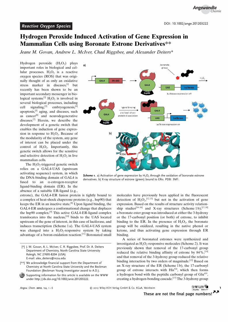
A series of boronated estrones were synthesized andinvestigated as H2O2-responsive molecules (Scheme 2). It waspreviously shown that removal of the 17-carbonyl groupreduced the relative binding affinity of estrone by 84%,[14]
and that removal of the 3-hydroxy group reduced the relativebinding interaction by two orders of magnitude.[15] Based onan X-ray structure of the ER (Scheme 1b), the 17-carbonylgroup of estrone interacts with His524, which then formsa hydrogen bond with the peptidic carbonyl group of Glu419,creating a hydrogen-bonding cascade.[17] The 3-hydroxy group
[*] J. M. Govan, A. L. McIver, C. R. Riggsbee, Prof. Dr. A. DeitersDepartment of Chemistry, North Carolina State UniversityRaleigh, NC 27695-8204 (USA)E-mail: [email protected]
[**] We acknowledge financial support from the Department ofChemistry at North Carolina State University and the BeckmanFoundation (Beckman Young Investigator award to A.D.).
Supporting information for this article is available on the WWWunder http://dx.doi.org/10.1002/anie.201203222.
Scheme 1. a) Activation of gene expression by H2O2 through the oxidation of boronate estronederivatives. b) X-ray structure of estrone (green) bound to ERa. PDB: 3M1.
AngewandteChemie
1Angew. Chem. 2012, 124, 1 – 6 � 2012 Wiley-VCH Verlag GmbH & Co. KGaA, Weinheim
These are not the final page numbers! � �
interacts through hydrogen bonds with Glu353, Arg394, anda water molecule within the binding pocket.[18] Therefore, thereplacement of either one of these groups with a phenyl orvinyl boronic acid ester derivative should render the estronemolecule biologically inactive.[14,15] If the boronation of eitherthe 3-position (3), or 17-position (4), or both (5) completelyinactivates the binding of the estrone derivative to the ER,then estrone activity should be restored through H2O2-mediated oxidation to a functional estrone molecule. Inaddition, the dehydroxy estrone 2 was used as a negativecontrol compound.
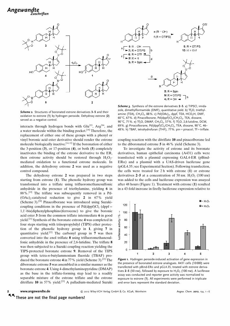
The dehydroxy estrone 2 was prepared in two stepsstarting from estrone (1). The phenolic hydroxy group wastransformed into a triflate using trifluoromethanesulfonicanhydride in the presence of triethylamine, yielding 6 in88%.[19] The triflate was subsequently removed in a Pd-(OAc)2-catalyzed reduction to give 2 in 67 % yield(Scheme 3).[20] Pinacolborane was introduced using Suzuki-coupling conditions in the presence of Pd(dppf)Cl2 (dppf =
1,1’-bis(diphenylphosphino)ferrocene) to give the boronicacid ester 3 from the common triflate intermediate 6 in goodyield.[21] Synthesis of the boronate estrone 4 was completed infour steps starting with triisopropylsilyl (TIPS) ether protec-tion of the phenolic hydroxy group in 1, giving 7 inquantitative yield.[22] The carbonyl group in 7 was thenconverted into the enol triflate 8 using trifluoromethanesul-fonic anhydride in the presence of 2,6-lutidine. The triflate 8was then subjected to a Suzuki coupling reaction yielding theTIPS-protected boronate estrone 9. Removal of the TIPSgroup with tetra-n-butylammonium fluoride (TBAF) pro-duced the boronate estrone 4 in 77% yield (Scheme 3).[22] Thediboronate estrone 5 was assembled in a similar manner as theboronate estrone 4. Using 4-dimethylaminopyridine (DMAP)as the base in the triflate-forming step lead to a readilyseparable mixture of the estrone triflate and the estroneditriflate 10 in 57% yield.[23] A palladium-mediated Suzuki
coupling reaction with the ditriflate 10 and pinacolborane ledto the diboronated estrone 5 in 46% yield (Scheme 3).
To investigate the activity of estrone and its boronatederivatives, human epithelial carcinoma (A431) cells weretransfected with a plasmid expressing GAL4-ER (pBind-ERa) and a plasmid with a UAS-driven luciferase gene(pGL4.35; see Experimental Section). Following transfection,the cells were treated for 2 h with estrone (1) or estronederivatives 2–5 at a concentration of 50 nm. H2O2 (100 nm)was added to the cells and luciferase expression was assayedafter 48 hours (Figure 1). Treatment with estrone (1) resultedin a 43-fold increase in firefly luciferase expression relative to
Scheme 2. Structures of boronated estrone derivatives 3–5 and theiroxidation to estrone (1) by hydrogen peroxide. Dehydroxy estrone (2)served as a negative control.
Scheme 3. Synthesis of the estrone derivatives 2–5. a) TIPSCl, imida-zole, dimethylformamide (DMF), quantitative yield. b) Tf2O, triethyl-amine (TEA), CH2Cl2, 88%. c) Pd(OAc)2, dppf, TEA, HCO2H, DMF,60 8C, 67%. d) Pinacolborane, Pd(dppf)Cl2/CH2Cl2, TEA, dioxane,90 8C, 71%. e) Tf2O, DMAP, CH2Cl2, 57%. f) Tf2O, 2,6-lutidine, DCM,69%. g) Pinacolborane, Pd(dppf)Cl2/CH2Cl2, TEA, dioxane, 90 8C, 46–48%. h) TBAF, tetrahydrofuran (THF), 77%. pin= pinacol; Tf= triflate.
Figure 1. Hydrogen peroxide-induced activation of gene expression inthe presence of boronated estrone analogues. A431 cells (10000) weretransfected with pBind-ERa and pGL4.35, treated with estrone deriva-tives 2–5 (50 nm), followed by exposure to H2O2 (100 nm). A luciferaseassay was conducted and reporter gene activity was normalized toexposure to estrone (1). All experiments were performed in triplicateand error bars represent the standard deviation.
.AngewandteZuschriften
2 www.angewandte.de � 2012 Wiley-VCH Verlag GmbH & Co. KGaA, Weinheim Angew. Chem. 2012, 124, 1 – 6� �
These are not the final page numbers!
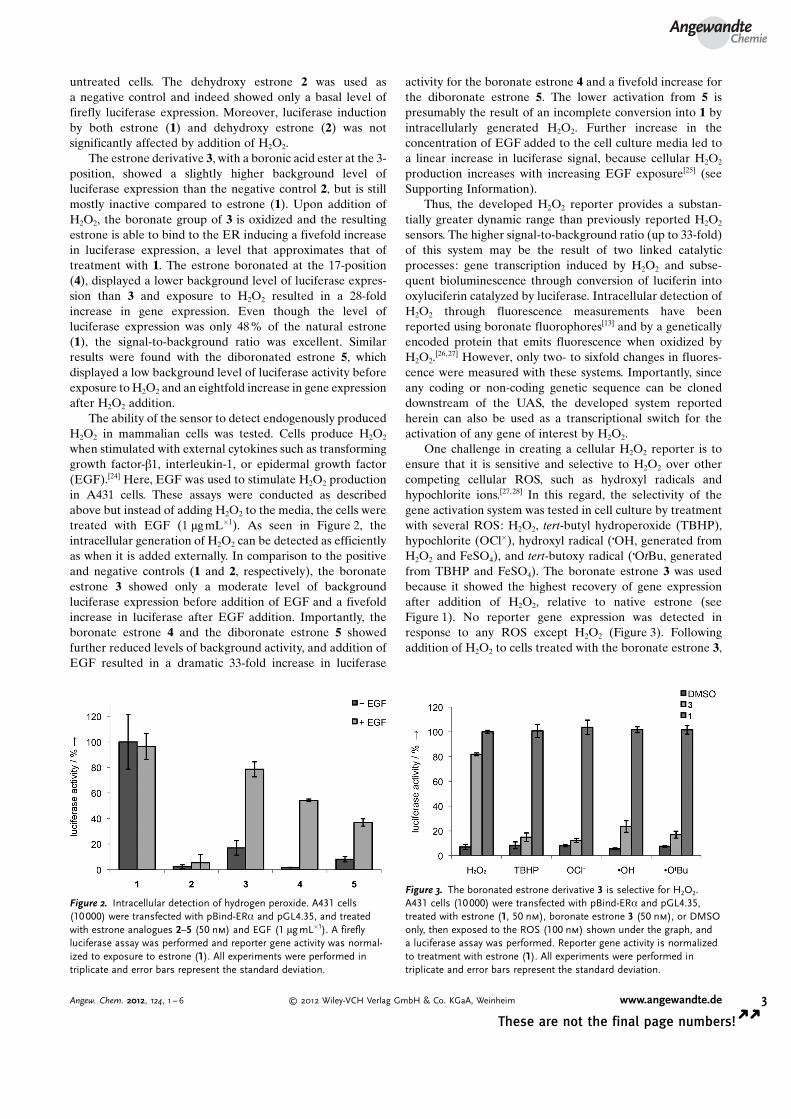
untreated cells. The dehydroxy estrone 2 was used asa negative control and indeed showed only a basal level offirefly luciferase expression. Moreover, luciferase inductionby both estrone (1) and dehydroxy estrone (2) was notsignificantly affected by addition of H2O2.
The estrone derivative 3, with a boronic acid ester at the 3-position, showed a slightly higher background level ofluciferase expression than the negative control 2, but is stillmostly inactive compared to estrone (1). Upon addition ofH2O2, the boronate group of 3 is oxidized and the resultingestrone is able to bind to the ER inducing a fivefold increasein luciferase expression, a level that approximates that oftreatment with 1. The estrone boronated at the 17-position(4), displayed a lower background level of luciferase expres-sion than 3 and exposure to H2O2 resulted in a 28-foldincrease in gene expression. Even though the level ofluciferase expression was only 48 % of the natural estrone(1), the signal-to-background ratio was excellent. Similarresults were found with the diboronated estrone 5, whichdisplayed a low background level of luciferase activity beforeexposure to H2O2 and an eightfold increase in gene expressionafter H2O2 addition.
The ability of the sensor to detect endogenously producedH2O2 in mammalian cells was tested. Cells produce H2O2
when stimulated with external cytokines such as transforminggrowth factor-b1, interleukin-1, or epidermal growth factor(EGF).[24] Here, EGF was used to stimulate H2O2 productionin A431 cells. These assays were conducted as describedabove but instead of adding H2O2 to the media, the cells weretreated with EGF (1 mgmL�1). As seen in Figure 2, theintracellular generation of H2O2 can be detected as efficientlyas when it is added externally. In comparison to the positiveand negative controls (1 and 2, respectively), the boronateestrone 3 showed only a moderate level of backgroundluciferase expression before addition of EGF and a fivefoldincrease in luciferase after EGF addition. Importantly, theboronate estrone 4 and the diboronate estrone 5 showedfurther reduced levels of background activity, and addition ofEGF resulted in a dramatic 33-fold increase in luciferase
activity for the boronate estrone 4 and a fivefold increase forthe diboronate estrone 5. The lower activation from 5 ispresumably the result of an incomplete conversion into 1 byintracellularly generated H2O2. Further increase in theconcentration of EGF added to the cell culture media led toa linear increase in luciferase signal, because cellular H2O2
production increases with increasing EGF exposure[25] (seeSupporting Information).
Thus, the developed H2O2 reporter provides a substan-tially greater dynamic range than previously reported H2O2
sensors. The higher signal-to-background ratio (up to 33-fold)of this system may be the result of two linked catalyticprocesses: gene transcription induced by H2O2 and subse-quent bioluminescence through conversion of luciferin intooxyluciferin catalyzed by luciferase. Intracellular detection ofH2O2 through fluorescence measurements have beenreported using boronate fluorophores[13] and by a geneticallyencoded protein that emits fluorescence when oxidized byH2O2.
[26,27] However, only two- to sixfold changes in fluores-cence were measured with these systems. Importantly, sinceany coding or non-coding genetic sequence can be cloneddownstream of the UAS, the developed system reportedherein can also be used as a transcriptional switch for theactivation of any gene of interest by H2O2.
One challenge in creating a cellular H2O2 reporter is toensure that it is sensitive and selective to H2O2 over othercompeting cellular ROS, such as hydroxyl radicals andhypochlorite ions.[27,28] In this regard, the selectivity of thegene activation system was tested in cell culture by treatmentwith several ROS: H2O2, tert-butyl hydroperoxide (TBHP),hypochlorite (OCl�), hydroxyl radical (COH, generated fromH2O2 and FeSO4), and tert-butoxy radical (COtBu, generatedfrom TBHP and FeSO4). The boronate estrone 3 was usedbecause it showed the highest recovery of gene expressionafter addition of H2O2, relative to native estrone (seeFigure 1). No reporter gene expression was detected inresponse to any ROS except H2O2 (Figure 3). Followingaddition of H2O2 to cells treated with the boronate estrone 3,
Figure 2. Intracellular detection of hydrogen peroxide. A431 cells(10000) were transfected with pBind-ERa and pGL4.35, and treatedwith estrone analogues 2–5 (50 nm) and EGF (1 mg mL�1). A fireflyluciferase assay was performed and reporter gene activity was normal-ized to exposure to estrone (1). All experiments were performed intriplicate and error bars represent the standard deviation.
Figure 3. The boronated estrone derivative 3 is selective for H2O2.A431 cells (10000) were transfected with pBind-ERa and pGL4.35,treated with estrone (1, 50 nm), boronate estrone 3 (50 nm), or DMSOonly, then exposed to the ROS (100 nm) shown under the graph, anda luciferase assay was performed. Reporter gene activity is normalizedto treatment with estrone (1). All experiments were performed intriplicate and error bars represent the standard deviation.
AngewandteChemie
3Angew. Chem. 2012, 124, 1 – 6 � 2012 Wiley-VCH Verlag GmbH & Co. KGaA, Weinheim www.angewandte.de
These are not the final page numbers! � �
a luciferase response almost identical to that of native-estrone-induced levels was detected. However, exposure toTBHP, OCl� , COH, or COtBu, instead of H2O2, only resulted inbackground levels of gene expression. To confirm that theseresults are the consequence of a highly selective oxidation ofthe boronate estrone 3, in vitro oxidation reactions wereanalyzed by GC (see Supporting Information, Figure S5).Furthermore, a modified cell-based assay was performedwhere the estrone and boronate estrone 3 were incubatedwith the ROS reagents prior to addition to the cells. In thisassay, if the ROS oxidizes 3, it should do so before beingintroduced into the cell, regardless of its lifetime. Confirmingour previous results, selective activation of luciferase activitywas detected exclusively in the presence of H2O2 and no otherROS reagent (see Supporting Information, Figure S6).Together, these results indicate that the estrone derivative 3,in conjunction with a genetically encoded reporter, is highlyspecific for H2O2 and can differentiate it from other ROS withan exceptionally high signal-to-background ratio.
In summary, we have developed a genetically encodedgene activation system that selectively responds to H2O2. Thismethod can be used for the activation of any gene of interest.A central component of this system is a novel boronateestrone “cofactor” that is cell permeable but inactive untiloxidized by H2O2. The oxidation step converts the inactiveboronate estrone into estrone, which induces transcriptionalactivation of the gene of interest, for example, a luciferasereporter gene. The sensor was able to detect H2O2 that waseither added to the cellular medium or generated endoge-nously through growth factor-induced cellular H2O2 produc-tion. Importantly, the system is highly specific for H2O2 and isnot activated by any other reactive oxygen species. In contrastto previously reported intracellular H2O2 sensors, this systemdisplays a substantially larger dynamic range of output signal.Moreover, it is conceivable that this system could be adaptedto other orthogonal, ligand-induced transcription factors toactivate genes of interest in response to an H2O2 stimulus. Forexample, in addition to transcriptional activators, fusionproteins of the ER have been used in the conditional controlof Cre recombinase,[29] the I-Sec1 restriction enzyme,[30] Flperecombinase,[10] and interferon regulatory factor-3.[31] Thus,these proteins and others could also be regulated by intra-cellular H2O2 levels using boronate estrone derivatives.
Experimental SectionEstrone-induced gene expression in mammalian cells: A431 humanepithelial carcinoma cells were grown at 37 8C and 5% CO2 inDulbecco�s modified Eagle�s medium (Hyclone), supplemented with10% fetal bovine serum (Hyclone) and 10% streptomycin/penicillin(MP Biomedicals). Cells were passaged into a 96-well plate (200 mLper well, 10 000 cells per well) and transfected with pBind-ERa
(0.15 mg, Promega) and pGL4.35 (0.15 mg, Promega) using Lipofect-amine (Invitrogen) according to the manufacturer�s protocol. Alltransfections were performed in triplicate. After a 16 h incubation,the medium was replaced with DMEM growth media containing theestrone derivatives. The cells were then treated with H2O2 (100 nm) orEGF (1 mgmL�1) and incubated for 48 h at 37 8C and 5% CO2.Luciferase expression was determined with a Bright Glo-LuciferaseReporter Assay system (Promega) using a Biotek Synergy 4 micro-
plate reader. For each of the triplicates, the data were averaged andstandard deviations were calculated.
Received: April 26, 2012Revised: June 28, 2012Published online: && &&, &&&&
.Keywords: biosensors · estrogen receptor · estrone ·gene expression · hydrogen peroxide
[1] D. Banerjee, U. K. Madhusoodanan, S. Nayak, J. Jacob, Clin.Chim. Acta 2003, 334, 205 – 209.
[2] a) M. E. Rice, Neuroscientist 2011, 17, 389 – 406; b) E. Veal, A.Day, Antioxid. Redox Signaling 2011, 15, 147 – 151.
[3] L. Holmquist, G. Stuchbury, M. Steele, G. M�nch, J. NeuralTransm. Suppl. 2007, 72, 39 – 41.
[4] M. H. Nasr-Esfahani, J. R. Aitken, M. H. Johnson, Development1990, 109, 501 – 507.
[5] G. Nindl, N. R. Peterson, E. F. Hughes, L. R. Waite, M. T.Johnson, Biomed. Sci. Instrum. 2004, 40, 123 – 128.
[6] M. L�pez-L�zaro, Cancer Lett. 2007, 252, 1 – 8.[7] J. Lee, S. Giordano, J. Zhang, Biochem. J. 2012, 441, 523 – 540.[8] M. Nichols, J. M. Rientjes, C. Logie, A. F. Stewart, Mol.
Endocrinol. 1997, 11, 950 – 961.[9] W. B. Pratt, M. J. Welsh, Semin. Cell Biol. 1994, 5, 83 – 93.
[10] N. L. Hunter, R. B. Awatramani, F. W. Farley, S. M. Dymecki,Genesis 2005, 41, 99 – 109.
[11] a) H. Kuivila, R. Wiles, J. Am. Chem. Soc. 1955, 77, 4830 – 4834;b) H. Kuivila, A. Armour, J. Am. Chem. Soc. 1957, 79, 5659 –5662; c) J. L. Major Jourden, S. Cohen, Angew. Chem. 2010, 122,6947 – 6949; Angew. Chem. Int. Ed. 2010, 49, 6795 – 6797.
[12] S. G. Rhee, T. S. Chang, W. Jeong, D. Kang, Mol. Cells 2010, 29,539 – 549.
[13] B. C. Dickinson, C. Huynh, C. J. Chang, J. Am. Chem. Soc. 2010,132, 5906 – 5915.
[14] B. T. Zhu, G. Z. Han, J. Y. Shim, Y. Wen, X. R. Jiang, Endo-crinology 2006, 147, 4132 – 4150.
[15] H. Fang, W. Tong, L. M. Shi, R. Blair, R. Perkins, W. Branham,B. S. Hass, Q. Xie, S. L. Dial, C. L. Moland, D. M. Sheehan,Chem. Res. Toxicol. 2001, 14, 280 – 294.
[16] a) W. Tong, R. Perkins, R. Strelitz, E. R. Collantes, S. Keenan,W. J. Welsh, W. S. Branham, D. M. Sheehan, Environ. HealthPerspect. 1997, 105, 1116 – 1124; b) V. C. Jordan, S. Mittal, B.Gosden, R. Koch, M. E. Lieberman, Environ. Health Perspect.1985, 61, 97 – 110.
[17] M. Gangloff, M. Ruff, S. Eiler, S. Duclaud, J. M. Wurtz, D.Moras, J. Biol. Chem. 2001, 276, 15059 – 15065.
[18] D. M. Tanenbaum, Y. Wang, S. P. Williams, P. B. Sigler, Proc.Natl. Acad. Sci. USA 1998, 95, 5998 – 6003.
[19] T. Furuya, A. E. Strom, T. Ritter, J. Am. Chem. Soc. 2009, 131,1662 – 1663.
[20] S. Cacchi, P. Ciattini, E. Morera, G. Ortar, Tetrahedron Lett.1986, 27, 5541 – 5544.
[21] V. Ahmed, Y. Liu, C. Silvestro, S. D. Taylor, Bioorg. Med. Chem.2006, 14, 8564 – 8573.
[22] F. Jourdan, M. Leese, W. Dohle, E. Ferrandis, S. Newman, S.Chander, A. Purohit, B. Potter, J. Med. Chem. 2011, 54, 4863 –4879.
[23] D. A. Holt, M. A. Levy, D. L. Ladd, H. J. Oh, J. M. Erb, J. I.Heaslip, M. Brandt, B. W. Metcalf, J. Med. Chem. 1990, 33, 937 –942.
[24] Y. S. Bae, S. W. Kang, M. S. Seo, I. C. Baines, E. Tekle, P. B.Chock, S. G. Rhee, J. Biol. Chem. 1997, 272, 217 – 221.
[25] J. L. Macdonald-Obermann, D. Piwnica-Worms, L. J. Pike, Proc.Natl. Acad. Sci. USA 2012, 109, 137 – 142.
.AngewandteZuschriften
4 www.angewandte.de � 2012 Wiley-VCH Verlag GmbH & Co. KGaA, Weinheim Angew. Chem. 2012, 124, 1 – 6� �
These are not the final page numbers!
[26] V. V. Belousov, A. F. Fradkov, K. A. Lukyanov, D. B. Staroverov,K. S. Shakhbazov, A. V. Terskikh, S. Lukyanov, Nat. Methods2006, 3, 281 – 286.
[27] K. N. Markvicheva, D. S. Bilan, N. M. Mishina, A. Y. Gorokho-vatsky, L. M. Vinokurov, S. Lukyanov, V. V. Belousov, Bioorg.Med. Chem. 2011, 19, 1079 – 1084.
[28] M. C. Chang, A. Pralle, E. Y. Isacoff, C. J. Chang, J. Am. Chem.Soc. 2004, 126, 15392 – 15393.
[29] R. Feil, J. Brocard, B. Mascrez, M. LeMeur, D. Metzger, P.Chambon, Proc. Natl. Acad. Sci. USA 1996, 93, 10887 – 10890.
[30] A. Hartlerode, S. Odate, I. Shim, J. Brown, R. Scully, PLoS One2011, 6, e16501.
[31] L. Yao, X. Yan, H. Dong, D. R. Nelson, C. Liu, X. Li, Virol. J.2011, 8, 445.
AngewandteChemie
5Angew. Chem. 2012, 124, 1 – 6 � 2012 Wiley-VCH Verlag GmbH & Co. KGaA, Weinheim www.angewandte.de
These are not the final page numbers! � �
Zuschriften
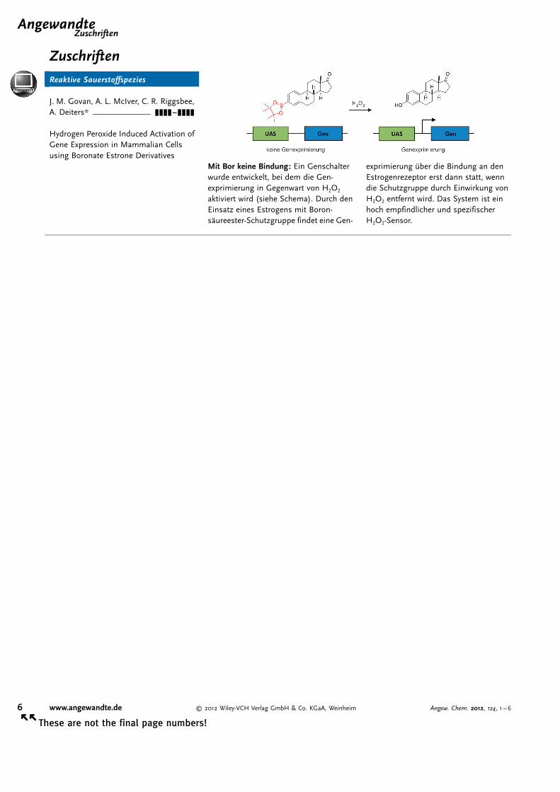
Reaktive Sauerstoffspezies
J. M. Govan, A. L. McIver, C. R. Riggsbee,A. Deiters* &&&&—&&&&
Hydrogen Peroxide Induced Activation ofGene Expression in Mammalian Cellsusing Boronate Estrone Derivatives
Mit Bor keine Bindung: Ein Genschalterwurde entwickelt, bei dem die Gen-exprimierung in Gegenwart von H2O2
aktiviert wird (siehe Schema). Durch denEinsatz eines Estrogens mit Boron-s�ureester-Schutzgruppe findet eine Gen-
exprimierung �ber die Bindung an denEstrogenrezeptor erst dann statt, wenndie Schutzgruppe durch Einwirkung vonH2O2 entfernt wird. Das System ist einhoch empfindlicher und spezifischerH2O2-Sensor.
.AngewandteZuschriften
6 www.angewandte.de � 2012 Wiley-VCH Verlag GmbH & Co. KGaA, Weinheim Angew. Chem. 2012, 124, 1 – 6� �
These are not the final page numbers!