Embed Size (px)
Citation preview
Hydrogel microfluidics for the patterningof pluripotent stem cellsS. Cosson & M. P. Lutolf
School of Life Sciences, Institute of Bioengineering and Laboratory of Stem Cell Bioengineering, Ecole Polytechnique Federale deLausanne (EPFL), CH-1015 Lausanne, Switzerland.
Biomolecular signaling is of utmost importance in governing many biological processes such as thepatterning of the developing embryo where biomolecules regulate key cell-fate decisions. In vivo, thesefactors are presented in a spatiotemporally tightly controlled fashion. Although state-of-the-art microfluidictechnologies allow precise biomolecule delivery in time and space, long-term (stem) cell culture at themicro-scale is often far from ideal due to medium evaporation, limited space for cell growth or shear stress.To overcome these challenges, we here introduce a concept based on hydrogel microfluidics for decouplingconventional, macro-scale cell culture from precise biomolecule delivery through a gel layer. Wedemonstrate the spatiotemporally controlled neuronal commitment of mouse embryonic stem cells viadelivery of retinoic acid gradients. This technique should be useful for testing the effect of dose and timing ofbiomolecules, singly or in combination, on stem cell fate.
Spatiotemporally variable signaling cues control cellular and multicellular behavior in many crucial bio-logical systems. A case in point is the concentration- and time-dependent display of biomolecules termedmorphogens that locally control key cell-fate decisions such as lineage commitment during the patterning
of the early embryo1. Insights gained from developmental biology studies have also spurred advances in differ-entiating pluripotent stem cell such as embryonic stem cells (ESC) into various specialized cell types2. However,typical ESC differentiation protocols, relying on the formation of cell aggregates termed embryoid bodies (EB),expose cells to bulk culture conditions that crudely recapitulate the intricate biomolecule display found duringembryogenesis. Although this method allows at least to some extent the temporal recapitulation of embryonicgene expression patterns in vitro, developing EBs lack an embryo-like spatial organization3. The delivery ofgraded biomolecules by traditional methods such as micropipettes or the Boyden chamber is rather limited inmimicking natural biomolecule presentation.
The emergence of microfluidic technology has revolutionized the generation of in vitro model systems byaffording very precise, picoliter-scale fluid handling, parallelization of experiments and minimization of reagentconsumption4,5. Microfluidic chips also offer unprecedented means to systematically probe the role of micro-environmental signals on stem cell fate in high-throughput6 and with exquisite spatiotemporal resolution7,8.Application of such microsystems have for example allowed to polarize single EBs9 or direct neural progenitordifferentiation by soluble gradients10.
However, state-of-the-art microfluidic culture systems are often not ideal for long-term (stem) cell culture11,12.Firstly, since cells are continuously exposed to fluid flow in most microsystems, the constant removal of autocrinesignals and shear stress may be problematic13, even though more complex shear-free systems have beenreported14–19. Secondly, microsystems are typically made of poly(dimethylsiloxane) (PDMS), a material that,despite its many advantages for microfabrication and its excellent gas permeability20, suffers from susceptibility toliquid evaporation, protein adsorption from the medium21, leaching of non-reacted compounds and hydrophobicrecovery22,23. Thirdly, it is not trivial to functionalize PDMS surfaces with biomolecules in order, for example, tomimic some of the natural interactions of stem cells with their microenvironment that are often critical inmaintaining stem cell function24. To overcome some of these issues, researchers have started to incorporatehydrogels into PDMS microfluidic chips20, for example to generate gradients within 3D scaffolds25 or shield cellsfrom flow and shear stresses26,27. In some cases, PDMS has been entirely replaced by hydrogels to generate‘biomicrofluidic’ networks3,20,28 or scaffolds for tissue engineering21,29,30. However, despite these advances, keychallenges related to micro-scale cell culture remain: The limited space available for cell growth and tissuedevelopment or the difficulty to perform routine cell culture manipulation such as passaging cells or removecells from chips for downstream analyses have not yet been overcome31.
OPEN
SUBJECT AREAS:STEM-CELL
BIOTECHNOLOGY
BIOINSPIRED MATERIALS
Received28 January 2014
Accepted10 March 2014
Published25 March 2014
Correspondence andrequests for materials
should be addressed toM.P.L. (matthias.
SCIENTIFIC REPORTS | 4 : 4462 | DOI: 10.1038/srep04462 1
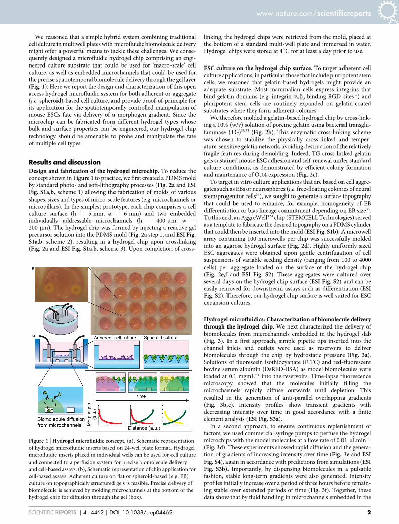
We reasoned that a simple hybrid system combining traditionalcell culture in multiwell plates with microfluidic biomolecule deliverymight offer a powerful means to tackle these challenges. We conse-quently designed a microfluidic hydrogel chip comprising an engi-neered culture substrate that could be used for ‘macro-scale’ cellculture, as well as embedded microchannels that could be used forthe precise spatiotemporal biomolecule delivery through the gel layer(Fig. 1). Here we report the design and characterization of this openaccess hydrogel microfluidic system for both adherent or aggregate(i.e. spheroid)-based cell culture, and provide proof-of-principle forits application for the spatiotemporally controlled manipulation ofmouse ESCs fate via delivery of a morphogen gradient. Since themicrochip can be fabricated from different hydrogel types whosebulk and surface properties can be engineered, our hydrogel chiptechnology should be amenable to probe and manipulate the fateof multiple cell types.
Results and discussionDesign and fabrication of the hydrogel microchip. To reduce theconcept shown in Figure 1 to practice, we first created a PDMS moldby standard photo- and soft-lithography processes (Fig. 2a and ESIFig. S1a,b, scheme 1) allowing the fabrication of molds of variousshapes, sizes and types of micro-scale features (e.g. microchannels ormicropillars). In the simplest prototype, each chip comprises a cellculture surface (h 5 5 mm, ø 5 6 mm) and two embeddedindividually addressable microchannels (h 5 400 mm, w 5
200 mm). The hydrogel chip was formed by injecting a reactive gelprecursor solution into the PDMS mold (Fig. 2a step 1, and ESI Fig.S1a,b, scheme 2), resulting in a hydrogel chip upon crosslinking(Fig. 2a and ESI Fig. S1a,b, scheme 3). Upon completion of cross-
linking, the hydrogel chips were retrieved from the mold, placed atthe bottom of a standard multi-well plate and immersed in water.Hydrogel chips were stored at 4uC for at least a day prior to use.
ESC culture on the hydrogel chip surface. To target adherent cellculture applications, in particular those that include pluripotent stemcells, we reasoned that gelatin-based hydrogels might provide anadequate substrate. Most mammalian cells express integrins thatbind gelatin domains (e.g. integrin avb3 binding RGD sites32) andpluripotent stem cells are routinely expanded on gelatin-coatedsubstrates where they form adherent colonies.
We therefore molded a gelatin-based hydrogel chip by cross-link-ing a 10% (w/v) solution of porcine gelatin using bacterial transglu-taminase (TG)28,33 (Fig. 2b). This enzymatic cross-linking schemewas chosen to stabilize the physically cross-linked and temper-ature-sensitive gelatin network, avoiding destruction of the relativelyfragile features during demolding. Indeed, TG-cross-linked gelatingels sustained mouse ESC adhesion and self-renewal under standardculture conditions, as demonstrated by efficient colony formationand maintenance of Oct4 expression (Fig. 2c).
To target in vitro culture applications that are based on cell aggre-gates such as EBs or neurospheres (i.e. free-floating colonies of neuralstem/progenitor cells34), we sought to generate a surface topographythat could be used to enhance, for example, homogeneity of EBdifferentiation or bias lineage commitment depending on EB size35.To this end, an AggreWellTM chip (STEMCELL Technologies) servedas a template to fabricate the desired topography on a PDMS cylinderthat could then be inserted into the mold (ESI Fig. S1b). A microwellarray containing 100 microwells per chip was successfully moldedinto an agarose hydrogel surface (Fig. 2d). Highly uniformly sizedESC aggregates were obtained upon gentle centrifugation of cellsuspensions of variable seeding density (ranging from 100 to 4000cells) per aggregate loaded on the surface of the hydrogel chip(Fig. 2e,f and ESI Fig. S2). These aggregates were cultured overseveral days on the hydrogel chip surface (ESI Fig. S2) and can beeasily removed for downstream assays such as differentiation (ESIFig. S2). Therefore, our hydrogel chip surface is well suited for ESCexpansion cultures.
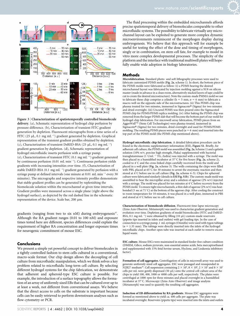
Hydrogel microfluidics: Characterization of biomolecule deliverythrough the hydrogel chip. We next characterized the delivery ofbiomolecules from microchannels embedded in the hydrogel slab(Fig. 3). In a first approach, simple pipette tips inserted into thechannel inlets and outlets were used as reservoirs to deliverbiomolecules through the chip by hydrostatic pressure (Fig. 3a).Solutions of fluorescein isothiocyanate (FITC) and red-fluorescentbovine serum albumin (DsRED-BSA) as model biomolecules wereloaded at 0.1 mgmL21 into the reservoirs. Time-lapse fluorescencemicroscopy showed that the molecules initially filling themicrochannels rapidly diffuse outwards until depletion. Thisresulted in the generation of anti-parallel overlapping gradients(Fig. 3b,c). Intensity profiles show transient gradients withdecreasing intensity over time in good accordance with a finiteelement analysis (ESI Fig. S3a).
In a second approach, to ensure continuous replenishment offactors, we used commercial syringe pumps to perfuse the hydrogelmicrochips with the model molecules at a flow rate of 0.01 mLmin21
(Fig. 3d). These experiments showed rapid diffusion and the genera-tion of gradients of increasing intensity over time (Fig. 3e and ESIFig. S4), again in accordance with predictions from simulations (ESIFig. S3b). Importantly, by dispensing biomolecules in a pulsatilefashion, stable long-term gradients were also generated. Intensityprofiles initially increase over a period of three hours before remain-ing stable over extended periods of time (Fig. 3f). Together, thesedata show that by fluid handling in microchannels embedded in the
Figure 1 | Hydrogel microfluidic concept. (a), Schematic representation
of hydrogel microfluidic inserts based on 24-well plate format. Hydrogel
microfluidic inserts placed in individual wells can be used for cell culture
and connected to a perfusion system for precise biomolecule delivery
and cell-based assays. (b), Schematic representation of chip application for
cell-based assays. Adherent culture on flat or spheroid-based (e.g. EB)
culture on topographically structured gels is feasible. Precise delivery of
biomolecule is achieved by molding microchannels at the bottom of the
hydrogel chip for diffusion through the gel (box).
www.nature.com/scientificreports
SCIENTIFIC REPORTS | 4 : 4462 | DOI: 10.1038/srep04462 2
hydrogel chip, biomolecules can be delivered to build up transient orstable gradients.
Spatiotemporally controlled neural differentiation of ESCs viagraded retinoic acid delivery. To validate this versatile cell cultureplatform, we tested the spatiotemporally controlled induction ofneurogenic differentiation of ESC under serum-free cultureconditions (N2B27 medium36) by delivery of a gradient of themorphogen retinoic acid (RA) (ESI Fig. S5). To probe induction ofneurogenic differentiation in situ, we used a Sox1-GFP reporter ESCline36. Uniform EBs composed of circa 400 ESCs were exposed to RAgradients for two, six, eight or twelve hours and kept in culture for anadditional three days (Fig. 4). Bright-field and fluorescent imageswere acquired in mosaıque mode at day four of differentiation. Imageanalysis was performed to quantify Sox1 expression in individual EBson the entire microwell array. Stitched images show a strikingposition- and thus concentration-dependent effect of RA on EB
size and Sox1-expression with EBs close to the RA source showingstrongest neural induction (Fig. 4a–d and ESI Fig. S6). EBs close tothe RA source are significantly larger (216 6 41 mm) than far awayfrom the source (124 6 42 mm) (ESI Fig. S7). The difference in size islikely due to differential cell proliferation that precedes commitment.The GFP expression profiles along the RA gradient was found totransition from a relative flat line for short exposure times (twoand six hours, Fig. 4e,f), whereas the profile became increasinglysteep for longer exposures (eight and twelve hours, Fig. 4g,h).Statistical analysis revealed a significant dose dependency of GFPexpression for RA gradients delivery for six hours or longer.
These data suggest that the temporal modulation of RA deliveryinfluences neurogenic induction in our in vitro model. It is wellknown that morphogen signaling in vivo is highly complex, not onlyspatially but also temporally1. For example, recent in vivo visualiza-tion of RA in zebrafish embryos via genetically encoded probes forRA allowed for the first time to detect short lived (, six hours) RA
Figure 2 | Fabrication of hydrogel chips and application to ESC culture. (a), Fabrication of hydrogel microfluidic inserts. 1. Mold assembly and hydrogel
precursor injection. 2. Gelation, mold disassembly and hydrogel microfluidic insert recovery. 3. Transfer into multiwell plate. A photograph of a chip is
shown. 4. Immersion in culture media, cell seeding and connection to perfusion system for cell-based assays. Photograph of hydrogel chips with
embedded microchannel visualized by food dye. (b), Schematic representation of enzymatically cross-linked gelatin hydrogel system. (c), Adherent
mouse ESC culture on hydrogel chip surface. Colony morphology and expression of Oct4 (GFP) suggests maintenance of mouse ESC pluripotency. (d),
Micrographs of topographically structured hydrogel chips for spheroid-based cultures. Scale bar, 400 mm (e), Micrographs of cell aggregates formed by
seeding and centrifuging 100, 400, 1000 or 4000 cells per microwell and subsequent overnight culture. Scale bar, 100 mm (f), Graphical representation of
size distribution of cell aggregate obtained by seeding 100, 400, 1000 and 4000 cells/microwell. Uniform aggregates are obtained with the AggreWellTM-
based patterned gel surface.
www.nature.com/scientificreports
SCIENTIFIC REPORTS | 4 : 4462 | DOI: 10.1038/srep04462 3
gradients (ranging from two to six nM) during embryogenesis37.Although the RA gradient ranges (0.01 to 100 nM) and exposuretimes tested here are different, species differences could explain therequirement of higher RA concentration and longer exposure timesfor neurogenic commitment of mouse ESC.
ConclusionsWe present a simple yet powerful concept to deliver biomolecules ina tightly controlled fashion to stem cells cultured in a conventional,macro-scale format. Our chip design allows the decoupling of cellculture from microfluidic manipulation, which we think solves a keyproblem related to microfluidic long-term cell culture. By selectingdifferent hydrogel systems for the chip fabrication, we demonstratethat adherent and spheroid-type ESC culture is possible. Forexample, the introduction of a surface topography allows the forma-tion of an array of uniformly sized EBs that can be cultured over up toat least a week, not different from conventional assays. We believethat the direct access to cells on the substrate is important becausecells can be easily retrieved to perform downstream analyses such asflow cytometry or PCR.
The fluid processing within the embedded microchannels affordsprecise spatiotemporal delivery of biomolecules comparable to othermicrofluidic systems. The possibility to fabricate virtually any micro-channel layout can be exploited to generate more complex dynamicmicroenvironments reminiscent of the morphogen display duringembryogenesis. We believe that this approach will for example beuseful for testing the effect of the dose and timing of morphogens,singly or in combination, on stem cell fate, for example to model invitro more complex developmental processes. The simplicity of theplatform and the interface with traditional multiwell plates will hope-fully enable wide adoption in biology laboratories.
MethodsMicrofabrication. Standard photo- and soft lithography processes were used tofabricate customized PDMS molds (Fig. 2a, scheme 1). In short, the bottom piece ofthe PDMS molds were fabricated as follow: (i) a PDMS bearing the desiredmicrochannel layout was fabricated by injection molding against a SU8 on siliconmaster (made in advance in a clean room, alternatively stacked layers of tape could becut to create the desired microstructure). Note the custom-made PMMA mold we useto fabricate these chip comprise a cylinder (h 5 5 mm, ø 5 6 mm) to fabricate amacro-well on the opposite side of the microstructures. (ii) This PDMS chip wasplasma treated for two minutes, immersed in SigmacoteH (Sigma) for two minutesand dried overnight. (iii) Uncured PDMS was then poured onto the SigmacotedPDMS chip for PDMS/PDMS replica molding. (iv) After baking the PDMS chip wasremoved from the larger PDMS slab that will become the bottom part of our mold forhydrogel chip fabrication. For microwell array fabrication, PDMS pieces from anAggreWellTM (Stem Cell Technologies) were plasma-treated, immersed inSigmacoteH (Sigma) for two minutes, dried overnight and used for PDMS/PDMSmolding. The resulting PDMS pieces were punched (ø 5 6 mm) and inserted into thetop part of the PDMS mold (the PDMS chip mentioned above).
Hydrogel microfluidic chip fabrication. A detailed fabrication procedure can befound in the electronic supplementary information (ESI), Figure S1. Briefly, foradherent cell culture, the PDMS mold was assembled (Fig. 2a, Scheme 1) and a gelatinsolution (10% w/v, porcine high strength, Fluka) supplemented with bacterialtransglutaminase (1 UmL21 TG, Zedira) was injected with a syringe. The mold wasthen placed in a humidified incubator at 37uC for five hours (Fig. 2a, scheme 2),cooled to 4uC and the cross-linked chips carefully recovered from the mold andplaced in a well plate (Fig. 2a, scheme 3). The wells containing the chips were filledwith PBS, placed at 65uC for 30 minutes to arrest the enzymatic reaction and thenstored at 4uC before use in cell culture (Fig. 2a, scheme 4–5). Chips for spheroidculture were fabricated similarly (details in ESI Fig. S1b). The custom-made mold wasassembled to bear the micropillar array and a solution of agarose (2% w/v) injectedwith a syringe. The mold was placed for ten minutes at 4uC before recovery from thePDMS mold. To ensure tight microchannels, a thin slab of agarose (2% w/v) was heat-bonded (3 sec at 71uC) at the bottom of the agarose chip. After cooling the constructto room temperature for 10 minutes, it was placed in a well plate, immersed in PBSand stored at 4uC before use in cell culture.
Characterization of biomolecule diffusion. Fluorescent time-lapse microscopy(Zeiss Axio Observer, Metamorph) was used to characterize gradient generation andevolution over time. Depletion gradients of model biomolecules (FITC and DsRED-BSA, 0.1 mg mL21) were obtained by filling (10 mL) custom-made reservoirs(pipette tips inserted in inlets and outlets) with gel-loading tips. In the case ofconnection to a syringe pump, 1 mL syringes (Omnifix) were used with tygon tubings(ø 5 1.35 mm). The tubings were directly inserted into the inlets of the hydrogelmicrofluidic chips. Another open tube was inserted at each outlet to remove excessliquid waste.
ESC culture. Mouse ESCs were maintained in standard feeder-free culture condition(DMEM, Gibco, sodium pyruvate, non-essential amino acids, beta mercaptoethanoland supplemented with 15% fetal bovine serum, Hyclone, and Leukemia inhibitoryfactor).
Formation of cell aggregates. Centrifugation of cells in microwell array was used togenerate uniformly sized cell aggregates. ESC were passaged and resuspended inN2B27 medium36. Cell suspension containing 2 3 106, 8 3 106, 2 3 107 and 8 3 107
cells per mL were gently dispensed (50 mL) onto the central cell culture area of thechips to yield 100, 400, 1000 or 4000 cells per well, respectively. The plates werecentrifuged at 1000 rpm for three minutes and placed overnight in a humidifiedincubator at 37uC. Microscopy (Zeiss Axio Observer) and image analysis(Metamorph) was used to quantify the resulting cell aggregates.
Induction of EB differentiation by RA gradients. Mouse ESC aggregates wereformed as mentioned above to yield ca. 400 cells per aggregate. The plate wasincubated overnight. Reservoirs (pipette tips) were inserted into the inlets and outlets
Figure 3 | Characterization of spatiotemporally controlled biomoleculedelivery. (a), Schematic representation of hydrogel chip perfusion by
pressure difference. (b), Characterization of transient FITC gradient
generation by depletion. Fluorescent micrographs from a time series of a
FITC (25 mL, 0.1 mg mL21) gradient generated by depletion. Graphical
representation of the transient gradient profiles obtained by depletion.
(c), Characterization of transient DsRED-BSA (25 mL, 0.1 mg mL21)
gradient generation by depletion. (d), Schematic representation of
hydrogel microfluidic inserts perfusion with a syringe pump.
(e), Characterization of transient FITC (0.1 mg mL21) gradient generated
by continuous perfusion (0.01 mL min21). Continuous perfusion yields
gradients with increasing intensities over time. (f), Characterization of
stable DsRED-BSA (0.1 mg mL21) gradient generated by perfusion with a
syringe pump at defined intervals (one minute at 0.01 mL min21 every 30
minutes). The micrographs and respective intensity profiles demonstrate
that stable gradient profiles can be generated by replenishing the
biomolecule solution within the microchannel at given time intervals.
Gradient profiles were measured across a single plane (right above the
hydrogel surface), as depicted by the red dashed line in the schematic
representation of the device. Scale bar, 200 mm.
www.nature.com/scientificreports
SCIENTIFIC REPORTS | 4 : 4462 | DOI: 10.1038/srep04462 4
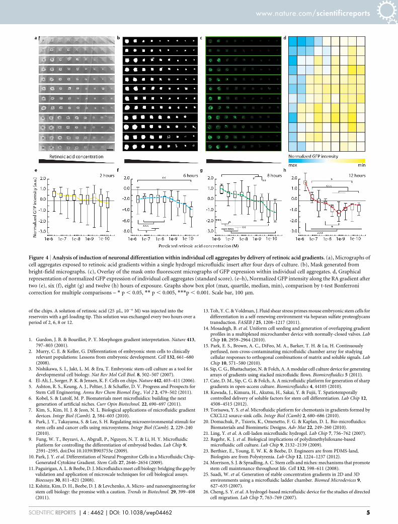
of the chips. A solution of retinoic acid (25 mL, 1026 M) was injected into thereservoirs with a gel-loading tip. This solution was exchanged every two hours over aperiod of 2, 6, 8 or 12.
1. Gurdon, J. B. & Bourillot, P. Y. Morphogen gradient interpretation. Nature 413,797–803 (2001).
2. Murry, C. E. & Keller, G. Differentiation of embryonic stem cells to clinicallyrelevant populations: Lessons from embryonic development. Cell 132, 661–680(2008).
3. Nishikawa, S. I., Jakt, L. M. & Era, T. Embryonic stem-cell culture as a tool fordevelopmental cell biology. Nat Rev Mol Cell Biol. 8, 502–507 (2007).
4. El-Ali, J., Sorger, P. K. & Jensen, K. F. Cells on chips. Nature 442, 403–411 (2006).5. Ashton, R. S., Keung, A. J., Peltier, J. & Schaffer, D. V. Progress and Prospects for
Stem Cell Engineering. Annu Rev Chem Biomol Eng., Vol 2 2, 479–502 (2011).6. Kobel, S. & Lutolf, M. P. Biomaterials meet microfluidics: building the next
generation of artificial niches. Curr Opin Biotechnol. 22, 690–697 (2011).7. Kim, S., Kim, H. J. & Jeon, N. L. Biological applications of microfluidic gradient
devices. Integr Biol (Camb). 2, 584–603 (2010).8. Park, J. Y., Takayama, S. & Lee, S. H. Regulating microenvironmental stimuli for
stem cells and cancer cells using microsystems. Integr Biol (Camb). 2, 229–240(2010).
9. Fung, W. T., Beyzavi, A., Abgrall, P., Nguyen, N. T. & Li, H. Y. Microfluidicplatform for controlling the differentiation of embryoid bodies. Lab Chip 9,2591–2595, doi:Doi 10.1039/B903753e (2009).
10. Park, J. Y. et al. Differentiation of Neural Progenitor Cells in a Microfluidic Chip-Generated Cytokine Gradient. Stem Cells 27, 2646–2654 (2009).
11. Paguirigan, A. L. & Beebe, D. J. Microfluidics meet cell biology: bridging the gap byvalidation and application of microscale techniques for cell biological assays.Bioessays 30, 811–821 (2008).
12. Kshitiz, Kim, D. H., Beebe, D. J. & Levchenko, A. Micro- and nanoengineering forstem cell biology: the promise with a caution. Trends in Biotechnol. 29, 399–408(2011).
13. Toh, Y. C. & Voldman, J. Fluid shear stress primes mouse embryonic stem cells fordifferentiation in a self-renewing environment via heparan sulfate proteoglycanstransduction. FASEB J 25, 1208–1217 (2011).
14. Mosadegh, B. et al. Uniform cell seeding and generation of overlapping gradientprofiles in a multiplexed microchamber device with normally-closed valves. LabChip 10, 2959–2964 (2010).
15. Park, E. S., Brown, A. C., DiFeo, M. A., Barker, T. H. & Lu, H. Continuouslyperfused, non-cross-contaminating microfluidic chamber array for studyingcellular responses to orthogonal combinations of matrix and soluble signals. LabChip 10, 571–580 (2010).
16. Sip, C. G., Bhattacharjee, N. & Folch, A. A modular cell culture device for generatingarrays of gradients using stacked microfluidic flows. Biomicrofluidics 5 (2011).
17. Cate, D. M., Sip, C. G. & Folch, A. A microfluidic platform for generation of sharpgradients in open-access culture. Biomicrofluidics 4, 44105 (2010).
18. Kawada, J., Kimura, H., Akutsu, H., Sakai, Y. & Fujii, T. Spatiotemporallycontrolled delivery of soluble factors for stem cell differentiation. Lab Chip 12,4508–4515 (2012).
19. Torisawa, Y. S. et al. Microfluidic platform for chemotaxis in gradients formed byCXCL12 source-sink cells. Integr Biol (Camb) 2, 680–686 (2010).
20. Domachuk, P., Tsioris, K., Omenetto, F. G. & Kaplan, D. L. Bio-microfluidics:Biomaterials and Biomimetic Designs. Adv Mat 22, 249–260 (2010).
21. Ling, Y. et al. A cell-laden microfluidic hydrogel. Lab Chip 7, 756–762 (2007).22. Regehr, K. J. et al. Biological implications of polydimethylsiloxane-based
microfluidic cell culture. Lab Chip 9, 2132–2139 (2009).23. Berthier, E., Young, E. W. K. & Beebe, D. Engineers are from PDMS-land,
Biologists are from Polystyrenia. Lab Chip 12, 1224–1237 (2012).24. Morrison, S. J. & Spradling, A. C. Stem cells and niches: mechanisms that promote
stem cell maintenance throughout life. Cell 132, 598–611 (2008).25. Saadi, W. et al. Generation of stable concentration gradients in 2D and 3D
environments using a microfluidic ladder chamber. Biomed Microdevices 9,627–635 (2007).
26. Cheng, S. Y. et al. A hydrogel-based microfluidic device for the studies of directedcell migration. Lab Chip 7, 763–769 (2007).
Figure 4 | Analysis of induction of neuronal differentiation within individual cell aggregates by delivery of retinoic acid gradients. (a), Micrographs of
cell aggregates exposed to retinoic acid gradients within a single hydrogel microfluidic insert after four days of culture. (b), Mask generated from
bright-field micrographs. (c), Overlay of the mask onto fluorescent micrographs of GFP expression within individual cell aggregates. d, Graphical
representation of normalized GFP expression of individual cell aggregates (standard score). (e–h), Normalized GFP intensity along the RA gradient after
two (e), six (f), eight (g) and twelve (h) hours of exposure. Graphs show box plot (max, quartile, median, min), comparison by t-test Bonferroni
correction for multiple comparisons – * p , 0.05, ** p , 0.005, ***p , 0.001. Scale bar, 100 mm.
www.nature.com/scientificreports
SCIENTIFIC REPORTS | 4 : 4462 | DOI: 10.1038/srep04462 5
27. Tan, D. C. W., Yung, L. Y. L. & Roy, P. Controlled microscale diffusion gradientsin quiescent extracellular fluid. Biomed Microdevices 12, 523–532 (2010).
28. Paguirigan, A. & Beebe, D. J. Gelatin based microfluidic devices for cell culture.Lab Chip 6, 407–413 (2006).
29. Cabodi, M. et al. A microfluidic biomaterial. JACS 127, 13788–13789 (2005).30. Choi, N. W. et al. Microfluidic scaffolds for tissue engineering. Nat Mater 6,
908–915 (2007).31. Toh, Y. C., Blagovic, K. & Voldman, J. Advancing stem cell research with
microtechnologies: opportunities and challenges. Integr Biol (Camb). 2, 305–325(2010).
32. Davis, G. E. Affinity of Integrins for Damaged Extracellular-Matrix - Alpha-V-Beta-3 Binds to Denatured Collagen Type-I through Rgd Sites. Biochem Bioph ResCo 182, 1025–1031 (1992).
33. Paguirigan, A. L. & Beebe, D. J. Protocol for the fabrication of enzymaticallycrosslinked gelatin microchannels for microfluidic cell culture. Nat Protoc 2,1782–1788 (2007).
34. Reynolds, B. A. & Weiss, S. Generation of Neurons and Astrocytes from IsolatedCells of the Adult Mammalian Central-Nervous-System. Science 255, 1707–1710(1992).
35. Ungrin, M. D., Joshi, C., Nica, A., Bauwens, C. & Zandstra, P. W. Reproducible,Ultra High-Throughput Formation of Multicellular Organization from SingleCell Suspension-Derived Human Embryonic Stem Cell Aggregates. Plos One 3,(2008).
36. Ying, Q. L., Stavridis, M., Griffiths, D., Li, M. & Smith, A. Conversion ofembryonic stem cells into neuroectodermal precursors in adherent monoculture.Nat Biotech 21, 183–186 (2003).
37. Shimozono, S., Iimura, T., Kitaguchi, T., Higashijima, S. & Miyawaki, A.Visualization of an endogenous retinoic acid gradient across embryonicdevelopment. Nature 496, 363 (2013).
AcknowledgmentsWe thank Austin Smith for providing the Sox1-GFP knock-in (46C) reporter mouse ESCline. We thank Yannick Devaud for testing hydrogels for microfluidic fabrication and SoniaHallen for help with optimizing ESC aggregate culture and differentiation. This work wasfunded by the EU FP7 Large-scale integrating project ‘PluriMes’ (http://www.plurimes.eu/)as well as a EURYI award to M.P.L.
Author contributionsS.C. and M.P.L. conceived the study, interpreted results and wrote the manuscript. S.C.performed all experiments.
Additional informationSupplementary information accompanies this paper at http://www.nature.com/scientificreports
Competing financial interests: The authors declare no competing financial interests.
How to cite this article: Cosson, S. & Lutolf, M.P. Hydrogel microfluidics for the patterningof pluripotent stem cells. Sci. Rep. 4, 4462; DOI:10.1038/srep04462 (2014).
This work is licensed under a Creative Commons Attribution-NonCommercial-NoDerivs 3.0 Unported license. To view a copy of this license,
visit http://creativecommons.org/licenses/by-nc-nd/3.0
www.nature.com/scientificreports
SCIENTIFIC REPORTS | 4 : 4462 | DOI: 10.1038/srep04462 6
![Advances in Microfluidics‐Based Assisted Reproductive ... · microfluidics has also been used for 3D cell culture and cryo-preservation.[12] Furthermore, droplet-based microfluidics](https://img.pdfslide.us/doc/110x75/5e831de01be17b7cdc733cfb/advances-in-microfluidicsabased-assisted-reproductive-microfluidics-has-also.jpg)