Embed Size (px)
Citation preview
J. Comp. Path. 1998 Vol. 118, IIi9 173
SHORT PAPER
m
:J C P
H y d r o c e p h a l u s A s s o c i a t e d w i t h Neospora caninum I n f e c t i o n in an A b o r t e d B o v i n e F e t u s
j. P. Dubey, B. Abbitt*, M.J. TopperS andJ. F. Edwards+ US Department of Agriculture, Agricultural Research Service, Livestock and Poultry Sciences
Institute, Parasite Biology and Epidemiology Laboratory, BARC-East, Building 1040, Beltsville, Maryland 20705-2350, * Texas Veterinary Medical Diagnostic Laboratory, P.O. Drawer 3040,
College Station, Texa.~ ~ 77841-3040, ~Division of Patholog), Walter Reed Army Institute of Research, Washington, D.C. 20306 and +Department of Veterinary P.athobiology,
Texas A & M University, College Station, Texas 77843-4467, USA
Summary This paper describes Neospora caninum-associated hydrocephalus in an aborted Hereford bovine fetus of 7 months' gestational age. Numerous tachyzoites were observed in areas of the cerebrum with lesions of non-suppurative necrotizing encephalitis.
Introduction
Neosporosis is a newly recognized disease of animals caused by the protozoan parasite, Neospora caninum, which until 1988 was misidentified as Toxoplasma gondii (Dubey et al., 1988). Neospora caninum causes mortality in dogs, cattle, sheep, goats, horses and deer, and is a major cause of abortion in dairy cattle in many countries (Dubey and Lindsay, 1996). We report here a case of hydrocephalus, associated with neosporosis, in an aborted bovine fetus.
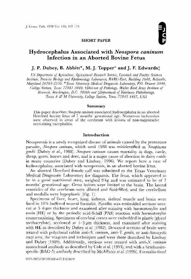
An aborted Hereford female calf was submitted to the Texas Veterinary Medical Diagnostic Laboratory for diagnosis. The fetus, which appeared to be in a good nutritional state, weighed 9 kg and was estimated to be of 7 months' gestational age. Gross lesions were limited to the brain. The lateral ventricles of the cerebrum were dilated and fluid-filled, and the cerebellum and medulla were hypoplastic (Fig. 1).
Specimens of liver, heart, lung, kidneys, skeletal muscle and brain were fixed in 10% buffered neutral formalin. Paraffin wax-embedded sections were cut at 5 6 gm thickness and examined after staining with haematoxylin and eosin (HE) or by the periodic acid-Schiff (PAS) reaction with haematoxylin counterstaining. Specimens of cerebral cortex were embedded in plastic (glycol methacrylate), sectioned at 1-3 tam thickness, and examined after staining with HE as described by Dubey et al. (1982). Dewaxed sections of brain were treated with polyclonal rabbit anti-)(, caninum, anti-T, gondii, or anti-Sarcocystis cruzi sera; the reagents and techniques used were those described by Lindsay and Dubey (1989). Additionally, sections were treated with anti-N, caninum monoclonal antibody as described by Cole et al. (1993), and with a bradyzoite- specific (BAG-5) antibody described by McAllister el al. (1996). Formalin-fixed
00~ 1 9975/98/020169 + 05 $ 12.00/0
1 7 0 J.P. Dubey e t al.
Fig. 1. Transverse hemisections of brain with the cerebellum on the left and the anterior brain to the right. Expansion of the lateral ventricles is shown.
specimens of cerebrum were post-fixed in 1% osmium tetroxide, dehydrated in a rising ethanol gradient, and placed in propylene oxide. The tissues were infiltrated with Epon, and polymerized in capsules with Epon at 70~ for 24h. Tissue blocks were sectioned, placed on grids, post-stained with 2% uranyl acetate and 1% lead citrate, and examined by transmission electron microscopy (TEM) with a Zeiss EM 109.
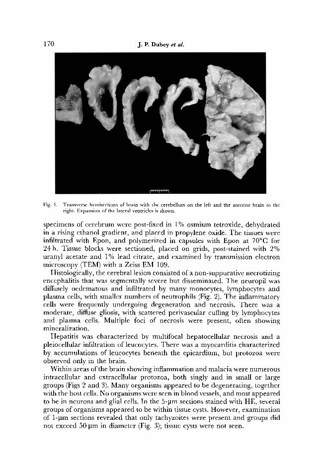
Histologically, the cerebral lesion consisted of a non-suppurative necrotizing encephalitis that was segmentally severe but disseminated. The neuropil was diffusely oedematous and infiltrated by many monocytes, lymphocytes and plasma cells, with smaller numbers of neutrophils (Fig. 2). The inflammatory cells were frequently undergoing degeneration and necrosis. There was a moderate, diffuse gliosis, with scattered perivascular cuffing by lymphocytes and plasma cells. Multiple foci of necrosis were present, often showing mineralization.
Hepatitis was characterized by multifocal hepatocellular necrosis and a pleiocellular infiltration of leucocytes. There was a myocarditis characterized by accumulations of leucocytes beneath the epicardium, but protozoa were observed only in the brain.
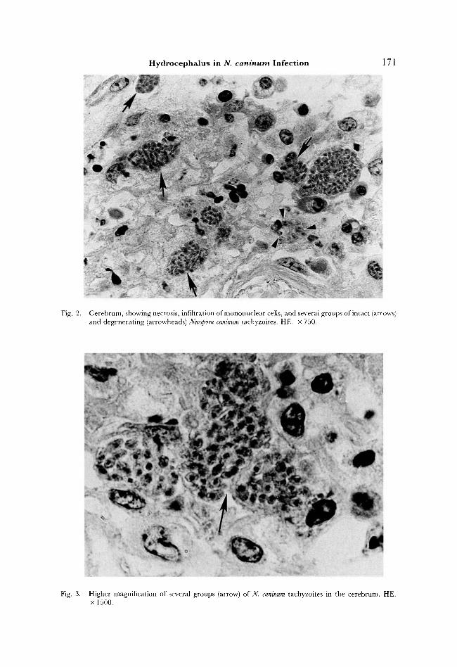
Within areas of the brain showing inflammation and malacia were numerous intracellular and extracellular protozoa, both singly and in small or large groups (Figs 2 and 3). Many organisms appeared to be degenerating, together with the host cells. No organisms were seen in blood vessels, and most appeared to be in neurons and glial cells. In the 5-gm sections stained with HE, several groups of organisms appeared to be within tissue cysts. However, examination of 1-gm sections revealed that only tachyzoites were present and groups did not exceed 50 gm in diameter (Fig. 3); tissue cysts were not seen.
Hydrocephalus in N. caninum Infection 171
Fig. 2. Cerebrum, showing necrosis, infiltration of mononuclear cells, and several groups of intact (arrows) and degenerating (arrowheads) Neospora caninum tachyzoites. HE. x 750.
Fig. 3. Higher magnification of several groups (arrow) of N. caninum tachyzoites in the cerebrum. HE. • 1500.
172 J.P. D u b e y et al.
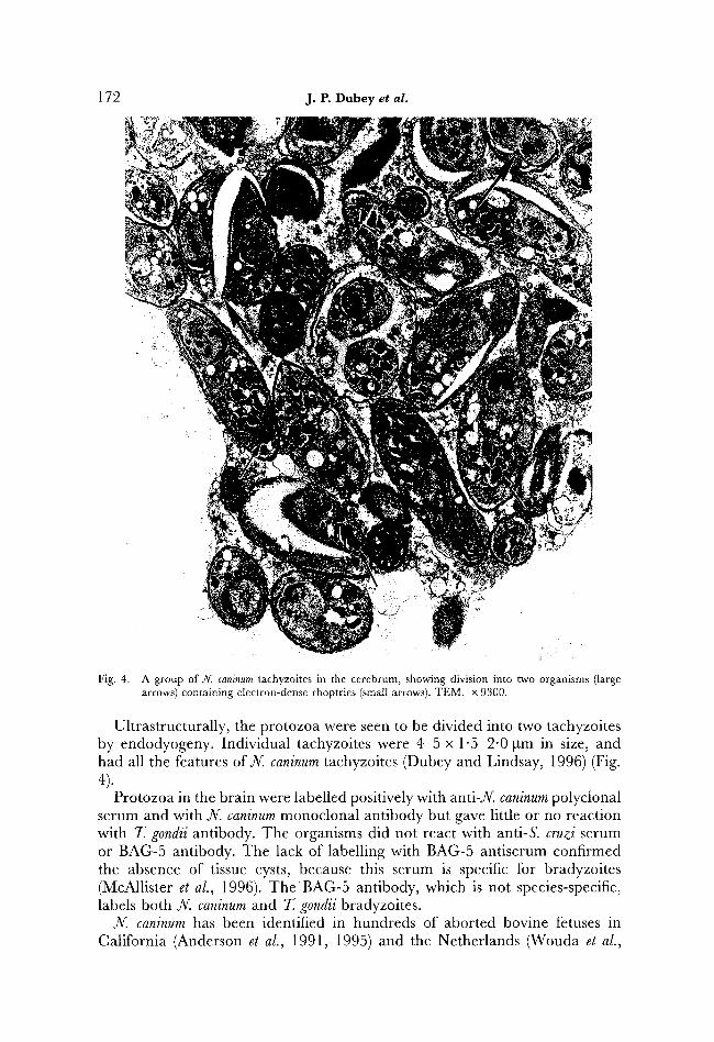
Fig. 4. A group of N. caninum tachyzoites in the cerebrum, showing division into two organisms (large arrows) containing electron-dense rhoptries (small arrows). TEM. x 9300.
Ultrastructurally, the protozoa were seen to be divided into two tachyzoites by endodyogeny. Individual tachyzoites were 4-5 x 1"5-2"0 gm in size, and had all the features of N. caninum tachyzoites (Dubey and Lindsay, 1996) (Fig. 4).
Protozoa in the brain were labelled positively with anti-N, caninum polyclonal serum and with N. caninum monoclonal antibody but gave little or no reaction with T. gondii antibody. The organisms did not react with anti-S, cruzi serum or BAG-5 antibody. The lack of labelling with BAG-5 antiserum confirmed the absence of tissue cysts, because this serum is specific for bradyzoites (McAllister et al., 1996). The BAG-5 antibody, which is not species-specific, labels both N. caninum and T. gondii bradyzoites.
N. caninum has been identified in hundreds of aborted bovine fetuses in California (Anderson et al., 1991, 1995) and the Netherlands (Wouda et al.,
Hydrocephalus in N. c a n i n u m Infection 173
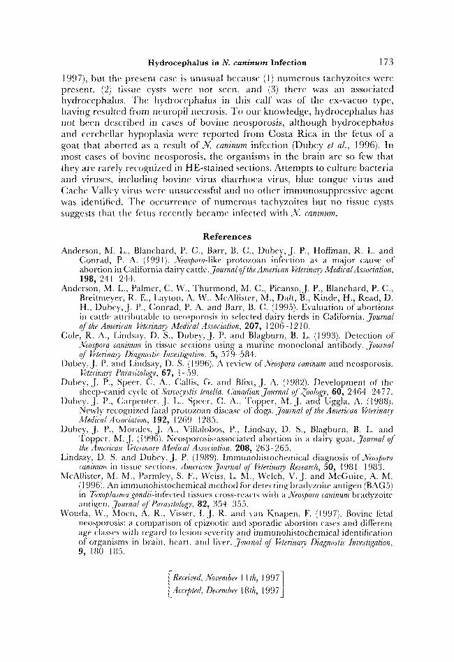
1997), bul the present case is unusual because (1) numerous tachyzoites were present, (2) tissue cysts were not seen, and (3) there was an associated hydrocephalus. The hydrocephalus in this calf" was of the ex-vacuo type, having resuhed from ncuropil necrosis. To our knowledge, hydrocephalus has not been described in cases of bovine neosporosis, although hydrocephalus and cerebellar hypoplasia were reported from Costa Rica in the fetus of a goat that aborted as a result of N. caninum infection (Dubey et al., 1996). In most cases of bovine neosporosis, the organisms in the brain are so few that they are rarely recognized in HE-stained sections. Attempts to culture bacteria and viruses, including bovine virus diarrhoea virus, blue tongue virus and Cache Valley virus were unsuccessful and no other immunosuppressive agent was identified. The occurrence of numerous tachyzoites but no tissue cysts suggests thal the fetus recently became int?eted with N. caninum.
R e f e r e n c e s
Anderson, M. L., Blanchard, P. C., Barr, B. C., Dubey, J. P., Hoflinan, R. L. and Conrad, P. A. (1991)..Xeospora-like protozoan infection as a major cause of abortion in California dairy cattle.journal o/the American Veterinary Medieal Assoeiation, 198, 241 244.
Anderson, M. L., Palmer, C. W., Thurmond, M. C., Picanso, J. P., Blanchard, P. C., Breitmeyer, R. E., l,ayton, A. W., McAllister, M., Daft, B., Kinde, H., Read, D. H., Dubey, J. P., Conrad, P. A. and Barr, B. C. (1995). Evaluation of abortions in cattle attributable 1o neosporosis in selected dairy herds in California. journal of the American l~terinarv Medical Association, 207, 1206 - 12 l 0.
Cole, R. A., Lindsay, D. S., Dubey, J. P. and Blagburn, B. L. (1993). Detection of .Xeospora caninum in tissue sections using a murine monoclonal antibody. Journal ~ Veterinmy Diagnostic Investigation, 5, 579-584.
Dubey, J. P. and Lindsay, D. S. (1996). A review ofNeo~pora caninum and neosporosis. I+terinar>, Parasitology, 67, 1-59.
Dubey, J. P., Speer, C. A., Callis, G. and Blixt, J. A. (1982). Development of lhe sheep-canid cycle of Sarcoc)~stis lenella. Canadian Journal q/Zoolog~, 60, 2464 2477.
Dubey, J. P., Carpenter, .J.L., Speer, C. A., Topper, M.J. and Uggla, A. (1988). Newly recognized tatal protozoan disease of dogs. Journal o.fthe American l~terinar~ A4edica[ A.ssociation, 192, 1269 1285.
Dubey, J. P., Morales, J. A., Villah)bos, P., Lindsay, D. S., Blagburn, B. L. and Topper, M..]. (1996). Neosporosis-associated abortion in a dairy goat. Journa[ oj the American Veterinary Medical Association, 208, 263- 265.
Lindsay, D. S. and Dul~ey,.l.P. (1989). hnmunohistochemical diagnosis ofNeospora caninum in tissue sections. American Journal ~" Veterinary Research, 50, 1981 - 1983.
McA|lister, M. M., Parmley, S. F., Weiss, L. M., Welch, V.J. and McGuire, A. M. (1996). An immunohistochemical method for detecting bradyzoite antigen (BAG5) in Toxoplasma gondii-infected tissues cross-reacts with a .Neospora caninum bradyzoite antigen. Journal qft~arasitologI,, 82, 354 355.
Wouda, W., Moen, A. R., Visser, I . J . R . and van Knapen, F. (1997). Bovine fetal neosporosis: a comparison of epizootic and sporadic abortion cases and different age classes with regard to lesion severity and irnmunohistochemical identitication of organisms in brain, heart, and liver, journal oJ Veterinary Diagnostic Investigation, 9, 180 185.
I Received, November 11 th, 1997] Accepted, December 18th, 1997J