Embed Size (px)
Citation preview
ROCHE MOLECULAR BIOCHEMICALS BIOCHEMICA · No. 1 n 1999
LIG
HT
CY
CLE
R
5CONTENTS
H
I
The most direct way to monitor PCR amplification for quantification and mutation detection.
Brian Erich Caplin1, Randy P. Rasmussen1, Philip S. Bernard2, and Carl T. Wittwer1,2
1 ARUP Institute for Clinical and Experimental Pathology, 500 Chipeta Way, Salt Lake City, UT 84108 2 Department of Pathology, University of Utah School of Medicine, 50 North Medical Drive, Salt Lake City, UT 84132
Introduction
The polymerase chain reaction (PCR) is perhaps themost powerful modern tool available for today'smolecular biologist. Its extraordinary sensitivityallows for the detection of only a few molecules ofDNA. The sensitivity of PCR is often complimented bythe specificity of hybridization techniques such asSouthern blotting or oligonucleotide hybridization.Amplification and hybridization techniques areusually performed separately. However, with therecent development of LightCycler™ technology, PCRamplification and hybridization probe detection occursimultaneously in homogeneous solution. That is,both amplification and hybridization analysis can pro-ceed in the same reaction. Because the LightCyclerInstrument uses rapid cycling techniques (12), theentire process is finished in 15–30 min.
Sequence detection with adjacent oligonucleotidesand fluorescence was suggested as early as 1985(6). However, it was not until 1997 that fluorescenthybridization analysis was demonstrated during PCR(11). All reagents for both amplification and detec-tion are added before temperature cycling is begun.Sequence-specific probe hybridization occursduring amplification, allowing real-time productidentification, quantification, and mutation detection(1, 2, 8, 11, 13, 14). The LightCycler is the only instru-ment currently available for real-time fluorescenthybridization probe analysis. Although other fluores-cent probes and dyes (including SYBR® Green I Dyeand TaqMan® Probes) can be used on the Light-Cycler Instrument (11), this article will focus on theunique characteristics and advantages of fluores-cent hybridization probes.
Hybridization probe basicsThe hybridization probe system consists of two fluo-rescently labeled oligonucleotides. A donor probelabeled with fluorescein at the 3' end absorbs lightfrom the blue LED of the LightCycler Instrument. Anadjacent acceptor probe absorbs resonance energy
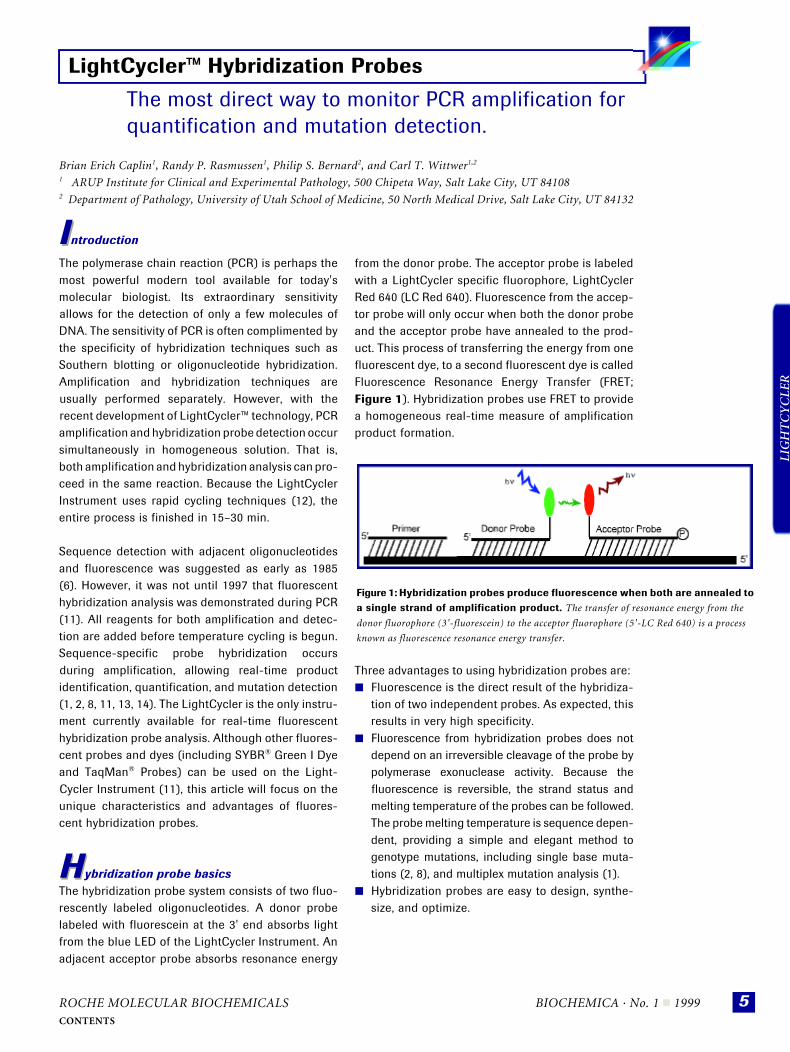
from the donor probe. The acceptor probe is labeledwith a LightCycler specific fluorophore, LightCyclerRed 640 (LC Red 640). Fluorescence from the accep-tor probe will only occur when both the donor probeand the acceptor probe have annealed to the prod-uct. This process of transferring the energy from onefluorescent dye, to a second fluorescent dye is calledFluorescence Resonance Energy Transfer (FRET;Figure 1). Hybridization probes use FRET to providea homogeneous real-time measure of amplificationproduct formation.
Three advantages to using hybridization probes are: ■ Fluorescence is the direct result of the hybridiza-
tion of two independent probes. As expected, thisresults in very high specificity.
■ Fluorescence from hybridization probes does notdepend on an irreversible cleavage of the probe bypolymerase exonuclease activity. Because thefluorescence is reversible, the strand status andmelting temperature of the probes can be followed.The probe melting temperature is sequence depen-dent, providing a simple and elegant method togenotype mutations, including single base muta-tions (2, 8), and multiplex mutation analysis (1).
■ Hybridization probes are easy to design, synthe-size, and optimize.
LightCycler™ Hybridization Probes
Figure 1: Hybridization probes produce fluorescence when both are annealed to a single strand of amplification product. The transfer of resonance energy from the
donor fluorophore (3'-fluorescein) to the acceptor fluorophore (5'-LC Red 640) is a process
known as fluorescence resonance energy transfer.

BIOCHEMICA · No. 1 n 1999 ROCHE MOLECULAR BIOCHEMICALS
LIG
HT
CY
CLE
R
6CONTENTS
H
Q
MHHybridization probe designThe design of hybridization probes is straight-forward. Following a few general principles willensure success:■ Hybridization probes should anneal adjacent to each
other on the same strand of product (Figure 1). ■ The spacing between adjacent hybridization
probes is optimally one base. However, excellentfluorescence transfer occurs with as many as fivebases between the two probes, and some observ-able fluorescence can occur with up to 20 inter-vening bases or more.
■ One probe is best labeled at the 3' end with fluo-rescein, and the other at the 5' end with LC Red640. To prevent extension of the LC Red 640labeled probe, the 3' end must be blocked byphosphorylation.
■ Probe Tm should be approximately 5–10°C higherthan the Tm of the primers. Usually, the probes are23–35 bases in length with a G+C content rangingfrom 38–60%.
■ Probe sequences that cause secondary structuresmust be avoided, as for normal PCR primers.
The optimal Tm difference between the two probeswill depend on the type of experiment that is beingperformed. For detection and quantification, the Tm ofthe hybridization probes should be the same (within2ºC of each other). For mutation detection, the bestmelting curves are obtained when the differencebetween probe Tm is 5–10ºC. The probe with thelowest stability should be positioned directly over themutation to be detected.
Quantification with hybridization probes Real-time or kinetic PCR is a powerful method forestimating the initial template copy number (7, 11,14). Fluorescence is acquired once each cycle andthe fluorescence is plotted against the cycle number.A typical titration experiment on the LightCyclerInstrument with hybridization probes is shown inFigure 2.
In addition to hybridization probes, the doublestranded DNA binding SYBR® Green I dye can alsobe used for analysis of PCR products (11, 14), evenfor quantification of low-copy transcripts (9). TheLight Cycler System is also compatible with dual-labeled TaqMan® Probes that are commonly labeledwith fluorescein (FAM) and rhodamine (TAMRA).
Mutation detection with hybridization probesHybridization probes provide a simple and elegantsystem for real-time detection of mutations, includ-ing single-base mutations (1, 2, 8). Only one reactionand one set of probes are required for genotypingwith the LightCycler System. A melting curve ofhybridization probe fluorescense produces a high-resolution “dynamic dot blot” that can easilydiscriminate even the most stable single basemismatches (2). Unlike a standard dot blot wherehybridization occurs at a single temperature, meltingcurve analysis on the LightCycler Instrument simpli-fies the optimization of probe hybridization withcontinuous monitoring of probe hybridization statusas the temperature changes. A single base mismatchunder the probe decreases the melting temperatureby as little as 3°C for G::T mismatches, to as great as7–10°C for A::C mismatches. Typically the probeshould be designed to produce the greatest temper-ature change between the wild type and mutantmelting curves. Figure 3 demonstrates a typicalderivate melting curve for single base genotyping.
Hybridization probe synthesisHybridization probes with a single label are easier tosynthesize and characterize than dual-labeled oligo-nucleotides such as exonuclease probes (TaqMan),hairpin probes (Molecular Beacons™), or hairpinprimers (Sunrise™ Primers). The single fluorescentlabel can be added during or after automated oligo-nucleotide synthesis.
Figure 2: Quantification of initial template copy number with hybridization probes. Samples of ten-fold serial dilutions of template
(human genomic DNA) were amplified using primers and hybridization
probes, specific for the human β-globin gene. Template copy numbers are
10 (30 pg), 102 (300 pg), 103 (3 ng), 104 (30 ng), and 105 (300 ng). The
cycling conditions were 95°C for 0, 55°C for 10 sec. and 72°C for 5 s.
Temperature transition rates were programmed at 20°C/s. The 45 cycle
PCR was completed in 20 min.

ROCHE MOLECULAR BIOCHEMICALS BIOCHEMICA · No. 1 n 1999
LIG
HT
CY
CLE
R
7CONTENTS
H
For fluorescein labeling, it is easiest to start with a flu-orescein-coupled CPG-support. Such supports areprelabeled with fluorescein and the oligonucleotide isextended in the 5'-direction during synthesis. Aftercleavage and deprotection, the result is a 3'-fluores-cein-labeled oligonucleotide. LightCycler Red 640(LC Red 640) is a special dye, optimized specificallyfor use as a fluorescence acceptor for hybridizationprobes. It is currently available for addition to amino-derivatized oligonucleotides. The N-hydroxysuccin-imide ester of LC Red 640 is reacted with a 5'-aminolinker attached to the desired oligonucleotide. Theresult is a 5'-labeled LC Red 640 probe. The 5'-labeledprobe must be phosphorylated on its 3' end to preventextension of the probe during the thermal cyclingreaction. This is best achieved by starting the oligo-nucleotide synthesis on a modified CPG-support. A3'-fluorescein labeled probe and a 5'- LC Red 640labeled probe make up a single hybridization probepair. Reverse phase HPLC of the labeled oligonucleo-tide is highly recommended for purification.
Hybridization probe characterizationProbe purity can be assessed by HPLC, PAGE gels,and/or the concentration ratio of dye to oligonucleo-tide. This ratio can be calculated from two experi-mental absorbance values:
1. The absorbance at 260 nm (A260).2. The absorbance at the absorbance maximum of
the dye (Adye).
The predicted absorbance of the unlabeled oligonu-cleotide at 260 nm (nmol/A260) is calculated fromnearest neighbor values [3] or conveniently fromcommercial software such as Oligo 4.0 (NationalBiosciences). Finally, [dye] = Adye/ εdye
[oligo] = [A260– (Adye x ε260(dye)/εdye)]/[106/(nmol/A260)]The ratio [dye]/[oligo] should be about 1.0, indicat-ing that on average, one dye molecule is present foreach oligonucleotide.
Table 1: LightCycler dye fluorescence constants** Spectral data obtained in 50 mM Tris, 3 mM MgCl2, pH 8.3.
Figure 3: Derivative melting curve (-dF/dT) showing single base genotyping. Samples are wild type (black) with a perfect
match to the hybridization probe and a melting temperature of
60°C, the mutation (red) with a C::A mismatch to the hybridiza-
tion probe and a melting temperature of 54°C, and a heterozygous
(yellow) sample with both wild type and mutant alleles.
Absorbance Emission Dye Maximum ε dye ε 260 (dye) Maximum
(nm) (M–1cm–1) (M–1cm–1) (nm)
Fluorescein 494 68,000 12,000 524LC Red 640 622 110,000 31,000 638

BIOCHEMICA · No. 1 n 1999 ROCHE MOLECULAR BIOCHEMICALS
LIG
HT
CY
CLE
R
8CONTENTS
SSummaryHybridization probes are simple to design and touse. They are effective in such powerful applicationsas real-time quantification and mutation detectionby high resolution melting curves. Rapid cycling andfluorescence monitoring allow complete amplifica-tion and analysis in less than 30 min. Although thisarticle has focused on hybridization probes, otherfluorescent dyes and probes can be used on theLightCycler System. For example, the use of SYBR®
Green I for real time analysis of PCR was first intro-duced on the LightCycler System (11). In addition,the most commonly used TaqMan probes can beanalyzed in real-time on this system.
References[1] Bernard, P.S., Ajioka, R.S., Kushner, J.P., and Wittwer,
C.T., 1998, Am. J. Pathol. 153: 1055–1061.
[2] Bernard, P., Lay. M., and Wittwer, C., 1998, Anal. Biochem.
255: 101–107.
[3] Borer, P.N. 1975, In: Handbook of Biochemistry and
Molecular Biology, Nucleic Acids (Fasman GD, ed.), 3rd
ed., Vol. 152, CRC Press, Boca Raton, p. 589.
[4] Brown, R.A., Lay, M.J., and Wittwer, C.T. 1998, In: Genetic
Engineering with PCR, (Horton RM, and Tait RC, eds.),
Horizon Scientific Press, Norfolk, England, pp. 57–70.
[5] DeSilva, D., Reiser, A., Herrmann, M., Tabiti, K., and
Wittwer, C. 1998, Biochemica 2, 12–15.
[6] Heller, M.J. and Morrison, L.E. 1985 In: Rapid Detection
and Identification of Infectious Agents, (Kingsbury DT,
and Falkow S, eds.), Academic Press, Inc., New York,
pp.245–256.
[7] Huguchi, R., Fockler, C., Dollinger, G., Watson, R., and
Gelfand, D.H. 1993, Bio/Technology 11: 1026–1030.
[8] Lay, M., Wittwer, C. 1997 Real-time fluorescence genotyp-
ing of factor V Leiden during rapid cycle PCR. Clin. Chem.
43:12. 2262–2267.
[9] Morrison, T.B., Weis, J.J. and Wittwer, C.T. 1998 Quantifi-
cation of low-copy transcripts by continuous SYBR® Green
I monitoring during amplification. BioTechniques 24: 954–
962.
[10]Ririe, K.M., Rasmussen, R.P., Wittwer, C.T. 1997 Product
Differentiation by Analysis of DNA Melting Curves During
Polymerase Chain Reaction. Anal. Biochem. 245: 154–160.
[11]Wittwer, C.T., Herrmann, M.G., Moss, A.A. and Rasmussen,
R.P. 1997 Continuous fluorescent monitoring of rapid cycle
DNA amplification. BioTechniques 22: 130–138.
[12]Wittwer, C.T., Reed,G.B., Ririe, K.M. 1994 Rapid cycle
DNA amplification in the Polymerase Chain Reaction.
Mullis, K.B., Ferre, F. and Gibbs, R.A., eds., Birkhauser,
Boston.
[13]Wittwer, C.T., Rierie, K.M., Andrew, R.V., David, D.A.,
Gundry, R.A. and Balis, U.J. 1997 The LightCyclerTM:
A microvolume multisample flourimeter with rapid tem-
perature control. BioTechniques 22: 176–181.
[14]Wittwer, C.T., Rierie, K., Rasmussen, R. 1998 Fluores-
cence monitoring of rapid cycle PCR for quantification in
Gene Quantification, Ferre, F., ed., Birkhauser, New York,
129–144.
The LightCyclerTM technology is licensed from Idaho TechnologyInc., Idaho, USA.