Embed Size (px)
Citation preview
U.P.B. Sci. Bull., Series B, Vol. 77, Iss. 1, 2015 ISSN 1454-2331
HYBRID COLLAGEN-CARBOXY-METHYLCELLULOSE/HYDROXYAPATITE COMPOSITE
MATERIALS FOR BONE TISSUE REGENERATION
Ioan-Avram NEDELCU1, Anton FICAI2, Denisa FICAI3, Georgeta VOICU4, Mădălina Georgiana ALBU5, Ecaterina ANDRONESCU6*
This work is devoted to the synthesis and characterisation of hybrid composite materials based on collagen (COLL), carboxymethylcellulose and hydroxyapatite (HA). The hybrid nature is assured by the presence of both 0D filling materials – HA and 1D reinforcing agent – carboxymethylcellulose (CMC). The obtained hybrid material, COLL-CMC/HA, is highly homogenous, CMC being well integrated into the COLL/HA matrix as it results from the microscopic analysis. This hybrid composite can be a proper candidate for bone grafting and support for drug delivery, especially for hard tissue repairing.
Keyword: collagen, hydroxyapatite; carboxymethylcellulose; composite materials; bone graft
1. Introduction Increasing needs of bone grafts lead to a higher interest for developing
materials for substitute and/or repairing of bone tissue. Nowadays are known four classes of materials for bone grafts: metals and alloys, ceramics and polymers, composite and nanocomposites and tissue engineered nanocomposites [1]. So far, despite increasing efforts, bone-like materials have not been yet obtained. In terms of developing materials with improved properties (similar to natural bones) the researchers focus their attention to develop collagen / hydroxyapatite based composite materials [2-9], being known that bone is a true composite material 1 Faculty of Applied Chemistry and Material Science, University POLITEHNICA of Bucharest,
Romania 2 Faculty of Applied Chemistry and Material Science, University POLITEHNICA of Bucharest,
Romania 3 Faculty of Applied Chemistry and Material Science, University POLITEHNICA of Bucharest,
Romania 4 Faculty of Applied Chemistry and Material Science, University POLITEHNICA of Bucharest,
Romania 5 National Research & Development Institute for Textiles and Leather (INCDTP) – Leather and
Footwear Research Institute, Bucharest, Romania 6 Faculty of Applied Chemistry and Material Science, University POLITEHNICA of Bucharest,
Romania, e-mail: [email protected]

4 I. Nedelcu, A. Ficai, Denisa Ficai, Georgeta Voicu, Mădălina Albu, Ecaterina Andronescu
mainly consisting on these components or composite materials with no or limited compositional similarity with bone [4, 10-18]. The main components used for the preparation of composite materials for bone repairing [19-32] are presented in Table 1. The interest for bone grafts is explained based on their high need, in present the need of bone being surpassed only by the need of blood [7].
Table 1. Main components of the bone grafts
Polymers Inorganics Collagen (COLL) Poly – L- lactic acid PL(L)A Poly(glycolic acid) – PGA Poly(lactic-co-glycolic) acid – PLGA Polyethylene – PE Poly(caprolactone) – PC Polyurethanes – PU Poly(3-hydroxybutyrate) Poly(3-hydroxyvalerate) Polyphosphazene Cellulose and derivatives Chitosan and chitin Polymethylmethacrylate and other acrilates
Hydroxyapatite (HA) Different calcium phosphates Calcium sulphate Bioglass Silica Aluminosilicates Alumina Zircona Carbon nanotube (single or multiwalled) – SWCNT and MWCNT Carbon fibre
Due to the compositional similarity with bone, composite materials based
on collagen and hydroxyapatite are intensively studied. There are two main ways to improve the properties of these materials: improving the morphology of the material on one hand, and adding adequate components on the other hand. Till now, a lot of researches were performed in the field of COLL/HA composite materials but, unfortunately, up today the mechanical properties of the bone were not obtained. Perhaps, the easiest way to improve the properties of the COLL/HA composite materials is to add a third component - X, as Table 2 presents.
Table 2. Types of COLL-HA-X ternary composites and role of the third component
Crt. No.
Third component - X Role of third component References
1 polyvinyl alcohol –PVA
Morphological changes of the composite materials
[9, 33]
2 collagen hydrolysate Stronger inorganic – organic interaction [34], 3 poly(D,L-lactic acid) NA* [35] 4 poly(D,L-lactic-co-
glycolic acid) – PLGA
NA* [36]
5 chitosan Antimicrobial activity [37] 6 magnetite Antitumoral activity when proper
electromagnetic field is applied (leading to hyperthermia)
[38]
* not studied or data not available
Hybrid collagen-carboxy-methylcellulose/hydroxyapatite composite materials bone tissue... 5
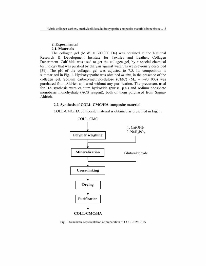
2. Experimental 2.1. Materials The collagen gel (M.W. ≈ 300,000 Da) was obtained at the National
Research & Development Institute for Textiles and Leather, Collagen Department. Calf hide was used to get the collagen gel, by a special chemical technology that was purified by dialysis against water, as we previously described [39]. The pH of the collagen gel was adjusted to 7.5. Its composition is summarized in Fig. 1. Hydroxyapatite was obtained in situ, in the presence of the collagen gel. Sodium carboxymethylcellulose (CMC) (Mw = ~90 000) was purchased from Aldrich and used without any purification. The precursors used for HA synthesis were calcium hydroxide (puriss. p.a.) and sodium phosphate monobasic monohydrate (ACS reagent), both of them purchased from Sigma-Aldrich.
2.2. Synthesis of COLL-CMC/HA composite material
COLL-CMC/HA composite material is obtained as presented in Fig. 1.
Fig. 1. Schematic representation of preparation of COLL-CMC/HA
Mineralization
Polymer weighing
1. Ca(OH)2 2. NaH2PO4
Cross-linking
Drying
COLL-CMC/HA
COLL, CMC
Glutaraldehyde
Purification
6 I. Nedelcu, A. Ficai, Denisa Ficai, Georgeta Voicu, Mădălina Albu, Ecaterina Andronescu
The first step was the homogenisation of the collagen gel and carboxymethylcellulose (COLL:CMC = 2:1 (w/w)) followed by mineralization with Ca(OH)2 and NaH2PO4
. H2O, as we previously presented [38, 40]. The mineralization was performed in order to obtain COLL:CMC:HA = 2:1:8 (w/w/w). Once mineralized, the sample is cross-linked with 1% (reported to the dry collagen) glutaraldehyde solution and dried at 20oC [41].
2.3. Methods X-ray diffraction analysis was performed using a Shimadzu XRD 6000
diffractometer at room temperature, using Cu Kα radiation. The samples were scanned in the range 2θ =10–70oC, with a scanning rate of 2oC/ min.
IR microscopy/spectroscopy was performed by using a Thermo FT-IR Nicolet iN10 MX microscope; the spectra were recorded in the wave number range of 400 – 4000 cm-1, with a resolution of 4cm-1. For a better identification of the peaks, the obtained spectra were resolved using a Gaussian-Lorentzian peak resolve procedure, with no baseline (for all the spectra, the baseline correction was previously done).
SEM analyses were performed on a HITACHI S2600N electron microscope with EDAX on samples covered with silver layer.
3. Results and discussion After synthesis and drying, COLL-CMC/HA composite material was
analyzed using X-ray diffraction (XRD), Fourier Transform – Infrared Spectroscopy and Microscopy (FTIR) and Scanning Electron Microscopy (SEM).
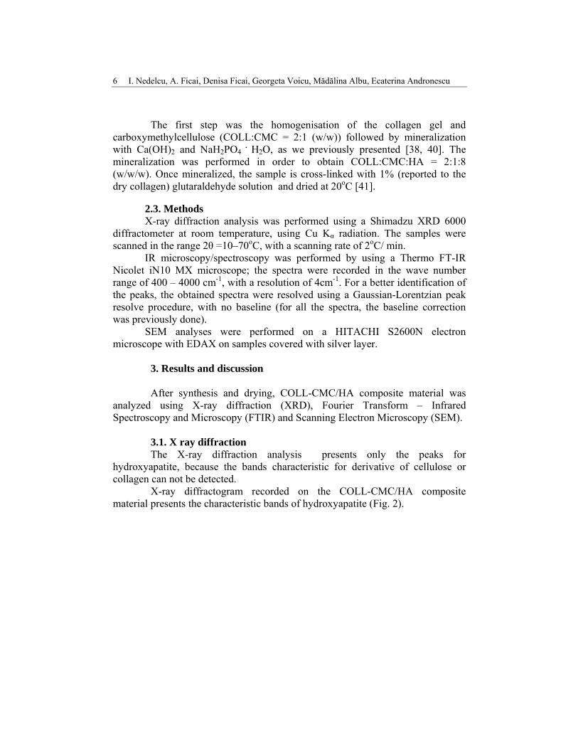
3.1. X ray diffraction The X-ray diffraction analysis presents only the peaks for
hydroxyapatite, because the bands characteristic for derivative of cellulose or collagen can not be detected.
X-ray diffractogram recorded on the COLL-CMC/HA composite material presents the characteristic bands of hydroxyapatite (Fig. 2).
Hybrid collagen-carboxy-methylcellulose/hydroxyapatite composite materials bone tissue... 7
Fig. 2. XRD diffraction pattern of COLL-CMC/HA
Due to the low crystallinity of the organic phase and low levels of
potential by-products, the characteristic bands of other crystalline phases are not identified beside the high content of collagen and CMC. Sodium chloride and different calcium phosphates (CaPs) can be identified as by-products. The presence of CaPs is a proof of proper synthesis conditions (especially pH and Ca:P ratio). Sodium chloride can be easily removed by washing the cross-linked composite with distilled water, as we have already presented [7].
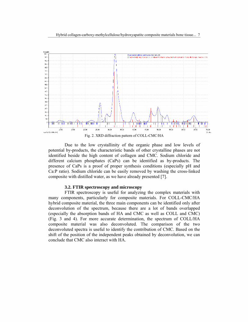
3.2. FTIR spectroscopy and microscopy FTIR spectroscopy is useful for analyzing the complex materials with
many components, particularly for composite materials. For COLL-CMC/HA hybrid composite material, the three main components can be identified only after deconvolution of the spectrum, because there are a lot of bands overlapped (especially the absorption bands of HA and CMC as well as COLL and CMC) (Fig. 3 and 4). For more accurate determination, the spectrum of COLL/HA composite material was also deconvoluted. The comparison of the two deconvoluted spectra is useful to identify the contribution of CMC. Based on the shift of the position of the independent peaks obtained by deconvolution, we can conclude that CMC also interact with HA.
8 I. Nedelcu, A. Ficai, Denisa Ficai, Georgeta Voicu, Mădălina Albu, Ecaterina Andronescu
Fig. 3. FTIR spectra of COLL-CMC/HA
Fig. 4. Deconvoluted FTIR spectra (715-1800cm-1) of COLL-CMC/HA and COLL/HA
Hybrid collagen-carboxy-methylcellulose/hydroxyapatite composite materials bone tissue... 9
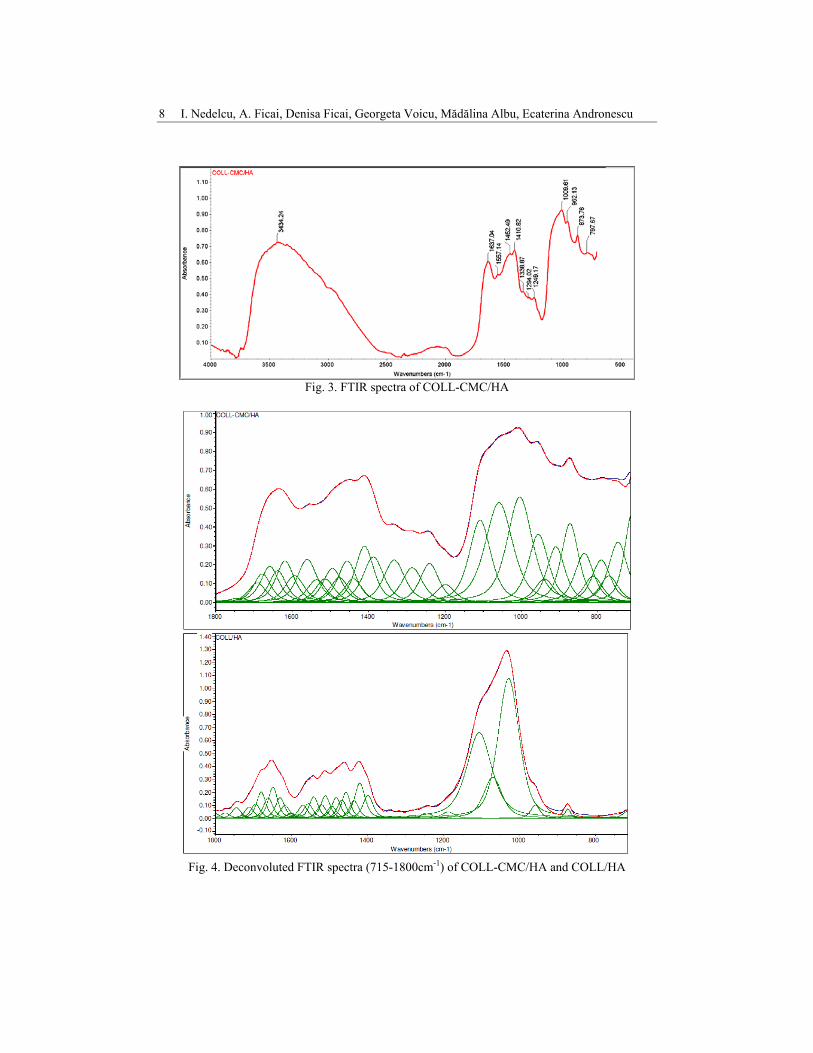
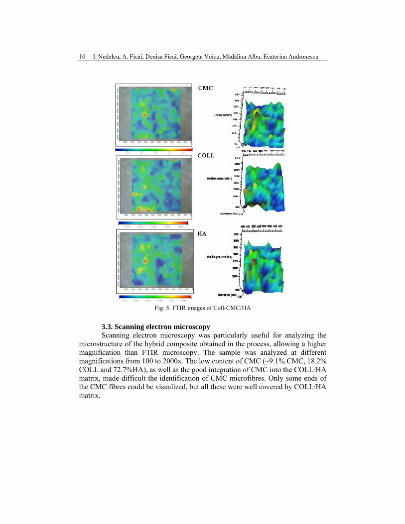
FTIR microscopy was also used to characterize the hybrid composite. For this reason, the sample was fractured and the resulting surface analyzed by FTIR spectroscopy. Once obtained the microscopic information, the FTIR maps at different wavelengths were obtained. In our case, the peaks of interests were:
• 715cm-1 for carboxymethylcellulose; • 1655cm-1 for collagen; • 1033cm-1 for hydroxyapatite.
According to FTIR maps (Fig. 5) the material is homogenous, which means that CMC microfibres are well dispersed into the COLL/HA matrix.
Abso
rban
ce (
Abs)
0.000.10
0.20
0.30
Wavenumbers (cm-1)3500 3000 2500 2000 1500 1000
Position (micrometers)=4269 µm,26100 µm, point #232
Abso
rban
ce (
Abs)
0.000.10
0.20
0.30
Wavenumbers (cm-1)3500 3000 2500 2000 1500 1000
Position (micrometers)=4269 µm,26100 µm, point #232
10 I. Nedelcu, A. Ficai, Denisa Ficai, Georgeta Voicu, Mădălina Albu, Ecaterina Andronescu
Fig. 5. FTIR images of Coll-CMC/HA
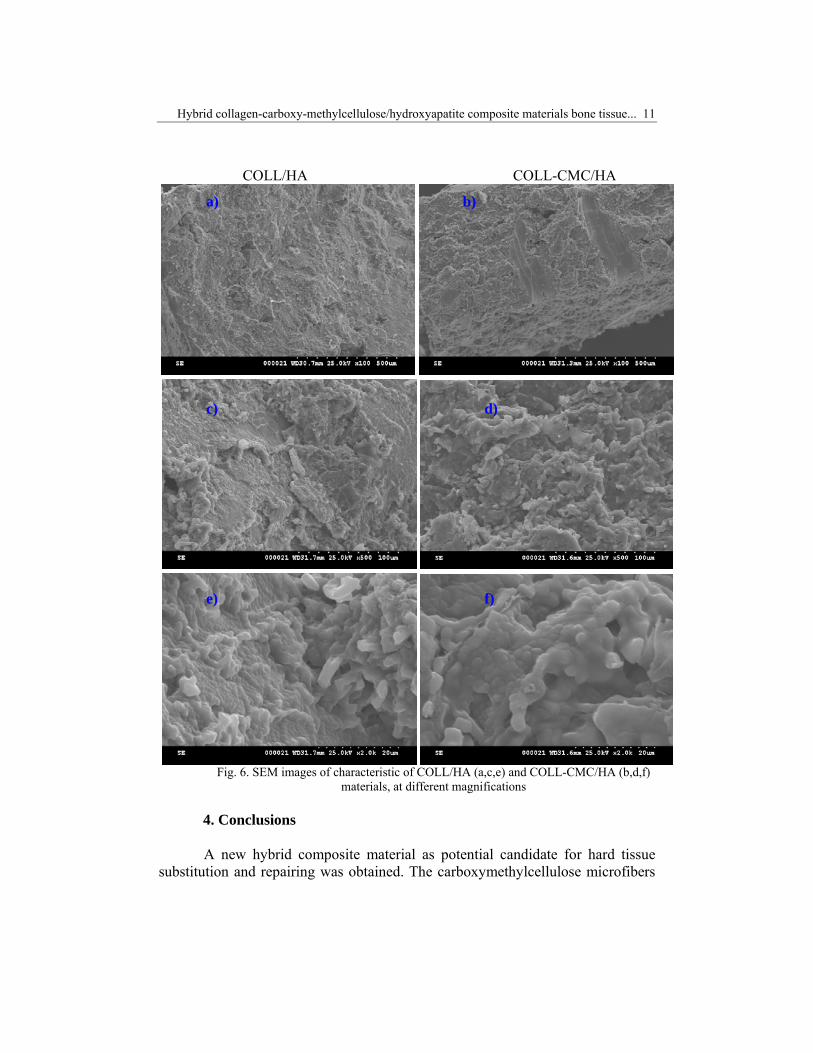
3.3. Scanning electron microscopy Scanning electron microscopy was particularly useful for analyzing the
microstructure of the hybrid composite obtained in the process, allowing a higher magnification than FTIR microscopy. The sample was analyzed at different magnifications from 100 to 2000x. The low content of CMC (~9.1% CMC, 18.2% COLL and 72.7%HA), as well as the good integration of CMC into the COLL/HA matrix, made difficult the identification of CMC microfibres. Only some ends of the CMC fibres could be visualized, but all these were well covered by COLL/HA matrix.
Hybrid collagen-carboxy-methylcellulose/hydroxyapatite composite materials bone tissue... 11
COLL/HA COLL-CMC/HA
Fig. 6. SEM images of characteristic of COLL/HA (a,c,e) and COLL-CMC/HA (b,d,f)
materials, at different magnifications 4. Conclusions A new hybrid composite material as potential candidate for hard tissue
substitution and repairing was obtained. The carboxymethylcellulose microfibers
a) b) c) d) e) f)
12 I. Nedelcu, A. Ficai, Denisa Ficai, Georgeta Voicu, Mădălina Albu, Ecaterina Andronescu
are well embedded in the mass of mineralized polymer, being difficult to be identified by SEM in the ternary composite based on collagen, carboxymethylcellulose and hydroxyapatite. Further in vitro and in vivo tests will clarify the possibility to use this material for bone grafting and substitution, as well as for drug delivery systems, especially in the case of bone cancer and osteoporosis.
R E F E R E N C E S 1. Murugan R, Ramakrishna S. Development of nanocomposites for bone grafting.
Composites Science and Technology 2005;65:2385–2406. 2. Cunniffe GM, Dickson GR, Partap S, Stanton KT, O'Brien FJ. Development and
characterisation of a collagen nano-hydroxyapatite composite scaffold for bone tissue engineering. Journal of Materials Science: Materials in Medicine 2009:1-6.
3. Fratzl P, Weinkamer R. Nature's hierarchical materials. Progress in Materials Science 2007 Nov;52(8):1263-1334.
4. Ilan DI, Ladd AL. Bone Graft Substitutes. Operative Techniques in Plastic and Reconstructive Surgery 2002;9(4):151-160.
5. Oprita EI, Moldovan L, Craciunescu O, Buzgariu W, Tardei C, Zarnescu O. A bioactive collagen-b-tricalcium phosphate scaffold for tissue engineering. Central European Journal of Biology 2006;1(1):61-72.
6. Ficai A, Andronescu E, Ficai D, Sonmez M, Albu MG, Voicu G. Mimicking the morphology of long bone. Central European Journal of Chemistry 2012 Dec;10(6):1949-1953.
7. Ficai A, Andronescu E, Voicu G, Ghitulica C, Vasile BS, Ficai D, et al. Self assembled collagen/ hydroxyapatite composite materials. Chemical Engineering Journal 2010;160(2):794-800.
8. Ficai A, Andronescu E, Voicu G, Manzu D, Ficai M. Layer by layer deposition of hydroxyapatite onto the collagen matrix. Materials Science & Engineering C-Materials for Biological Applications 2009 Aug 31;29(7):2217-2220.
9. Ficai M, Andronescu E, Ficai D, Voicu G, Ficai A. Synthesis and characterization of COLL-PVA/HA hybrid materials with stratified morphology. Colloids and Surfaces B: Biointerfaces 2010;81(2):614-619.
10. Arsilmaz FS, Rhan NO, Nsaldi EU, Durmus AS, Olakoglu NC. A polyethylene-high proportion hydroxyapatite implant and its investigation in vivo. Acta of Bioengineering and Biomechanics 2007;9(2).
11. Gay S, Arostegui S, Lemaitre J. Preparation and characterization of dense nanohydroxyapatite/PLLA composites. Materials Science and Engineering C 2009;29(1):172-177.
12. Murugan R, Ramakrishna S. Bioresorbable composite bone paste using polysaccharide based nano hydroxyapatite. Biomaterials 2004;25(17):3829-3835.
13. Ngiam M, Liao S, Patil AJ, Cheng Z, Chan CK, Ramakrishna S. The fabrication of nano-hydroxyapatite on PLGA and PLGA/collagen nanofibrous composite scaffolds and their effects in osteoblastic behavior for bone tissue engineering. Bone 2009;45(1):4-16.
14. Siriphannon P, Monvisade P. Poly(ethylene terephthalate)/hydroxyapatite biomaterials: Preparation, characterization, and in vitro bioactivity. Journal of Biomedical Materials Research Part A 2009 Feb;88A(2):464-469.
15. Zhang PB, Hong ZK, Yu T, Chen XS, Jing XB. In vivo mineralization and osteogenesis of nanocomposite scaffold of poly (lactide-co-glycolide) and hydroxyapatite surface-grafted with poly(L-lactide). Biomaterials 2009 Jan;30(1):58-70.
Hybrid collagen-carboxy-methylcellulose/hydroxyapatite composite materials bone tissue... 13
16. Miyazaki T, Ishikawa K, Shirosaki Y, Ohtsuki C. Organic-inorganic composites designed for biomedical applications. Biol Pharm Bull;36(11):1670-1675.
17. Panda NN, Jonnalagadda S, Pramanik K. Development and evaluation of cross-linked collagen-hydroxyapatite scaffolds for tissue engineering. Journal of Biomaterials Science-Polymer Edition 2013 Dec 1;24(18):2031-2044.
18. Kikuchi M. Hydroxyapatite/Collagen Bone-Like Nanocomposite. Biological & Pharmaceutical Bulletin 2013 Nov;36(11):1666-1669.
19. Gunatillake PA, Adhikari R. Biodegradable synthetic polymers for tissue engineering. European Cells & Materials 2003;5:1-16.
20. Jones JR. Review of bioactive glass: From Hench to hybrids. Acta Biomaterialia 2013 Jan;9(1):4457-4486.
21. Li ZL, Kong WN, Li XL, Xu C, He YQ, Gao JP, et al. Antibiotic-Containing Biodegradable Bead Clusters with Porous PLGA Coating as Controllable Drug-Releasing Bone Fillers. Journal of Biomaterials Science-Polymer Edition 2011;22(13):1713-1731.
22. Busenlechner D, Tangl S, Fitzl C, Bernhart T, Gruber R, Watzek G. Paste-like inorganic bone matrix: preclinical testing of a prototype preparation in the porcine calvaria. Clinical Oral Implants Research 2009 Oct;20(10):1099-1104.
23. La Gatta A, De Rosa A, Laurienzo P, Malinconico M, De Rosa M, Schiraldi C. A novel injectable poly(epsilon-caprolactone) calcium sulfate system for bone regeneration: Synthesis and characterization. Macromolecular Bioscience 2005 Nov 4;5(11):1108-1117.
24. Saraswathy G, Pal S, Rose C, Sastry TP. A novel bio-inorganic bone implant containing deglued bone, chitosan and gelatin. Bulletin of Materials Science 2001 Aug;24(4):415-420.
25. Alkhraisat MH, Rueda C, Jerez LB, Marino FT, Torres J, Gbureck U, et al. Effect of silica gel on the cohesion, properties and biological performance of brushite cement. Acta Biomaterialia 2010 Jan;6(1):257-265.
26. Dietrich E, Oudadesse H, Lucas-Girot A, Le Gal Y, Jeanne S, Cathelineau G. Effects of Mg and Zn on the surface of doped melt-derived glass for biomaterials applications. Applied Surface Science 2008 Nov 15;255(2):391-395.
27. Gerber T, Traykova T, Henkel KO, Bienengraeber V, Witt M, Koewitz J. Silica/calcium phosphate sol-gel derived bone grafting material and bone remodelling. An eight months in vivo study. Bioceramics 15 2003;240-2:411-414.
28. Birsan C, Ghitulica C, Andronescu E, Ionita C, Birsan M. Bioglasses in the SiO2-CaO-P2O5 system. In: Lau AKT, Lu J, Varadan VK, Chang FK, Tu JP, Lam PM, editors. International Conference on Multifunctional Materials and Structures; 2008 Jul 28-31; Hong Kong, PEOPLES R CHINA; 2008. p. 1063-1066.
29. Ghitulica C, Boretto GM, Andronescu E. Processing and characterization of zirconia reinforced composite ceramic foams. Revista De Chimie 2005 Dec;56(12):1218-1221.
30. Ghitulica C, Andronescu E, Dumitru G, Stoleriu S. Preparation of alumina and cordierite based porous ceramic materials. Euro Ceramics Viii, Pts 1-3 2004;264-268:2247-2250.
31. Ghitulica C, Andronescu E, Fantozzi G, Jorand Y. Alumina porous ceramic materials prepared by slip casting. Silicates Industriels 2002 Mar-Apr;67(3-4):33-36.
32. Ficai M, Andronescu E, Ficai A, Voicu G, Vasile BŞ. Poly bis-GMA/HA based hybrid composite materials. UPB Sci Bull, Series B 2011;73-84(1):75-.
33. Kobayashi H, Kato M, Taguchi T, Ikoma T, Miyashita H, Shimmura S, et al. Collagen immobilized PVA hydrogel-hydroxyapatite composites prepared by kneading methods as a material for peripheral cuff of artificial cornea. Materials Science & Engineering C-Biomimetic and Supramolecular Systems 2004 Dec 1;24(6-8):729-735.
34. Ficai A, Albu MG, Birsan M, Sonmez M, Ficai D, Trandafir V, et al. Collagen hydrolysate based collagen/hydroxyapatite composite materials. Journal of Molecular Structure 2013 Apr 10;1037:154-159.
14 I. Nedelcu, A. Ficai, Denisa Ficai, Georgeta Voicu, Mădălina Albu, Ecaterina Andronescu
35. Hu YY, Zhang C, Zhang SM, Xiong Z, Xu JQ. Development of a porous poly(L-lactic acid)/hydroxyapatite/collagen scaffold as a BMP delivery system and its use in healing canine segmental bone defect. Journal of Biomedical Materials Research Part A 2003 Nov 1;67A(2):591-598.
36. Li JS, Yuan XY, He F, Mak AFT. Hybrid coating of hydroxyapatite and collagen within poly(D,L-lactic-co-glycolic acid) scaffold. Journal of Biomedical Materials Research Part B-Applied Biomaterials 2008 Aug;86B(2):381-388.
37. Luo D, Sang L, Wang X, Xu S, Li X. Low temperature, pH-triggered synthesis of collagen-chitosan-hydroxyapatite nanocomposites as potential bone grafting substitutes. Materials Letters 2011;65(15-16):2395-2397.
38. Andronescu E, Ficai M, Voicu G, Ficai D, Maganu M, Ficai A. Synthesis and characterization of collagen/hydroxyapatite:magnetite composite material for bone cancer treatment. Journal of Materials Science - Materials in Medicine 2010;21(7):2237-2242.
39. Albu MG. Collagen gels and matrices for biomedical applications: Lambert Academic Publishing, Saarbrücken, Germany, 2011.
40. Ficai A, Andronescu E, Trandafir V, Ghitulica C, Voicu G. Collagen/hydroxyapatite composite obtained by electric field orientation. Materials Letters 2010 Feb 28;64(4):541-544.
41. Andronescu E, Voicu G, Ficai M, Mohora IA, Trusca R, Ficai A. Collagen/hydroxyapatite composite materials with desired ceramic properties. Journal of Electron Microscopy 2011 Jun;60(3):253-259.