Embed Size (px)
Citation preview

HVint: A Strategy for Identifying Novel Protein-Protein Interactions in Herpes Simplex VirusType 1*□S
Paul Ashford‡ ‡‡, Anna Hernandez‡ ‡‡§, Todd Michael Greco¶‡‡, Anna Buch�,Beate Sodeik�, Ileana Mihaela Cristea¶**, Kay Grunewald§ Adrian Shepherd‡,and Maya Topf‡**
Human herpesviruses are widespread human pathogenswith a remarkable impact on worldwide public health.Despite intense decades of research, the molecular de-tails in many aspects of their function remain to be fullycharacterized. To unravel the details of how these virusesoperate, a thorough understanding of the relationshipsbetween the involved components is key. Here, we pres-ent HVint, a novel protein-protein intraviral interactionresource for herpes simplex virus type 1 (HSV-1) integrat-ing data from five external sources. To assess each inter-action, we used a scoring scheme that takes into consid-eration aspects such as the type of detection method andthe number of lines of evidence. The coverage of the initialinteractome was further increased using evolutionary in-formation, by importing interactions reported for otherhuman herpesviruses. These latter interactions consti-tute, therefore, computational predictions for potentialnovel interactions in HSV-1. An independent experimentalanalysis was performed to confirm a subset of our pre-dicted interactions. This subset covers proteins thatcontribute to nuclear egress and primary envelopmentevents, including VP26, pUL31, pUL40, and the recentlycharacterized pUL32 and pUL21. Our findings support acoordinated crosstalk between VP26 and proteins such aspUL31, pUS9, and the CSVC complex, contributing to thedevelopment of a model describing the nuclear egressand primary envelopment pathways of newly synthesized
HSV-1 capsids. The results are also consistent with recentfindings on the involvement of pUL32 in capsid maturationand early tegumentation events. Further, they open thedoor to new hypotheses on virus-specific regulators ofpUS9-dependent transport. To make this repository ofinteractions readily accessible for the scientific commu-nity, we also developed a user-friendly and interactiveweb interface. Our approach demonstrates the power ofcomputational predictions to assist in the design of tar-geted experiments for the discovery of novel protein-pro-tein interactions. Molecular & Cellular Proteomics 15:10.1074/mcp.M116.058552, 2939–2953, 2016.
One important milestone toward understanding the com-plexity of viral infections is to unravel the interplay betweenviral proteins (the intraviral interactome). This is particularlyimportant for complex and large DNA viruses, such as humanherpesviruses, which have the ability to express a large num-ber of viral gene-products. For example, the genome ofHSV-1 encodes for more than 75 viral proteins, and the levelsof these proteins are temporally and spatially regulated duringthe progression of the viral infection (1). Human herpesvirusescause life-long infections and many human and animal dis-eases. The severity of symptoms ranges from cold sores,genital ulcers, and blisters to blindness and life-threateningconditions, including fatal encephalitis, meningitis and cancer(2, 3). Infections from herpesviruses are also a major threat toimmunosuppressed patients (e.g. infected by human immu-nodeficiency virus) and have been associated with Alzhei-mer’s disease (3, 4).
Protein interactome studies can reveal critical biologicalinformation and shed light on mechanisms underlying infec-tious diseases (5), supporting proteome-wide annotation (6, 7)and the development of therapeutic strategies (8, 9). Thecurrent methods used for building protein-protein interaction(PPI) networks mainly rely on known interactions and se-quence analysis (11–13). Recently the field has moved for-ward through the development of structural and functionalproteomics techniques that include fluorescence microscopyand Mass Spectrometry (MS)-based approaches (6, 14–16).
From the: ‡Institute of Structural and Molecular Biology, BirkbeckCollege, University of London, Malet Street, London, WC1E 7HX, UK;§Oxford Particle Imaging Centre, Division of Structural Biology, Well-come Trust Centre for Human Genetics, University of Oxford, Oxford,OX3 7BN, UK; ¶Department of Molecular Biology, Princeton Univer-sity, Lewis Thomas Laboratory, Washington Road, Princeton, NewJersey 08544; �Institute of Virology, Hannover Medical School, OE4310, Carl-Neuberg-Str. 1, D-30623, Hannover, Germany
Author’s Choice—Final version free via Creative CommonsCC-BY license.
Received February 15, 2016, and in revised form, June 30, 2016Published, MCP Papers in Press, July 6, 2016, DOI
10.1074/mcp.M116.058552Author contributions: P.A., A.H., A.S., and M.T. designed research;
P.A., A.H., T.M.G., and A.B. performed research; P.A., A.H., T.M.G.,A.B., B.S., I.M.C., K.G., A.S., and M.T. analyzed data; P.A., A.H.,T.M.G., A.B., B.S., I.M.C., K.G., A.S., and M.T. wrote the paper.
Research
Author’s Choice © 2016 by The American Society for Biochemistry and Molecular Biology, Inc.This paper is available on line at http://www.mcponline.org
crossmark
Molecular & Cellular Proteomics 15.9 2939

These techniques have helped to increase the coverage of theinteractome in the context of infection. Several public repos-itories of PPI data exist, such as IntAct (www.ebi.ac.uk/intact/)(15). Multiple evidence lines, depending on the nature of theinteraction itself and, importantly, the detection method used,can support each individual PPI. For example, evidence forPPIs can be derived from biochemical assays, such as YeastTwo-Hybrid (Y2H), CoImmunoprecipitation (Co-IP)1, in vitrobinding assays, and protein cross-linking, which can be thenanalyzed by MS. PPIs can also be derived from NuclearMagnetic Resonance (NMR), x-ray crystallography andElectron Microscopy techniques. Most resources include ev-idence manually extracted from the literature. Furthermore,databases that are not explicitly dedicated to storing PPI dataprovide additional valuable resources, such as the ProteinData Bank (PDB www.rcsb.org/) (16) and the Electron Micros-copy Data Bank (EMDB www.emdatabank.org/) (17), whichcontain structural information. Other databases gather PPIsbased on information from multiple resources (e.g. VirHostNetwww.virhostnet.prabi.fr/, STRING-DB http://string-db.org/)(20, 21). However, constructing a PPI network from disparatesources while ensuring trustworthiness and high coverage ischallenging. Experiments vary in terms of both reliability andability to discriminate between different categories of interac-tions, notably direct (physical) versus indirect interactions (e.g.proteins belonging to the same protein complex but withoutdirect physical contact) as well as transient versus stableinteractions (22). Moreover, for many nonmodel organisms,the number of known PPIs remains limited, and thus there isa need to develop hypotheses about additional PPIs that arenot yet supported by direct experimental evidence.
Computational prediction of PPIs (19–23) provide the op-portunity to maximize the coverage of interaction networks.These predictions often rely on sequence homology or ma-chine learning methods (24–26). Several studies have nowillustrated that transferring interaction data between closehomologous species (i.e. interologues mapping (31)) is a suit-able approach to expand PPI data for a given species. Com-putational methods for building and analyzing PPI networks
have the potential to identify novel candidates for future ex-perimental validation, thereby saving valuable time and re-sources (30, 32, 33).
In this study, we created HVint, a new database for intraviralPPIs for an important human pathogen, of herpes simplexvirus type 1 (HSV-1, also known as HHV-1), and derived theassociated PPI network. To accomplish this, PPIs reported forany stage of the “life cycle” were compiled, i.e. interactiondata both for proteins that are incorporated within extracellu-lar virion particles and proteins that are only expressed ininfected cells has been integrated. Information on the locationof these proteins within the different structural layers of thevirion particle (34, 35), namely the capsid, the tegument andthe viral envelope, was included in the annotation of theinteractions in HVint. To further expand the HVint database,we integrated the subsets of data from existing databases forany stage of the virus “life cycle”, implemented a method forscoring multiple lines of experimental evidence, and incorpo-rated homologous interactions from the other human herpes-viruses of the �-, �-, and �-herpesvirus subfamilies. As aresult, our network has significantly higher coverage thanprevious HSV-1 networks derived from existing databases(17, 18, 20, 36, 37), and it predicts novel interactions. Wevalidated several of these predictions by affinity purifica-tion-MS using primary human fibroblasts infected with anHSV-1 virus strain expressing the small capsid protein VP26tagged with green fluorescent protein (EGFP). Lastly, we de-veloped a user-friendly interactive web interface, which willallow the scientific community to readily access and analyzethe interaction data. Taken together, this work demonstratesthe value of data integration and homology transfer in pre-dicting previously uncovered interactions in a complex virussuch as HSV-1 and in guiding future experimental work.
EXPERIMENTAL PROCEDURES
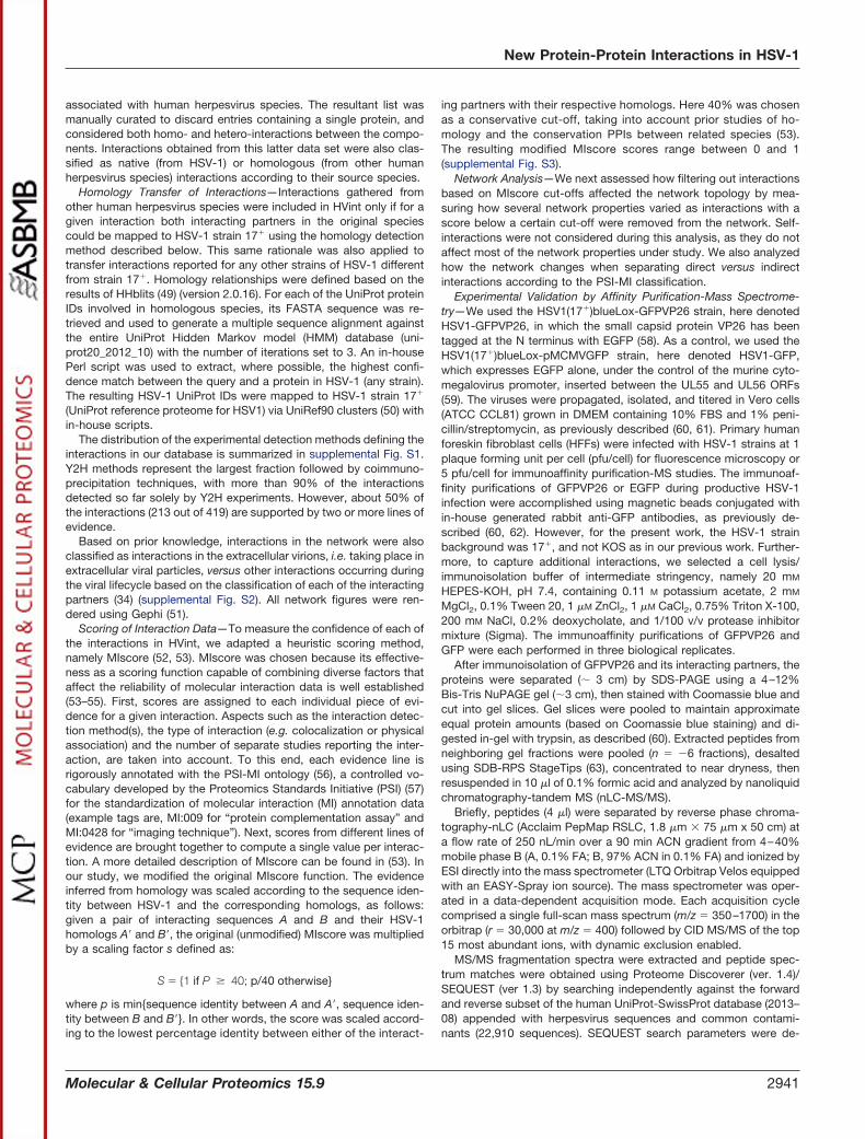
HVint Data Integration—The PPI data compiled to generate ournovel intra-viral protein interactome includes interactions identified bya range of experimental methods drawn from multiple sources (Figs.1 and 2). First, data from four publicly available protein interactiondatabases, IntAct (15) (June 2015), VirHostNet 2.0 (18) (June 2015),Database of Interacting Proteins (DIP) (36) (October 2015) andBioGRID (37) (October 2015) were collected (38–47). Custom Perlscripts were used to parse the data and to select “native” interactionsdirectly reported for HSV-1, and “homologous” interactions originallyreported for any of the other human herpesviruses, i.e. herpes simplexvirus type 2 (HSV-2, HHV-2), varicella-zoster virus (VZV, HHV-3),Epstein-Barr virus (EBV, HHV-4), human cytomegalovirus (HCMV,HHV-5), human herpesvirus 6 (HHV-6, A and B), human herpesvirus 7(HHV-7), or Kaposi’s sarcoma-associated herpesvirus (KSHV, HHV-8). The search was conducted using taxonomy identifiers (IDs) and allreported strains for each species were considered. Where possible,protein IDs, as reported in the above interaction databases, weremapped to UniProt (48) accession numbers using the UniProt IDmapping tool (e.g. DIP and BioGRID interactor IDs are from a varietyof databases, including UniProt (48), NCBI RefSeq, and EMBL/Gen-Bank/DDBJ). In addition we collected interactions derived from struc-tural evidence provided by the Protein Data Bank (PDB) (16) (June2015). The taxonomy browser in PDB was used to retrieve entries
1 The abbreviations used are: Co-IP, Co-Immunoprecipitation; PPI,protein-protein interaction; Y2H, Yeast Two-Hybrid; NMR, nuclearmagnetic resonance; EMDB, Electron Microscopy Data Bank; HSV-1,HHV-1, herpes simplex virus type 1, human herpesvirus 1; GFP,Enhanced Green Fluorescent Protein; DIP, Database of InteractingProteins; HSV-2, HHV-2, herpes simplex virus type 2, human herpes-virus 2; VZV, HHV-3, varicella-zoster virus, human herpesvirus 3; EBV,HHV-4, Epstein-Barr virus, human herpesvirus 4; HCMV, HHV-5,human cytomegalovirus, human herpesvirus 5; HHV-6, human her-pesvirus 6; HHV-7, human herpesvirus 7; KSHV, HHV-8, Kaposi’ssarcoma-associated herpesvirus, human herpesvirus 8; NCBI, Na-tional Center for Biotechnology Information; EMBL, European Bioin-formatics Institute; DDBJ, DNA Data Bank of Japan; PDB, ProteinData Bank; HMM, Hidden Markov model; PSI, Proteomics StandardsInitiative; MI, molecular interactions; CVSC, capsid vertex-specificcomponent; PFU, plaque forming unit.
New Protein-Protein Interactions in HSV-1
2940 Molecular & Cellular Proteomics 15.9
associated with human herpesvirus species. The resultant list wasmanually curated to discard entries containing a single protein, andconsidered both homo- and hetero-interactions between the compo-nents. Interactions obtained from this latter data set were also clas-sified as native (from HSV-1) or homologous (from other humanherpesvirus species) interactions according to their source species.
Homology Transfer of Interactions—Interactions gathered fromother human herpesvirus species were included in HVint only if for agiven interaction both interacting partners in the original speciescould be mapped to HSV-1 strain 17� using the homology detectionmethod described below. This same rationale was also applied totransfer interactions reported for any other strains of HSV-1 differentfrom strain 17�. Homology relationships were defined based on theresults of HHblits (49) (version 2.0.16). For each of the UniProt proteinIDs involved in homologous species, its FASTA sequence was re-trieved and used to generate a multiple sequence alignment againstthe entire UniProt Hidden Markov model (HMM) database (uni-prot20_2012_10) with the number of iterations set to 3. An in-housePerl script was used to extract, where possible, the highest confi-dence match between the query and a protein in HSV-1 (any strain).The resulting HSV-1 UniProt IDs were mapped to HSV-1 strain 17�
(UniProt reference proteome for HSV1) via UniRef90 clusters (50) within-house scripts.
The distribution of the experimental detection methods defining theinteractions in our database is summarized in supplemental Fig. S1.Y2H methods represent the largest fraction followed by coimmuno-precipitation techniques, with more than 90% of the interactionsdetected so far solely by Y2H experiments. However, about 50% ofthe interactions (213 out of 419) are supported by two or more lines ofevidence.
Based on prior knowledge, interactions in the network were alsoclassified as interactions in the extracellular virions, i.e. taking place inextracellular viral particles, versus other interactions occurring duringthe viral lifecycle based on the classification of each of the interactingpartners (34) (supplemental Fig. S2). All network figures were ren-dered using Gephi (51).
Scoring of Interaction Data—To measure the confidence of each ofthe interactions in HVint, we adapted a heuristic scoring method,namely MIscore (52, 53). MIscore was chosen because its effective-ness as a scoring function capable of combining diverse factors thataffect the reliability of molecular interaction data is well established(53–55). First, scores are assigned to each individual piece of evi-dence for a given interaction. Aspects such as the interaction detec-tion method(s), the type of interaction (e.g. colocalization or physicalassociation) and the number of separate studies reporting the inter-action, are taken into account. To this end, each evidence line isrigorously annotated with the PSI-MI ontology (56), a controlled vo-cabulary developed by the Proteomics Standards Initiative (PSI) (57)for the standardization of molecular interaction (MI) annotation data(example tags are, MI:009 for “protein complementation assay” andMI:0428 for “imaging technique”). Next, scores from different lines ofevidence are brought together to compute a single value per interac-tion. A more detailed description of MIscore can be found in (53). Inour study, we modified the original MIscore function. The evidenceinferred from homology was scaled according to the sequence iden-tity between HSV-1 and the corresponding homologs, as follows:given a pair of interacting sequences A and B and their HSV-1homologs A� and B�, the original (unmodified) MIscore was multipliedby a scaling factor s defined as:
S � �1 if P � 40; p/40 otherwise}
where p is min{sequence identity between A and A�, sequence iden-tity between B and B�}. In other words, the score was scaled accord-ing to the lowest percentage identity between either of the interact-
ing partners with their respective homologs. Here 40% was chosenas a conservative cut-off, taking into account prior studies of ho-mology and the conservation PPIs between related species (53).The resulting modified MIscore scores range between 0 and 1(supplemental Fig. S3).
Network Analysis—We next assessed how filtering out interactionsbased on MIscore cut-offs affected the network topology by mea-suring how several network properties varied as interactions with ascore below a certain cut-off were removed from the network. Self-interactions were not considered during this analysis, as they do notaffect most of the network properties under study. We also analyzedhow the network changes when separating direct versus indirectinteractions according to the PSI-MI classification.
Experimental Validation by Affinity Purification-Mass Spectrome-try—We used the HSV1(17�)blueLox-GFPVP26 strain, here denotedHSV1-GFPVP26, in which the small capsid protein VP26 has beentagged at the N terminus with EGFP (58). As a control, we used theHSV1(17�)blueLox-pMCMVGFP strain, here denoted HSV1-GFP,which expresses EGFP alone, under the control of the murine cyto-megalovirus promoter, inserted between the UL55 and UL56 ORFs(59). The viruses were propagated, isolated, and titered in Vero cells(ATCC CCL81) grown in DMEM containing 10% FBS and 1% peni-cillin/streptomycin, as previously described (60, 61). Primary humanforeskin fibroblast cells (HFFs) were infected with HSV-1 strains at 1plaque forming unit per cell (pfu/cell) for fluorescence microscopy or5 pfu/cell for immunoaffinity purification-MS studies. The immunoaf-finity purifications of GFPVP26 or EGFP during productive HSV-1infection were accomplished using magnetic beads conjugated within-house generated rabbit anti-GFP antibodies, as previously de-scribed (60, 62). However, for the present work, the HSV-1 strainbackground was 17�, and not KOS as in our previous work. Further-more, to capture additional interactions, we selected a cell lysis/immunoisolation buffer of intermediate stringency, namely 20 mM
HEPES-KOH, pH 7.4, containing 0.11 M potassium acetate, 2 mM
MgCl2, 0.1% Tween 20, 1 �M ZnCl2, 1 �M CaCl2, 0.75% Triton X-100,200 mM NaCl, 0.2% deoxycholate, and 1/100 v/v protease inhibitormixture (Sigma). The immunoaffinity purifications of GFPVP26 andGFP were each performed in three biological replicates.
After immunoisolation of GFPVP26 and its interacting partners, theproteins were separated (� 3 cm) by SDS-PAGE using a 4–12%Bis-Tris NuPAGE gel (�3 cm), then stained with Coomassie blue andcut into gel slices. Gel slices were pooled to maintain approximateequal protein amounts (based on Coomassie blue staining) and di-gested in-gel with trypsin, as described (60). Extracted peptides fromneighboring gel fractions were pooled (n � �6 fractions), desaltedusing SDB-RPS StageTips (63), concentrated to near dryness, thenresuspended in 10 �l of 0.1% formic acid and analyzed by nanoliquidchromatography-tandem MS (nLC-MS/MS).
Briefly, peptides (4 �l) were separated by reverse phase chroma-tography-nLC (Acclaim PepMap RSLC, 1.8 �m � 75 �m x 50 cm) ata flow rate of 250 nL/min over a 90 min ACN gradient from 4–40%mobile phase B (A, 0.1% FA; B, 97% ACN in 0.1% FA) and ionized byESI directly into the mass spectrometer (LTQ Orbitrap Velos equippedwith an EASY-Spray ion source). The mass spectrometer was oper-ated in a data-dependent acquisition mode. Each acquisition cyclecomprised a single full-scan mass spectrum (m/z � 350–1700) in theorbitrap (r � 30,000 at m/z � 400) followed by CID MS/MS of the top15 most abundant ions, with dynamic exclusion enabled.
MS/MS fragmentation spectra were extracted and peptide spec-trum matches were obtained using Proteome Discoverer (ver. 1.4)/SEQUEST (ver 1.3) by searching independently against the forwardand reverse subset of the human UniProt-SwissProt database (2013–08) appended with herpesvirus sequences and common contami-nants (22,910 sequences). SEQUEST search parameters were de-
New Protein-Protein Interactions in HSV-1
Molecular & Cellular Proteomics 15.9 2941

fined as the following: full trypsin specificity, maximum of 2 missedtrypsin cleavages, ion precursor mass tolerance of 10 ppm, fragmention mass tolerance of 0.5 Da, fixed modification of cysteine carbam-idomethylation, variable modifications of methionine oxidation, andserine, threonine, and tyrosine phosphorylation. The Proteome Dis-coverer workflow also included the precursor ions area detector nodefor MS1-based label-free quantification. Database search resultswere analyzed in Scaffold (ver. 4.6, Proteome Software, Inc) using theLFDR algorithm and a refinement search using the X! Tandem algo-rithm (Beavis Informatics), which specified the following additionalmodifications using the same search parameters as above: deamida-tion of glutamine and asparagine, acetylation of protein N termini. Theglobal peptide and protein FDR were controlled to � 1%, estimatedby reverse database search matches. The MS proteomics data havebeen deposited to the ProteomeXchange Consortium via the PRIDE(64) partner repository with the data set PXD003599 and 10.6019/PXD003599 identifier. Unweighted protein spectrum counts and cor-responding precursor area quantitative values were exported to Exceland additional filtering of the protein identifications was performed.Briefly, protein groups with � 10 spectra in at least two out of threereplicates were retained. For proteins identified in both GFPVP26 andthe GFP controls, the spectral count fold enrichment was calculatedfor each biological replicate as the ratio of the spectral counts in eachGFPVP26 replicate and the averaged GFP samples. Viral proteinswith an average enrichment ratio of � fivefold were retained. TheTop3 precursor area method, which uses the three most intensepeptides as a measure of protein concentration (65), was used toestimate the stoichiometry of viral proteins coisolated with GFPVP26.
Fluorescence Microscopy—Primary human fibroblast cells werecultured as described above, except on glass cover slips, and in-fected with HSV1-GFPVP26 at a multiplicity of infection of 1 pfu/cell.At 14 h postinfection, cells were fixed with 2% (v/v) paraformaldehydein PBS for 15 min at room temperature and prepared for imaging aspreviously described (60). Nuclei were stained with DAPI. The sub-cellular localization of GFPVP26 was analyzed using a confocal mi-croscope (Leica SP5) equipped with a 100x oil-immersion lens.
Experimental Design and Statistical Rationale—The HVint databasewas populated with non-redundant PPI data collated from five differ-ent public resources (IntAct, VirHostNet 2.0, DIP, BioGRID and PDB).In doing so, we distinguished between two different sets of PPI data:(1) PPIs directly reported in HSV-1 and (2) PPIs reported in otherhuman herpesviruses species. HHblits was used to establish orthol-ogy-relationships between proteins interacting in homologous humanherpesviruses and HSV-1 proteins. When HSV-1 homologous pro-teins were detected for both interacting partners in the source spe-cies, the interaction was transferred to the HSV-1 interactome andincluded in the HVint database. All PPI data in HVint was scoredunder the MIscore scheme, a heuristic scoring method designed tocapture the heterogeneity of PPI data. MIscore is computed takinginto account the annotation data for a given interaction. This annota-tion is defined by the HUPO PSI controlled vocabulary, which hasbeen implemented by the public database members of the IMExconsortium (56). The final normalized scores result from the weightedsum of three different terms representing (1) the number of differentcitations reporting the interaction, (2) the corresponding detectionmethod(s) used, and (3) the nature of the interaction (type). An earlierstudy compared the performance of MIscore with that of the normal-ized score implemented in Mentha, a large integrative database (notcontaining viral interactomes) with over 570,000 PPIs (66). It wastested on a benchmark of 500 high confidence human PPIs (positivedata set) and 397 noninteractions extracted from Negatome (negativedata set) (67). Around the optimal score cut-offs for each database(0.485 for MIscore, 0.343 for the Mentha score) MIscore was shown tohave greater accuracy, precision, and recall than the Mentha score
(0.76 versus 0.67, 0.70 versus 0.66 and 0.98 versus 0.85, respectively)(53). Our predicted interactions transferred from other human herpes-viruses were scaled down from their originally assigned MIscoreaccording to the sequence identity of each interacting protein to itscorresponding HSV-1 homolog. The resulting scores take valuesbetween 0 and 1 (inclusive), with larger values reflecting higher con-fidence in the interaction. This calibrated scale was designed to allowusers to fine-tune the selection of PPI data subsets in the database,based on their research purposes.
A subset of computationally predicted interactions was selected aspotential candidates for experimental validation by IP-MS. To identifythis subset, we studied how several parameters characterizing thetopology of the interactome changed when interactions scoring belowa given MIscore threshold were removed from the network. Thechanges in network parameters were assessed in the entire rangebetween 0 to 1 MIscore thresholds, in steps of 0.1. The IP-MSexperiments isolating HSV1-GFPVP26 were performed in three bio-logical replicates and compared against three biological IP-MS rep-licates of HSV1-GFP. In total six biological samples were subjected toGeLC-MS/MS analysis.
RESULTS
HVint Interactome—The computational and experimentalpipeline developed in this work (see Experimental Proce-dures, Fig. 1) resulted in a novel HVint interactome (Fig. 2).The data fed into the pipeline originated from five differentresources (IntAct, VirHostNet, DIP, BioGRID, and PDB) (Figs.1 and 3). PPIs reported for HSV-1 and all of the other humanherpesvirus species currently known (herpes simplex virustype 2, varicella-zoster virus, Epstein-Barr virus, human cyto-megalovirus, human herpesviruses 6 and 7, and Kaposi’ssarcoma-associated herpesvirus) were collected. The latterinteractions were transferred to the HSV-1 interactome onlywhen orthologs for both interacting partners in the sourcespecies (e.g. HCMV) could be found in the HSV-1 proteome.The resulting network comprises 73 nodes and 419 PPIs(including 36 self-interactions), the reliability of which wasassessed with a modified MIscore function (see Methods).The confidence values for the interactions in the networkrange from 0.147 to 0.972. Confidence values were computedusing a highly integrative and experimentally based scoringscheme that takes into account the type of interaction anddetection method used to detect the interaction, as well as thenumber of different studies providing support for the interac-tion. Interactions were divided into two different data setsaccording to the source species: native interactions (reportedin HSV-1) and homologous interactions (transferred from an-other human herpesvirus species). Homologous interactions(255 in total) account for �55% of the network, hence signif-icantly increasing the size of the network beyond that of thenative network (Fig. 4A). Twenty-nine interactions are presentin both native and homologous PPI data sets, indicating con-servation of PPIs between two or more herpesvirus species.These conserved herpesvirus PPIs involve 50 of the HSV-1proteins, which correspond to about 70% of the HSV-1 pro-teome (Fig. 4B). 98% (413) of these interactions were classi-fied as “direct interaction” or “physical association” by the
New Protein-Protein Interactions in HSV-1
2942 Molecular & Cellular Proteomics 15.9

PSI-MI annotation, meaning the respective experimental con-ditions indicate that this interaction either involves directphysical contact or that or the molecules are in closeproximity.
New Interactions—Predicted PPIs that so far have not beensupported by experimental evidence are of particular interest,as they point to putative biological mechanisms yet to beconfirmed. To investigate whether such predicted PPIs in-deed exist for HSV-1, a subset of high confidence homolo-gous interactions was subjected to experimental validation.This subset was selected from the interactions in HVint havinga confidence score greater than 0.4. The rationale behind thiscutoff was that the initially observed topological properties ofthe interactome were barely altered for confidence score cut-offs in the range between 0 and 0.4 (supplemental Fig. S4),but for cut-offs between 0.4 and 0.5 drastic changes in sev-eral network parameters (including clustering coefficient, cen-tralization, density, and average node degree) were observed.This calculated MIscore threshold is consistent with previ-ously reported estimates of optimal cut-offs for scores pre-dictions, within the range of 0.4 to 0.5 (53). We then retainedPPIs derived from homology predictions only (i.e. only in thehomologous data set). By definition, these homology-basedPPIs have been reported for other human herpesviruses butnot yet for HSV-1.
The resulting data set, referred hereafter as high-confi-dence homologous interactions subset, comprises a total of35 interactions (30 hetero-interactions and 5 self-interactions)among 28 different proteins (Fig. 5, supplemental Table S1).28 hetero-interactions out of the 30 are connected in a singleconnected component, i.e. a group of nodes in which a path
can be traced for any random pair of nodes (68). Theseinteractions involve 23 proteins that have been annotated asbeing involved mainly in transcription, capsid assembly, andnuclear capsid egress events (supplemental Table S2). Thetwo binary interactions outside the main graph involve theobligate hetero-dimer gH/gL, an essential component ofthe herpesvirus entry machinery (69), and the tegument pro-teins pUL7 and pUL51. The minor capsid protein pUL25 isalso present in the data set with only one self-interaction. It isworth emphasizing that these disconnected components arenevertheless connected to the rest of the network when thewhole interactome is considered (i.e. without filtering interac-tions). Our HVint interactome highlighted envelope proteinpUS9 to be a hub for several potential interactions. Its pre-dicted first neighbors, including VP26 (pUL35), the majorsmall capsid protein (SCP), pUL17, another minor-capsid pro-tein that interacts with pUL25, and the inner tegument proteinpUL36, that are known to function in capsid structure, matu-ration, and tegument association (70–74). As the function ofthese proteins has not been studied in the context of pUS9-mediated virion maturation and transport, they representpromising targets for future investigation.
Validation of New Interactions—To assess the informativevalue of our novel PPI network (Fig. 5), we first searched thepublished literature for experimental evidence that had notbeen used as an input to our HVint (i.e. has yet to be incor-porated within one of our source databases). We found recentexperimental evidence supporting a complex formation oftegument proteins pUL7 and pUL51; coimmunopurificationfollowed by MS detected this interaction, which was thenconfirmed and functionally characterized (75).
FIG. 1. Workflow of the procedure to generate and process protein-protein interaction data for the HVint database.
New Protein-Protein Interactions in HSV-1
Molecular & Cellular Proteomics 15.9 2943

Second, we conducted affinity purification MS-based pro-teomics experiments. From the homology-based network, weselected VP26 (pUL35) for affinity purification from a produc-tive HSV-1 infection as a respective replication-competentGFP-tagged virus was available (76). Primary human fibro-blast cells were infected with HSV1-GFP or HSV1-GFPVP26,which are HSV-1 (17�) strains expressing EGFP alone (con-trol) or EGFP-tagged VP26, respectively (c.f. ExperimentalProcedures and (76)). Wide field fluorescence microscopywas used to determine the subcellular localization of GF-PVP26 following infection (Fig. 6). We focused on 14 h postinfection (p.i.), a relatively late time point in infection whenunder our experimental conditions capsid and virus assemblyare in progress (Fig. 6C). Thus, this represents the stage ofinfection when VP26 is predominantly nuclear, and expectedto interact with other capsid components. Additionally, at thisstage, capsids containing VP26 are also starting to undergo
nuclear export, and thereby expected to associate with pro-teins involved in capsid egress. Both of these subsets ofinteractions, capsid-associated and nuclear egress proteins,were part of our predicted VP26 interactions (Fig. 5). The GFPtag was used for affinity isolation of GFPVP26 using specific,high-affinity anti-GFP antibodies under relatively stringentconditions (60).
The immuno-isolated proteins from cells infected withHSV1-GFP or HSV1-GFPVP26 were digested with trypsin andanalyzed by liquid chromatography-tandem MS. Viral proteinsdetected with at least fivefold greater spectral counts inHSV1-GFPVP26 compared with the HSV1-GFP control im-muno-isolations were considered high confidence candidateinteractions (supplemental Table S3). Although VP26 was iso-lated here using another HSV-1 strain and different lysis con-ditions when compared with a previous study ((60); c.f. Ex-perimental Procedures), 90% of the enriched interaction
FIG. 2. Circular layout of the novel PPI network. The respective node size indicates the number of interacting partners for each protein(degree). The edge width is scaled according to the confidence score associated with the interaction. Nodes are color-coded according to therespective protein location in the virion particles - gray: protein has not been detected in virions by MS in Loret et al. 2008 (34); cyan - capsidor capsid-associated protein; orange - tegument protein; yellow - envelope glycoprotein; dark blue - other envelope protein (not glycoproteins).
New Protein-Protein Interactions in HSV-1
2944 Molecular & Cellular Proteomics 15.9
candidates were the same, pointing to reproducible findings.Importantly, our experiment confirmed two of the three pre-dicted direct PPIs of VP26, specifically with pUL31 and pUL40(Fig. 6D, red edges). The third predicted association withpUS9 was the only predicted direct association not detectedunder these lysis conditions so far. However, we cannot ex-clude the possibility that pUS9 is a bona fide interaction thatwas not maintained in the applied lysis buffer. Given the
relatively small size of US9 (�10 kDa and 90 amino acids), itsdetectability by mass spectrometry would be more challeng-ing than either pUL31 or pUL40. In fact, an in silico trypticdigestion of HSV1 US9 predicts only 1 - 3 tryptic peptidesto be detected under our instrument configuration andmethodology.
Because immuno-affinity purification of GFPVP26 is ex-pected to also identify indirect interacting partners, the pro-
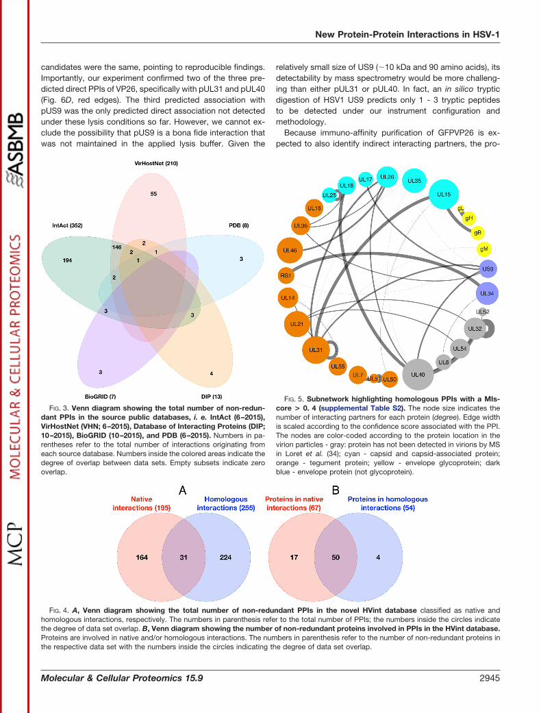
FIG. 3. Venn diagram showing the total number of non-redun-dant PPIs in the source public databases, i. e. IntAct (6–2015),VirHostNet (VHN; 6–2015), Database of Interacting Proteins (DIP;10–2015), BioGRID (10–2015), and PDB (6–2015). Numbers in pa-rentheses refer to the total number of interactions originating fromeach source database. Numbers inside the colored areas indicate thedegree of overlap between data sets. Empty subsets indicate zerooverlap.
FIG. 4. A, Venn diagram showing the total number of non-redundant PPIs in the novel HVint database classified as native andhomologous interactions, respectively. The numbers in parenthesis refer to the total number of PPIs; the numbers inside the circles indicatethe degree of data set overlap. B, Venn diagram showing the number of non-redundant proteins involved in PPIs in the HVint database.Proteins are involved in native and/or homologous interactions. The numbers in parenthesis refer to the number of non-redundant proteins inthe respective data set with the numbers inside the circles indicating the degree of data set overlap.
FIG. 5. Subnetwork highlighting homologous PPIs with a MIs-core > 0. 4 (supplemental Table S2). The node size indicates thenumber of interacting partners for each protein (degree). Edge widthis scaled according to the confidence score associated with the PPI.The nodes are color-coded according to the protein location in thevirion particles - gray: protein has not been detected in virions by MSin Loret et al. (34); cyan - capsid and capsid-associated protein;orange - tegument protein; yellow - envelope glycoprotein; darkblue - envelope protein (not glycoprotein).
New Protein-Protein Interactions in HSV-1
Molecular & Cellular Proteomics 15.9 2945
teins coisolated with GFPVP26 were compared with the pre-dicted, second order VP26 interactions (via pUL40 andpUL31, see Fig. 5). Indeed, the IP-MS experimental data setidentified the proteins pUL32, pUL18, pUL26, pUL52, pUL21,and pUL10 (gM). Binary interactions between pUL40 andeach of pUL32, pUL18, pUL26, and pUL52, as well as be-tween pUL31 and each of pUL26, pUL21 and gM are pre-dicted by our high-confidence homologous HVint subnetwork(MIscore 0.4). Additionally, the interaction between pUL31,a nuclear egress protein, and pUL32, a protein suggested tocontribute in efficient localization of newly synthesized cap-sids to nuclear replication compartments and DNA packag-ing, is present in our homology-transferred data set but withscores below the 0.4 cut-off. Overall, the experimental im-muno-affinity purification experiments provide compellingsupport for the homologous HVint network.
Interactive Web-based HVint Interface—All the data inHVint, including pairwise interactions, lines of evidence, andscores, are available for download in a standard format formolecular interactions data, namely PSI-MITAB 2.5. Addition-ally, we provide a user-friendly and intuitive interface tobrowse the data in a more straightforward and intuitive man-ner (http://topf-group.ismb.lon.ac.uk/hvint). The HVint inter-face displays an interactive graphical representation of thenetwork and allows easy access to annotation data for indi-vidual nodes and edges. These can be selected by clicking onthem or, in the case of nodes, specifying their UniProt ID in theprovided search tool. The subnetwork created by the firstneighbors of the selected element is then presented, togetherwith its associated data (including a list of interactions, con-fidence scores and supporting evidence) in tabular form.Other functionality of the interface includes the ability to filterinteractions based on user-defined confidence thresholds.
DISCUSSION
HVint was originally populated with data from five majorexternal public resources. Although some overlap exists in thedata coming from these databases, each contributes multipleunique interactions. A recurrent issue in interactomics studiesis the lack of network coverage, which severely limits theinformative power of the resulting networks (77, 78). There-fore, in retrieving data from the five resources, we distin-guished between two data sets. One data set contained na-tive interactions reported as directly detected for HSV-1, andthe second one was composed of homologous interactionsdetected in any of the other human herpesvirus species,covering all three herpesvirus subfamilies. The latter data setwas then used to predict PPIs in HSV-1 based on orthologyrelationships between the interacting partners in the homolo-gous species and HSV-1 proteins. Integrating these data in-creased the network coverage by more than twofold. Moreimportantly, it provides a set of putative novel interactions tobe validated in HSV-1. To provide a measure of the qualityand reliability of the data in the database, we implemented the
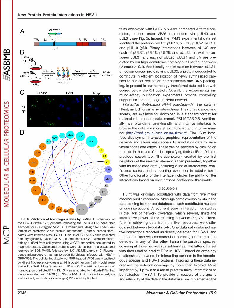
FIG. 6. Validation of homologous PPIs by IP-MS. A, Schematic ofthe HSV-1 (strain 17�) genome indicating the locus (UL35 gene) thatencodes for GFP-tagged VP26. B, Experimental design for IP-MS val-idation of predicted VP26 protein interactions. Primary human fibro-blasts were infected with HSV1 GFP or HSV1 GFPVP26, then collectedand cryogenically lysed. GFPVP26 and control GFP were immuno-affinity purified from cell lysates using �-GFP antibodies conjugated tomagnetic beads. Coisolated proteins were eluted from the beads andresolved by SDS-PAGE, followed by nLC-MS/MS analysis. C, Fluores-cence microscopy of human foreskin fibroblasts infected with HSV1-GFPVP26. The cellular localization of GFP-tagged VP26 was visualizedby direct fluorescence (green) at 14 h post-infection (hpi). Nuclei werestained by DAPI (blue). Scale bar � 20 �m. D, The HVint subnetwork ofhomologous predicted PPIs (Fig. 5) was annotated to indicate PPIs thatwere coisolated with VP26 (pUL35) by IP-MS. Both direct (red edges)and indirect, secondary (blue edges) PPIs are highlighted.
New Protein-Protein Interactions in HSV-1
2946 Molecular & Cellular Proteomics 15.9

MIscore (53) scoring function in our protocol. MIscore inte-grates data from multiple experiments that report a giveninteraction to calculate an overall confidence score, which isnormalized to the interval [0, 1]. It calculates weighted scoresbased on key variables: experimental detection method(s)(e.g. biophysical, imaging), type of interaction (e.g. physicalassociation, colocalization), and number of different scientificpublications reporting it. In our implementation we gave lowerweighting to orthology-transferred interactions by penalizingtheir original score in a sequence-identity dependent manner.An advantage of implementing this scoring scheme was thatit yielded a calibrated series of scores (ranging from 0 to 1),which allowed for fine-tuned selection of sets of interactionsbased on their confidence levels. We used this approach toselect a subset of high-confidence interactions for furtherexperimental testing.
Overall, our interactome, including 419 interactions,achieved a notably larger coverage than any individual sourcedatabase alone. This set is split between 195 native and 255homologous interactions, with a small subset of them over-lapping between the two (31 interactions), and it covers all 77proteins in the HSV-1 reference proteome (as reported inUniProt (48)). The data integration framework used in thisstudy (Fig. 1) is automated and relatively simple, which willallow future updates to be implemented easily as new inter-action data becomes available.
The prevalence of Y2H experiments in protein-protein in-teraction studies (79) is reflected in the large fraction of inter-actions obtained by this method in HVint (�90%, 380 inter-actions). This is because of the ability of Y2H experiments todetermine physical pairwise interactions between potentiallyinteracting partners, and from the feasibility of conductingboth small- and large-scale screenings. Since the publicationof the first large-scale protein interactions map for S. cerevi-siae in 2000 (80), multiple studies have followed and severalcaveats have been raised regarding the interpretation andreliability of Y2H data, especially when obtained from high-throughput screenings (81). The main concern is the largerfalse positive and false negative rates than in previous tradi-tional small-scale approaches. This has demanded a contin-ued optimization of the technique (as shown in recent reports(79, 81–83)), and the development of a range of alternativehybrid strategies to complement Y2H data sets (79). Theseapproaches have minimized the error rate in more recent data,suggesting that in the future the reliability of Y2H data couldbe assessed on a per data set basis in the future. HVintincludes data derived from Y2H experiments across a broadperiod of time. Consequently, we could not yet, at present,systematically assess the quality of each data set (as this isnot reflected in the source databases). The use of MIscore asa scoring method is advantageous in this respect; it assignscomparatively low scores to interactions detected by Y2Hexperiments, thereby representing a conservative approach inassessing the confidence of these interactions.
To test the predictive power of our computationally derivedinteraction data set, we subjected a subset of PPIs withhigh-confidence scores to experimental validation. To thisend, a number of homologous-only interactions that scoredabove 0.4 were selected. We focused our own validationanalysis on VP26 (encoded by UL35), because it is a proteinwhose functions are still unclear, and because several HSV-1strains in the F, 17� and KOS background have been gener-ated, in which VP26 has been successfully tagged with fluo-rescent proteins without or with minor impairment of HSV-1propagation (58, 60, 84–87). Hexamers of VP26 are locatedon top of the capsid hexons, which are hexamers of VP5, butnot on the pentons, which are pentamers of VP5. VP26 istherefore perfectly placed on the capsid surface for interac-tions with other viral and cellular components (74, 76, 88, 89).Mutants of pseudorabies virus (PRV), a porcine �-herpesvirus,or of HSV-1, that lack VP26, are less neuroinvasive and neu-rovirulent and grow to lower titers than their respective pa-rental strains (85, 90–92). VP26 can interact with the dyneinlight chains of the Tctex family in Y2H and coIP experiments.However, it is not required for efficient dynein-mediated trans-port toward the nucleus during cell entry, and dynein does notbind to un-tegumented capsids that expose VP26 over theirentire surface (74, 76, 84, 93). Homologous small capsidproteins of other herpesviruses have been shown to contrib-ute to capsid stability (94, 95).
Our HVint homologous subnetwork predicts that VP26 isassociated with pUL31 and pUL40 (Fig. 7). This is supportedby our experimental immuno-affinity isolation studies. VP26 ispresent in the mature virion particle, whereas pUL31 andpUL40 are not part of it (34, 88, 89, 96–99). Hence theseinteractions may be particularly relevant in understanding therole of VP26 in the viral life cycle. Notably, VP26 was alsocoisolated with pUL34, one of the known binding partners ofpUL31 (supplemental Table S3). Together, pUL31 and pUL34form the so-called nuclear egress complex, which is anchoredto the inner nuclear membrane by the C-terminal domain ofpUL34 and faces the nuclear lumen (99, 100). Given theconservation of the interaction between pUL31 and pUL34and their respective orthologs across the entire Herpesviridaefamily (101), many studies have explored their roles in theearly steps of the capsid assembly and the nuclear egresspathway. Besides being recruited directly to pUL34 at theinner nuclear envelope, pUL31 has also been reported tointeract via its N-terminal domain to newly synthesized nu-clear capsids (102). The pUL31-bound capsids are thenthought to be translocated to the inner nuclear membrane,where they associate via UL31 with pUL34 to mediate capsidegress from the nucleoplasm to the cytosol (102). Overall, thelocalization of VP26 to the capsid surface except the vertices,its documented roles in capsid trafficking, and our computa-tional and experimental findings of VP26 association withnuclear egress and CVSC complexes, suggests an as yetunknown role for VP26 in enforcing capsid stability and coor-
New Protein-Protein Interactions in HSV-1
Molecular & Cellular Proteomics 15.9 2947
dinating intra-nuclear capsid trafficking events (Fig. 7). Fur-ther, the recent in situ analyses of the NEC architecture re-vealed a curved hexagonal lattice for PRV (103, 104). AspUL31 and pUL34 are highly conserved in the Herpesviridaethis architecture is highly likely to be evident in HSV-1 as well.Therefore, an attractive hypothesis is that VP26 hexamers andpUL31/34 hexamers interact directly with each other. Future,in situ analysis will need to show whether this is in fact thecase.
Another pair of viral proteins, pUL39 and pUL40, whichconstitute the small and the large subunits (RR1 and RR2,respectively) of the heterotetrameric HSV-1 ribonucleotidereductase complex (105–108), were also coisolated with VP26by IP-MS analysis. Intriguingly, the latter was also predictedto interact with VP26 in our homologous subnetwork. In ad-dition to this enzymatic function, several studies have high-lighted a role for pUL39 as a virulence factor (106, 108–110),for instance by interfering with apoptotic cascades (106, 109–111). This again suggests a potential role for VP26 in the earlyevents of virus replication. Our experimental validation sup-
ports the interaction between VP26 via pUL40 to pUL39 andfurther to each of pUL32, pUL18 (VP23) and one or moreproducts of the HSV-1 UL26 gene, which encodes for anautocatalytic protease that is processed into the scaffoldproteins VP24 and VP21 (112, 113). Notably, all these inter-actions have been predicted in our homologous subnetwork.Further, a recent study has provided new insights into apossible role of pUL32 as a chaperone-like protein capable ofmodulating capsid maturation and tegument acquisition byinteracting with the capsid proteins pUL6 (the portal protein),pUL25, and pUL38 (VP19c) as well as the inner tegumentprotein pUL36 (114).
Among the membrane proteins, our HVint interactome pro-vides a particularly high number of novel interactions forpUS9. pUS9 is a small type II membrane protein of about 10kDa that localizes to the trans-Golgi network and axonal ves-icles of neuronal cells (115–117). Our HSV-1 interactome pre-dicts interactions between pUS9 and the capsid proteinsVP26 and pUL17, as well as the inner-tegument proteinpUL36. pUL17 and pUL25 form the Capsid Vertex-specific
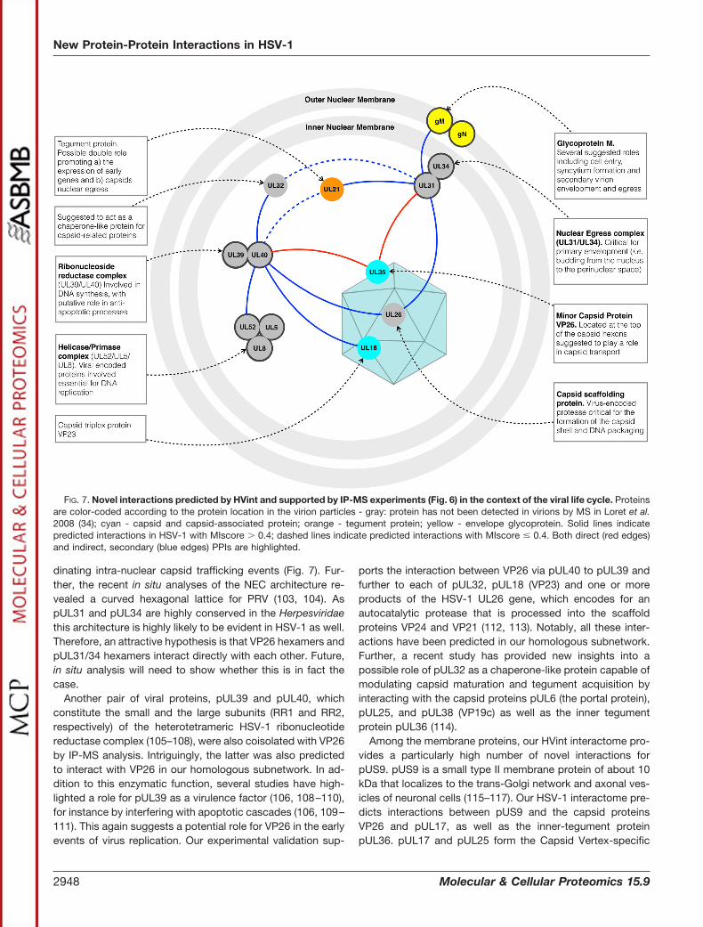
FIG. 7. Novel interactions predicted by HVint and supported by IP-MS experiments (Fig. 6) in the context of the viral life cycle. Proteinsare color-coded according to the protein location in the virion particles - gray: protein has not been detected in virions by MS in Loret et al.2008 (34); cyan - capsid and capsid-associated protein; orange - tegument protein; yellow - envelope glycoprotein. Solid lines indicatepredicted interactions in HSV-1 with MIscore 0.4; dashed lines indicate predicted interactions with MIscore � 0.4. Both direct (red edges)and indirect, secondary (blue edges) PPIs are highlighted.
New Protein-Protein Interactions in HSV-1
2948 Molecular & Cellular Proteomics 15.9

component (CVSC) located on the pentons edges and pUL17and pUL25 are both required for stable DNA packaging intocapsids prior to nuclear egress (70, 118, 119). pUL36 is aninner protein known to be involved in early tegumentationevents together with its binding partner pUL37 (72, 84). Likethe CVSC, the complex pUL36-pUL37 is also located aroundpenton vertices. Interestingly, PPIs and coordinated eventsbetween pUL36-pUL37 and CVSC have been previouslyshown (72). Together with the heterodimer gE/gI, pUS9 me-diates anterograde axonal transport of viral tegument, glyco-proteins, and virions of �-herpesviruses in neurons, but theseproteins are not essential for replication in epithelial or fibro-blasts cells (40–42). Although studies on the gE/gI and pUS9-mediated transport of PRV structures show that PRV is trans-ported along axons as fully assembled virions in transportvesicles, the data on HSV-1 regarding the subviral and viralparticles being targeted to axons is still controversial (116,117, 120). Because the HSV-1 or PRV proteins that mightcontribute to these differences are not known, our novel pre-dicted interactions from HVint generate new hypotheses onpotentially virus-specific regulators of US9-dependent an-terograde transport.
The nodes outside the main cluster of the 35 new interac-tions involve those forming two heterodimers conservedacross the entire Herpesviridae family, namely gH/gL andpUL51-pUL7, and one self-interaction for pUL25. The com-plex gH/gL was structurally solved in HSV-2 by x-ray crystal-lography in 2010 (121). Given the high confidence of theexperimental data and the degree of sequence similarity be-tween HSV-1 and HSV-2 gH/gL sequences, the existence ofthis complex in HSV-1 is undisputed (121–123). Although itsspecific role is yet to be fully defined, it is likely involved inregulating the activity of the membrane fusion glycoprotein gB(121).
Finally, complex formation between pUL51 and pUL7, aspredicted in our HVint database, was confirmed recently byaffinity purification and tandem MS experiments (75). At thetime of writing, these data were not integrated into the sourcePPI databases used in this work. pUL51 and pUL7 are bothconserved across the whole Herpesviridae family. pUL51 is anN terminus membrane-anchored tegument protein (124–126)with no apparent enzymatic function, suggested to play a rolein viral egress, viral envelopment and epithelial cell-to-cellspread (127, 128). Its interaction with pUL7 seems to berequired for efficient recruitment of the latter to cytoplasmicmembranes leading to its incorporation into the virion. Bothproteins partially colocalize in cytoplasmic membranes to-gether, as observed in the case of pUS9, with glycoprotein gE(128). This implies that the complex is non-obligate and eachprotein could carry out independent functional activities.pUL7 was also found inside the nuclear and cytoplasmiccompartments in the absence of pUL51, and interactionsbetween pUL7 and capsid proteins have been reported. As inthe case of pUS9, the complex between pUL51 and pUL7
supports the idea that viral membrane proteins are principalagents in tegumentation and secondary envelopment pro-cesses. By establishing interactions with capsid and capsid-associated proteins they guide progeny virion particles todifferent cytoplasmic compartments (e.g. trans Golgi networkor endosomes) until they reach the plasma membrane (42,129–134).
CONCLUSIONS
Here we introduce the HVint database, an integrative re-source for HSV-1 protein interaction data. The database con-tains interactions experimentally detected in HSV-1 speciesand computationally predicted using evolutionary information.Interactions in the database have been scored, thus confi-dently providing novel predicted interactions. Consequently,HVint can be used as a tool to prioritize candidate interac-tions for future experimental testing. The protocol used tocreate and populate the database was kept simple, hencefacilitating database updates. In the future, our aim is tofurther improve our methods for assessing the reliability ofprotein interaction data and to include more advanced fea-tures in the HVint graphical interface to accommodateusers’ needs. We also plan to extend the current databaseto other human herpesvirus species and to virus-hostinteractions.
Acknowledgments—We thank Dr. David Houldershaw for com-puter support. We thank Drs. Tzviya Zeev-Ben-Mordehai andDaven Vasishtan for helpful discussions. We thank Ben Diner,Marni Crow, and Minghao Li for technical assistance with controlisolations.
* The work was supported by the MRC [G0600084] (M.T.) and[MR/M019292/1] (M.T., K.G.), the Leverhulme Trust [RPG-2012–519](M.T., K.G.), the Niedersachsen Research Network on Neuroinfecti-ology (NRENNT) of the Ministry of Science and Culture of LowerSaxony, Germany (A.B., B.S.), the Wellcome Trust [090895/Z/09/Z,090532/Z/09/Z, 107806/Z/15/Z,] (K.G.), the HFSP [RGY0079/2009-C](K.G., M.T., I.C.], and grants from the National Institutes of Health(AI102187 and GM114141) (I.M.C), NJCCR fellowship to T.M.G. Thecontent is solely the responsibility of the authors and does not nec-essarily represent the official views of the National Institutes of Health.
□S This article contains supplemental material.** To whom correspondence should be addressed: Institute of
Structural and Molecular Biology, Birkbeck College, Malet St., Lon-don WC1E 7HX, United Kingdom. Tel.: �44-(0)20 7079 0886; E-mail:[email protected]; [email protected].
‡‡ These authors contributed equally to this work.
REFERENCES
1. Engel, E. A., Song, R., Koyuncu, O. O., and Enquist, L. W. (2015) Inves-tigating the biology of alpha herpesviruses with MS-based proteomics.Proteomics 15, 1943–1956
2. Brady, R. C., and Bernstein, D. I. (2004) Treatment of herpes simplex virusinfections. Antiviral Res. 61, 73–81
3. Chentoufi, A. A., and Benmohamed, L. (2012) Mucosal herpes immunityand immunopathology to ocular and genital herpes simplex virus infec-tions. Clin. Dev. Immunol. 2012, 149135
4. Lovheim, H., Gilthorpe, J., Adolfsson, R., Nilsson, L. G., and Elgh, F. (2014)Reactivated herpes simplex infection increases the risk of Alzheimer’sdisease. Alzheimers Dement. 11, 593–599
New Protein-Protein Interactions in HSV-1
Molecular & Cellular Proteomics 15.9 2949

5. Rollenhagen, C., Lathrop, M. J., Macura, S. L., Doncel, G. F., and Asin,S. N. (2014) Herpes simplex virus type-2 stimulates HIV-1 replication incervical tissues: implications for HIV-1 transmission and efficacy ofanti-HIV-1 microbicides. Mucosal Immunol. 7, 1165–1174
6. Marcoux, J., and Cianferani, S. (2015) Towards integrative structural massspectrometry: Benefits from hybrid approaches. Methods 89, 4–12
7. Stelzl, U., Worm, U., Lalowski, M., Haenig, C., Brembeck, F. H., Goehler,H., Stroedicke, M., Zenkner, M., Schoenherr, A., Koeppen, S., Timm, J.,Mintzlaff, S., Abraham, C., Bock, N., Kietzmann, S., Goedde, A., Tok-soz, E., Droege, A., Krobitsch, S., Korn, B., Birchmeier, W., Lehrach, H.,and Wanker, E. E. (2005) A human protein-protein interaction network:A resource for annotating the proteome. Cell 122, 957–968
8. Deng, M., Zhang, K., Mehta, S., Chen, T., and Sun, F. (2002) Prediction ofprotein function using protein-protein interaction data. Proc. IEEE Com-put. Soc. Bioinform. Conf. 1, 197–206
9. Jaeger, S., and Aloy, P. (2012) From protein interaction networks to noveltherapeutic strategies. IUBMB Life 64, 529–537
10. de Chassey, B., Meyniel-Schicklin, L., Vonderscher, J., Andre, P., andLotteau, V. (2014) Virus-host interactomics: new insights and opportu-nities for antiviral drug discovery. Genome Med. 6, 115
11. Nourani, E., Khunjush, F., and Durmus, S. (2015) Computational ap-proaches for prediction of pathogen-host protein-protein interactions.Front. Microbiol. 6, 94
12. Kshirsagar, M., Carbonell, J., and Klein-Seetharaman, J. (2012) Tech-niques to cope with missing data in host-pathogen protein interactionprediction. Bioinformatics 28, i466–i472
13. Durmus Tekir, S. D., and Ulgen, K. O. (2013) Systems biology of pathogen-host interaction: Networks of protein-protein interaction within patho-gens and pathogen-human interactions in the post-genomic era. Bio-technol. J. 8, 85–96
14. Greco, T. M., Diner, B. A., and Cristea, I. M. (2014) The Impact of MassSpectrometry–Based Proteomics on Fundamental Discoveries in Virol-ogy. Annu. Rev. Virol. 1, 581–604
15. DeBlasio, S. L., Chavez, J. D., Alexander, M. M., Ramsey, J., Eng, J. K.,Mahoney, J., Gray, S. M., Bruce, J. E., and Cilia, M. (2015) Visualizationof host-polerovirus interaction topologies using Protein Interaction Re-porter technology. J. Virol. 90, 1973–1987
16. Ramisetty, S. R., and Washburn, M. P. (2011) Unraveling the dynamics ofprotein interactions with quantitative mass spectrometry. Crit. Rev.Biochem. Mol. Biol. 46, 216–228
17. Orchard, S., Ammari, M., Aranda, B., Breuza, L., Briganti, L., Broackes-Carter, F., Campbell, N. H., Chavali, G., Chen, C., Del-Toro, N., Dues-bury, M., Dumousseau, M., Galeota, E., Hinz, U., Iannuccelli, M., Jag-annathan, S., Jimenez, R., Khadake, J., Lagreid, A., Licata, L., Lovering,R. C., Meldal, B., Melidoni, A. N., Milagros, M., Peluso, D., Perfetto, L.,Porras, P., Raghunath, A., Ricard-Blum, S., Roechert, B., Stutz, A.,Tognolli, M., van Roey, K., Cesareni, G., and Hermjakob, H. (2014) TheMIntAct project - IntAct as a common curation platform for 11 molecularinteraction databases. Nucleic Acids Res. 42, D358–D63
18. Berman, H. M. (2000) The Protein Data Bank. Nucleic Acids Res. 28,235–242
19. Lawson, C. L., Baker, M. L., Best, C., Bi, C., Dougherty, M., Feng, P., vanGinkel, G., Devkota, B., Lagerstedt, I., Ludtke, S. J., Newman, R. H.,Oldfield, T. J., Rees, I., Sahni, G., Sala, R., Velankar, S., Warren, J.,Westbrook, J. D., Henrick, K., Kleywegt, G. J., Berman, H. M., and Chiu,W. (2011) EMDataBank.org: unified data resource for CryoEM. NucleicAcids Res. 39, D456–64
20. Guirimand, T., Delmotte, S., and Navratil, V. (2015) VirHostNet 2.0: surfingon the web of virus/host molecular interactions data. Nucleic Acids Res.43, D583–D7
21. Szklarczyk, D., Franceschini, A., Wyder, S., Forslund, K., Heller, D.,Huerta-Cepas, J., Simonovic, M., Roth, A., Santos, A., Tsafou, K. P.,Kuhn, M., Bork, P., Jensen, L. J., and von Mering, C. (2015) STRINGv10: protein-protein interaction networks, integrated over the tree of life.Nucleic Acids Res. 43, D447–D52
22. Budayeva, H. G., and Cristea, I. M. (2014) A mass spectrometry view ofstable and transient protein interactions. Adv. Exp. Med. Biol. 806,263–282
23. McDowall, M. D., Scott, M. S., and Barton, G. J. (2009) PIPs: humanprotein-protein interaction prediction database. Nucleic Acids Res. 37,D651–D6
24. Hosur, R., Peng, J., Vinayagam, A., Stelzl, U., Xu, J., Perrimon, N., Bien-kowska, J., and Berger, B. (2012) A computational framework for boost-ing confidence in high-throughput protein-protein interaction datasets.Genome Biol. 13, R76
25. Zahiri, J., Bozorgmehr, J. H., and Masoudi-Nejad, A. (2013) Computa-tional prediction of protein-protein interaction networks: algorithms andresources. Curr. Genomics 14, 397–414
26. Hosur, R., Xu, J., Bienkowska, J., and Berger, B. (2011) iWRAP: Aninterface threading approach with application to prediction of cancer-related protein-protein interactions. J. Mol. Biol. 405, 1295–1310
27. Singh, R., Park, D., Xu, J., Hosur, R., and Berger, B. (2010) Struct2Net: aweb service to predict protein-protein interactions using a structure-based approach. Nucleic Acids Res. 38, W508–W15
28. You, Z. H., Chan, K. C. C., and Hu, P. (2015) Predicting protein-proteininteractions from primary protein sequences using a novel multi-scalelocal feature representation scheme and the random forest. PLoS ONE10, e0125811
29. Zhang, Y. N., Pan, X. Y., Huang, Y., and Shen, H. Bin (2011) Adaptivecompressive learning for prediction of protein-protein interactions fromprimary sequence. J. Theor. Biol. 283, 44–52
30. Folador, E. L., Hassan, S. S., Lemke, N., Barh, D., Silva, A., Ferreira, R. S.,and Azevedo, V. (2014) An improved interolog mapping-based compu-tational prediction of protein–protein interactions with increased net-work coverage. Integr. Biol. 6, 1080–1087
31. Yu, H., Luscombe, N. M., Lu, H. X., Zhu, X., Xia, Y., Han, J. D. J., Bertin,N., Chung, S., Vidal, M., and Gerstein, M. (2004) Annotation transferbetween genomes: protein-protein interologs and protein-DNA regu-logs. Genome Res. 14, 1107–1118
32. Murakami, Y., and Mizuguchi, K. (2014) Homology-based prediction ofinteractions between proteins using Averaged One-Dependence Esti-mators. BMC Bioinformatics 15, 213
33. Saeed, R., and Deane, C. (2008) An assessment of the uses of homolo-gous interactions. Bioinformatics 24, 689–695
34. Loret, S., Guay, G., and Lippe, R. (2008) Comprehensive characterizationof extracellular herpes simplex virus type 1 virions. J. Virol. 82,8605–8618
35. Grunewald, K., Desai, P., Winkler, D. C., Heymann, J. B., Belnap, D. M.,Baumeister, W., and Steven, A. C. (2003) Three-dimensional structure ofherpes simplex virus from cryo-electron tomography. Science 302,1396–1398
36. Xenarios, I., Fernandez, E., Salwinski, L., Duan, X. J., Thompson, M. J.,Marcotte, E. M., and Eisenberg, D. (2001) DIP: The Database of Inter-acting Proteins: 2001 update. Nucleic Acids Res. 29, 239–241
37. Chatr-Aryamontri, A., Breitkreutz, B. J., Oughtred, R., Boucher, L., Hei-nicke, S., Chen, D., Stark, C., Breitkreutz, A., Kolas, N., O’Donnell, L.,Reguly, T., Nixon, J., Ramage, L., Winter, A., Sellam, A., Chang, C.,Hirschman, J., Theesfeld, C., Rust, J., Livstone, M. S., Dolinski, K., andTyers, M. (2015) The BioGRID interaction database: 2015 update. Nu-cleic Acids Res. 43, D470–D8
38. Calderwood, M. A., Venkatesan, K., Xing, L., Chase, M. R., Vazquez, A.,Holthaus, A. M., Ewence, A. E., Li, N., Hirozane-Kishikawa, T., Hill, D. E.,Vidal, M., Kieff, E., and Johannsen, E. (2007) Epstein-Barr virus andvirus human protein interaction maps. Proc. Natl. Acad. Sci. U.S.A. 104,7606–7611
39. Panagiotidis, C. A., Lium, E. K., and Silverstein, S. J. (1997) Physical andfunctional interactions between herpes simplex virus immediate-earlyproteins ICP4 and ICP27. J. Virol. 71, 1547–1557
40. Rozen, R., Sathish, N., Li, Y., and Yuan, Y. (2008) Virion-wide proteininteractions of Kaposi’s sarcoma-associated herpesvirus. J. Virol. 82,4742–4750
41. Taylor, T. J., and Knipe, D. M. (2004) Proteomics of herpes simplex virusreplication compartments: association of cellular DNA replication, re-pair, recombination, and chromatin remodeling proteins with ICP8.J. Virol. 78, 5856–5866
42. Vittone, V., Diefenbach, E., Triffett, D., Douglas, M. W., Cunningham, A. L.,and Diefenbach, R. J. (2005) Determination of interactions betweentegument proteins of herpes simplex virus type 1. J. Virol. 79,9566–9571
43. To, A., Bai, Y., Shen, A., Gong, H., Umamoto, S., Lu, S., and Liu, F. (2011)Yeast two hybrid analyses reveal novel binary interactions betweenhuman cytomegalovirus-encoded virion proteins. PLoS ONE 6, e17796
New Protein-Protein Interactions in HSV-1
2950 Molecular & Cellular Proteomics 15.9
44. Lee, J. H., Vittone, V., Diefenbach, E., Cunningham, A. L., and Diefenbach,R. J. (2008) Identification of structural protein-protein interactions ofherpes simplex virus type 1. Virology 378, 347–354
45. Uetz, P., Dong, Y. A., Zeretzke, C., Atzler, C., Baiker, A., Berger, B.,Rajagopala, S. V., Roupelieva, M., Rose, D., Fossum, E., and Haas, J.(2006) Herpesviral protein networks and their interaction with the humanproteome. Science 311, 239–242
46. Stellberger, T., Hauser, R., Baiker, A., Pothineni, V. R., Haas, J., and Uetz,P. (2010) Improving the yeast two-hybrid system with permutatedfusions proteins: the Varicella Zoster Virus interactome. ProteomeSci. 8, 8
47. Fossum, E., Friedel, C. C., Rajagopala, S. V., Titz, B., Baiker, A., Schmidt,T., Kraus, T., Stellberger, T., Rutenberg, C., Suthram, S., Bandyopad-hyay, S., Rose, D., von Brunn, A., Uhlmann, M., Zeretzke, C., Dong,Y. A., Boulet, H., Koegl, M., Bailer, S. M., Koszinowski, U., Ideker, T.,Uetz, P., Zimmer, R., and Haas, J. (2009) Evolutionarily conservedherpesviral protein interaction networks. PLoS Pathog. 5, e1000570
48. The UniProt Consortium (2014) UniProt: a hub for protein information.Nucleic Acids Res. 43, D204–D212
49. Remmert, M., Biegert, A., Hauser, A., and Soding, J. (2012) HHblits:lightning-fast iterative protein sequence searching by HMM-HMM align-ment. Nat. Methods 9, 173–175
50. Suzek, B. E., Wang, Y., Huang, H., McGarvey, P. B., and Wu, C. H. (2015)UniRef clusters: a comprehensive and scalable alternative for improvingsequence similarity searches. Bioinformatics 31, 926–932
51. Bastian, M., Heymann, S., and Jacomy, M. (2009) in International AAAIConference on Weblogs and Social Media
52. Aranda, B., Blankenburg, H., Kerrien, S., Brinkman, F. S. L., Ceol, A.,Chautard, E., Dana, J. M., De Las Rivas, J., Dumousseau, M., Galeota,E., Gaulton, A., Goll, J., Hancock, R. E. W., Isserlin, R., Jimenez, R. C.,Kerssemakers, J., Khadake, J., Lynn, D. J., Michaut, M., O’Kelly, G.,Ono, K., Orchard, S., Prieto, C., Razick, S., Rigina, O., Salwinski, L.,Simonovic, M., Velankar, S., Winter, A., Wu, G., Bader, G. D., Cesareni,G., Donaldson, I. M., Eisenberg, D., Kleywegt, G. J., Overington, J.,Ricard-Blum, S., Tyers, M., Albrecht, M., and Hermjakob, H. (2011)PSICQUIC and PSISCORE: accessing and scoring molecular interac-tions. Nat. Methods 8, 528–529
53. Villaveces, J. M., Jimenez, R. C., Porras, P., Del-Toro, N., Duesbury, M.,Dumousseau, M., Orchard, S., Choi, H., Ping, P., Zong, N. C., Askenazi,M., Habermann, B. H., and Hermjakob, H. (2015) Merging and scoringmolecular interactions utilising existing community standards: tools,use-cases and a case study. Database 2015, bau131
54. Lopez-Blanco, J. R., and Chacon, P. (2015) Structural modeling fromelectron microscopy data. Wiley Interdiscip. Rev. Comput. Mol. Sci. 5,62–81
55. Mora, A., and Donaldson, I. M. (2012) Effects of protein interaction dataintegration, representation and reliability on the use of network proper-ties for drug target prediction. BMC Bioinformatics 13, 294
56. Kerrien, S., Orchard, S., Montecchi-Palazzi, L., Aranda, B., Quinn, A. F.,Vinod, N., Bader, G. D., Xenarios, I., Wojcik, J., Sherman, D., Tyers, M.,Salama, J. J., Moore, S., Ceol, A., Chatr-Aryamontri, A., Oesterheld, M.,Stumpflen, V., Salwinski, L., Nerothin, J., Cerami, E., Cusick, M. E.,Vidal, M., Gilson, M., Armstrong, J., Woollard, P., Hogue, C., Eisenberg,D., Cesareni, G., Apweiler, R., and Hermjakob, H. (2007) Broadening thehorizon–level 2.5 of the HUPO-PSI format for molecular interactions.BMC Biol. 5, 44
57. Deutsch, E. W., Albar, J. P., Binz, P.-A., Eisenacher, M., Jones, A. R.,Mayer, G., Omenn, G. S., Orchard, S., Vizcaíno, J. A., and Hermjakob,H. (2015) Development of data representation standards by the humanproteome organization proteomics standards initiative. J. Am. Med.Inform. Assoc. 22, 495–506
58. Nagel, C. H., Dohner, K., Binz, A., Bauerfeind, R., and Sodeik, B. (2012)Improper tagging of the non-essential small capsid protein VP26 impairsnuclear capsid egress of herpes simplex virus. PLoS ONE 7, e44177
59. Snijder, B., Sacher, R., Ramo, P., Liberali, P., Mench, K., Wolfrum, N.,Burleigh, L., Scott, C. C., Verheije, M. H., Mercer, J., Moese, S., Heger,T., Theusner, K., Jurgeit, A., Lamparter, D., Balistreri, G., Schelhaas, M.,De Haan, C. A. M., Marjomaki, V., Hyypia, T., Rottier, P. J. M., Sodeik,B., Marsh, M., Gruenberg, J., Amara, A., Greber, U., Helenius, A., andPelkmans, L. (2012) Single-cell analysis of population context advancesRNAi screening at multiple levels. Mol. Syst. Biol. 8, 579
60. Rowles, D. L., Tsai, Y. C., Greco, T. M., Lin, A. E., Li, M., Yeh, J., andCristea, I. M. (2015) DNA methyltransferase DNMT3A associates withviral proteins and impacts HSV-1 infection. Proteomics 15,1968–1982
61. Lin, A. E., Greco, T. M., Dohner, K., Sodeik, B., and Cristea, I. M. (2013) Aproteomic perspective of inbuilt viral protein regulation: pUL46 tegu-ment protein is targeted for degradation by ICP0 during herpes simplexvirus type 1 infection. Mol. Cell. Proteomics 12, 3237–3252
62. Cristea, I. M., Williams, R., Chait, B. T., and Rout, M. P. (2005) Fluorescentproteins as proteomic probes. Mol. Cell. Proteomics 4, 1933–1941
63. Kulak, N. a, Pichler, G., Paron, I., Nagaraj, N., and Mann, M. (2014)Minimal, encapsulated proteomic-sample processing applied to copy-number estimation in eukaryotic cells. Nat. Methods 11, 319–324
64. Vizcaíno, J. A., Csordas, A., Del-Toro, N., Dianes, J. A., Griss, J., Lavidas,I., Mayer, G., Perez-Riverol, Y., Reisinger, F., Ternent, T., Xu, Q. W.,Wang, R., and Hermjakob, H. (2016) 2016 update of the PRIDE data-base and its related tools. Nucleic Acids Res. 44, D447–D56
65. Silva, J. C., Gorenstein, M. V., Li, G. Z., Vissers, J. P. C., and Geromanos,S. J. (2006) Absolute quantification of proteins by LCMSE: a virtue ofparallel MS acquisition. Mol. Cell. Proteomics 5, 144–156
66. Calderone, A., Castagnoli, L., and Cesareni, G. (2013) mentha: a resourcefor browsing integrated protein-interaction networks. Nat. Methods 10,690–691
67. Blohm, P., Frishman, G., Smialowski, P., Goebels, F., Wachinger, B.,Ruepp, A., and Frishman, D. (2014) Negatome 2.0: a database ofnon-interacting proteins derived by literature mining, manual annotationand protein structure analysis. Nucleic Acids Res. 42, D396–D400
68. Winterbach, W., Van Mieghem, P., Reinders, M., Wang, H., and de Ridder,D. (2013) Topology of molecular interaction networks. BMC Syst. Biol. 7,90
69. Connolly, S. A., Jackson, J. O., Jardetzky, T. S., and Longnecker, R.(2011) Fusing structure and function: a structural view of the herpesvirusentry machinery. Nat. Rev. Microbiol. 9, 369–381
70. Toropova, K., Huffman, J. B., Homa, F. L., and Conway, J. F. (2011) Theherpes simplex virus 1 UL17 protein is the second constituent of thecapsid vertex-specific component required for DNA packaging andretention. J. Virol. 85, 7513–7522
71. Trus, B. L., Homa, F. L., Booy, F. P., Newcomb, W. W., Thomsen, D. R.,Cheng, N., Brown, J. C., and Steven, A. C. (1995) Herpes simplex viruscapsids assembled in insect cells infected with recombinant baculovi-ruses: structural authenticity and localization of VP26. J. Virol. 69,7362–7366
72. Cardone, G., Newcomb, W. W., Cheng, N., Wingfield, P. T., Trus, B. L.,Brown, J. C., and Steven, A. C. (2012) The UL36 Tegument Protein ofHerpes Simplex Virus 1 Has a Composite Binding Site at the CapsidVertices. J. Virol. 86, 4058–4064
73. Zhou, Z. H., He, J., Jakana, J., Tatman, J. D., Rixon, F. J., and Chiu, W.(1995) Assembly of VP26 in herpes simplex virus-1 inferred from struc-tures of wild-type and recombinant capsids. Nat. Struct. Biol. 2,1026–1030
74. Douglas, M. W., Diefenbach, R. J., Homa, F. L., Miranda-Saksena, M.,Rixon, F. J., Vittone, V., Byth, K., and Cunningham, A. L. (2004) HerpesSimplex Virus Type 1 Capsid Protein VP26 Interacts with Dynein LightChains RP3 and Tctex1 and Plays a Role in Retrograde Cellular Trans-port. J. Biol. Chem. 279, 28522–28530
75. Roller, R. J., and Fetters, R. (2015) The Herpes Simplex Virus 1 UL51Protein Interacts with the UL7 Protein and Plays a Role in Its Recruit-ment into the Virion. J. Virol. 89, 3112–3122
76. Dohner, K., Radtke, K., Schmidt, S., and Sodeik, B. (2006) Eclipse phaseof herpes simplex virus type 1 infection: Efficient dynein-mediatedcapsid transport without the small capsid protein VP26. J. Virol. 80,8211–8224
77. Navlakha, S., and Kingsford, C. (2010) The power of protein interactionnetworks for associating genes with diseases. Bioinformatics 26,1057–1063
78. Marras, E., Travaglione, A., Chaurasia, G., Futschik, M., and Capobianco,E. (2010) Inferring modules from human protein interactome classes.BMC Syst. Biol. 4, 102
79. Bruckner, A., Polge, C., Lentze, N., Auerbach, D., and Schlattner, U. (2009)Yeast two-hybrid, a powerful tool for systems biology. Int. J. Mol. Sci.10, 2763–2788
New Protein-Protein Interactions in HSV-1
Molecular & Cellular Proteomics 15.9 2951

80. Uetz, P., Giot, L., Cagney, G., Mansfield, T. A., Judson, R. S., Knight, J. R.,Lockshon, D., Narayan, V., Srinivasan, M., Pochart, P., Qureshi-Emili,A., Li, Y., Godwin, B., Conover, D., Kalbfleisch, T., Vijayadamodar, G.,Yang, M., Johnston, M., Fields, S., and Rothberg, J. M. (2000) A com-prehensive analysis of protein-protein interactions in Saccharomycescerevisiae. Nature 403, 623–627
81. Grunenfelder, B., and Winzeler, E. A. (2002) Treasures and traps in ge-nome-wide data sets: case examples from yeast. Nat. Rev. Genet. 3,653–661
82. Chen, Y. C., Rajagopala, S. V., Stellberger, T., and Uetz, P. (2010) Ex-haustive benchmarking of the yeast two-hybrid system. Nat. Methods 7,667–668
83. Sprinzak, E., Sattath, S., and Margalit, H. (2003) How reliable are experi-mental protein-protein interaction data? J. Mol. Biol. 327, 919–923
84. Desai, P., Sexton, G. L., Huang, E., and Person, S. (2008) Localization ofherpes simplex virus type 1 UL37 in the Golgi complex requires UL36but not capsid structures. J. Virol. 82, 11354–11361
85. Nagel, C. H., Dohner, K., Fathollahy, M., Strive, T., Borst, E. M., Mes-serle, M., and Sodeik, B. (2008) Nuclear egress and envelopment ofherpes simplex virus capsids analyzed with dual-color fluorescenceHSV1(17�). J. Virol. 82, 3109–3124
86. de Oliveira, A. P., Glauser, D. L., Laimbacher, A. S., Strasser, R., Schraner,E. M., Wild, P., Ziegler, U., Breakefield, X. O., Ackermann, M., andFraefel, C. (2008) Live visualization of herpes simplex virus type 1compartment dynamics. J. Virol. 82, 4974–4990
87. Sugimoto, K., Uema, M., Sagara, H., Tanaka, M., Sata, T., Hashimoto, Y.,and Kawaguchi, Y. (2008) Simultaneous tracking of capsid, tegument,and envelope protein localization in living cells infected with triply fluo-rescent herpes simplex virus 1. J. Virol. 82, 5198–5211
88. Desai, P., Akpa, J. C., and Person, S. (2003) Residues of VP26 of herpessimplex virus type 1 that are required for its interaction with capsids.J. Virol. 77, 391–404
89. Antinone, S. E., Shubeita, G. T., Coller, K. E., Lee, J. I., Haverlock-Moyns, S., Gross, S. P., and Smith, G. A. (2006) The Herpesviruscapsid surface protein, VP26, and the majority of the tegument pro-teins are dispensable for capsid transport toward the nucleus. J. Virol.80, 5494–5498
90. Desai, P., and Person, S. (1998) Incorporation of the green fluorescentprotein into the herpes simplex virus type 1 capsid. J. Virol. 72,7563–7568
91. Krautwald, M., Maresch, C., Klupp, B. G., Fuchs, W., and Mettenleiter,T. C. (2008) Deletion or green fluorescent protein tagging of the pUL35capsid component of pseudorabies virus impairs virus replication in cellculture and neuroinvasion in mice. J. Gen. Virol. 89, 1346–1351
92. Kobayashi, R., Kato, A., Oda, S., Koyanagi, N., Oyama, M., Kozuka-Hata,H., Arii, J., and Kawaguchi, Y. (2015) Function of the Herpes SimplexVirus 1 Small Capsid Protein VP26 Is Regulated by Phosphorylation ata Specific Site. J. Virol. 89, 6141–6147
93. Radtke, K., Kieneke, D., Wolfstein, A., Michael, K., Steffen, W., Scholz, T.,Karger, A., and Sodeik, B. (2010) Plus- and minus-end directed micro-tubule motors bind simultaneously to herpes simplex virus capsidsusing different inner tegument structures. PLoS Pathog. 6, 1–20
94. Dai, X., Yu, X., Gong, H., Jiang, X., Abenes, G., Liu, H., Shivakoti, S., Britt,W. J., Zhu, H., Liu, F., and Zhou, Z. H. (2013) The smallest capsidprotein mediates binding of the essential tegument protein pp150 tostabilize DNA-containing capsids in human cytomegalovirus. PLoS Pat-hog. 9, e1003525
95. Dai, X., Gong, D., Xiao, Y., Wu, T.-T., Sun, R., and Zhou, Z. H. (2015)CryoEM and mutagenesis reveal that the smallest capsid protein ce-ments and stabilizes Kaposi’s sarcoma-associated herpesvirus capsid.Proc. Natl. Acad. Sci. U.S.A. 112, E649–E56
96. Booy, F. P., Trus, B. L., Newcomb, W. W., Brown, J. C., Conway, J. F., andSteven, A. C. (1994) Finding a needle in a haystack: detection of a smallprotein (the 12-kDa VP26) in a large complex (the 200-MDa capsid ofherpes simplex virus). Proc. Natl. Acad. Sci. U.S.A. 91, 5652–5656
97. Chen, D. H., Jakana, J., McNab, D., Mitchell, J., Zhou, Z. H., Dougherty, M.,Chiu, W., and Rixon, F. J. (2001) The pattern of tegument-capsid interac-tion in the herpes simplex virus type 1 virion is not influenced by the smallhexon-associated protein VP26. J. Virol. 75, 11863–11867
98. Yang, K., Wills, E., Lim, H. Y., Zhou, Z. H., and Baines, J. D. (2014)Association of herpes simplex virus pUL31 with capsid vertices and
components of the capsid vertex-specific complex. J. Virol. 88,3815–3825
99. Reynolds, A. E., Ryckman, B. J., Baines, J. D., Zhou, Y., Liang, L., andRoller, R. J. (2001) U(L)31 and U(L)34 proteins of herpes simplex virustype 1 form a complex that accumulates at the nuclear rim and isrequired for envelopment of nucleocapsids. J. Virol. 75, 8803–8817
100. Wills, E., Mou, F., and Baines, J. D. (2009) The U(L)31 and U(L)34 geneproducts of herpes simplex virus 1 are required for optimal localizationof viral glycoproteins D and M to the inner nuclear membranes ofinfected cells. J. Virol. 83, 4800–4809
101. Schnee, M., Ruzsics, Z., Bubeck, A., and Koszinowski, U. H. (2006)Common and specific properties of herpesvirus UL34/UL31 proteinfamily members revealed by protein complementation assay. J. Virol.80, 11658–11666
102. Funk, C., Ott, M., Raschbichler, V., Nagel, C. H., Binz, A., Sodeik, B.,Bauerfeind, R., and Bailer, S. M. (2015) The Herpes Simplex VirusProtein pUL31 Escorts Nucleocapsids to Sites of Nuclear Egress, aProcess Coordinated by Its N-Terminal Domain. PLoS Pathog. 11,e1004957
103. Hagen, C., Dent, K. C., Zeev-Ben-Mordehai, T., Grange, M., Bosse, J. B.,Whittle, C., Klupp, B. G., Siebert, C. A., Vasishtan, D., Bauerlein, F. J. B.,Cheleski, J., Werner, S., Guttmann, P., Rehbein, S., Henzler, K., Dem-merle, J., Adler, B., Koszinowski, U., Schermelleh, L., Schneider, G.,Enquist, L. W., Plitzko, J. M., Mettenleiter, T. C., and Grunewald, K.(2015) Structural Basis of Vesicle Formation at the Inner Nuclear Mem-brane. Cell 163, 1692–1701
104. Zeev-Ben-Mordehai, T., Weberru�, M., Lorenz, M., Cheleski, J., Hellberg,T., Whittle, C., El Omari, K., Vasishtan, D., Dent, K. C., Harlos, K.,Franzke, K., Hagen, C., Klupp, B. G., Antonin, W., Mettenleiter, T. C.,and Grunewald, K. (2015) Crystal Structure of the Herpesvirus NuclearEgress Complex Provides Insights into Inner Nuclear Membrane Re-modeling. Cell Rep. 13, 2645–2652
105. Conner, J., Marsden, H., and Clements, J. B. (1994) Ribonucleotide re-ductase of herpesviruses. Rev. Med. Virol. 4, 25–34
106. Dufour, F., Bertrand, L., Pearson, A., Grandvaux, N., and Langelier, Y.(2011) The ribonucleotide reductase R1 subunits of herpes simplex virus1 and 2 protect cells against poly(I � C)-induced apoptosis. J. Virol. 85,8689–8701
107. Wnuk, S. F., and Robins, M. J. (2006) Ribonucleotide reductase inhibitorsas anti-herpes agents. Antiviral Res. 71, 122–126
108. Chabaud, S., Lambert, H., Sasseville, A. M. J., Lavoie, H., Guilbault, C.,Massie, B., Landry, J., and Langelier, Y. (2003) The R1 subunit of herpessimplex virus ribonucleotide reductase has chaperone-like activity sim-ilar to Hsp27. FEBS Lett. 545, 213–218
109. Langelier, Y., Bergeron, S., Chabaud, S., Lippens, J., Guilbault, C.,Sasseville, A. M. J., Denis, S., Mosser, D. D., and Massie, B. (2002) TheR1 subunit of herpes simplex virus ribonucleotide reductase protectscells against apoptosis at, or upstream of, caspase-8 activation. J. Gen.Virol. 83, 2779–2789
110. Chabaud, S., Sasseville, A. M. J., Elahi, S. M., Caron, A., Dufour, F.,Massie, B., and Langelier, Y. (2007) The ribonucleotide reductase do-main of the R1 subunit of herpes simplex virus type 2 ribonucleotidereductase is essential for R1 antiapoptotic function. J. Gen. Virol. 88,384–394
111. Guo, H., Kaiser, W. J., and Mocarski, E. S. (2015) Manipulation of apo-ptosis and necroptosis signaling by herpesviruses. Med. Microbiol.Immunol. 204, 439–448
112. Sheaffer, A. K., Newcomb, W. W., Brown, J. C., Gao, M., Weller, S. K., andTenney, D. J. (2000) Evidence for controlled incorporation of herpes sim-plex virus type 1 UL26 protease into capsids. J. Virol. 74, 6838–6848
113. Thomsen, D. R., Newcomb, W. W., Brown, J. C., and Homa, F. L. (1995)Assembly of the herpes simplex virus capsid: requirement for the car-boxyl-terminal twenty-five amino acids of the proteins encoded by theUL26 and UL26.5 genes. J. Virol. 69, 3690–3703
114. Albright, B. S., Kosinski, A., Szczepaniak, R., Cook, E. a, Stow, N. D.,Conway, J. F., and Weller, S. K. (2015) The Putative Herpes SimplexVirus 1 Chaperone Protein UL32 Modulates Disulfide Bond Formationduring Infection. J. Virol. 89, 443–453
115. Lyman, M. G., Feierbach, B., Curanovic, D., Bisher, M., and Enquist, L. W.(2007) Pseudorabies virus Us9 directs axonal sorting of viral capsids.J. Virol. 81, 11363–11371
New Protein-Protein Interactions in HSV-1
2952 Molecular & Cellular Proteomics 15.9
116. Snyder, A., Polcicova, K., and Johnson, D. C. (2008) Herpes simplex virusgE/gI and US9 proteins promote transport of both capsids and virionglycoproteins in neuronal axons. J. Virol. 82, 10613–10624
117. Tomishima, M. J., and Enquist, L. W. (2001) A conserved alpha-herpes-virus protein necessary for axonal localization of viral membrane pro-teins. J. Cell Biol. 154, 741–752
118. Trus, B. L., Newcomb, W. W., Cheng, N., Cardone, G., Marekov, L., Homa,F. L., Brown, J. C., and Steven, A. C. (2007) Allosteric Signaling and aNuclear Exit Strategy: Binding of UL25/UL17 Heterodimers to DNA-Filled HSV-1 Capsids. Mol. Cell 26, 479–489
119. Coller, K. E., Lee, J. I. H., Ueda, A., and Smith, G. A. (2007) The capsid andtegument of the alphaherpesviruses are linked by an interaction be-tween the UL25 and VP1/2 proteins. J. Virol. 81, 11790–11797
120. Kramer, T., and Enquist, L. W. (2012) Alphaherpesvirus infection disruptsmitochondrial transport in neurons. Cell. Host Microbe 11, 504–514
121. Chowdary, T. K., Cairns, T. M., Atanasiu, D., Cohen, G. H., Eisenberg,R. J., and Heldwein, E. E. (2010) Crystal structure of the conservedherpesvirus fusion regulator complex gH-gL. Nat. Struct. Mol. Biol. 17,882–888
122. Stampfer, S. D., and Heldwein, E. E. (2013) Stuck in the middle: structuralinsights into the role of the gH/gL heterodimer in herpesvirus entry. Curr.Opin. Virol. 3, 13–19
123. Eisenberg, R. J., Atanasiu, D., Cairns, T. M., Gallagher, J. R., Krummen-acher, C., and Cohen, G. H. (2012) Herpes virus fusion and entry: a storywith many characters. Viruses 4, 800–832
124. Klupp, B. G., Granzow, H., Klopfleisch, R., Fuchs, W., Kopp, M., Lenk, M.,and Mettenleiter, T. C. (2005) Functional analysis of the pseudorabiesvirus UL51 protein. J. Virol. 79, 3831–3840
125. Daikoku, T., Ikenoya, K., Yamada, H., Goshima, F., and Nishiyama, Y.(1998) Identification and characterization of the herpes simplex virustype 1 UL51 gene product. J. Gen. Virol. 79, 3027–3031
126. Lenk, M., Visser, N., and Mettenleiter, T. C. (1997) The pseudorabies virusUL51 gene product is a 30-kilodalton virion component. J. Virol. 71,5635–5638
127. Nozawa, N., Kawaguchi, Y., Tanaka, M., Kato, A., Kato, A., Kimura, H.,and Nishiyama, Y. (2005) Herpes simplex virus type 1 UL51 protein isinvolved in maturation and egress of virus particles. J. Virol. 79,6947–6956
128. Roller, R. J., Haugo, A. C., Yang, K., and Baines, J. D. (2014) The HerpesSimplex Virus 1 UL51 Gene Product Has Cell Type-Specific Functionsin Cell-to-Cell Spread. J. Virol. 88, 4058–4068
129. Kopp, M., Granzow, H., Fuchs, W., Klupp, B., and Mettenleiter, T. C.(2004) Simultaneous deletion of pseudorabies virus tegument proteinUL11 and glycoprotein M severely impairs secondary envelopment.J. Virol. 78, 3024–3034
130. Leege, T., Fuchs, W., Granzow, H., Kopp, M., Klupp, B. G., and Metten-leiter, T. C. (2009) Effects of simultaneous deletion of pUL11 and gly-coprotein M on virion maturation of herpes simplex virus type 1. J. Virol.83, 896–907
131. Meckes, D. G., and Wills, J. W. (2007) Dynamic interactions of the UL16tegument protein with the capsid of herpes simplex virus. J. Virol. 81,13028–13036
132. Yeh, P. C., Meckes, D. G., and Wills, J. W. (2008) Analysis of the interac-tion between the UL11 and UL16 tegument proteins of herpes simplexvirus. J. Virol. 82, 10693–10700
133. Kopp, M., Granzow, H., Fuchs, W., Klupp, B. G., Mundt, E., Karger, A., andMettenleiter, T. C. (2003) The pseudorabies virus UL11 protein is a virioncomponent involved in secondary envelopment in the cytoplasm. J. Vi-rol. 77, 5339–5351
134. Loomis, J. S., Courtney, R. J., and Wills, J. W. (2003) Binding partners forthe UL11 tegument protein of herpes simplex virus type 1. J. Virol. 77,11417–11424
New Protein-Protein Interactions in HSV-1
Molecular & Cellular Proteomics 15.9 2953