Embed Size (px)
Citation preview

20
Cuneiform Osteotomy
FRANCIS LYNCH
While the use of the cuneiform osteotomy for surgical repair of hallux abducto valgus has never gained sig- nificant notoriety, there has been a recent heightened interest in this procedure as a potentially valuable tool in the correction of both the adult and juvenile forms of the deformity. As such, this chapter presents specific guidelines for the efficacious application of the cunei- form osteotomy in the repair of select cases of hallux abducto valgus.
HISTORICAL REVIEW
The deformity of hallux ahducto valgus is not a new concept. In fact, the deformity has drawn the attention of clinicians and surgeons for more than a century. Interestingly, however, there is little reported use of the cuneiform osteotomy in the surgical repair of hal- lux abducto valgus. More commonly, surgical inter- vention at this level has been reserved for the repair of flexible flatfoot1 and metatarsus adductus.2-4
The first reported use of the cuneiform osteotomy in the repair of hallux abducto valgus was in 1908 by Riedl.5,6 He recommended a closing wedge osteotomy of the medial cuneiform to address an "atavistic" de- formity. This procedure would clearly be technically demanding and is rarely, if ever, performed today.
In 1910, Young7 initially reported on the use of the opening wedge cuneiform osteotomy for the correc- tion of hallux abducto valgus. Some 42 years later, Bonney and MacNab8 applied a similar technique to realign the metatarsocuneiform joint without disturb- ing the proximal metatarsal epiphysis.
In 1935, Cotton1 described an opening wedge oste- otomy of the medial cuneiform with insertion of allo-
genic femoral graft as a sagittal plane structural correc- tion of the depressed medial column of the arch in pes planus. He reported favorably on the stabilization of the medial column to the supporting surface with the use of this procedure.
In 1958, Joplin9 modified his sling procedure for the correction of "splay foot," originally described in 1950, to routinely include an opening wedge osteot- omy of the first cuneiform. A year later, Fowler et al.2
advocated the use of the opening wedge cuneiform osteotomy for the correction of residual forefoot ad- duction in the treatment of talipes equinovarus.
Graver,10 in 1978, reported on the use of the cunei- form osteotomy in the correction of metatarsus pri- mus adductus. Using an autograft constructed of a tri- angular piece of bone fashioned from the resected medial eminence of the first metatarsal head and in- serted into the cuneiform osteotomy, he claimed ex- cellent reduction of the metatarsus primus adductus component of the bunion, with a significantly reduced healing time and a decreased incidence of long-term complications such as metatarsalgia.
In 1986, Bicardi and Frankel11 reported on the use of the biplane cuneiform osteotomy for the surgical repair of juvenile metatarsus primus varus. They advo- cated the use of an appropriately sized wedge of ho- mologous or autogenous bone graft that was wider medially and dorsally and fixated with a bone staple; they thought that this procedure addressed the apex of the deformity, which was the obliquity of the metatar- socuneiform articulation, accounting for the increase in metatarsus primus varus in conjunction with the lack of sagittal plane stability of the first ray. They fur- ther advocated the concomitant use of the Hohmann osteotomy bunionectomy to alter the proximal articu-
285
286 HALLUX VALGUS AND FOREFOOT SURGERY
lar set angle (PASA) by realigning the adapted articular cartilage of the first metatarsal head, and also recom- mended that the capital fragment be transposed later- ally for additional intermetatarsal angle correction, and plantarly, to overcome the elevation of the first metatarsal that is often seen with a hypermobile first ray.
Bicardi and Frankel believed that this procedure preserved the length of the first metatarsal, and by increasing the height of the distal-medial arch and the forward inclination of the metatarsocuneiform joint in the sagittal plane, enhanced the durability of correc- tion against recurrence from continued pronatory stress in the flatfoot.
Using pre- and postoperative electrodynagraphy (EDG), Bicardi and Frankel noted significant altera- tions in the weight-bearing of the forefoot postopera- tively. Interestingly, the first metatarsal bore weight relatively earlier in the stance phase of gait and also loaded to a greater extent than preoperatively. They were also impressed with the overall reduction and alignment of the first metatarsophalangeal joint (MPJ). As such, the authors concluded that this particular technique was superior for intermetatarsal angle re- duction and realignment of the first metatarsocu- neiform joint.
INDICATIONS
The following represent indications for the use of the cuneiform osteotomy in the repair of hallux abducto valgus:
1. Structural increase in the first intermetatarsal angle or metatarsus primus adductus as a result of ata- vism or increased obliquity of the distal articular facet of the medial cuneiform
2. Absence of deformity of the first metatarsal proper—no hyperadduction of the long axis of the first metatarsal relative to its base
3. Medial column adductus with or without concomi- tant metatarsus primus adductus
4. Hypermobility of the first ray with resultant eleva- tus and instability
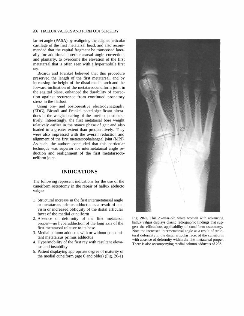
5. Patient displaying appropriate degree of maturity of the medial cuneiform (age 6 and older) (Fig. 20-1)
Fig. 20-1. This 25-year-old white woman with advancing hallux valgus displays classic radiographic findings that sug- gest the efficacious applicability of cuneiform osteotomy. Note the increased intermetatarsal angle as a result of struc- tural deformity in the distal articular facet of the cuneiform with absence of deformity within the first metatarsal proper. There is also accompanying medial column adductus of 25°.
CUNEIFORM OSTEOTOMY 287
EFFECTS
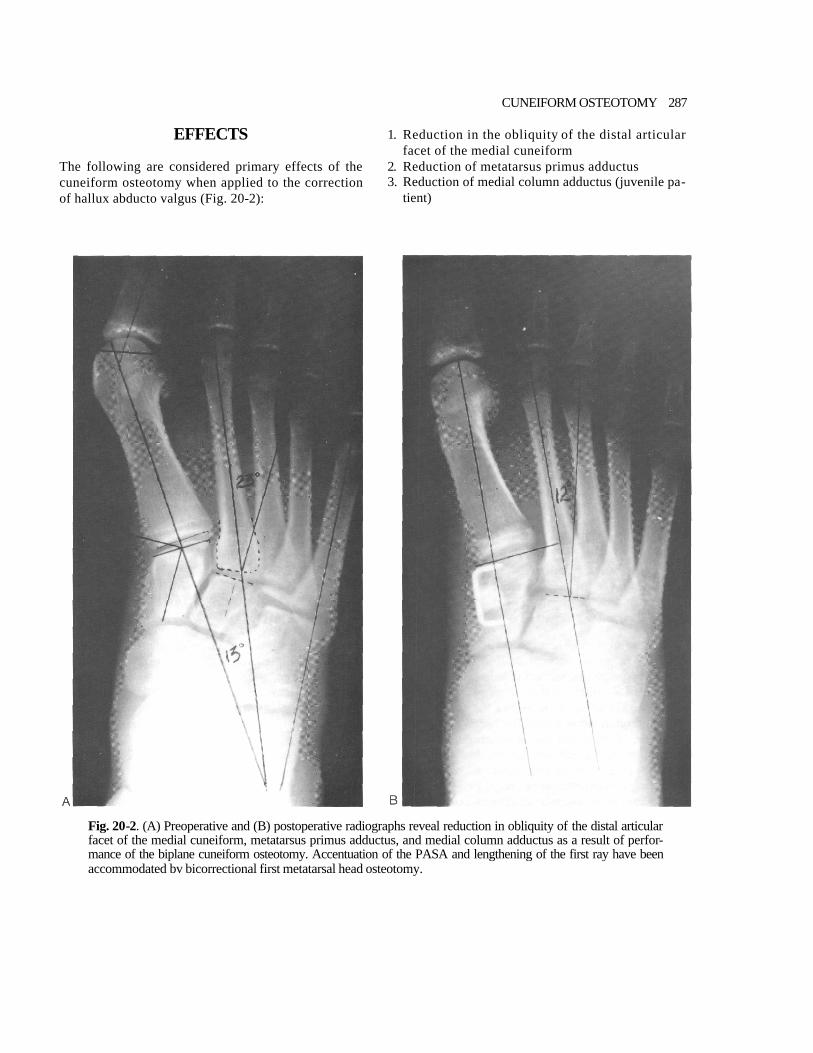
The following are considered primary effects of the cuneiform osteotomy when applied to the correction of hallux abducto valgus (Fig. 20-2):
1. Reduction in the obliquity of the distal articular facet of the medial cuneiform
2. Reduction of metatarsus primus adductus 3. Reduction of medial column adductus (juvenile pa-
tient)
Fig. 20-2. (A) Preoperative and (B) postoperative radiographs reveal reduction in obliquity of the distal articular facet of the medial cuneiform, metatarsus primus adductus, and medial column adductus as a result of perfor- mance of the biplane cuneiform osteotomy. Accentuation of the PASA and lengthening of the first ray have been accommodated bv bicorrectional first metatarsal head osteotomy.
288 HALLUX VALGUS AND FOREFOOT SURGERY
4. Plantar flexion of the first ray 5. Lengthening of the first ray 6. Accentuation of PASA deformity of the first MPJ
SURGICAL TECHNIQUES AND APPLICATIONS
The cuneiform osteotomy is reserved for the older child and adult. In children much less than the age of 6 years the ossification of the first cuneiform has not proceeded to an extent that would allow appropriate dissection and osteotomy at this level.3
It is imperative that the surgical procedure be car- ried out in a certain manner for optimal benefit. The sequence of steps in the performance of the proce- dure are as follows:
1. Complete release of the first MPJ. 2. Biplane cuneiform osteotomy with grafting and fix-
ation. 3. Transpositional, PASA-realigning, and first metatar-
sal-shortening osteotomy as necessary. 4. Adductor tendon transfer, capsulorrhaphy, and
concomitant rebalancing of the first MPJ as deemed appropriate.
5. Performance of ancillary surgical procedures deemed necessary for the overall elimination of associated pedal pathology. This may include lengthening of the achilles tendon, gastrocnemius recession, surgical repair of flexible flatfoot, or ad- ditional adjunctive repair of metatarsus adductus.
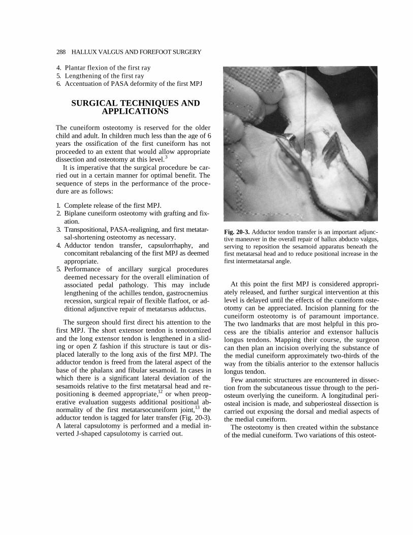
The surgeon should first direct his attention to the first MPJ. The short extensor tendon is tenotomized and the long extensor tendon is lengthened in a slid- ing or open Z fashion if this structure is taut or dis- placed laterally to the long axis of the first MPJ. The adductor tendon is freed from the lateral aspect of the base of the phalanx and fibular sesamoid. In cases in which there is a significant lateral deviation of the sesamoids relative to the first metatarsal head and re- positioning is deemed appropriate,12 or when preop- erative evaluation suggests additional positional ab- normality of the first metatarsocuneiform joint,13 the adductor tendon is tagged for later transfer (Fig. 20-3). A lateral capsulotomy is performed and a medial in- verted J-shaped capsulotomy is carried out.
Fig. 20-3. Adductor tendon transfer is an important adjunc- tive maneuver in the overall repair of hallux abducto valgus, serving to reposition the sesamoid apparatus beneath the first metatarsal head and to reduce positional increase in the first intermetatarsal angle.
At this point the first MPJ is considered appropri- ately released, and further surgical intervention at this level is delayed until the effects of the cuneiform oste- otomy can be appreciated. Incision planning for the cuneiform osteotomy is of paramount importance. The two landmarks that are most helpful in this pro- cess are the tibialis anterior and extensor hallucis longus tendons. Mapping their course, the surgeon can then plan an incision overlying the substance of the medial cuneiform approximately two-thirds of the way from the tibialis anterior to the extensor hallucis longus tendon.
Few anatomic structures are encountered in dissec- tion from the subcutaneous tissue through to the peri- osteum overlying the cuneiform. A longitudinal peri- osteal incision is made, and subperiosteal dissection is carried out exposing the dorsal and medial aspects of the medial cuneiform.
The osteotomy is then created within the substance of the medial cuneiform. Two variations of this osteot-
CUNEIFORM OSTEOTOMY 289
omy have been used to date and warrant some degree of explanation.
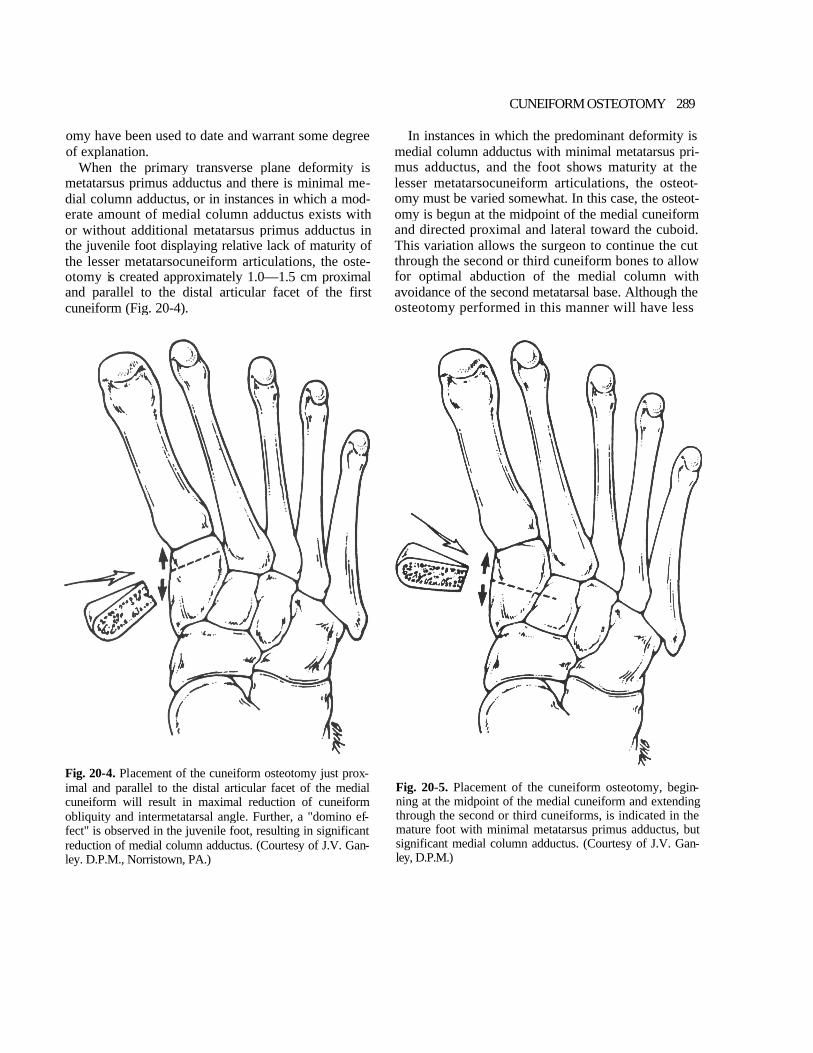
When the primary transverse plane deformity is metatarsus primus adductus and there is minimal me- dial column adductus, or in instances in which a mod- erate amount of medial column adductus exists with or without additional metatarsus primus adductus in the juvenile foot displaying relative lack of maturity of the lesser metatarsocuneiform articulations, the oste- otomy is created approximately 1.0—1.5 cm proximal and parallel to the distal articular facet of the first cuneiform (Fig. 20-4).
In instances in which the predominant deformity is medial column adductus with minimal metatarsus pri- mus adductus, and the foot shows maturity at the lesser metatarsocuneiform articulations, the osteot- omy must be varied somewhat. In this case, the osteot- omy is begun at the midpoint of the medial cuneiform and directed proximal and lateral toward the cuboid. This variation allows the surgeon to continue the cut through the second or third cuneiform bones to allow for optimal abduction of the medial column with avoidance of the second metatarsal base. Although the osteotomy performed in this manner will have less
Fig. 20-4. Placement of the cuneiform osteotomy just prox- imal and parallel to the distal articular facet of the medial cuneiform will result in maximal reduction of cuneiform obliquity and intermetatarsal angle. Further, a "domino ef- fect" is observed in the juvenile foot, resulting in significant reduction of medial column adductus. (Courtesy of J.V. Gan- ley. D.P.M., Norristown, PA.)
Fig. 20-5. Placement of the cuneiform osteotomy, begin- ning at the midpoint of the medial cuneiform and extending through the second or third cuneiforms, is indicated in the mature foot with minimal metatarsus primus adductus, but significant medial column adductus. (Courtesy of J.V. Gan- ley, D.P.M.)
290 HALLUX VALGUS AND FOREFOOT SURGERY
direct effect on the first intermetatarsal angle proper, it has better ability overall to abduct the entire medial column when this surgical maneuver is deemed of primary importance (Fig. 20-5).
With the osteotomy held open the desired amount, an allogenic corticocancellous graft of appropriate proportions is created and inserted. This graft should be wider dorsally and medially to allow for plantar flexion of the medial column in conjunction with the transverse plane correction. The graft is then secured with Kirschner wire (K-wire) or power staple fixation and remodeled as necessary.
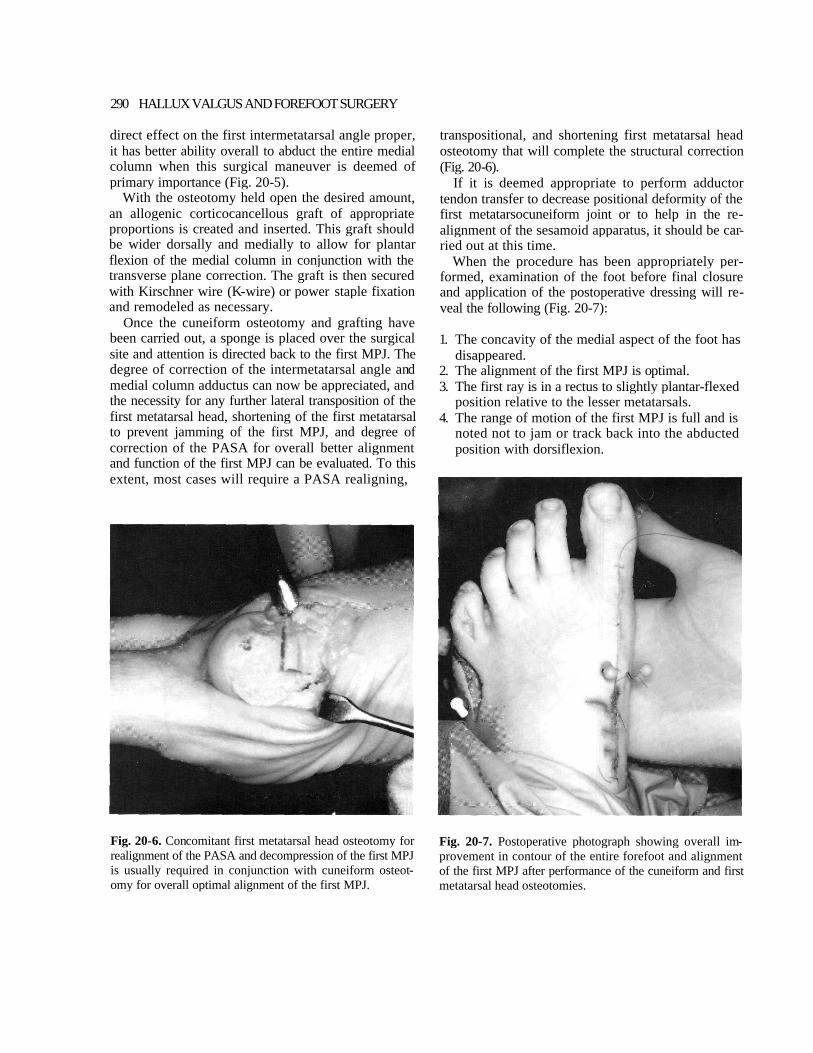
Once the cuneiform osteotomy and grafting have been carried out, a sponge is placed over the surgical site and attention is directed back to the first MPJ. The degree of correction of the intermetatarsal angle and medial column adductus can now be appreciated, and the necessity for any further lateral transposition of the first metatarsal head, shortening of the first metatarsal to prevent jamming of the first MPJ, and degree of correction of the PASA for overall better alignment and function of the first MPJ can be evaluated. To this extent, most cases will require a PASA realigning,
transpositional, and shortening first metatarsal head osteotomy that will complete the structural correction (Fig. 20-6).
If it is deemed appropriate to perform adductor tendon transfer to decrease positional deformity of the first metatarsocuneiform joint or to help in the re- alignment of the sesamoid apparatus, it should be car- ried out at this time.
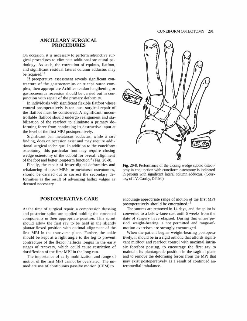
When the procedure has been appropriately per- formed, examination of the foot before final closure and application of the postoperative dressing will re- veal the following (Fig. 20-7):
1. The concavity of the medial aspect of the foot has disappeared.
2. The alignment of the first MPJ is optimal. 3. The first ray is in a rectus to slightly plantar-flexed
position relative to the lesser metatarsals. 4. The range of motion of the first MPJ is full and is
noted not to jam or track back into the abducted position with dorsiflexion.
Fig. 20-6. Concomitant first metatarsal head osteotomy for realignment of the PASA and decompression of the first MPJ is usually required in conjunction with cuneiform osteot- omy for overall optimal alignment of the first MPJ.
Fig. 20-7. Postoperative photograph showing overall im- provement in contour of the entire forefoot and alignment of the first MPJ after performance of the cuneiform and first metatarsal head osteotomies.
CUNEIFORM OSTEOTOMY 291
ANCILLARY SURGICAL PROCEDURES
On occasion, it is necessary to perform adjunctive sur- gical procedures to eliminate additional structural pa- thology. As such, the correction of equinus, flatfoot, and significant residual lateral column adductus may be required.12
If preoperative assessment reveals significant con- tracture of the gastrocnemius or triceps surae com- plex, then appropriate Achilles tendon lengthening or gastrocnemius recession should be carried out in con- junction with repair of the primary deformity.
In individuals with significant flexible flatfoot whose control postoperatively is tenuous, surgical repair of the flatfoot must be considered. A significant, uncon- trollable flatfoot should undergo realignment and sta- bilization of the rearfoot to eliminate a primary de- forming force from continuing its destructive input at the level of the first MPJ postoperatively.
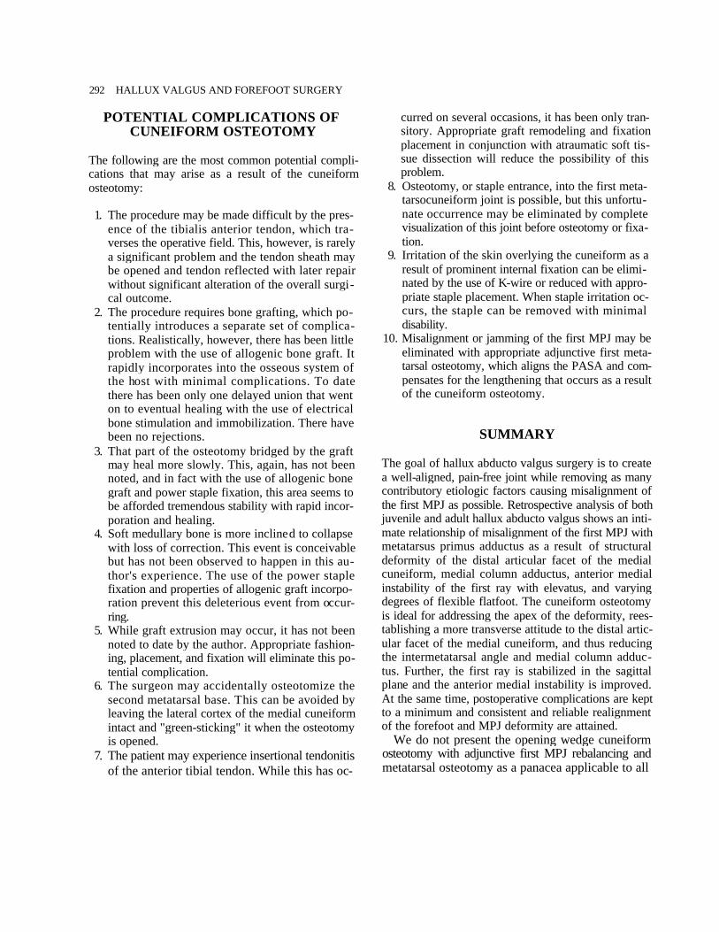
Significant pan metatarsus adductus, while a rare finding, does on occasion exist and may require addi- tional surgical technique. In addition to the cuneiform osteotomy, this particular foot may require closing wedge osteotomy of the cuboid for overall alignment of the foot and better long-term function14 (Fig. 20-8).
Finally, the repair of lesser digital deformities and rebalancing of lesser MPJs, or metatarsal osteotomies, should be carried out to correct the secondary de- formities as the result of advancing hallux valgus as deemed necessary.
Fig. 20-8. Performance of the closing wedge cuboid osteot- omy in conjunction with cuneiform osteotomy is indicated in patients with significant lateral column adductus. (Cour- tesy of J.V. Ganley, D.P.M.)
POSTOPERATIVE CARE
At the time of surgical repair, a compression dressing and posterior splint are applied holding the corrected components in their appropriate position. This splint should allow the first ray to be held in the slightly plantar-flexed position with optimal alignment of the first MPJ in the transverse plane. Further, the ankle should be kept at a right angle to the leg to prevent contracture of the flexor hallucis longus in the early stages of recovery, which could cause restriction of dorsiflexion of the first MPJ in the long run.
The importance of early mobilization and range of motion of the first MPJ cannot be overstated. The im- mediate use of continuous passive motion (CPM) to
encourage appropriate range of motion of the first MPJ postoperatively should be entertained.15
The sutures are removed in 14 days, and the splint is converted to a below-knee cast until 6 weeks from the date of surgery have elapsed. During this entire pe- riod, weight-bearing is not permitted and range-of- motion exercises are strongly encouraged.
When the patient begins weight-bearing postopera- tively, it should be in a rigid orthotic that affords signifi- cant midfoot and rearfoot control with maximal intrin- sic forefoot posting, to encourage the first ray to maintain its plantargrade position in the sagittal plane and to remove the deforming forces from the MPJ that may exist postoperatively as a result of continued an- teromedial imbalance.
292 HALLUX VALGUS AND FOREFOOT SURGERY
POTENTIAL COMPLICATIONS OF CUNEIFORM OSTEOTOMY
The following are the most common potential compli- cations that may arise as a result of the cuneiform osteotomy:
1. The procedure may be made difficult by the pres- ence of the tibialis anterior tendon, which tra- verses the operative field. This, however, is rarely a significant problem and the tendon sheath may be opened and tendon reflected with later repair without significant alteration of the overall surgi- cal outcome.
2. The procedure requires bone grafting, which po- tentially introduces a separate set of complica- tions. Realistically, however, there has been little problem with the use of allogenic bone graft. It rapidly incorporates into the osseous system of the host with minimal complications. To date there has been only one delayed union that went on to eventual healing with the use of electrical bone stimulation and immobilization. There have been no rejections.
3. That part of the osteotomy bridged by the graft may heal more slowly. This, again, has not been noted, and in fact with the use of allogenic bone graft and power staple fixation, this area seems to be afforded tremendous stability with rapid incor- poration and healing.
4. Soft medullary bone is more inclined to collapse with loss of correction. This event is conceivable but has not been observed to happen in this au- thor's experience. The use of the power staple fixation and properties of allogenic graft incorpo- ration prevent this deleterious event from occur- ring.
5. While graft extrusion may occur, it has not been noted to date by the author. Appropriate fashion- ing, placement, and fixation will eliminate this po- tential complication.
6. The surgeon may accidentally osteotomize the second metatarsal base. This can be avoided by leaving the lateral cortex of the medial cuneiform intact and "green-sticking" it when the osteotomy is opened.
7. The patient may experience insertional tendonitis of the anterior tibial tendon. While this has oc-
curred on several occasions, it has been only tran- sitory. Appropriate graft remodeling and fixation placement in conjunction with atraumatic soft tis- sue dissection will reduce the possibility of this problem.
8. Osteotomy, or staple entrance, into the first meta- tarsocuneiform joint is possible, but this unfortu- nate occurrence may be eliminated by complete visualization of this joint before osteotomy or fixa- tion.
9. Irritation of the skin overlying the cuneiform as a result of prominent internal fixation can be elimi- nated by the use of K-wire or reduced with appro- priate staple placement. When staple irritation oc- curs, the staple can be removed with minimal disability.
10. Misalignment or jamming of the first MPJ may be eliminated with appropriate adjunctive first meta- tarsal osteotomy, which aligns the PASA and com- pensates for the lengthening that occurs as a result of the cuneiform osteotomy.
SUMMARY
The goal of hallux abducto valgus surgery is to create a well-aligned, pain-free joint while removing as many contributory etiologic factors causing misalignment of the first MPJ as possible. Retrospective analysis of both juvenile and adult hallux abducto valgus shows an inti- mate relationship of misalignment of the first MPJ with metatarsus primus adductus as a result of structural deformity of the distal articular facet of the medial cuneiform, medial column adductus, anterior medial instability of the first ray with elevatus, and varying degrees of flexible flatfoot. The cuneiform osteotomy is ideal for addressing the apex of the deformity, rees- tablishing a more transverse attitude to the distal artic- ular facet of the medial cuneiform, and thus reducing the intermetatarsal angle and medial column adduc- tus. Further, the first ray is stabilized in the sagittal plane and the anterior medial instability is improved. At the same time, postoperative complications are kept to a minimum and consistent and reliable realignment of the forefoot and MPJ deformity are attained.
We do not present the opening wedge cuneiform osteotomy with adjunctive first MPJ rebalancing and metatarsal osteotomy as a panacea applicable to all
CUNEIFORM OSTEOTOMY 293
foot types. However, in the appropriately applied clini- cal setting, a consistently reliable result can be at- tained, and modern podiatric surgeons must consider this procedure as a valuable addition to their surgical armamentarium.
REFERENCES
1. Cotton F: J Foot statistics and surgery. Trans N Engl Surg Soc 18:181, 1935
2. Fowler B, Brooks AL, Parrish TF: The cavovarus foot. J Bone Joint Surg 41A:757, 1959
3. Hofmann AA, Constine RM, McBride GG, Coleman SS: Osteotomy of the first cuneiform as treatment of resid- ual adduction of the fore part of the foot in club foot. J Bone Joint Surg 66A:985, 1984
4. Lincoln CR, Wood KE, Bugg El: Metatarsus varus cor- rected by open wedge osteotomy of the first cuneiform bone. Orthop Clin North Am 7:795, 1976
5. Helal B: Surgery for adolescent hallux valgus. Clin Orthop 157:50, 1981
6. Kelikian H: Hallux Valgus, Allied Deformities of the Forefoot and Metatarsalgia. WB Saunders. Philadelphia, 1965
7. Young JD: A new operation for adolescent hallux valgus. Univ Pa Med Bull 23:459, 1910
8. Bonney G, McNab I: Hallux valgus and hallux rigidus: a critical survey of operative results. J Bone Joint Surg 34B:366. 1952
9. Joplin RJ: Some common foot disorders amenable to surgery. Am Acad Orthop Surg 15:144, 1958
10. Graver HH: Cuneiform osteotomy in correction of meta- tarsus primus adductus. J Am Podiatry Assoc 68:111, 1978
11. Bicardi BE, Frankel JP: Biplane cuneiform osteotomy for juvenile metatarsus primus varus. J Foot Surg 25:472, 1986
12. Mahan KT, Yu GV: Juvenile and adolescent hallux valgus. p. 61. In McGlamry ED, McGlamry R (eds): Doctors Hos- pital Podiatry Institute Surgical Seminar Syllabus. Doc- tors Hospital Podiatric Education and Research Institute, Atlanta, 1985
13. Pressman MM, Stano GW, Krantz MK, Novicki DC: Cor- rection of hallux valgus with positionally increased in- termetatarsal angle. J Am Podiatr Med Assoc 76:611,1986
14. Grumbine N: Cuboid osteotomy. Seminar lecture, Baha Project, Los Angeles, 1986
15. Kaczander B: The podiatric application of continuous passive motion: a preliminary report. J Am Podiatr Med Assoc 81:631, 1991