Embed Size (px)
DESCRIPTION
Humic Acid References at NIH
Citation preview

HUMIC ACID
BROAD-SPECTRUM EFFICACY NATIONAL INSTITUTES OF HEALTH
Laub BioChemicals Corporation 1401 Quail St., Suite 121
Newport Beach, CA 92660
August 2001 – January 2002

-i-

-ii-
FRONTISPIECE: Electron micrograph of the Ebola virus.
-iii-
Forward
This Report presents the results of toxicology, cell proliferation, and efficacytesting work carried out on natural-product and synthetic humate materials in2001-2002 by contract laboratories of the Virology Branch of the Antiviral Researchand Antimicrobial Chemistry Program (Dr. Christopher Tseng, Program Officer),Division of Microbiology and Infectious Diseases (DMID) Screening and TestingProgram for Antiviral, Immunomodulatory, Antitumor and/or Drug DeliveryActivities, National Institutes of Allergy and Infectious Diseases (NIAID), under theauspices of the National Institutes of Health (NIH, Bethesda, Maryland). Efficacydata are presented for five herpes viruses, three influenza viruses, and twohemorrhagic fever viruses.
The toxicology data are reported as TC50 values, that is, toxic concentrations ofdrug that result in 50% cell toxicity. Cell proliferation data are reported as CP50
values, that is, concentrations of drug that produce a 50% decrease in cellproliferation. Drug efficacy data are given as IC50 and IC90 values, that is,inhibitory concentrations of drug that are efficacious in preventing infection of 50%or 90% of the cells treated. All concentrations are in units of µg/mL (correspondingto parts per million by weight/volume, ppm).
Natural-product humic acid is coded “HA”; while synthetic humates are coded“xxx”, where xxx represents the starting material employed in the syntheticprocess–CA: caffeic acid; CGA: chlorogenic acid; HGA: homogentisic acid.
Natural-product and synthetic humates are protected by U.S. patents(5,946,445; 6,569,416; 6,524,566; 6,524,567; 6,534,049; 6,576,229) and other U.S.and international patents and patents pending assigned to Laub BioChemicalsCorp.
-iv-
Table of Contents
Forward ................................................................................................................. iii
Toxicity AssaysHFF, MDCK, LLC-MK2 Cells
Methodology .................................................................................................... 1
Results .............................................................................................................. 2
Cell Proliferation (Viability) AssaysHFF, Daudi Cells
Methodology .................................................................................................... 4
Results .............................................................................................................. 4
Herpes VirusesHerpes Simplex 1, Herpes Simplex 2, HumanCytomegalovirus, and Varicella Zoster Virus with HFFCells; Epstein-Barr Virus with Daudi Cells
Methodology .................................................................................................... 6
Preparation of Human Foreskin Fibroblast (HFF) Cells ........................ 6
Cytopathic Effect Inhibition Assay (CPE) for Herpes SimplexViruses (HSV), Human Cytomegalovirus (HCMV), and VaricellaZoster Virus (VZV) ..................................................................................... 6
Efficacy Assay for Epstein-Barr Virus (EBV) .......................................... 7
Virus ................................................................................................. 7
Cell Lines .......................................................................................... 7
ELISA Assay .................................................................................... 7
Reference Compounds ............................................................................... 7
Results .............................................................................................................. 7
-v-
Influenza VirusesInfluenza A/New Caledonia/20/99 (H1N1), InfluenzaA/Pan-ama/2007/99 (H3N2), Influenza A/NWS/33 (H1N1),Influenza A/PR/8/34 (H1N1), Influenza A/Shangdong/09/93 (H3N2), Influenza A/Sydney/05/97 (H3N2),Influenza B/Beijing/184/93, Influenza B/Harbin/07/94, andInfluenza B/Hong Kong/5/72 with MDCK Cells
Methodology .................................................................................................... 12
Viruses and Cell Lines Used in Primary Screening ................................ 12
Methods for Assay of Antiviral Activity ................................................... 12
Inhibition of Viral Cytopathic Effect (CPE) .................................... 12
Increase in Neutral Red (NR) Dye Uptake ..................................... 12
Decrease in Virus Yield (VY) ........................................................... 12
Secondary Test .......................................................................................... 13
Reference Compound ................................................................................. 13
Results .............................................................................................................. 13
Influenza VirusesLive-Animal Trial: Synthetic Humic Acid CA withInfluenza A/Shang-dong/09/93 (H3N2)
Methodology .................................................................................................... 20
Animals ...................................................................................................... 20
Compound .................................................................................................. 20
Determination of Arterial Oxygen Saturation (SaO2) ............................. 20
Lung Virus Titer Determinations ............................................................. 20
Experiment Design, Toxicity Determination ........................................... 20
Experiment Design, Antiviral Experiment .............................................. 20
Statistical Evaluation ................................................................................ 21
Results .............................................................................................................. 21
Toxicity Determination ............................................................................. 21
Antiviral Experiment ................................................................................ 21
Summary and Conclusions .......................................................................... 22

-vi-
Hemorrhagic Fever VirusesPichinde Virus (Strain An 4763) with BSC-1 Cells; PuntaToro A Virus (Strain Adames) with LLC-MK2 Cells
Methodology .................................................................................................... 30
Viruses and Cell Lines Used in Primary Drug Screening ....................... 30
Methods for Assay of Antiviral Activity ................................................... 30
Reference Compound ................................................................................. 30
Results .............................................................................................................. 30
TOXICITY ASSAYS
HFF CellsMDCK Cells
LLC-MK2 Cells

-1-
Toxicity Assays
Methodology
The Neutral Red method of assaying for drug toxicity was carried out in roughlythe same manner for all cell lines tested; that employed for human foreskinfibroblast (HFF) cells utilized in the herpes work is provided below as arepresentative example.
Twenty-four hours prior to assay, HFF cells were plated into 96-well plates at aconcentration of 2.5 x 10⁴ cells per well. After 24 h, the medium was aspirated and125 µL of drug was added to the first row of wells and then diluted serially 1:5 usingthe Cetus Liquid Handling System in a manner similar to that used in the CPEassay (see p. 7). After drug addition, the plates were incubated for seven days in aCO2 incubator at 37°C. At this time the medium+drug was aspirated and 200µL/well of 0.01% neutral red in PBS was added. This was incubated in the CO2
incubator for 1 h. The dye was aspirated and the cells were washed using a NuncPlate Washer. After removing the PBS, 200 µg/well of 50% EtOH/1% glacial aceticacid (in H2O) was added. The plates were rotated for 15 min and the opticaldensities were read at 540 nm on a plate reader.
Visual observation was employed to confirm cell toxicity during the course ofinfluenza and punta toro virus efficacy testing. Thus, during the cytopathic effect(CPE) inhibition tests, two additional wells of uninfected cells treated with eachconcentration of test compound were run in parallel with the infected, treated wells.At the time CPE was determined microscopically the toxicity control cells were alsoexamined microscopically for any changes in cell appearance compared to normalcontrol cells run in the same plate. These changes became manifest asenlargement, granularity, cells with ragged edges, a filmy appearance, rounding,detachment from the surface of the well, or other changes. The changes were givena designation of T (100% toxic), PVH (partially toxic-very heavy - 80%), PH(partially toxic-heavy - 60%), P (partially toxic - 40%), Ps (partially toxic-slight -20%), or 0 (no toxicity - 0%), conforming to the degree of cytotoxicity seen. A 50%cytotoxic concentration (TC50) was determined by regression analysis of these data.
-2-
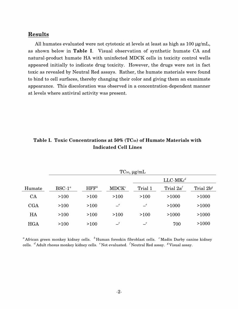
Results
All humates evaluated were not cytotoxic at levels at least as high as 100 µg/mL,as shown below in Table I. Visual observation of synthetic humate CA andnatural-product humate HA with uninfected MDCK cells in toxicity control wellsappeared initially to indicate drug toxicity. However, the drugs were not in facttoxic as revealed by Neutral Red assays. Rather, the humate materials were foundto bind to cell surfaces, thereby changing their color and giving them an exanimateappearance. This discoloration was observed in a concentration-dependent mannerat levels where antiviral activity was present.
Table I. Toxic Concentrations at 50% (TC50) of Humate Materials withIndicated Cell Lines
TC50, µg/mL
LLC-MK2d
Humate BSC-1a HFFb MDCKc Trial 1 Trial 2af Trial 2bg
CA >100 >100 >100 >100 >1000 >1000
CGA >100 >100 –e –e >1000 >1000
HA >100 >100 >100 >100 >1000 >1000
HGA >100 >100 –e –e 700 >1000
a African green monkey kidney cells. b Human foreskin fibroblast cells. c Madin Darby canine kidneycells. d Adult rhesus monkey kidney cells. e Not evaluated. f Neutral Red assay. g Visual assay.
CELL PROLIFERATION(VIABILITY) ASSAYS
HFF CellsDaudi Cells
-4-
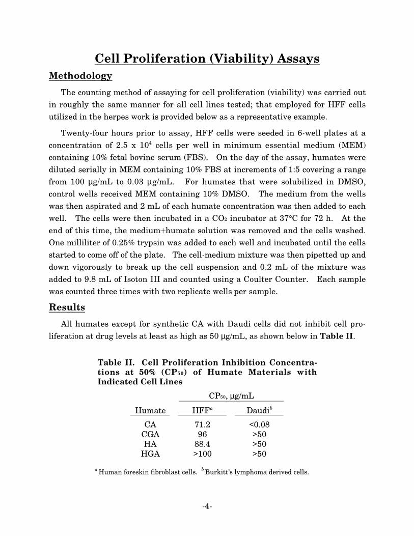
Cell Proliferation (Viability) AssaysMethodology
The counting method of assaying for cell proliferation (viability) was carried outin roughly the same manner for all cell lines tested; that employed for HFF cellsutilized in the herpes work is provided below as a representative example.
Twenty-four hours prior to assay, HFF cells were seeded in 6-well plates at aconcentration of 2.5 x 10⁴ cells per well in minimum essential medium (MEM)containing 10% fetal bovine serum (FBS). On the day of the assay, humates werediluted serially in MEM containing 10% FBS at increments of 1:5 covering a rangefrom 100 µg/mL to 0.03 µg/mL. For humates that were solubilized in DMSO,control wells received MEM containing 10% DMSO. The medium from the wellswas then aspirated and 2 mL of each humate concentration was then added to eachwell. The cells were then incubated in a CO2 incubator at 37°C for 72 h. At theend of this time, the medium+humate solution was removed and the cells washed.One milliliter of 0.25% trypsin was added to each well and incubated until the cellsstarted to come off of the plate. The cell-medium mixture was then pipetted up anddown vigorously to break up the cell suspension and 0.2 mL of the mixture wasadded to 9.8 mL of Isoton III and counted using a Coulter Counter. Each samplewas counted three times with two replicate wells per sample.
Results
All humates except for synthetic CA with Daudi cells did not inhibit cell pro-liferation at drug levels at least as high as 50 µg/mL, as shown below in Table II.
Table II. Cell Proliferation Inhibition Concentra-tions at 50% (CP50) of Humate Materials withIndicated Cell Lines
CP50, µg/mL
Humate HFFa Daudib
CA 71.2 <0.08CGA 96 >50HA 88.4 >50
HGA >100 >50
a Human foreskin fibroblast cells. b Burkitt’s lymphoma derived cells.
HERPES VIRUSES
Herpes Simplex Virus Type 1 (HSV-1)
Herpes Simplex Virus Type 2 (HSV-2)
Epstein-Barr Virus (EBV)
Human Cytomegalovirus (HCMV)
Varicella Zoster Virus (VZV)
-6-
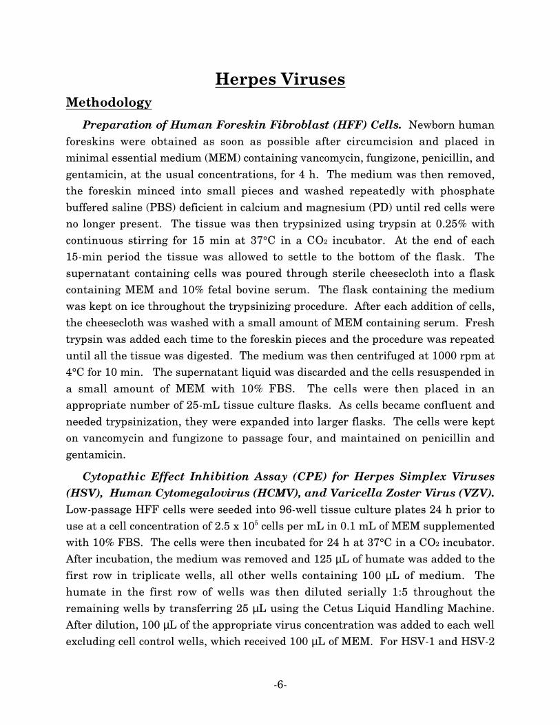
Herpes VirusesMethodology
Preparation of Human Foreskin Fibroblast (HFF) Cells. Newborn humanforeskins were obtained as soon as possible after circumcision and placed inminimal essential medium (MEM) containing vancomycin, fungizone, penicillin, andgentamicin, at the usual concentrations, for 4 h. The medium was then removed,the foreskin minced into small pieces and washed repeatedly with phosphatebuffered saline (PBS) deficient in calcium and magnesium (PD) until red cells wereno longer present. The tissue was then trypsinized using trypsin at 0.25% withcontinuous stirring for 15 min at 37°C in a CO2 incubator. At the end of each15-min period the tissue was allowed to settle to the bottom of the flask. Thesupernatant containing cells was poured through sterile cheesecloth into a flaskcontaining MEM and 10% fetal bovine serum. The flask containing the mediumwas kept on ice throughout the trypsinizing procedure. After each addition of cells,the cheesecloth was washed with a small amount of MEM containing serum. Freshtrypsin was added each time to the foreskin pieces and the procedure was repeateduntil all the tissue was digested. The medium was then centrifuged at 1000 rpm at4°C for 10 min. The supernatant liquid was discarded and the cells resuspended ina small amount of MEM with 10% FBS. The cells were then placed in anappropriate number of 25-mL tissue culture flasks. As cells became confluent andneeded trypsinization, they were expanded into larger flasks. The cells were kepton vancomycin and fungizone to passage four, and maintained on penicillin andgentamicin.
Cytopathic Effect Inhibition Assay (CPE) for Herpes Simplex Viruses(HSV), Human Cytomegalovirus (HCMV), and Varicella Zoster Virus (VZV).Low-passage HFF cells were seeded into 96-well tissue culture plates 24 h prior touse at a cell concentration of 2.5 x 10⁵ cells per mL in 0.1 mL of MEM supplementedwith 10% FBS. The cells were then incubated for 24 h at 37°C in a CO2 incubator.After incubation, the medium was removed and 125 µL of humate was added to thefirst row in triplicate wells, all other wells containing 100 µL of medium. Thehumate in the first row of wells was then diluted serially 1:5 throughout theremaining wells by transferring 25 µL using the Cetus Liquid Handling Machine.After dilution, 100 µL of the appropriate virus concentration was added to each wellexcluding cell control wells, which received 100 µL of MEM. For HSV-1 and HSV-2

-7-
assays, the virus concentration utilized was 1000 PFU's per well. For CMV andVZV assays, the virus concentration added was 2500 PFU per well. The plates werethen incubated at 37°C in a CO2 incubator for 3 days for HSV-1 and HSV-2, 10 daysfor VZV, or 14 days for CMV. After the incubation period, the medium wasaspirated and the cells stained with a 0.1% crystal violet solution for 4 h. The stainwas then removed and the plates rinsed using tap water until all excess stain wasremoved. The plates were allowed to dry for 24 h and then read on a BioTek PlateReader at 620 nm.
Efficacy Assay for Epstein-Barr Virus (EBV).
Virus. There are two prototypes of infectious EBV. One is exemplified by thevirus derived from supernatant fluids of the P3HR-1 cell line. This cell lineproduces nontransforming virus that induces the production of early antigen (EA)and viral capsid antigen (VCA) after primary infection or superinfection of B celllines. The other prototype is exemplified by the B-95-8 virus. This virusimmortalizes cord blood lymphocytes and induces tumors in marmosets. It doesnot, however, induce an abortive productive infection even in cell lines harboringEBV genome copies. The virus used in the assays of this work was P3HR-1.
Cell Lines. Daudi is a low level producer that contains 152 EBV genomecopies/cell. These cells respond to superinfection by EBV by expressing EA andVCA. This cell line was maintained in RPMI-1640 medium supplemented by 10%FBS, L-glutamine and 100 µg/mL gentamicin. The cultures were fed twice weeklyand the cell concentration adjusted to 3 x 10⁵/mL. The cells were kept at 37°C in ahumidified atmosphere with 5% CO2.
ELISA Assay. Daudi cells were infected and treated with drug as describedabove. The cultures were incubated for 4 days at 37°C. The cells were counted,washed and brought to the desired final concentration. For each dilution of drug,cells were added to triplicate wells of a 96-well plate and air dried. The cells werethen fixed for 20 min in an acetic acid/ethanol solution. A monoclonal antibody toEBV VCA was added and the cells were incubated for 1 h, followed by an incubationwith horseradish peroxidase labeled goat anti-mouse IgG1 for 30 min. Plates wererinsed with PBS/Tween20 between incubations. Substrate containing O-phenylenediamine, citrate buffer and hydrogen peroxide was added to each well,and the plates were covered and gently shaken for 10 min. The reaction was

-8-
stopped by adding 3N sulfuric acid, following which the plates were read on amicroplate reader at 492 nm.
Reference Compounds. Acyclovir (Glaxo SmithKline) was the referencecompound employed in the HSV-1, HSV-2, VZV, and EBV efficacy testing work.Ganciclovir (Roche) was the reference compound used with HCMV.
Results
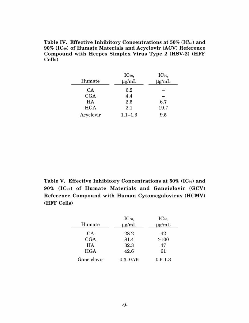
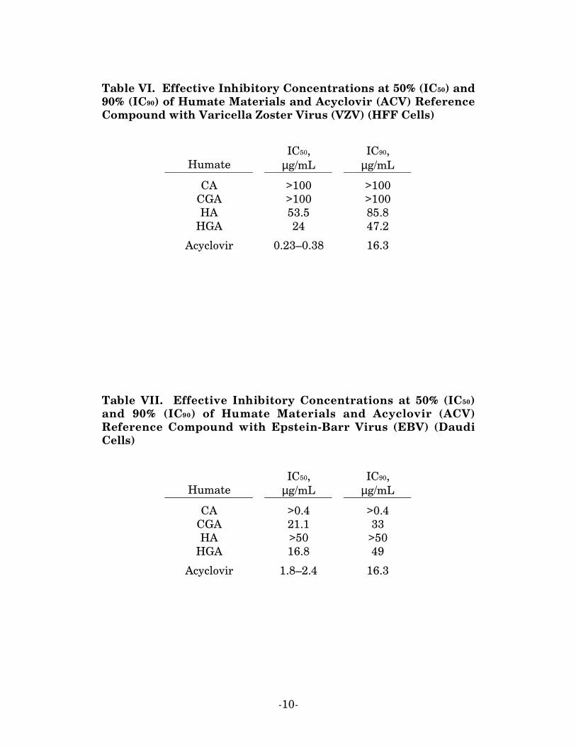
The efficacy data for all humates with the five herpes viruses examined in thiswork are provided in the following tables. As shown, synthetic humates CA andHGA were found to be effective against HSV-1 and HSV-2, and their efficacyapproached that of Acyclovir. Humate CA was somewhat effective against humancytomegalovirus, while synthetic HGA was equally so against Varicella Zostervirus. Humate CA was very highly effective against Epstein-Barr virus.
Table III. Effective Inhibitory Concentrations at 50% (IC50) and90% (IC90) of Humate Materials and Acyclovir (ACV) ReferenceCompound with Herpes Simplex Virus Type 1 (HSV-1) (HFFCells)
HumateIC50,
µg/mLIC90,
µg/mL
CA 6 17.3CGA 15.1 –HA 4.7 13.1
HGA 16.9 51.6
Acyclovir 1.2–1.6 7.9
-9-
Table IV. Effective Inhibitory Concentrations at 50% (IC50) and90% (IC90) of Humate Materials and Acyclovir (ACV) ReferenceCompound with Herpes Simplex Virus Type 2 (HSV-2) (HFFCells)
HumateIC50,
µg/mLIC90,
µg/mL
CA 6.2 –CGA 4.4 –HA 2.5 6.7
HGA 2.1 19.7Acyclovir 1.1–1.3 9.5
Table V. Effective Inhibitory Concentrations at 50% (IC50) and90% (IC90) of Humate Materials and Ganciclovir (GCV)Reference Compound with Human Cytomegalovirus (HCMV)(HFF Cells)
HumateIC50,
µg/mLIC90,
µg/mL
CA 28.2 42CGA 81.4 >100HA 32.3 47
HGA 42.6 61
Ganciclovir 0.3–0.76 0.6-1.3
-10-
Table VI. Effective Inhibitory Concentrations at 50% (IC50) and90% (IC90) of Humate Materials and Acyclovir (ACV) ReferenceCompound with Varicella Zoster Virus (VZV) (HFF Cells)
HumateIC50,
µg/mLIC90,
µg/mL
CA >100 >100CGA >100 >100HA 53.5 85.8
HGA 24 47.2
Acyclovir 0.23–0.38 16.3
Table VII. Effective Inhibitory Concentrations at 50% (IC50)and 90% (IC90) of Humate Materials and Acyclovir (ACV)Reference Compound with Epstein-Barr Virus (EBV) (DaudiCells)
HumateIC50,
µg/mLIC90,
µg/mL
CA >0.4 >0.4CGA 21.1 33HA >50 >50
HGA 16.8 49
Acyclovir 1.8–2.4 16.3

INFLUENZA VIRUSES
Influenza A/New Caledonia/20/99 (H1N1)Influenza A/Panama/2007/99 (H3N2)
Influenza A/NWS/33 (H1N1)Influenza A/PR/8/34 (H1N1)
Influenza A/Shangdong/09/93 (H3N2)Influenza A/Sydney/05/97 (H3N2)
Influenza B/Beijing/184/93Influenza B/Harbin/07/94
Influenza B/Hong Kong/5/72
-12-
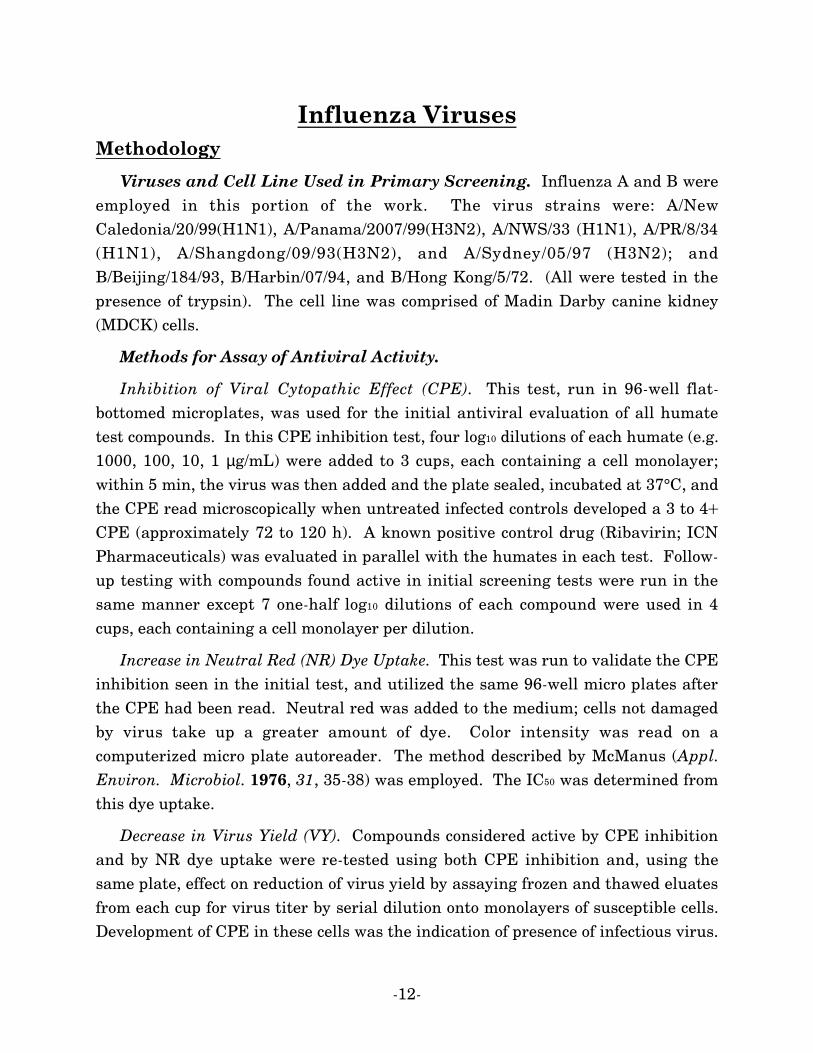
Influenza VirusesMethodology
Viruses and Cell Line Used in Primary Screening. Influenza A and B wereemployed in this portion of the work. The virus strains were: A/NewCaledonia/20/99(H1N1), A/Panama/2007/99(H3N2), A/NWS/33 (H1N1), A/PR/8/34(H1N1), A/Shangdong/09/93(H3N2), and A/Sydney/05/97 (H3N2); andB/Beijing/184/93, B/Harbin/07/94, and B/Hong Kong/5/72. (All were tested in thepresence of trypsin). The cell line was comprised of Madin Darby canine kidney(MDCK) cells.
Methods for Assay of Antiviral Activity.
Inhibition of Viral Cytopathic Effect (CPE). This test, run in 96-well flat-bottomed microplates, was used for the initial antiviral evaluation of all humatetest compounds. In this CPE inhibition test, four log10 dilutions of each humate (e.g.1000, 100, 10, 1 µg/mL) were added to 3 cups, each containing a cell monolayer;within 5 min, the virus was then added and the plate sealed, incubated at 37°C, andthe CPE read microscopically when untreated infected controls developed a 3 to 4+CPE (approximately 72 to 120 h). A known positive control drug (Ribavirin; ICNPharmaceuticals) was evaluated in parallel with the humates in each test. Follow-up testing with compounds found active in initial screening tests were run in thesame manner except 7 one-half log10 dilutions of each compound were used in 4cups, each containing a cell monolayer per dilution.
Increase in Neutral Red (NR) Dye Uptake. This test was run to validate the CPEinhibition seen in the initial test, and utilized the same 96-well micro plates afterthe CPE had been read. Neutral red was added to the medium; cells not damagedby virus take up a greater amount of dye. Color intensity was read on acomputerized micro plate autoreader. The method described by McManus (Appl.Environ. Microbiol. 1976, 31, 35-38) was employed. The IC50 was determined fromthis dye uptake.
Decrease in Virus Yield (VY). Compounds considered active by CPE inhibitionand by NR dye uptake were re-tested using both CPE inhibition and, using thesame plate, effect on reduction of virus yield by assaying frozen and thawed eluatesfrom each cup for virus titer by serial dilution onto monolayers of susceptible cells.Development of CPE in these cells was the indication of presence of infectious virus.
-13-
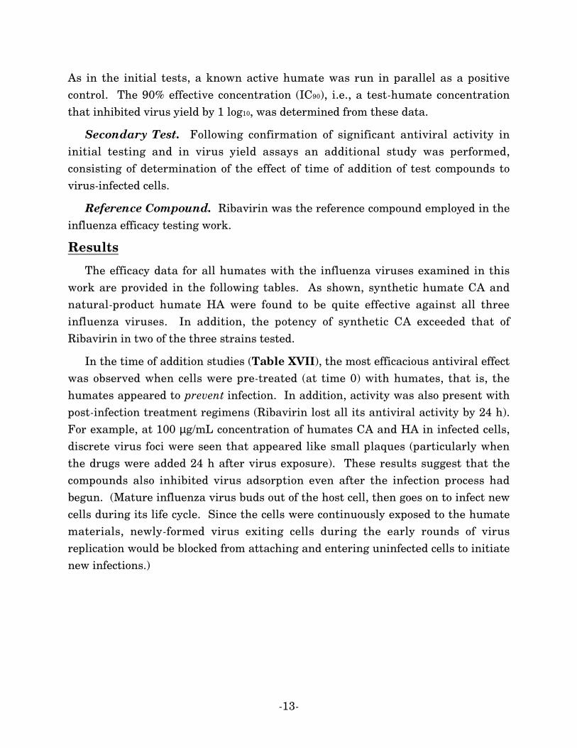
As in the initial tests, a known active humate was run in parallel as a positivecontrol. The 90% effective concentration (IC90), i.e., a test-humate concentrationthat inhibited virus yield by 1 log10, was determined from these data.
Secondary Test. Following confirmation of significant antiviral activity ininitial testing and in virus yield assays an additional study was performed,consisting of determination of the effect of time of addition of test compounds tovirus-infected cells.
Reference Compound. Ribavirin was the reference compound employed in theinfluenza efficacy testing work.
Results
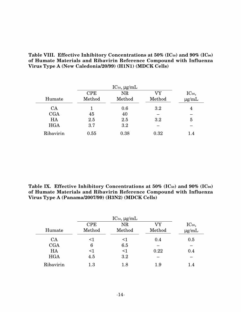
The efficacy data for all humates with the influenza viruses examined in thiswork are provided in the following tables. As shown, synthetic humate CA andnatural-product humate HA were found to be quite effective against all threeinfluenza viruses. In addition, the potency of synthetic CA exceeded that ofRibavirin in two of the three strains tested.
In the time of addition studies (Table XVII), the most efficacious antiviral effectwas observed when cells were pre-treated (at time 0) with humates, that is, thehumates appeared to prevent infection. In addition, activity was also present withpost-infection treatment regimens (Ribavirin lost all its antiviral activity by 24 h).For example, at 100 µg/mL concentration of humates CA and HA in infected cells,discrete virus foci were seen that appeared like small plaques (particularly whenthe drugs were added 24 h after virus exposure). These results suggest that thecompounds also inhibited virus adsorption even after the infection process hadbegun. (Mature influenza virus buds out of the host cell, then goes on to infect newcells during its life cycle. Since the cells were continuously exposed to the humatematerials, newly-formed virus exiting cells during the early rounds of virusreplication would be blocked from attaching and entering uninfected cells to initiatenew infections.)
-14-
Table VIII. Effective Inhibitory Concentrations at 50% (IC50) and 90% (IC90)of Humate Materials and Ribavirin Reference Compound with InfluenzaVirus Type A (New Caledonia/20/99) (H1N1) (MDCK Cells)
IC50, µg/mL
HumateCPE
MethodNR
MethodVY
MethodIC90,
µg/mL
CA 1 0.6 3.2 4CGA 45 40 – –HA 2.5 2.5 3.2 5
HGA 3.7 3.2 – –
Ribavirin 0.55 0.38 0.32 1.4
Table IX. Effective Inhibitory Concentrations at 50% (IC50) and 90% (IC90)of Humate Materials and Ribavirin Reference Compound with InfluenzaVirus Type A (Panama/2007/99) (H3N2) (MDCK Cells)
IC50, µg/mL
HumateCPE
MethodNR
MethodVY
MethodIC90,
µg/mL
CA <1 <1 0.4 0.5CGA 6 6.5 – –HA <1 <1 0.22 0.4
HGA 4.5 3.2 – –
Ribavirin 1.3 1.8 1.9 1.4

-15-
Table X. Effective Inhibitory Concentrations at 50% (IC50) of HumateMaterials and Ribavirin Reference Compound with Influenza Virus Type A(NWS/33) (H1N1) (MDCK Cells)
IC50, µg/mL
HumateCPE
MethodNR
MethodVY
Method
CA 0.65-1 0.55-0.85 –CGA – – –HA 1.3 1.3 –
HGA 18 17 –
Ribavirin 5-6.0 4.6-6.5 –
Table XI. Effective Inhibitory Concentrations at 50% (IC50) of HumateMaterials and Ribavirin Reference Compound with Influenza Virus Type A(PR/8/34) (H1N1) (MDCK Cells)
IC50, µg/mL
HumateCPE
MethodNR
MethodVY
Method
CA 8.5 10 –CGA – – –HA 14 18 –
HGA 18 18 –
Ribavirin 9 12 –
-16-
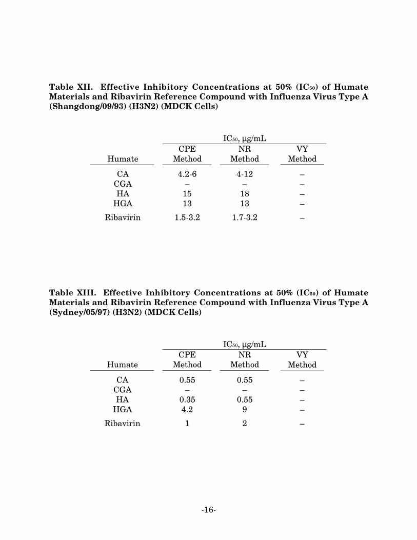
Table XII. Effective Inhibitory Concentrations at 50% (IC50) of HumateMaterials and Ribavirin Reference Compound with Influenza Virus Type A(Shangdong/09/93) (H3N2) (MDCK Cells)
IC50, µg/mL
HumateCPE
MethodNR
MethodVY
Method
CA 4.2-6 4-12 –CGA – – –HA 15 18 –
HGA 13 13 –
Ribavirin 1.5-3.2 1.7-3.2 –
Table XIII. Effective Inhibitory Concentrations at 50% (IC50) of HumateMaterials and Ribavirin Reference Compound with Influenza Virus Type A(Sydney/05/97) (H3N2) (MDCK Cells)
IC50, µg/mL
HumateCPE
MethodNR
MethodVY
Method
CA 0.55 0.55 –CGA – – –HA 0.35 0.55 –
HGA 4.2 9 –
Ribavirin 1 2 –

-17-
Table XIV. Effective Inhibitory Concentrations at 50% (IC50) and 90% (IC90)of Humate Materials and Ribavirin Reference Compound with InfluenzaVirus Type B (Beijing/184/93) (MDCK Cells)
IC50, µg/mL
HumateCPE
MethodNR
MethodVY
MethodIC90,
µg/mL
CA <1 <1 0.55 0.75CGA 5.5 4.7 – –HA <1 <1 0.5 2.5
HGA 3.2 3.2 – –
Ribavirin 1 1.5 0.5 1
Table XV. Effective Inhibitory Concentrations at 50% (IC50) of HumateMaterials and Ribavirin Reference Compound with Influenza Virus Type B(Harbin/07/94) (MDCK Cells)
IC50, µg/mL
HumateCPE
MethodNR
MethodVY
Method
CA 1.3 0.85 –CGA – – –HA 0.7 0.65 –
HGA 7 7 –
Ribavirin 0.85 1.1 –

-18-
Table XVI. Effective Inhibitory Concentrations at 50% (IC50) of HumateMaterials and Ribavirin Reference Compound with Influenza Virus Type B(Hong Kong/5/72) (MDCK Cells)
IC50, µg/mL
HumateCPE
MethodNR
MethodVY
Method
CA 3.2-23 4.2-19 –CGA – – –HA 3.2 5 –
HGA 3.2 3.8 –
Ribavirin 1.2-1.8 1.8-1.8 –
Table XVII. Effect of Time of Addition on Efficacy of Humate Materialsand Ribavirin Reference Compound against Influenza Virus Type A (NewCaledonia/20/99) (H1N1) (MDCK Cells)
IC50, µg/mL:Visual–Neutral Red Methods
Time ofAddition, h
HumateCA
HumateHA Ribavirin
0 6.5–8 5.5–5.5 7.5–61 12–15 14–15 6–5.52 18–18 16–17 7–84 18–18 10–10 7–78 16–17 14–14 9–12
24 22–25 48–55 >100–>100

INFLUENZA VIRUSES
Live-Animal Trial:
Hepsyl® CA with Influenza A/Shangdong/09/93 (H3N2)

-20-
Influenza Viruses: Live-Animal Trial
Synthetic humate CA exhibited significant in vitro activity against influenza Aand B viruses, with IC50 values ranging from 0.4 to 12 µg/mL, and TC50 values of150 µg/mL or greater. These data prompted a live-animal trial for this compoundagainst influenza A (Shangdong/09/93) (H3N2) in mice. Since no information wasknown regarding the tolerance of this compound in mice, a preliminary toxicitydetermination was run using a maximal dose (100 mg/kg/day). It was decided touse an intraperitoneal (i.p.) treatment route and a treatment schedule of twice dailyfor 5 days beginning 4 h pre-virus exposure in order to maximize any potentialantiviral effect.
Methodology
Animals. Female 18-21 g specific pathogen-free BALB/c mice were obtainedfrom Harlan Sprague-Dowley, Inc. (Indianapolis, IN). They were quarantined 24 hprior to use and fed Wayne LabBlox and tap water ad libitum.
Compound. Humate CA was dissolved in sterile physiological saline for use inthis study. Ribavirin, used as a known positive control, was obtained from ICNPharmaceuticals Inc. (Costa Mesa, CA). It also was dissolved in saline. Bothsolutions were stored at 4°C until used.
Determination of Arterial Oxygen Saturation (SaO2). The effects ofinfluenza virus on arterial oxygen saturation (SaO2) were determined using theOhmeda Biox 3740 pulse oximeter (Ohmeda, Louisville, OH). The ear probeattachment was used, the probe placed on the thigh of the animal, with the slowinstrument mode selected. Readings were made after a 30-sec stabilization time oneach animal.
Lung Virus Titer Determinations. Each mouse lung was homogenized andvarying dilutions assayed in triplicate for infectious virus in MDCK cells.
Experiment Design, Toxicity Determination. Two mice were injected i.p.with 100 mg/kg/day of humate CA twice daily for 5 days. The animals wereweighed daily and observed for death for 10 days.
Experiment Design, Antiviral Experiment. Groups of 19 mice were infectedintranasally with virus and treated i.p. with humate CA at dosages of 50, 25, or 12.5mg/kg/day or with Ribavirin at a dose of 75 mg/kg/day. Treatment was twice daily
-21-
for 5 days beginning 4 h pre-virus exposure. As controls, 35 infected mice weretreated with saline in parallel to the above. Ten mice in each treated group and 20saline-treated controls were observed for death for 21 days and SaO2 levelsascertained in days 3-11. An additional 3 mice from each treated group and 5 micefrom the saline controls were killed in days 3, 6, and 9 and their lungs removed,assigned a consolidation score ranging from 0 (normal) to 4 (maximal, 100% plumcoloration), weighed, and assayed for virus titer. Toxicity controls were included foreach treatment group consisting of 3 mice per dosage. These were weighed prior tostart of treatment and again 18 h after final treatment, and observed for death for21 days. A group of 12 normal controls were also included; 3 were weighed inparallel with the toxicity controls and SaO2 levels determined with the infectedanimals. Three additional mice were killed on days 3, 6, and 9 to providebackground lung data.
Statistical Evaluation. Survivor number differences were analyzed by chisquare analysis with Yates’ correction. Changes in mean day to death, lungweights, SaO2 levels, and virus titers were evaluated using the t test. Lung scoreswere analyzed using the Wilcoxon ranked sum analysis.
Results
Toxicity Determination. The preliminary toxicity data are summarized inTable XVIII. No animals died during treatment with 100 mg/kg/day of humateCA, but major weight loss (1.8 g) was seen, indicating the compound was not welltolerated at this dose. In view of these results, the maximum dosage used in theantiviral experiment was 50 mg/kg/day.
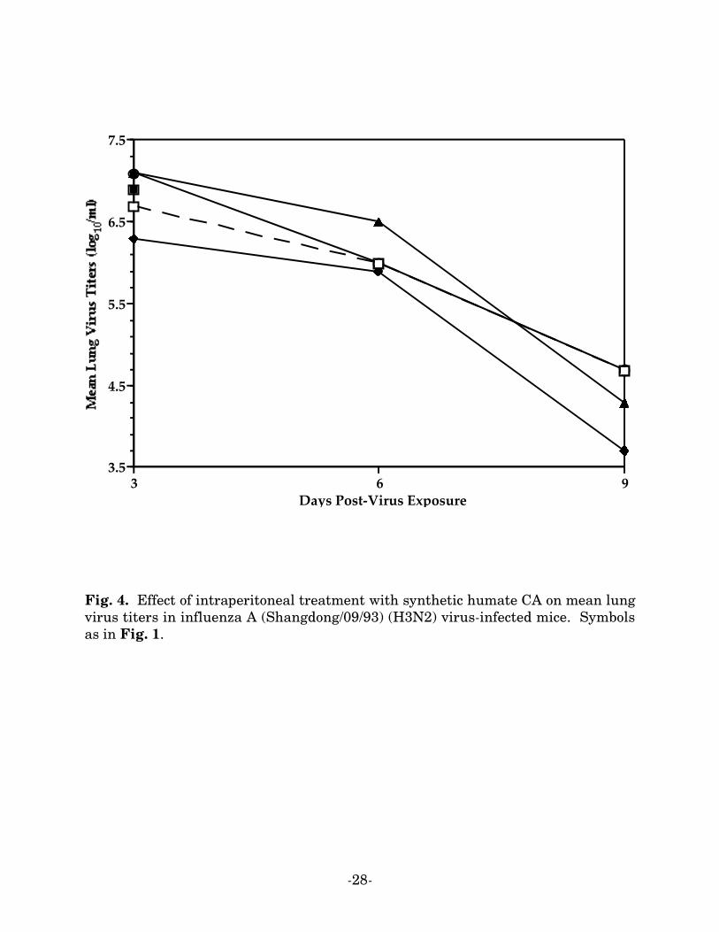
Antiviral Experiment. The results of this experiment are summarized inTable XIX and in Figs. 1-4. Treatment with humate CA did indeed appear toexhibit some inhibitory effect on this influenza infection at 25 and 12.5 mg/kg/daydosages. This was seen by 30-40% increases in survivors, lessened decline in SaO2
(Fig. 1), and inhibition of lung scores and weight (Figs. 2-3). Interestingly, thehighest dosage, 50 mg/kg/day, although not lethal to the toxicity control animals,appeared to enhance the virus infection as seen by a shortened mean day to death(Table XX) and markedly lowered SaO2 levels (Fig. 1). It will also be noted thatthe mice in the group to be sacrificed for lung parameters had all died prior to day 6when treated with the 50 mg/kg/day dose.
-22-
Weight loss was still observed in toxicity control mice receiving both the 50 and25 mg/kg/day dose of humate CA, but weight gain occurred at the 12.5 mg/kg/daydose (Table XX). Ribavirin appeared well tolerated at the 75 mg/kg/day dose usedin this study.
Ribavirin exerted the positive activity expected, preventing any deaths fromoccurring, markedly lessening SaO2 decline, inhibiting lung consolidation, andreducing lung virus titers.
As discussed earlier, humate CA was significantly inhibitory to influenza A andB viruses in vitro; in anticipation of this experiment the compound was tested invitro also against the influenza A/Shangdong/09/93 (H3N2) virus used in thisanimal experiment. The IC50 values were 6 and 12 µg/mL using visual and neutralred endpoints, respectively, as presented in the preceding Section. These were up to10-fold less potent than seen using new clinical isolates, but still indicates thecompound was inhibitory to the virus.
Summary and Conclusions
Mice infected with a lethal dose of influenza A/Shangdong/09/93 (H3N2) viruswere treated i.p. with 50, 25, or 12.5 mg/kg/day of synthetic humate CA.Treatments were twice daily for 5 days beginning 4 h pre-virus exposure. The highdose appeared to enhance the virus infection, presumably due to a sub-lethaltoxicity. The lower doses were somewhat inhibitory to the infection as seen byincreased numbers of survivors, lessened SaO2 decline, and inhibition of lungconsolidation. Ribavirin, included as a positive control exerted the inhibitory effectexpected at the 75 mg/kg/day dose used.
Although only one humate material was employed against a single inflenzastrain in this work, the data nevertheless indicate that humates do in fact showsome promise as potential influenza inhibitors. Using a lower dosage, altering thetreatment schedule to once or three times daily, and testing other humates againstother influenza virus strains might in fact provide quite substantially different andimproved therapeutic efficacy.

-23-
Table XVIII. Preliminary Toxicity Determination ofIntraperitoneally-Administered Synthetic Humate CAa inYoung Adult Miceb
TreatmentcDosage,
mg/kg/daySurvivors/
TotalMean Host
Weight Changed, g
Humate CA 100 2/2 -1.8
a Drug diluent: sterile saline. b Female BALB/c mice, 18-21 g. c Treatment schedule:bid x 5 beg; 4-h pre-virus exposure. Experiment duration: 10 days. d Differencebetween initial weight and weight 18 hours after final treatment.

-24-
Table XIX. Effect of Intraperitoneal Treatment of Synthetic Humate CAa on Influenza Virus Type A(Shangdong/09/93) (H3N2) Infection in Miceb
Toxicity Controls Infected Treated Mice
TreatmentcDosage,
mg/kg/daySurvivors/
Total
Mean HostWeight
Changed, gSurvivors/
Total
Mean Dayto Deathe
± SD
Mean Day11 SaO2,% ± SD
Humate CA 50 3/3 -0.8 0/10 6.5 ± 4.2 76.4 ± 4.6
25 3/3 -0.7 3/10f 10.3 ± 4.8 79.8 ± 4.6
12.5 3/3 0.2 4/10g 10.2 ± 2.8 81.7 ± 4.9f
Ribavirin 75 3/3 0.1 10/10h >21.0 ± 0.0h 88.2 ± 1.6h
Saline – – – 0/20 9.2 ± 3.5 76.9 ± 3.8
Normal Controls – 3/3 0.6 5/5 >21.0 ± 0.0 89.4 ± 2.2
a Diluent: sterile saline. b Female BALB/c mice, 18-21 g. c Treatment schedule: bid x 5 beg; 4-h pre-virus exposure. Experimentduration: 21 days. d Difference between initial weight and weight 18 hours after final treatment. e Mean day to death of micedying prior to day 21. f P<0.05; g P<0.01; h P<0.001, compared to saline-treated controls.

-25-
Fig. 1. Effect of intraperitoneal treatment with synthetic humate CA on arterialoxygen saturation in influenza A (Shangdong/09/93) (H3N2) virus-infected mice.Filled squares: 50 mg/kg/day humate CA; filled circles: 25; triangles: 12.5.Diamonds: 75 mg/kg/day Ribavirin. Squares: saline; circles: normal controls.
3 4 5 6 7 8 9 10 1175
80
85
90
Days Post-Virus Exposure

-26-
Fig. 2. Effect of intraperitoneal treatment with synthetic humate CA on mean lungscores in influenza A (Shangdong/09/93) (H3N2) virus-infected mice. Symbols as inFig. 1.
0 3 6 90
1
2
3
4
Days Post-Virus

-27-
Fig. 3. Effect of intraperitoneal treatment with synthetic humate CA on mean lungweights in influenza A (Shangdong/09/93) (H3N2) virus-infected mice. Symbols asin Fig. 1.
0 3 6 9125
165
205
245
285
325
Days Post-Virus Exposure
-28-
Fig. 4. Effect of intraperitoneal treatment with synthetic humate CA on mean lungvirus titers in influenza A (Shangdong/09/93) (H3N2) virus-infected mice. Symbolsas in Fig. 1.
3 6 93.5
4.5
5.5
6.5
7.5
Days Post-Virus Exposure

HEMORRHAGIC FEVER VIRUSES
Pichinde Virus/An 4763
Punta Toro A Virus/Adames
-30-
Hemorrhagic Fever VirusesMethodology
Viruses and Cell Lines Used in Primary Screening. Pichinde and PuntaToro A viruses were employed in this portion of the work. The virus strains wereAn 4763 and Adames, respectively. The cell lines were African green monkeykidney cells (BSC-1; Pichinde virus) and adult Rhesus monkey kidney cells (LLC-MK2; Punta Toro A virus).
Methods for Assay of Antiviral Activity. The methodologies employed withthe hemorrhagic fever viruses (inhibition of viral cytopathic effect–visual CPE;increase in neutral red dye uptake–NR; time-of-addition study) were identical tothose used with influenza viruses described in the preceding Section. Values ofIC100 were determined by virus titer.
Reference Compound. Ribavirin was again the reference compound employedin the efficacy testing work.
Results
The efficacy data for all humate materials with the hemorrhagic fever virusesexamined in this work are provided in the following tables. As shown, the humatesexhibited subtantial efficacy against both hemorrhagic fever viruses, synthetic CAand natural-product HA particularly so against Punta Toro A virus. Virus titerexperiments established that the IC50 and IC100 values for the former material werein fact 5-27 µg/mL and 270 µg/mL, respectively. The addition of humates 1 h beforevirus exposure, at the time of virus exposure, and 1 h after virus exposure resultedin similar levels of inhibition of viral infection (Table XXII). When the humatematerials were added at 2 h after virus exposure or longer, both CA and HA wereonly weakly inhibitory. These data, reminiscent of the findings for influenzaviruses (Table XVII), suggest that some early event in the virus replication cyclewas inhibited; previous work has established that the operative mechanism is infact the inhibition of viral fusion.

-31-
Table XX. Effective Inhibitory Concentrations at 50% (IC50) ofHumate Materials and Ribavirin Reference Compound withPichinde Virus (BSC-1 Cells)
IC50, µg/mL
HumateCPE
MethodNR
Method
CA <1 <1CGA <1 <1HA <1 <1
HGA <1 <1
Ribavirin 0.3 <1
Table XXI. Effective Inhibitory Concentrations at 50% (IC50) and 100%(IC100) of Humate Materials and Ribavirin Reference Compound withPunta Toro A Virus (LLC-MK2 Cells)
IC50, µg/mL IC100, µg/mL
HumateCPE
MethodNR
Method Virus Titer
CA 5 27 270CGA 10 100 –HA 5 15 378
HGA 10 15 –
Ribavirin 5 5 –
-32-
Table XXII. Effect of Time of Addition on Efficacy of Humate Materials andRibavirin Reference Compound against Punta Toro A Virus (LLC-MK2
Cells)
IC50, µg/mLVisual–Neutral Red Methods
Time ofAddition, h Humate CA Humate HA Ribavirin
-1 25–100 30–50 10–60 10–3 30–7 4–31 25–40 30–30 4–52 39–75 30–60 8–54 80–>100 100–>100 4–96 80–100 100–50 10–88 >100–>100 >100–>100 8–10
12 >100–>100 >100–>100 8–1224 >100–>100 >100–>100 20–35