Embed Size (px)
Citation preview
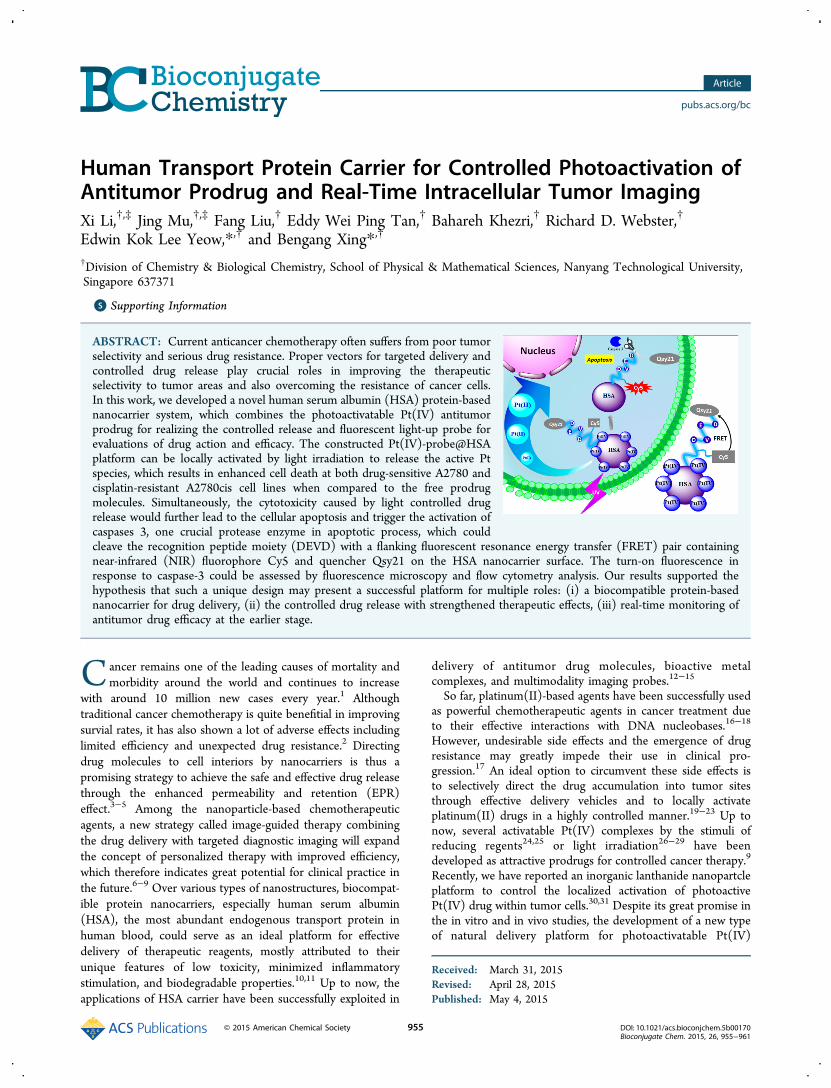
Human Transport Protein Carrier for Controlled Photoactivation ofAntitumor Prodrug and Real-Time Intracellular Tumor ImagingXi Li,†,‡ Jing Mu,†,‡ Fang Liu,† Eddy Wei Ping Tan,† Bahareh Khezri,† Richard D. Webster,†
Edwin Kok Lee Yeow,*,† and Bengang Xing*,†
†Division of Chemistry & Biological Chemistry, School of Physical & Mathematical Sciences, Nanyang Technological University,Singapore 637371
*S Supporting Information
ABSTRACT: Current anticancer chemotherapy often suffers from poor tumorselectivity and serious drug resistance. Proper vectors for targeted delivery andcontrolled drug release play crucial roles in improving the therapeuticselectivity to tumor areas and also overcoming the resistance of cancer cells.In this work, we developed a novel human serum albumin (HSA) protein-basednanocarrier system, which combines the photoactivatable Pt(IV) antitumorprodrug for realizing the controlled release and fluorescent light-up probe forevaluations of drug action and efficacy. The constructed Pt(IV)-probe@HSAplatform can be locally activated by light irradiation to release the active Ptspecies, which results in enhanced cell death at both drug-sensitive A2780 andcisplatin-resistant A2780cis cell lines when compared to the free prodrugmolecules. Simultaneously, the cytotoxicity caused by light controlled drugrelease would further lead to the cellular apoptosis and trigger the activation ofcaspases 3, one crucial protease enzyme in apoptotic process, which couldcleave the recognition peptide moiety (DEVD) with a flanking fluorescent resonance energy transfer (FRET) pair containingnear-infrared (NIR) fluorophore Cy5 and quencher Qsy21 on the HSA nanocarrier surface. The turn-on fluorescence inresponse to caspase-3 could be assessed by fluorescence microscopy and flow cytometry analysis. Our results supported thehypothesis that such a unique design may present a successful platform for multiple roles: (i) a biocompatible protein-basednanocarrier for drug delivery, (ii) the controlled drug release with strengthened therapeutic effects, (iii) real-time monitoring ofantitumor drug efficacy at the earlier stage.
Cancer remains one of the leading causes of mortality andmorbidity around the world and continues to increase
with around 10 million new cases every year.1 Althoughtraditional cancer chemotherapy is quite benefitial in improvingsurvial rates, it has also shown a lot of adverse effects includinglimited efficiency and unexpected drug resistance.2 Directingdrug molecules to cell interiors by nanocarriers is thus apromising strategy to achieve the safe and effective drug releasethrough the enhanced permeability and retention (EPR)effect.3−5 Among the nanoparticle-based chemotherapeuticagents, a new strategy called image-guided therapy combiningthe drug delivery with targeted diagnostic imaging will expandthe concept of personalized therapy with improved efficiency,which therefore indicates great potential for clinical practice inthe future.6−9 Over various types of nanostructures, biocompat-ible protein nanocarriers, especially human serum albumin(HSA), the most abundant endogenous transport protein inhuman blood, could serve as an ideal platform for effectivedelivery of therapeutic reagents, mostly attributed to theirunique features of low toxicity, minimized inflammatorystimulation, and biodegradable properties.10,11 Up to now, theapplications of HSA carrier have been successfully exploited in
delivery of antitumor drug molecules, bioactive metalcomplexes, and multimodality imaging probes.12−15
So far, platinum(II)-based agents have been successfully usedas powerful chemotherapeutic agents in cancer treatment dueto their effective interactions with DNA nucleobases.16−18
However, undesirable side effects and the emergence of drugresistance may greatly impede their use in clinical pro-gression.17 An ideal option to circumvent these side effects isto selectively direct the drug accumulation into tumor sitesthrough effective delivery vehicles and to locally activateplatinum(II) drugs in a highly controlled manner.19−23 Up tonow, several activatable Pt(IV) complexes by the stimuli ofreducing regents24,25 or light irradiation26−29 have beendeveloped as attractive prodrugs for controlled cancer therapy.9
Recently, we have reported an inorganic lanthanide nanopartcleplatform to control the localized activation of photoactivePt(IV) drug within tumor cells.30,31 Despite its great promise inthe in vitro and in vivo studies, the development of a new typeof natural delivery platform for photoactivatable Pt(IV)
Received: March 31, 2015Revised: April 28, 2015Published: May 4, 2015
Article
pubs.acs.org/bc
© 2015 American Chemical Society 955 DOI: 10.1021/acs.bioconjchem.5b00170Bioconjugate Chem. 2015, 26, 955−961
prodrug combined with simultaneous evaluation of thecorresponding antitumor activities for improved tumorspecificity and minimum drug resistance is of clinicalimportance, and relevant investigations have not been fullyexploited so far.Herein, we introduce a novel and effective drug release
system consisting of a specific photoactivatable Pt(IV) prodrugand an apoptosis-responsive probe upon their conjugation withHSA protein carrier. Such light-responsive nanoconjugates canselectively trigger the localized activation of Pt(IV) prodrug,thus greatly improving the anticancer effect of platinum-basedtherapeutic agents in tumor cells. More importantly, theproposed nanocarriers also provide promising advantages byallowing simultaneous real-time imaging of controlled drugrelease and evaluation of the corresponding antitumor activities.
■ RESULTS AND DISCUSSIONPreparation and Characterization of Photoactive
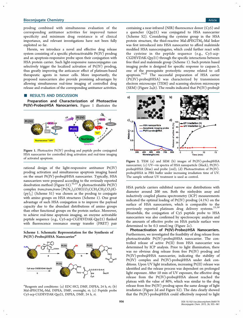
Pt(IV)-Probe@HSA Nanocarriers. Figure 1 illustrates the
rational design of the light-responsive antitumor Pt(IV)prodrug activation and simultaneous apoptosis imaging basedon the smart Pt(IV)-probe@HSA nanocarrier. Typically, HSAnanocarriers were prepared according to the reviously reporteddesolvation method (Figure S1).32,33 A photoactivatable Pt(IV)complex trans,trans,trans-[Pt(N3)2(OH)(O2CCH2CH2CO2H)-(py)2] (Scheme S1) was chosen as the prodrug to conjugatewith amino groups on HSA structures (Scheme 1). One greatadvantage of such HSA conjugation is to improve the payloadcapacity due to the abundant distributions of amino groupsthan other functional groups on the protein surface. Moreover,to achieve real-time apoptosis imaging, an enzyme activatablepeptide sequence (e.g., Cy5-acp-CGDEVDAK-Qsy21) flankedwith fluorescence resonance energy transfer (FRET) pair
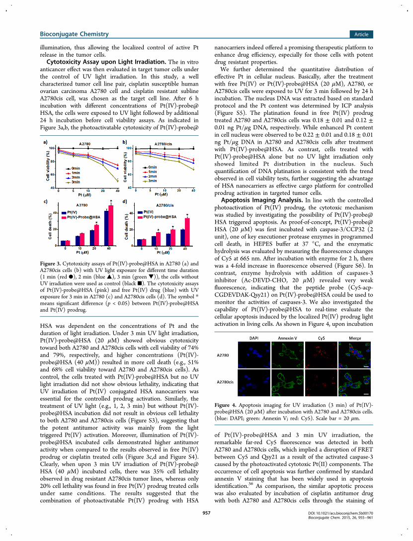
containing a near-infrared (NIR) fluorescence donor (Cy5) anda quencher (Qsy21) was conjugated to HSA nanocarrier(Scheme S2). Considering the cysteine group in the HSAprotein structure, the thiol-reactive Mal-dPEGTM6-Mal linkerwas first introduced into HSA nanocarrier to afford maleimidemodified HSA nanoconjugates, which could further react withthe cysteine in the peptide sequence (e.g., Cy5-acp-CGDEVDAK-Qsy21) through the specific interactions betweenfree thiol and maleimide group (Scheme 1). Such protein basedimaging probe is designed for specific response to caspase-3,one of the prerequisite proteolytic enzyme related to cellapoptosis.34,35 The successful preparation of HSA carrier(Pt(IV)-probe@HSA) was characterized by transmissionelectron microscope (TEM) and scanning electron microscope(SEM) (Figure 2a,b). The results indicated that Pt(IV)-probe@
HSA particle carriers exhibited narrow size distributions withdiameter around 200 nm. Both the ninhydrin assay andinductively coupled plasma spectrometry (ICP) measurementsindicated the optimal loading of Pt(IV) prodrug (4.1%) on thesurface of HSA nanocarriers, which is comparable to thepreviously reported platinum drug delivery systems.23,30
Meanwhile, the conjugation of Cy5 peptide probe to HSAnanocarriers was also confirmed by spectroscopic analysis andthe amounts of effective probe on HSA particle surface weredetermined to be 0.3 nmol/mg (Figure 2c).
Photoactivation of Pt(IV)-Probe@HSA Nanocarriers.Furthermore, we investigated the feasibility of drug release fromphotoactivatable Pt(IV)-probe@HSA nanocarrier. The con-trolled release of active Pt(II) from HSA nanocarrier wasdetermined by ICP analysis. Prior to light illumination, therewas no obvious drug release from free Pt(IV) prodrug andPt(IV)-probe@HSA nanocarrier, indicating the stability ofPt(IV) complex and Pt(IV)-probe@HSA under dark con-ditions. Upon UV light irradiation, increasing Pt(II) release wasidentified and the release process was dependent on prolongedlight exposure. After 10 min of UV exposure, the effective drugrelease from the Pt(IV)-probe@HSA almost reached theplateau with the value of 80%, which was similar to the drugrelease from free Pt(IV) prodrug upon the same dosage of lightirradiation (Figure 2d and Figure S2). The data clearly showedthat the Pt(IV)-probe@HSA could effectively respond to light
Figure 1. Photoactive Pt(IV) prodrug and peptide probe conjugatedHSA nanocarrier for controlled drug activation and real-time imagingof activated apoptosis.
Scheme 1. Schematic Representation for the Synthesis ofPt(IV)-Probe@HSA Nanocarriera
aReagents and conditions: (a) EDC·HCl, DMF, DIPEA, 24 h, rt; (b)Mal-dPEGTM6-Mal, DIPEA, DMF, overnight, rt; (c) Peptide probeCy5-acp-CGDEVDAK-Qsy21, DIPEA, DMF, 24 h, rt.
Figure 2. TEM (a) and SEM (b) images of Pt(IV)-probe@HSAnanocarrier; (c) UV−vis spectra of HSA nanoparticle (black), Pt(IV)-probe@HSA (blue) and probe (red); (d) Photoactivation of Pt(IV)-probe@HSA in PBS buffer under increasing irradiation time of UV.The sample without UV treatment is used as control.
Bioconjugate Chemistry Article
DOI: 10.1021/acs.bioconjchem.5b00170Bioconjugate Chem. 2015, 26, 955−961
956
illumination, thus allowing the localized control of active Ptrelease in the tumor cells.Cytotoxicity Assay upon Light Irradiation. The in vitro
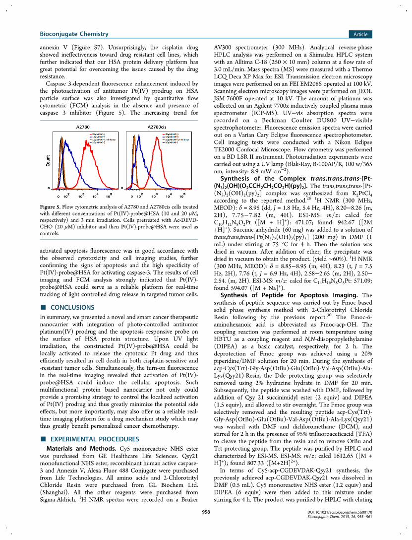
anticancer effect was then evaluated in target tumor cells underthe control of UV light irradiation. In this study, a wellcharacterized tumor cell line pair, cisplatin susceptible humanovarian carcinoma A2780 cell and cisplatin resistant sublineA2780cis cell, was chosen as the target cell line. After 6 hincubation with different concentrations of Pt(IV)-probe@HSA, the cells were exposed to UV light followed by additional24 h incubation before cell viability assays. As indicated inFigure 3a,b, the photoactivatable cytotoxicity of Pt(IV)-probe@
HSA was dependent on the concentrations of Pt and theduration of light irradiation. Under 3 min UV light irradiation,Pt(IV)-probe@HSA (20 μM) showed obvious cytotoxicitytoward both A2780 and A2780cis cells with cell viability of 74%and 79%, respectively, and higher concentrations (Pt(IV)-probe@HSA (40 μM)) resulted in more cell death (e.g., 51%and 68% cell viability toward A2780 and A2780cis cells). Ascontrol, the cells treated with Pt(IV)-probe@HSA but no UVlight irradiation did not show obvious lethality, indicating thatUV irradiation of Pt(IV) conjugated HSA nanocarriers wasessential for the controlled prodrug activation. Similarly, thetreatment of UV light (e.g., 1, 2, 3 min) but without Pt(IV)-probe@HSA incubation did not result in obvious cell lethalityto both A2780 and A2780cis cells (Figure S3), suggesting thatthe potent antitumor activity was mainly from the lighttriggered Pt(IV) activation. Moreover, illumination of Pt(IV)-probe@HSA incubated cells demonstrated higher antitumoractivity when compared to the results observed in free Pt(IV)prodrug or cisplatin treated cells (Figure 3c,d and Figure S4).Clearly, when upon 3 min UV irradiation of Pt(IV)-probe@HSA (40 μM) incubated cells, there was 35% cell lethalityobserved in drug resistant A2780cis tumor lines, whereas only20% cell lethality was found in free Pt(IV) prodrug treated cellsunder same conditions. The results suggested that thecombination of photoactivatable Pt(IV) prodrug with HSA
nanocarriers indeed offered a promising therapeutic platform toenhance drug efficiency, especially for those cells with potentdrug resistant properties.We further determined the quantitative distribution of
effective Pt in cellular nucleus. Basically, after the treatmentwith free Pt(IV) or Pt(IV)-probe@HSA (20 μM), A2780, orA2780cis cells were exposed to UV for 3 min followed by 24 hincubation. The nucleus DNA was extracted based on standardprotocol and the Pt content was determined by ICP analysis(Figure S5). The platination found in free Pt(IV) prodrugtreated A2780 and A2780cis cells was 0.18 ± 0.01 and 0.12 ±0.01 ng Pt/μg DNA, respectively. While enhanced Pt contentin cell nucleus were observed to be 0.22 ± 0.01 and 0.18 ± 0.01ng Pt/μg DNA in A2780 and A2780cis cells after treatmentwith Pt(IV)-probe@HSA. As contrast, cells treated withPt(IV)-probe@HSA alone but no UV light irradiation onlyshowed limited Pt distribution in the nucleus. Suchquantification of DNA platination is consistent with the trendobserved in cell viability tests, further suggesting the advantageof HSA nanocarriers as effective cargo platform for controlledprodrug activation in targeted tumor cells.
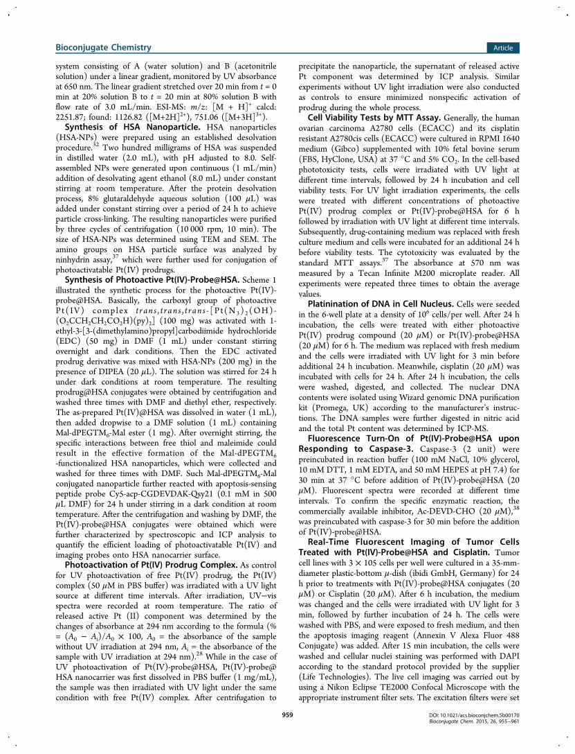
Apoptosis Imaging Analysis. In line with the controlledphotoactivation of Pt(IV) prodrug, the cytotoxic mechanismwas studied by investigating the possibility of Pt(IV)-probe@HSA triggered apoptosis. As proof-of-concept, Pt(IV)-probe@HSA (20 μM) was first incubated with caspase-3/CCP32 (2unit), one of key executioner protease enzymes in programmedcell death, in HEPES buffer at 37 °C, and the enzymatichydrolysis was evaluated by measuring the fluorescence changesof Cy5 at 665 nm. After incubation with enzyme for 2 h, therewas a 4-fold increase in fluorescence observed (Figure S6). Incontrast, enzyme hydrolysis with addition of caspases-3inhibitor (Ac-DEVD-CHO, 20 μM) revealed very weakfluorescence, indicating that the peptide probe (Cy5-acp-CGDEVDAK-Qsy21) on Pt(IV)-probe@HSA could be used tomonitor the activities of caspases-3. We also investigated thecapability of Pt(IV)-probe@HSA to real-time evaluate thecellular apoptosis induced by the localized Pt(IV) prodrug lightactivation in living cells. As shown in Figure 4, upon incubation
of Pt(IV)-probe@HSA and 3 min UV irradiation, theremarkable far-red Cy5 fluorescence was detected in bothA2780 and A2780cis cells, which implied a disruption of FRETbetween Cy5 and Qsy21 as a result of the activated caspase-3caused by the photoactivated cytotoxic Pt(II) components. Theoccurrence of cell apoptosis was further confirmed by standardannexin V staining that has been widely used in apoptosisidentification.36 As comparison, the similar apoptotic processwas also evaluated by incubation of cisplatin antitumor drugwith both A2780 and A2780cis cells through the staining of
Figure 3. Cytotoxicity assays of Pt(IV)-probe@HSA in A2780 (a) andA2780cis cells (b) with UV light exposure for different time duration(1 min (red ●), 2 min (blue ▲), 3 min (green ▼)), the cells withoutUV irradiation were used as control (black ■). The cytotoxicity assaysof Pt(IV)-probe@HSA (pink) and free Pt(IV) drug (blue) with UVexposure for 3 min in A2780 (c) and A2780cis cells (d). The symbol *means significant difference (p < 0.05) between Pt(IV)-probe@HSAand Pt(IV) prodrug.
Figure 4. Apoptosis imaging for UV irradiation (3 min) of Pt(IV)-probe@HSA (20 μM) after incubation with A2780 and A2780cis cells.(blue: DAPI; green: Annexin V; red: Cy5). Scale bar = 20 μm.
Bioconjugate Chemistry Article
DOI: 10.1021/acs.bioconjchem.5b00170Bioconjugate Chem. 2015, 26, 955−961
957
annexin V (Figure S7). Unsurprisingly, the cisplatin drugshowed ineffectiveness toward drug resistant cell lines, whichfurther indicated that our HSA protein delivery platform hasgreat potential for overcoming the issues caused by the drugresistance.Caspase 3-dependent fluorescence enhancement induced by
the photoactivation of antitumor Pt(IV) prodrug on HSAparticle surface was also investigated by quantitative flowcytometric (FCM) analysis in the absence and presence ofcaspase 3 inhibitor (Figure 5). The increasing trend for
activated apoptosis fluorescence was in good accordance withthe observed cytotoxicity and cell imaging studies, furtherconfirming the signs of apoptosis and the high specificity ofPt(IV)-probe@HSA for activating caspase-3. The results of cellimaging and FCM analysis strongly indicated that Pt(IV)-probe@HSA could serve as a reliable platform for real-timetracking of light controlled drug release in targeted tumor cells.
■ CONCLUSIONSIn summary, we presented a novel and smart cancer therapeuticnanocarrier with integration of photo-controlled antitumorplatinum(IV) prodrug and the apoptosis responsive probe onthe surface of HSA protein structure. Upon UV lightirradiation, the constructed Pt(IV)-probe@HSA could belocally activated to release the cytotoxic Pt drug and thusefficiently resulted in cell death in both cisplatin-sensitive and-resistant tumor cells. Simultaneously, the turn-on fluorescencein the real-time imaging revealed that activation of Pt(IV)-probe@HSA could induce the cellular apoptosis. Suchmultifunctional protein based nanocarrier not only couldprovide a promising strategy to control the localized activationof Pt(IV) prodrug and thus greatly minimize the potential sideeffects, but more importantly, may also offer us a reliable real-time imaging platform for a drug mechanism study which maythus greatly benefit personalized cancer chemotherapy.
■ EXPERIMENTAL PROCEDURESMaterials and Methods. Cy5 monoreactive NHS ester
was purchased from GE Healthcare Life Sciences. Qsy21monofunctional NHS ester, recombinant human active caspase-3 and Annexin V, Alexa Fluor 488 Conjugate were purchasedfrom Life Technologies. All amino acids and 2-ChlorotritylChloride Resin were purchased from GL Biochem Ltd.(Shanghai). All the other reagents were purchased fromSigma-Aldrich. 1H NMR spectra were recorded on a Bruker
AV300 spectrometer (300 MHz). Analytical reverse-phaseHPLC analysis was performed on a Shimadzu HPLC systemwith an Alltima C-18 (250 × 10 mm) column at a flow rate of3.0 mL/min. Mass spectra (MS) were measured with a ThermoLCQ Deca XP Max for ESI. Transmission electron microscopyimages were performed on an FEI EM208S operated at 100 kV.Scanning electron microscopy images were performed on JEOLJSM-7600F operated at 10 kV. The amount of platinum wascollected on an Agilent 7700x inductively coupled plasma massspectrometer (ICP-MS). UV−vis absorption spectra wererecorded on a Beckman Coulter DU800 UV−visiblespectrophotometer. Fluorescence emission spectra were carriedout on a Varian Cary Eclipse fluorescence spectrophotometer.Cell imaging tests were conducted with a Nikon EclipseTE2000 Confocal Microscope. Flow cytometry was performedon a BD LSR II instrument. Photoirradiation experiments werecarried out using a UV lamp (Blak-Ray, B-100AP/R, 100 w/365nm, intensity: 8.9 mW cm−2).
Synthesis of the Complex trans,trans,trans-[Pt-(N3)2(OH)(O2CCH2CH2CO2H)(py)2]. The trans,trans,trans-[Pt-(N3)2(OH)2(py)2] complex was synthesized from K2PtCl4according to the reported method.26 1H NMR (300 MHz,MEOD): δ = 8.95 (dd, J = 1.8 Hz, 5.4 Hz, 4H), 8.20−8.26 (m,2H), 7.75−7.82 (m, 4H). ESI-MS: m/z: calcd forC10H12N8O2Pt ([M + H]+): 471.07; found: 942.67 ([2M+H]+). Succinic anhydride (60 mg) was added to a solution oftrans,trans,trans-[Pt(N3)2(OH)2(py)2] (200 mg) in DMF (1mL) under stirring at 75 °C for 4 h. Then the solution wasdried in vacuum. After addition of ether, the precipitate wasdried in vacuum to obtain the product. (yield ∼60%). 1H NMR(300 MHz, MEOD): δ = 8.85−8.95 (m, 4H), 8.23 (t, J = 7.5Hz, 2H), 7.76 (t, J = 6.9 Hz, 4H), 2.58−2.65 (m, 2H), 2.50−2.54. (m, 2H). ESI-MS: m/z: calcd for C14H16N8O5Pt: 571.09;found 594.07 ([M + Na]+).
Synthesis of Peptide for Apoptosis Imaging. Thesynthesis of peptide sequence was carried out by Fmoc basedsolid phase synthesis method with 2-Chlorotrityl ChlorideResin following by the previous report.30 The Fmoc-6-aminohexanoic acid is abbreviated as Fmoc-acp-OH. Thecoupling reaction was performed at room temperature usingHBTU as a coupling reagent and N,N-diisopropylethylamine(DIPEA) as a basic catalyst, respectively, for 2 h. Thedeprotection of Fmoc group was achieved using a 20%piperidine/DMF solution for 20 min. During the synthesis ofacp-Cys(Trt)-Gly-Asp(OtBu)-Glu(OtBu)-Val-Asp(OtBu)-Ala-Lys(Qsy21)-Resin, the Dde protecting group was selectivelyremoved using 2% hydrazine hydrate in DMF for 20 min.Subsequently, the peptide was washed with DMF, followed byaddition of Qsy 21 succinimidyl ester (2 equiv) and DIPEA(1.5 equiv), and allowed to stir overnight. The Fmoc group wasselectively removed and the resulting peptide acp-Cys(Trt)-Gly-Asp(OtBu)-Glu(OtBu)-Val-Asp(OtBu)-Ala-Lys(Qsy21)was washed with DMF and dichloromethane (DCM), andstirred for 2 h in the presence of 95% trifluoroaceticacid (TFA)to cleave the peptide from the resin and to remove OtBu andTrt protecting group. The peptide was purified by HPLC andcharacterized by ESI-MS. ESI-MS: m/z: calcd 1612.65 ([M +H]+); found 807.33 ([M+2H]2+).In terms of Cy5-acp-CGDEVDAK-Qsy21 synthesis, the
previously achieved acp-CGDEVDAK-Qsy21 was dissolved inDMF (0.5 mL). Cy5 monoreactive NHS ester (1.2 equiv) andDIPEA (6 equiv) were then added to this mixture understirring for 4 h. The product was purified by HPLC with eluting
Figure 5. Flow cytometric analysis of A2780 and A2780cis cells treatedwith different concentrations of Pt(IV)-probe@HSA (10 and 20 μM,respectively) and 3 min irradiation. Cells pretreated with Ac-DEVD-CHO (20 μM) inhibitor and then Pt(IV)-probe@HSA were used ascontrols.
Bioconjugate Chemistry Article
DOI: 10.1021/acs.bioconjchem.5b00170Bioconjugate Chem. 2015, 26, 955−961
958
system consisting of A (water solution) and B (acetonitrilesolution) under a linear gradient, monitored by UV absorbanceat 650 nm. The linear gradient stretched over 20 min from t = 0min at 20% solution B to t = 20 min at 80% solution B withflow rate of 3.0 mL/min. ESI-MS: m/z: [M + H]+ calcd:2251.87; found: 1126.82 ([M+2H]2+), 751.06 ([M+3H]3+).Synthesis of HSA Nanoparticle. HSA nanoparticles
(HSA-NPs) were prepared using an established desolvationprocedure.32 Two hundred milligrams of HSA was suspendedin distilled water (2.0 mL), with pH adjusted to 8.0. Self-assembled NPs were generated upon continuous (1 mL/min)addition of desolvating agent ethanol (8.0 mL) under constantstirring at room temperature. After the protein desolvationprocess, 8% glutaraldehyde aqueous solution (100 μL) wasadded under constant stirring over a period of 24 h to achieveparticle cross-linking. The resulting nanoparticles were purifiedby three cycles of centrifugation (10 000 rpm, 10 min). Thesize of HSA-NPs was determined using TEM and SEM. Theamino groups on HSA particle surface was analyzed byninhydrin assay,37 which were further used for conjugation ofphotoactivatable Pt(IV) prodrugs.Synthesis of Photoactive Pt(IV)-Probe@HSA. Scheme 1
illustrated the synthetic process for the photoactive Pt(IV)-probe@HSA. Basically, the carboxyl group of photoactivePt( IV) complex t ran s , t r an s , t r an s - [P t(N3) 2(OH)-(O2CCH2CH2CO2H)(py)2] (100 mg) was activated with 1-ethyl-3-[3-(dimethylamino)propyl]carbodiimide hydrochloride(EDC) (50 mg) in DMF (1 mL) under constant stirringovernight and dark conditions. Then the EDC activatedprodrug derivative was mixed with HSA-NPs (200 mg) in thepresence of DIPEA (20 μL). The solution was stirred for 24 hunder dark conditions at room temperature. The resultingprodrug@HSA conjugates were obtained by centrifugation andwashed three times with DMF and diethyl ether, respectively.The as-prepared Pt(IV)@HSA was dissolved in water (1 mL),then added dropwise to a DMF solution (1 mL) containingMal-dPEGTM6-Mal ester (1 mg). After overnight stirring, thespecific interactions between free thiol and maleimide couldresult in the effective formation of the Mal-dPEGTM6-functionalized HSA nanoparticles, which were collected andwashed for three times with DMF. Such Mal-dPEGTM6-Malconjugated nanoparticle further reacted with apoptosis-sensingpeptide probe Cy5-acp-CGDEVDAK-Qsy21 (0.1 mM in 500μL DMF) for 24 h under stirring in a dark condition at roomtemperature. After the centrifugation and washing by DMF, thePt(IV)-probe@HSA conjugates were obtained which werefurther characterized by spectroscopic and ICP analysis toquantify the efficient loading of photoactivatable Pt(IV) andimaging probes onto HSA nanocarrier surface.Photoactivation of Pt(IV) Prodrug Complex. As control
for UV photoactivation of free Pt(IV) prodrug, the Pt(IV)complex (50 μM in PBS buffer) was irradiated with a UV lightsource at different time intervals. After irradiation, UV−visspectra were recorded at room temperature. The ratio ofreleased active Pt (II) component was determined by thechanges of absorbance at 294 nm according to the formula (%= (A0 − Ai)/A0 × 100, A0 = the absorbance of the samplewithout UV irradiation at 294 nm, Ai = the absorbance of thesample with UV irradiation at 294 nm).28 While in the case ofUV photoactivation of Pt(IV)-probe@HSA, Pt(IV)-probe@HSA nanocarrier was first dissolved in PBS buffer (1 mg/mL),the sample was then irradiated with UV light under the samecondition with free Pt(IV) complex. After centrifugation to
precipitate the nanoparticle, the supernatant of released activePt component was determined by ICP analysis. Similarexperiments without UV light irradiation were also conductedas controls to ensure minimized nonspecific activation ofprodrug during the whole process.
Cell Viability Tests by MTT Assay. Generally, the humanovarian carcinoma A2780 cells (ECACC) and its cisplatinresistant A2780cis cells (ECACC) were cultured in RPMI 1640medium (Gibco) supplemented with 10% fetal bovine serum(FBS, HyClone, USA) at 37 °C and 5% CO2. In the cell-basedphototoxicity tests, cells were irradiated with UV light atdifferent time intervals, followed by 24 h incubation and cellviability tests. For UV light irradiation experiments, the cellswere treated with different concentrations of photoactivePt(IV) prodrug complex or Pt(IV)-probe@HSA for 6 hfollowed by irradiation with UV light at different time intervals.Subsequently, drug-containing medium was replaced with freshculture medium and cells were incubated for an additional 24 hbefore viability tests. The cytotoxicity was evaluated by thestandard MTT assays.37 The absorbance at 570 nm wasmeasured by a Tecan Infinite M200 microplate reader. Allexperiments were repeated three times to obtain the averagevalues.
Platinination of DNA in Cell Nucleus. Cells were seededin the 6-well plate at a density of 106 cells/per well. After 24 hincubation, the cells were treated with either photoactivePt(IV) prodrug compound (20 μM) or Pt(IV)-probe@HSA(20 μM) for 6 h. The medium was replaced with fresh mediumand the cells were irradiated with UV light for 3 min beforeadditional 24 h incubation. Meanwhile, cisplatin (20 μM) wasincubated with cells for 24 h. After 24 h incubation, the cellswere washed, digested, and collected. The nuclear DNAcontents were isolated using Wizard genomic DNA purificationkit (Promega, UK) according to the manufacturer’s instruc-tions. The DNA samples were further digested in nitric acidand the total Pt content was determined by ICP-MS.
Fluorescence Turn-On of Pt(IV)-Probe@HSA uponResponding to Caspase-3. Caspase-3 (2 unit) werepreincubated in reaction buffer (100 mM NaCl, 10% glycerol,10 mM DTT, 1 mM EDTA, and 50 mM HEPES at pH 7.4) for30 min at 37 °C before addition of Pt(IV)-probe@HSA (20μM). Fluorescent spectra were recorded at different timeintervals. To confirm the specific enzymatic reaction, thecommercially available inhibitor, Ac-DEVD-CHO (20 μM),38
was preincubated with caspase-3 for 30 min before the additionof Pt(IV)-probe@HSA.
Real-Time Fluorescent Imaging of Tumor CellsTreated with Pt(IV)-Probe@HSA and Cisplatin. Tumorcell lines with 3 × 10⁵ cells per well were cultured in a 35-mm-diameter plastic-bottom μ-dish (ibidi GmbH, Germany) for 24h prior to treatments with Pt(IV)-probe@HSA conjugates (20μM) or Cisplatin (20 μM). After 6 h incubation, the mediumwas changed and the cells were irradiated with UV light for 3min, followed by further incubation of 24 h. The cells werewashed with PBS, and were exposed to fresh medium, and thenthe apoptosis imaging reagent (Annexin V Alexa Fluor 488Conjugate) was added. After 15 min incubation, the cells werewashed and cellular nuclei staining was performed with DAPIaccording to the standard protocol provided by the supplier(Life Technologies). The live cell imaging was carried out byusing a Nikon Eclipse TE2000 Confocal Microscope with theappropriate instrument filter sets. The excitation filters were set
Bioconjugate Chemistry Article
DOI: 10.1021/acs.bioconjchem.5b00170Bioconjugate Chem. 2015, 26, 955−961
959
as 364, 488, and 633 nm for DAPI, Annexin V, and Cy5,respectively.Flow Cytometric Analysis. Cells were seeded in 6-well
plates at a density of 3.0 × 10⁵ cells per well in RPMI 1640medium and incubated for 24 h. The medium was replacedwith Pt(IV)-probe@HSA (10, 20 μM) containing medium andincubated for 6 h, followed by UV irradiation for 3 min andfurther incubation for 24 h. The cells were harvested withtrypsin treatment, washed with PBS for 3 times. Thefluorescence intensity was quantified by FACS Calibur flowcytometer (BD Biosciences, USA), and the results wereanalyzed with FlowJo 7.6.1 software.
■ ASSOCIATED CONTENT*S Supporting InformationThe complete chemical synthesis of prodrug complex and theprobe Cy5-acp-CGDEVDAK-Qsy21, characterization of HSAparticles, activation analysis of Pt(IV) prodrug by UV−visspectra, phototoxicity of UV irradiation to cells, cell cytotoxicityof cisplatin, platination of DNA, fluorescence spectra of Pt(IV)-probe@HSA after incubation with caspase-3, and fluorescentapoptosis imaging of cisplatin. The Supporting Information isavailable free of charge on the ACS Publications website atDOI: 10.1021/acs.bioconjchem.5b00170.
■ AUTHOR INFORMATIONCorresponding Authors*E-mail: [email protected]. Phone: 65-63168759.*E-mail: [email protected]. Phone: 65-63168758.Author Contributions‡Xi Li and Jing Mu contributed to this work equally.NotesThe authors declare no competing financial interest.
■ ACKNOWLEDGMENTSThe authors acknowledge Start-Up Grant (SUG), A*STARPSF Grant (SERC1121202008), RG64/10, RG11/13, COSresearch collaboration award in Nanyang TechnologicalUniversity, Singapore.
■ ABBREVIATIONSHSA, human serum albumin; Pt(IV), prodrug, trans,trans,trans-[Pt(N3)2(OH)(O2CCH2CH2CO2H)(py)2]; ICP, inductivelycoupled plasma spectrometry; TEM, transmission electronmicroscope; SEM, scanning electron microscope
■ REFERENCES(1) Peer, D., Karp, J. M., Hong, S., Farokhzad, O. C., Margalit, R., andLanger, R. (2007) Nanocarriers as an emerging platform for cancertherapy. Nat. Nanotechnol. 2, 751−760.(2) Szakacs, G., Paterson, J. K., Ludwig, J. A., Booth-Genthe, C., andGottesman, M. M. (2006) Targeting multidrug resistance in cancer.Nat. Rev. Drug Discovery 5, 219−234.(3) Davis, M. E., and Shin, D. M. (2008) Nanoparticle therapeutics:an emerging treatment modality for cancer. Nat. Rev. Drug Discovery 7,771−782.(4) Wang, A. Z., Langer, R., and Farokhzad, O. C. (2012)Nanoparticle delivery of cancer drugs. Annu. Rev. Med. 63, 185−198.(5) Ng, K. K., Lovell, J. F., and Zheng, G. (2011) Lipoprotein-inspired nanoparticles for cancer theranostics. Acc. Chem. Res. 44,1105−1113.(6) Bu, L., Ma, X., Tu, Y., Shen, B., and Cheng, Z. (2013) Opticalimage-guided cancer therapy. Curr. Pharm. Biotechnol. 14, 723−732.
(7) Kim, J., Piao, Y., and Hyeon, T. (2009) Multifunctionalnanostructured materials for multimodal imaging, and simultaneousimaging and therapy. Chem. Soc. Rev. 38, 372−390.(8) Chen, Q., Wang, C., Cheng, L., He, W., Cheng, Z., and Liu, Z.(2014) Protein modified upconversion nanoparticles for imaging-guided combined photothermal and photodynamic therapy. Bio-materials 35, 2915−2923.(9) Huang, Y., He, S., Cao, W., Cai, K., and Liang, X.-J. (2012)Biomedical nanomaterials for imaging-guided cancer therapy. Nano-scale 4, 6135−6149.(10) Elzoghby, A. O., Samy, W. M., and Elgindy, N. A. (2012)Protein-based nanocarriers as promising drug and gene deliverysystems. J. Controlled Release 161, 38−49.(11) Liu, F., Mu, J., and Xing, B. (2015) Recent advances on thedevelopment of pharmacotherapeutic agents on the basis of humanserum albumin. Curr. Pharm. Des. 21, 1866−1888.(12) Kratz, F. (2014) A clinical update of using albumin as a drugvehicle-A commentary. J. Controlled Release 190, 331−336.(13) Zhang, S., Kucharski, C., Doschak, M. R., Sebald, W., andUludag , H. (2010) Polyethylenimine−PEG coated albumin nano-particles for BMP-2 delivery. Biomaterials 31, 952−963.(14) Zheng, Y.-R., Suntharalingam, K., Johnstone, T. C., Yoo, H., Lin,W., Brooks, J. G., and Lippard, S. J. (2014) Pt (IV) prodrugs designedto bind non-covalently to human serum albumin for drug delivery. J.Am. Chem. Soc. 136, 8790−8798.(15) Yang, M., Hoppmann, S., Chen, L., and Cheng, Z. (2012)Human serum albumin conjugated biomolecules for cancer molecularimaging. Curr. Pharm. Des. 18, 1023−1031.(16) Galanski, M., Jakupec, M. A., and Keppler, B. K. (2005) Updateof the preclinical situation of anticancer platinum complexes: noveldesign strategies and innovative analytical approaches. Curr. Med.Chem. 12, 2075−2094.(17) Kelland, L. (2007) The resurgence of platinum-based cancerchemotherapy. Nat. Rev. Cancer 7, 573−584.(18) Jung, Y., and Lippard, S. J. (2007) Direct cellular responses toplatinum-induced DNA damage. Chem. Rev. 107, 1387−1407.(19) Butler, J. S., and Sadler, P. J. (2013) Targeted delivery ofplatinum-based anticancer complexes. Curr. Opin. Chem. Biol. 17, 175−188.(20) Wang, X., and Guo, Z. (2013) Targeting and delivery ofplatinum-based anticancer drugs. Chem. Soc. Rev. 42, 202−224.(21) Min, Y., Mao, C. Q., Chen, S., Ma, G., Wang, J., and Liu, Y.(2012) Combating the drug resistance of cisplatin using a platinumprodrug based delivery system. Angew. Chem., Int. Ed. 51, 6742−6747.(22) Berners-Price, S. J. (2011) Activating platinum anticancercomplexes with visible light. Angew. Chem., Int. Ed. 50, 804−805.(23) Pichler, V., Mayr, J., Heffeter, P., Domotor, O., Enyedy, E. A.,Hermann, G., Groza, D., Kollensperger, G., Galanksi, M., and Berger,W. (2013) Maleimide-functionalised platinum (IV) complexes as asynthetic platform for targeted drug delivery. Chem. Commun. 49,2249−2251.(24) Graf, N., and Lippard, S. J. (2012) Redox activation of metal-based prodrugs as a strategy for drug delivery. Adv. Drug Delivery Rev.64, 993−1004.(25) Hall, M. D., and Hambley, T. W. (2002) Platinum (IV)antitumour compounds: their bioinorganic chemistry. Coord. Chem.Rev. 232, 49−67.(26) Farrer, N. J., Woods, J. A., Salassa, L., Zhao, Y., Robinson, K. S.,Clarkson, G., Mackay, F. S., and Sadler, P. J. (2010) A potent trans-diimine platinum anticancer complex photoactivated by visible light.Angew. Chem., Int. Ed. 49, 8905−8908.(27) Wong, D. Y. Q., Yeo, C. H. F., and Ang, W. H. (2014) Immuno-chemotherapeutic platinum (IV) prodrugs of cisplatin as multimodalanticancer agents. Angew. Chem., Int. Ed. 126, 6870−6874.(28) Mackay, F. S., Woods, J. A., Heringova, P., Kasparkova, J.,Pizarro, A. M., Moggach, S. A., Parsons, S., Brabec, V., and Sadler, P. J.(2007) A potent cytotoxic photoactivated platinum complex. Proc.Natl. Acad. Sci. U.S.A. 104, 20743−20748.
Bioconjugate Chemistry Article
DOI: 10.1021/acs.bioconjchem.5b00170Bioconjugate Chem. 2015, 26, 955−961
960
(29) Zhao, Y., Farrer, N. J., Li, H., Butler, J. S., McQuitty, R. J.,Habtemariam, A., Wang, F., and Sadler, P. J. (2013) De novogeneration of singlet oxygen and ammine ligands by photoactivation ofa platinum anticancer complex. Angew. Chem., Int. Ed. 125, 13878−13882.(30) Min, Y., Li, J., Liu, F., Yeow, E. K., and Xing, B. (2014) Near-infrared light-mediated photoactivation of a platinum antitumorprodrug and simultaneous cellular apoptosis imaging by upconver-sion-luminescent nanoparticles. Angew. Chem., Int. Ed. 126, 1030−1034.(31) Min, Y., Li, J., Liu, F., Padmanabhan, P., Yeow, E. K., and Xing,B. (2014) Recent advance of biological molecular imaging based onlanthanide-doped upconversion-luminescent nanomaterials. Nanoma-terials 4, 129−154.(32) Langer, K., Balthasar, S., Vogel, V., Dinauer, N., Von Briesen, H.,and Schubert, D. (2003) Optimization of the preparation process forhuman serum albumin (HSA) nanoparticles. Int. J. Pharm. 257, 169−180.(33) Ulbrich, K., Michaelis, M., Rothweiler, F., Knobloch, T.,Sithisarn, P., Cinatl, J., and Kreuter, J. (2011) Interaction of folate-conjugated human serum albumin (HSA) nanoparticles with tumourcells. Int. J. Pharm. 406, 128−134.(34) Lovell, J. F., Liu, T. W., Chen, J., and Zheng, G. (2010)Activatable photosensitizers for imaging and therapy. Chem. Rev. 110,2839−2857.(35) Huang, X., Swierczewska, M., Choi, K. Y., Zhu, L., Bhirde, A.,Park, J., Kim, K., Xie, J., Niu, G., and Lee, K. C. (2012) Multipleximaging of an intracellular proteolytic cascade by using a broad-spectrum nanoquencher. Angew. Chem., Int. Ed. 51, 1625−1630.(36) Dumont, E., Reutelingsperger, C., Smits, J., Daemen, M.,Doevendans, P., Wellens, H., and Hofstra, L. (2001) Real-timeimaging of apoptotic cell-membrane changes at the single-cell level inthe beating murine heart. Nat. Med. 7, 1352−1355.(37) Yang, Y., Aw, J., Chen, K., Liu, F., Padmanabhan, P., Hou, Y.,Cheng, Z., and Xing, B. (2011) Enzyme-responsive multifunctionalmagnetic nanoparticles for tumor intracellular drug delivery andimaging. Chem.Asian J. 6, 1381−1389.(38) McMillan, E. M., and Quadrilatero, J. (2011) Differentialapoptosis-related protein expression, mitochondrial properties,proteolytic enzyme activity, and DNA fragmentation between skeletalmuscles. Am. J. Physiol. Regul. Integr. Comp. Physiol. 300, R531−R543.
Bioconjugate Chemistry Article
DOI: 10.1021/acs.bioconjchem.5b00170Bioconjugate Chem. 2015, 26, 955−961
961