Embed Size (px)
Citation preview
Human TCR alpha/beta+ CD4-CD8- Double-Negative
T Cells in Patients with Autoimmune
Lymphoproliferative Syndrome Express Restricted
Vbeta TCR Diversity and Are Clonally Related to
CD8+ T Cells.
Anne Bristeau-Leprince, Veronique Mateo, Annick Lim, Aude
Magerus-Chatinet, Eric Solary, Alain Fischer, Frederic Rieux-Laucat,
Marie-Lise Gougeon
To cite this version:
Anne Bristeau-Leprince, Veronique Mateo, Annick Lim, Aude Magerus-Chatinet, Eric Solary,et al.. Human TCR alpha/beta+ CD4-CD8- Double-Negative T Cells in Patients with Au-toimmune Lymphoproliferative Syndrome Express Restricted Vbeta TCR Diversity and AreClonally Related to CD8+ T Cells.. Journal of Immunology, American Association of Immu-nologists, 2008, 181 (1), pp.440-8. <pasteur-00293060>
HAL Id: pasteur-00293060
https://hal-pasteur.archives-ouvertes.fr/pasteur-00293060
Submitted on 3 Jan 2009
HAL is a multi-disciplinary open accessarchive for the deposit and dissemination of sci-entific research documents, whether they are pub-
lished or not. The documents may come fromteaching and research institutions in France orabroad, or from public or private research centers.

L’archive ouverte pluridisciplinaire HAL, estdestinee au depot et a la diffusion de documentsscientifiques de niveau recherche, publies ou non,
emanant des etablissements d’enseignement et derecherche francais ou etrangers, des laboratoirespublics ou prives.
1
Revised JI-08-00603-FL
Human TCR α/ß+ CD4-CD8- double-negative T cells in patients with autoimmune
lymphoproliferative syndrome (ALPS) express restricted Vß TCR diversity
and are clonally related to CD8+ T cells1
[Running title: Repertoire Diversity and CD8 origin of DN T cells in ALPS]
Anne Bristeau-Leprince*,†, Véronique Mateo‡, Annick Lim*,†, Aude Magerus-Chatinet‡, Eric
Solary§, Alain Fischer‡, Frédéric Rieux-Laucat‡ and Marie-Lise Gougeon*,† ,2
* Antiviral Immunity, Biotherapy and Vaccine Unit, Infection and Epidemiology Department,
Institut Pasteur, 28 rue du Dr. Roux, 75015 Paris
† INSERM U668, F-75015 Paris, France
‡ INSERM U768, Paris F-75015; Université René Descartes-Paris 5, Paris F-75006, Hôpital
Necker-Enfants Malades, 149 rue de Sèvres, 75015 Paris, France
§ UMR INSERM U866, F-21079 Dijon, France; IFR-100 University of Burgundy, F-21079 Dijon,
France, CHU, Dijon Cedex, France
Corresponding author:
Marie-Lise Gougeon, Tel: + 33145688907, Fax +33145688909, email :[email protected]
Keywords : Human, T cells, Autoimmunity, Immunodeficiency, Repertoire development
2
Abstract
The peripheral expansion of α/ß+-CD4-CD8- double negative (DN) T cells in patients with
autoimmune lymphoproliferative syndrome (ALPS) is a consistent feature of this disease, and part
of the diagnostic criteria of ALPS. The origin of these cells remains undetermined. They could
derive from mature T cells that have lost co-receptor expression, or represent a special minor cell
lineage. To investigate relationship of DN and single positive (SP) T cells in ALPS, we used
Immunoscope technology to analyze the TCRVß repertoire diversity of sorted DN and SP T cells,
and we performed CDR3 sequence analyses of matching clonotypes. We show that DN T cells
express all the Vß gene families that are used by their SP counterparts, though they dominantly use
some Vß genes. Analysis of CDR3 length distribution revealed a diverse polyclonal TCR repertoire
for sorted CD4+ T cells, whereas both DN and CD8+ T cells showed a skewed TCR repertoire with
oligoclonal expansions throughout most of the Vß families. CDR3 sequencing of matching
clonotypes revealed a significant sharing of CDR3 sequences from selected Vß-Jß transcripts
between DN and CD8+ T cells. Altogether, these data strongly argue for a CD8 origin of DN T cells
in ALPS.
3
Introduction
The autoimmune lymphoproliferative syndrome (ALPS)3 (also known as Canale-Smith syndrome)
is one of the first human inherited disorders of apoptosis to be described (1,2). It is a rare disorder
of disrupted lymphocyte homeostasis caused by an impaired Fas/CD95-mediated apoptosis,
resulting in chronic lymphoproliferation and a breakdown in immunologic tolerance (3-7). The
main features of this disease are chronic splenomegaly and lymphadenopathy of early onset,
hypergammaglobulinemia (IgG and IgA), autoimmune manifestations, and expanded populations of
T-cell receptor (TCR)-α/ß+ CD3+CD4-CD8- T cells, called double negative T cells (DN T cells) (3-
7). The genetic defect responsible for this syndrome is in most cases a mutation in the Fas gene.
Fas, a member of the tumor necrosis factor (TNF) receptor superfamily, induces apoptosis of
lymphocytes when triggered by its ligand (FasL). The interaction between Fas and FasL leads to the
formation of the death-inducing signaling complex, which initiates a cascade of caspases that
culminates in apoptosis (8). The Fas apoptotic pathway is crucial for the downregulation of the
immune response, and loss-of-function mutations in the Fas (TNFRSF6) or FasL gene results in
chronic lymphoid hyperplasia and autoimmunity, as seen in mice with lpr and gld mutations (9) and
in patients with ALPS (2). ALPS is classified according to underlying genetic defect (10). In ALPS
type I, heterozygous Fas mutations (ALPS type Ia) (11,12) or heterozygous FasL mutation (ALPS
type Ib) (13) are usually associated with a partial defect in apoptosis mediated by Fas and its ligand.
ALPS type II, which is characterized by resistance to Fas-mediated apoptosis, despite the presence
of normal Fas and FasL, has been found in patients with caspase-10 mutations (14). Somatic
heterozygous mutations of Fas were found to cause a sporadic form of ALPS (ALPS type III) by
allowing lymphoid precursors to resist to normal process of cell death(15).
Lymphoproliferation results from the gradual accumulation of lymphocytes that have not undergone
normal programmed cell death, and expansion of an unusual population of peripheral DN T cells is
striking. In the blood, DN T cells constitute less than 1% in normal individuals but can reach up to
4
40% in patients with ALPS (4,16). These DN T cells express αß TCR chains, they exhibit an
unusual phenotype including expression of the CD45RA isoform (B220) and altered cell surface O-
glycans (16), and they are high producers of IL-10, in contrast to DN T cells from normal
individuals (17,18). Besides elevated IL-10, ALPS patients exhibit a Th2 cytokine profile that may
favor autoantibody production (19). The origin of DN T cells remains elusive. They could be either
previously activated mature T cells that have lost CD8 or CD4 co-receptor expression, or a special
minor cell lineage selectively expanded owing to defect in Fas signaling. To investigate the
relationships of DN T cells and the single positive (SP) T cell populations (CD4+ and CD8+), we
have analyzed the TCR diversity and characterized complementarity determining regions 3 (CDR3)
of TCR Vß chains in sorted DN and SP CD4+ and SP CD8+ T cells in patients with ALPS type 1a.
5
Materials and methods
Patients and samples
Blood samples were obtained from three patients with ALPS type Ia. All patients, or their parents,
provided written informed consent, validated by the Ethics Committee (Comité Consultatif pour la
Protection des Personnes en Recherche Biomédicale) from the Necker Hospital. Table 1
summarizes the clinical features of the patients.
Flow cytometry and sorting of double-negative T cells
Peripheral blood mononuclear cells (PBMC) were isolated from freshly drawn heparin-treated
blood by means of Ficoll-hypaque density gradient centrifugation. The proportion of DN T cells
was determined by flow cytometry on PBMC following staining with monoclonal antibodies (CD3-
APC, CD4-PE, CD8-FITC and TCRαß) purchased from Becton Dickinson. DN T cells (CD4-CD8-
CD3+TCRαß+) were identified as shown in Fig. 1A. Sorting of DN T CD3+CD4-CD8- cells from the
three patients was performed with a FACSstar with 98% purity. Sorted DN T cells therefore contain
a majority of αβT cells. Possible presence of γδ T cells within sorted DN T cells does not skew the
data since repertoire analyses were performed with Vß-specific primers.
Determination of Vß gene usage and Immunoscope analysis
Determination of Vß gene usage and Immunoscope analysis was performed as described in A. Lim
et al. (20). Briefly, total RNA was extracted using the RNeasy mini-kit (Qiagen, Courtaboeuf,
France) according to the manufacturers specifications. cDNA was then prepared by RNA reverse-
transcription with 0.5µg/µl oligo (dT)17 and 200 U of Superscript II reverse transcriptase
(Invitrogen, Cergy Pontoise, France). An aliquot of cDNA synthesis reaction was amplified with
each of the 24 TCR Vβ family-specific primers, together with a TCR Cβ primer and a Minor
Groove Binder (MGB)–TaqMan probe (Applied Biosystems). Real-time quantitative PCR was
carried out in an ABI7300 device (Applied Biosystems, Foster City, CA). In a second approach,
two microliters of each of these amplification reactions were used as template in run-off reactions
6
using a nested fluorescent primer specific for the Cß segment. In this reaction, all PCR products
were copied into fluorescently labeled single-stranded DNA fragments, irrespective of their TCR Jβ
usage or CDR3 sequence. These fluorescent products were separated on an ABI-PRISM 3730 DNA
analyzer (Applied Biosystems). The size and intensity of each band were analyzed with
Immunoscope software (21,22). Fluorescence intensity was plotted in arbitrary units on the y-axis,
whereas the x-axis corresponds to CDR3 length in amino acids. The Gaussian distribution of the
different CDR3 lengths is characteristic of normal Vß repertoire.
DNA cloning and sequencing analysis of TCR transcripts
Vß/Cß PCR were carried out on cDNA with 5 U Pfu polymerase (Stratagene) for 25 cycles. PCR
products were then cloned in pCR®4Blunt-TOPO vector (Invitrogen Life Technologies). For
sequencing purposes, PCR was carried out directly on LacZ- colonies with Taq polymerase
(Promega, Madison, MI) (23). Sequencing was carried out with the Big Dye Terminator Reaction
Kit (Applied Biosystem) and analyzed on an ABI PRISM 3730 DNA Analyzer.
7
Results
Clinical Features
Demographic and clinical features of the study patients are shown in Table 1. Patients presented
with ALPS, characterized by a lymphoproliferative syndrome consisting in massive splenomegaly
(splenectomy was performed at age 12 years in P1 because of hypersplenism), and
lymphadenopathy consisting in multiple lymph nodes sometimes larger than 2 cm (P1).
Autoimmune manifestations, were detected in P1 and P3, consisting in urticarial rash and arthralgia
in P1, uveitis, glomerulopathy and autoimmune thrombocytopenia in P3. Hypergammaglobulinemia
was found in all three patients, associated with hyper IgA in P1 and P3, and normal or low (P3)
serum IgM level. This was associated with a heterozygous mutation in the Fas gene, and a defect in
anti-CD95 monoclonal antibody induced-apoptosis, as previously described for such patients (12).
In all cases, an elevated proportion of TCRaß+/CD4-/CD8- DN T cells was detected in peripheral
blood, the size of this population varying from one patient to another (Table 1) (12).
Purification and immunophenotypic characteristics of DN T cells in ALPS patients
The proportion of DN T cells in the study patients ranged from 4.4% to 17.2% of peripheral blood
a/ß T cells (Fig. 1A). SP and DN T cells were purified from patients’ PBMC by positive selection of
CD3+ T cells with magnetic beads, followed by FACS sorting of three subsets: CD3+CD4+ SP,
CD3+CD8+ SP, CD3+CD4-CD8- DN T cells. DN T cells from these patients exhibited the phenotype
of mature “end stage” cells i.e. TCRhigh, CD2+, CD5+, CD27++, CD28+, CD45RA+RO- CD31+,
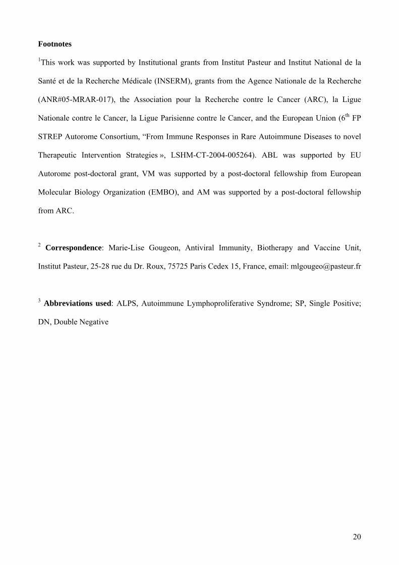
CD62Ldull, CXCR-5-, CD57+, CD11b (data not shown). Furthermore, analysis of the cytotoxic
granule content showed that ex vivo DN T-cells from ALPS patients were mostly GrzA positive (for
example 90.2% for P2) while GrzB was barely detected (Fig. 1B, 1C). In contrast, CD8+
T-cells
from the same individual significantly over-expressed GrzA and GrzB, as compared to control, and
granzymes were detected in CD4+
lymphocytes too (Fig. 1B, 1C). These data suggest that DN T
cells are unable to exert a cytolytic granule-dependent activity. In addition, as it was demonstrated
recently (24), cytotoxic granules-dependent cell-death in SP T cells may compensate for Fas-
8
deficiency in ALPS.
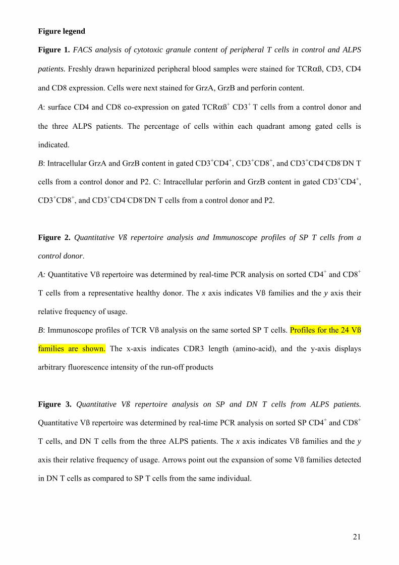
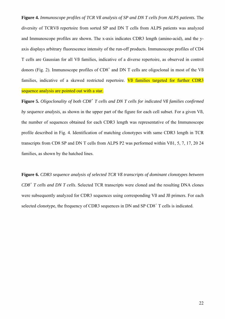
The TCR Vβ-gene usage of α/ß DN T cells from ALPS patients quantitatively differs from that of SP
cells
The expression of 24 Vß gene families was examined by RT-PCR amplification of cDNA derived
from sorted subpopulations. Representative data are shown for a control donor (Fig. 2A) and three
patients (Fig. 3). Qualitatively, repertoire diversity was conserved in all three subsets from ALPS
patients, and in particular DN T cells expressed all Vß-gene families that were used by CD4+ and
CD8+ SP cells (Fig. 3). However, quantitative differences in the expression of several Vßs were
apparent in each of the three patients, with preferential expression of some Vßs by DN T cells. For
example, Vß2 and Vß7 families were preferentially expressed in DN T cells from P1 (Vß2: 25% in
DN T cells vs 12% and 8% in SP CD8 and CD4 SP T cells respectively; Vß7: 30% in DN T cells vs
12.1% and 2.5% in SP CD8 and CD4 SP T cells respectively). Similarly, a preferential usage of
Vß17 and Vß7 genes was found in DN T cells from P2 and P3 respectively (Fig. 3).
CD8 SP and DN T cells from ALPS patients express a skewed TCR Vß repertoire, in contrast to
CD4 T cells
Analysis of CDR3 length distribution in sorted CD4 T cells from ALPS patients showed for all Vß-
Cß rearrangements Gaussian Immunoscope profiles (representative profiles are shown in Fig. 4),
which is the hallmark of a polyclonal T cell repertoire observed in control donors (Fig. 2) (20). In
sharp contrast, SP CD8 T cells from ALPS patients displayed skewed Immunoscope profiles with
oligoclonal expansions for some Vß families (Fig. 4), such as Vß2, Vß8, Vß13a, Vß14, Vß17,
Vß18, and Vß20 for P1, Vß1, Vß2, Vß5, Vß8, Vß13b, Vß17, Vß22, for P2, and Vß1 and Vß13b for
P3 (Fig. 4). This restricted diversity was not observed in sorted SP CD8 T cells from healthy donors
(Fig. 2), as previously reported (20). DN T cells from ALPS patients displayed highly limited
clonality with characteristic clonotypes for some Vß families (eg Vß2, Vß7, Vß13a, Vß13b, Vß14,
Vß20 for P1, Vß1, Vß5 and Vß22 for P2) while others showed more diverse patterns with specific
9
T cell expansions (for example Vß1, Vß21, Vß22 and Vß23 for P1, Vß2, Vß8, Vß13a, Vß22 for P2,
Vß1, Vß13b, Vß14, Vß18 for P3 (Fig. 4). Oligoclonality of both CD8 SP and DN T cells was
confirmed by sequence analysis selecting within Vß families matching clonotypes that had the same
CDR3 length in TCR transcripts from CD8 SP and DN T cells (as exemplified for patient 2 in Fig.
5). Table 2 indicates the frequency of unique in-frame CDR3 sequences within the total analyzed
selected Vß TCR clonotypes from CD4 SP, CD8 SP and DN T cells from the three patients. In DN
T cells from P1, as few as 14 unique CDR3 sequences out of 88 Vß2 TCR, 10 out of 72 Vß14 TCR,
39 out of 230 Vß18 TCR and 36 out of 233 Vß23 TCR were identified. A total of 682 CDR3
sequences were generated from selected transcripts, among which only 124 were unique (18%),
confirming Vß gene skewing among DN T cells. In contrast, paired CD4 SP T cells revealed
polyclonal frequency of CDR3 sequences with almost no repeat (66 unique CDR3 sequences out of
75 Vß2 TCR and 78 out of 80 Vß8 TCR), as found on a total of 155 CDR3 sequences among which
144 were unique (93%) (Table 2). Oligoclonality of SP CD8 T cells suggested by Immunoscope
profiles was confirmed by sequence analysis, the frequency of unique CDR3 sequences being
intermediate between SP CD4 and DN T cells, 34 unique CDR3 sequences out of 80 Vß2 TCR, 21
out of 84 Vß14 TCR, 94 out of 196 Vß18 TCR and 103 out of 216 Vß23 TCR. Overall, a total of
631 CDR3 sequences were generated from selected transcripts, among which 275 were unique
(44%). A similar skewed oligoclonal frequency of CDR3 sequences was found in patients 2 and 3
DN T cells (Table 2).
CDR3 sequence analysis of TCR transcripts containing common clonotypes in DN T and SP CD8 T
cells
To address whether DN T cells may originate from SP CD8 T cells, we identified dominant
clonotypes in DN T cells and selected clonotypes with similar CDR3 size in SP CD8 T cells for
sequence analysis (e.g. Vß7 in P2, Fig. 5). Selected TCR transcripts of common clonotypes were
cloned and the resulting DNA clones were subsequently analyzed for CDR3 sequences using
corresponding Vß and Jß primers. Fig. 6 shows that for each selected clonotype, CDR3 sequences
10
were detected at very high frequency in DN T cells, consistent with the oligoclonality of the Vß
repertoire. For example, the Vß7 clonotype in P2 included two major CDR3 sequences,
SLRQGLSTEA and SQGTGGNQPQ (76% and 6.4% of total analyzed sequences respectively).
Importantly, both CDR3 sequences were also detected in SP CD8 T cells, but with a much lower
frequency (4.8% and 0.4% of total analyzed sequences respectively) (Fig. 6). Similarly, the Vß13
clonotype in P3 was dominated by an 11aa CDR3 sequence (SSDSGGSYNEQ), which represented
53% of the clones in that gene family. This CDR3 sequence was also detected in SP CD8 T cells,
though at a lower frequency, as expected. In P1, The 9 aa CDR3 sequence RPGLPNPEA was
detected both in DN and SP CD8 T cells. Overall, the Immunoscope analysis and CDR3 sequencing
experiments argue for the hypothesis that DN T cells from ALPS patients may represent, at least in
part, originally SP CD8 T cells that down-modulated the co-receptor CD8. In addition, the extent of
oligoclonality together with phenotypic features of DN T cells suggest recent antigenic activation
by a limited set of antigens, possibly self antigens.
11
Discussion
The expansion of α/ß-DN T cells in peripheral blood of ALPS patients is a consistent feature of this
disease, and part of the diagnostic criteria of ALPS (1,10). DN T cell expansion is also evident
histologically in secondary lymphoid organs, particularly in the paracortical areas of lymph nodes,
where they accumulate and express markers of activated T cells, i.e. ki67+, CD45R0-CD45RA+
(25). Lymphoproliferation in ALPS is associated with chronic generalized activation, as
demonstrated by upregulation of HLA-DR expression on peripheral CD3 T cells as well as high
levels of activation markers such as soluble interleukine-2 receptor, soluble CD30 and IL10 in sera
of ALPS patients (7,26,27). Consistent with their unregulated peripheral proliferation and possible
recognition of self antigens, DN T cells exhibit the phenotype of mature antigen-experienced
cytotoxic T cells, i.e. TCRhigh, CD2+, CD5+, CD27++, CD28+, CD45RA+RO- CD31+, CD62Ldull,
CXCR-5-, CD57+, CD11b16. The uniform phenotype of a/ß-DN T cells from ALPS patients is
similar to a/ß-DN T cells from mice with defective Fas (lpr) (7,16,28) and different from the minor
a/ß-DN T cell compartment in healthy subjects that contains multiple subpopulations (29,30). To
investigate the origin of DN T cells from ALPS patients, we analyzed with Immunoscope
technology the TCR Vß repertoire diversity of sorted DN T cells and SP CD4 and CD8 T cells, and
we performed sequence analyses of the CDR3 region within Vß families showing, on Immunoscope
profiles, common clonotypes between DN and SP T cells. This study shows for the first time that
DN T cells from ALPS patients share with CD8 T cells unique CDR3 sequences across several
TCR Vß families, arguing for the CD8 origin of DN T cells.
Vß gene family usage was examined by quantitative RT-PCR amplification of cDNA derived from
sorted DN and SP T cells. Though the Vß-gene usage of DN T cells did not dramatically differ from
that of SP T cells in ALPS patients – i.e. DN T cells expressed all the Vß gene families that were
used by the SP counterparts, preferential expression of some Vß was found in DN T cells as
compared to autologous SP T cells. However, Vß usage skewing in DN T cells varied from one
patient to the other. Analysis of CDR3 length distribution revealed dramatic differences between all
12
three subsets. A Gaussian distribution of CDR3 peaks for all Vß-Cß rearrangements, the hallmark
of a polyclonal TCR repertoire, was obtained in sorted SP CD4 T cells from the ALPS patients. In
contrast, DN and SP CD8 T cells displayed skewed TCR-Vß Immunoscope profiles, with
oligoclonal or monoclonal expansions throughout most of the Vß families, indicating a contracted
TCR-Vβ repertoire. Oligoclonality of both SP CD8 and DN T cells was confirmed by sequence
analysis selecting, within Vß families, matching clonotypes that had the same CDR3 length in TCR
transcripts. For example, in DN T cells from P1, a total of 682 CDR3 sequences were generated
from selected transcripts, among which only 18% were unique, in contrast to SP CD4 T cells that
showed 93% of unique CDR3 sequences. The frequency of unique CDR3 sequences was
intermediate (44%) in SP CD8 T cells from P1. Similar data were found for patients 2 and 3. That
expanded DN T cells originate from SP CD8 T cells is suggested by several instances of shared
CDR3 sequences from selected Vß-Jß transcripts. For example, the Vß7 clonotype in DN T cells
from P2 was dominated by two major CDR3 sequences (SLRQGLSTEA and SQGTGGNQPQ) that
were both detected in SP CD8 T cells. We report similar findings for Vß13 clonotype in P3, or Vß
23 clonotype in P1. Interestingly, CDR3 sequences that are shared between DN and CD8 T cells
could not be detected in CD4 T cells. Therefore, from this study, we conclude that DN T cells from
ALPS patients are oligoclonally expanded, and they originate from SP CD8 T cells, as suggested by
shared Vß CDR3 sequences within several Vß families. Differences in Vß gene usage by DN as
compared to CD8 T cells suggest that the former originate from the expansion of a minor CD8
subset. Overall, this conclusion differs from the one proposed in a recent study, focusing on CDR3
spectratyping profiles of sorted DN and SP T cells from a single ALPS patient. The authors
concluded that DN are not clonally related to SP T cells because of the lack of CDR3 size matching
of the oligoclonal expansions detected on spectratype profiles from the three subsets (31). However,
the lack of CDR3 sequence analysis in this study precludes any valid conclusion on the origin of
DN T cells.
The accumulation of DN T cells in murine models of ALPS, i.e. mice lacking expression of Fas
13
(lpr) or Fas-ligand (gld), suggests that this subset might represent a subpopulation destined for
apoptosis in normal mice (9). It would arise from high affinity interactions of TCR with
peptide/MHC, and would accumulate to vast numbers in the absence of Fas-induced apoptosis (32).
The SP T cell origin of DN T cells has been suggested by several findings, including their bearing
of a demethylated CD8 gene (33,34), and their absence in mice lacking expression of class I MHC
(35). In ALPS patients, the homogenous phenotype of DN T cells, that characterizes mature
antigen-experienced cytotoxic T cells, suggest their CD8 T cell origin (16). Interestingly, the
peculiar expression pattern of cytolytic proteins by DN T cells makes them unable to exert a
cytolytic granule-dependent activity. Indeed, they strongly express granzyme A, whereas granzyme
B and perforin are barely detected, as opposed to granule expression in SP T cells (24) (and this
study). As recently discussed (24) the repressor mechanism that down-regulates expression of
granzyme B and perforin while sparing granzyme A in DN T cells may involve IL-10 [highly
produced by DN T-cells (17,36)] previously shown to repress granzyme B mRNA expression in
cytotoxic lymphocytes (37). This repressor mechanism could be involved in the modulation of the
CD4 or CD8 co-receptors (38).
The mechanisms involved in peripheral expansion of DN T cells in ALPS patients are not fully
understood. Holzelova et al. have shown that resistance to Fas-mediated apoptosis would confer a
selective advantage to mutant cells, leading to their accumulation and evolution towards the
phenotype of DN T cells (15). The recent observation that in vivo triggering of mitochondrial
apoptosis by rapamycin in murine ALPS induces a dramatic decrease in DN T cells (39) supports
this hypothesis. The lack of co-receptors in DN T cells from ALPS patients may also contribute to
their accumulation. Indeed, it has been reported in a TCR transgenic murine model that co-receptor
engagement controls expansion of normal T cells, and in the absence of co-receptor T cells survive
chronic stimulation and express B220, as seen in ALPS disease (40). In the present study, DN T
cells appeared to be oligoclonally expanded, as evidenced by CDR3 length distribution. This extent
of oligoclonality together with phenotypic features suggesting historical antigen encounter and
14
recent activation of large proportions of these cells may reflect stimulation of DN T cells by a
limited yet ubiquitous set of antigens. The antigen specificities recognized by the TCR of DN T
cells have not been defined, but the general assumption is that they will include self-antigens.
Interestingly, this study shows for the first time that CD8 T cells from ALPS patients (children and
young adults) exhibit a restricted TCR diversity, with oligoclonal and monoclonal expansions
throughout most of the Vß families, and redundant CDR3 sequences. This is reminiscent of
repertoire contraction and oligoclonal expansions seen in aged individuals, and supposed to be a
heterogeneous phenomenon including impaired homeostasis and antigen-driven stimulation (41).
The conserved CDR3 sequences between CD8 T cells and DN T cells reported here suggest that
they may recognize identical peptide sequences, and argue for their filiation. It has been proposed
that the defect in killing of lpr CD8 T cells following stimulation by self-antigen, would lead to the
modulation of CD8 co-receptor and accumulation as IL-10 secreting anergic cells (42,43). The
comparative molecular analysis of CDR3 distribution and sequencing of CD8 T cells and DN T
cells in the present study extends for the first time this hypothesis to the human disease.
15
References
1. Bidere, N., H. C. Su, and M. J. Lenardo. 2006. Genetic disorders of programmed cell death
in the immune system. Annu. Rev. Immunol. 24:321-352
2. Rieux-Laucat, F., F. Le Deist and A. Fischer. 2003. Autoimmune lymphoproliferative
syndrome : genetic defects of apoptosis pathways. Cell Death Diff. 10:124-133
3. Sneller, M. C., S. E. Strauss, J. S Jaffe, T. A Fleisher, M. Stetler-Stevenson, and W.
Strober. 1992. A novel lymphoproliferative/autoimmune syndrome resembling murine lpr/gld
disease. J. Clin. Invest. 90:334-341
4. Rieux-Laucat, F., F. Le Deist, C. Hivroz, M. C. Sneller, S. E. Straus, E. S. Jaffe, T. A.
Jaffe, T. A. Fleisher, M. Stetler-Stevenson, and W. Strober. 1995. Mutations in Fas associated with
human lymphoproliferative syndrome and autoimmunity. Science 268:1347-1349.
5. Fisher, G. H., F. J. Rosenberg, S. E. Straus, J. K. Dale, L. A. Middleton, A. Y. Lin, W.
Strober, M. J. Lenardo, and J. M Puck. 1995. Dominant interfering Fas gene mutations impair
apoptosis in a human autoimmune lymphoproliferative syndrome. Cell 81:935-946.
6. Drappa, J., A. K. Vaishnaw, K. E. Sullivan, J. L. Chu, and K. B. Elkon. 1996. Fas gene
mutation in the Canale-Smith syndrome, an inherited lymphoproliferative disorder associated with
autoimmunity. N. Eng. J. Med. 335:1643-1649
7. Bettinardi, A., D. Brugnoni, E. Quiros-Roldan, A. Malagoli, S. La Grutta, A. Correra, and
L. D. Notarangelo. 1997. Missense mutations in the Fas gene resulting in autoimmune
lymphoproliferative syndrome : a molecular and immunological analysis. Blood 89:902-909
8. Krammer, P.H. 2000. CD95’s deadly mission in the immune system. Nature 407 :789-795
9. Nagata, S. and T. Suda. 1995. Fas and Fas ligand: lpr and gld mutations. Immunol. Today
16:39-43.
10. Rieux-Laucat, F., A. Fischer, and F. Le Deist. 2003. Cell-death signaling and human
disease. Curr. Opin. Immunol. 15:325-331.
11. Jackson, C. E., R. E. Fischer, A. P. Hsu, S. M Anderson, Y. Choi, J Wang, J. K Dale, T. A.
16
Fleisher, L. A. Middleton, M. C. Sneller, M. J. Lenardo, S. E Straus, and J. M Puck. 1999.
Autoimmune lymphoproliferative syndrome with defective Fas : genotype influences penetrance.
Am. J. Hum. Genet. 64 :1002-1014
12. Rieux-Laucat, F., S. Blachere, S. Danielan, J. P. De Villartay, M. Oleastro, E. Solary, B.
Bader-Meunier, P. Arkwright, C. Pondare, F. Bernaudin, H. Chapel, S. Nielsen, M. Berrah, A.
Fischer and F. Le Deist, 1999. Lymphoproliferative syndrome with autoimmunity: A possible
genetic basis for dominant expression of the clinical manifestations. Blood 94:2575-2582.
13. Wu, J., J. Wilson, J. He, L. Xiang, P. H. Schur, and J. D. Mountz, 1996. Fas ligand
mutation in a patient with systemic lupus erythematosus and lymphoproliferative disease. J. Clin.
Invest. 98 :1107-1113
14. Wang, J., L. Zheng, A. Lobito, F. K. Chan, J. Dale, M. Sneller, X. Yao, J. M. Puck, S. E.
Straus, and M. J. Lenardo. 1999. Inherited human Caspase 10 mutations underlie defective
lymphocyte and dendritic cell apoptosis in autoimmune lymphoproliferative syndrome type II. Cell
98:47-58.
15. Holzelova, E., C. Vonarbourg, M. C. Stolzenberg, P. D. Arkwright, F. Selz, A. M. Prieur,
S. Blanche, J. Bartunkova, E. Vilmer, A. Fischer, F. Le Deist, and F. Rieux-Laucat. 2004.
Autoimmune lymphoproliferative syndrome with somatic Fas mutations. N. Engl. J. Med.
351:1409-1418.
16. Bleesing, J. J., M. R Brown, J. K Dale, S. E Straus, M. J. Lenardo, J. M. Puck, T. P.
Atkinson, and T. A. Fleisher, 2001. TcR-alpha/beta(+) CD4(-)CD8(-) T cells in humans with the
autoimmune lymphoproliferative syndrome express a novel CD45 isoform that is analogous to
murine B220 and represents a marker of altered O-glycan biosynthesis. Clin. Immunol. 100:314-
324.
17. Lopatin, U., X. Yao, R. K. Williams, J. J. Bleesing, J. K Dale, D. Wong, J. Teruya-
Feldstein, J. S. Fritz, M. R. Morrow, I. Fuss, M. C Sneller, M. Raffeld, T. A. Fleisher, J. M. Puck,
W. Strober, and S. E. Straus. 2001. Increases in circulating and lymphoid tissue interleukin-10 in
autoimmune lymphoproliferative syndrome are associated with disease expression. Blood 97:3161-
17
3170.
18. Ohga S, A. Nomura, Y. Takahata , K. Ihara, H. Takada, H. Wakiguchi, Y. Kudo, and T.
Hara. 2002. Dominant expression of interleukin 10 but not interferon gamma in CD4(-)CD8(-)alpha
betaT cells of autoimmune lymphoproliferative syndrome. Br. J. Haematol. 119:535-538.
19. Fuss, I.J., W. Strober, J.K. Dale, S. Fritz, G. R. Pearlstein, J. M. Puck, M. J. Lenardo, and
S.E. Straus. 1997. Characteristic T helper 2 T cell cytokine abnormalities in autoimmune
lymphoproliferative syndrome, a syndrome marked by defective apoptosis and humoral
autoimmunity. J. Immunol.158:1912-1918.
20. Lim, A., V. Baron, L. Ferradini, M. Bonneville, P. Kourilsky, and C. Pannetier, 2002.
Combination of MHC-peptide multimer-based T cell sorting with the Immunoscope permits
sensitive ex vivo quantitation and follow-up of human CD8+ T cell immune responses. J. Immunol.
Methods 261:177-194.
21. Pannetier, C., M. Cochet, S. Darche, A. Casrouge, M. Zöller, P. Kourilsky. 1993. The
sizes of the CDR3 hypervariable regions of the murine T-cell receptor beta chains vary as a
function of the recombined germ-line segments. Proc. Natl. Acad. Sci. U S A. 90:4319–4323.
22. Pannetier, C., J. Even, and P. Kourilsky. 1995. T-cell repertoire diversity and clonal
expansions in normal and clinical samples. Immunol. Today 16:176–181.
23. Bousso, P., A. Casrouge, J. D. Altman, M. Haury, J. Kanellopoulos, J. P. Abastado, and P.
Kourilsky. 1998. Individual variations in the murine T cell response to a specific peptide reflect
variability in naive repertoires. Immunity 9:169-78.
24. Mateo, V., M. Ménager, G. de Saint-Basile, M. C. Stolzenberg, B. Roquelaure, N. Andre,
B. Florkin, F. le Deist, C. Picard, A. Fischer, and F. Rieux-Laucat, 2007. Perforin-dependent
apoptosis functionally compensates Fas-deficiency in activation-induced cell-death of human T-
lymphocytes. Blood 110 :4285-4292
25. Rieux-Laucat, F. 2006. Inherited and acquired death receptor defects in human
autoimmune lymphoproliferative syndrome. Curr. Dir. Autoimmun. 9:18-36.
26. Le Deist, F., J. F Emile, F. Rieux-Laucat, M. Benkerrou, I. Roberts, N. Brousse, and A.
18
Fischer. 1996. Clinical, immunological, and pathological consequences of Fas-deficient conditions.
Lancet 348 :719-723
27. Sneller, M. C., J. Wang, J. K. Dale, W. Strober, L. A. Middelton, Y. Choi, T. A. Fleisher,
M. S. Lim, E. S. Jaffe, J. M. Puck, M. J. Lenardo, and S. E. Straus, 1997. Clinical, immunologic,
and genetic features of an autoimmune lymphoproliferative syndrome associated with abnormal
lymphocyte apoptosis. Blood 89:1341-1348
28. Morse, H.C., W. F. Davidson, R. A. Yetter, E. D. Murphy, J. B. Roth, and R. L. Coffman.
1982. Abnormalities induced by the mutant gene lpr: expansion of a unique lymphocyte subset. J.
Immunol. 129:2612-2615
29. Reimann, J. 1991. Double-negative (CD4-CD8-), TCR alpha beta-expressing, peripheral T
cells. Scand. J. Immunol. 34:679-688
30. Fischer, K., S. Voelkl, J. Heymann, G. K. Przybylski, K. Mondal, M. Laumer, L. Kunz-
Schughart, C. A. Schmidt, R. Andreesen, and A. Mackensen. 2005. Isolation and characterization of
human antigen-specific TCRαß+CD4-CD8- double negative regulatory T cells. Blood 105:2828-
2835
31. Marlies, A., G. Udo, B. Juergen, S. Bernd, M. Herrmann, and J. P. Haas. 2007. The
expanded double negative T cell populations of a patient with ALPS are not clonally related to
CD4+ or to CD8+ T cells. Autoimmunity 40:299-301
32. Mixter, P. F., J. Q. Russell, G. J. Morrissette, C. Charland, D. Aleman-Hoey, and R. C.
Budd. 1999. A model for the origin of TCR-alphabeta+ CD4-CD8- B220+ cells based on high
affinity TCR signals. J. Immunol. 162:5747-56
33. Landolfi, M. M., N. Van Houten, J. Q. Russell, R. Scollay, J.R. Parnes, and R.C. Budd,
1993. CD2-CD4-CD8- lymph node T lymphocytes in MRL lpr/lpr mice are derived from a
CD2+CD4+CD8+ thymic precursor. J. Immunol. 151:1086-1091
34. Wu, L., M. Pearse, M. Egerton, H. Petrie, and R. Scollay. 1990. CD4-CD8- thymocytes
that express the T cell receptor may have previously expressed CD8. Int. Immunol. 2:51-56.
35. Mixter, P.F., J. Q Russell, F. H. Durie, and R. C. Budd. 1995. Decreased CD4-CD8- TCR-
19
αβ+ cells in lpr/lpr mice lacking b2-microglobulin. J. Immunol. 154:2063-2067
36. Ohga, S., A. Nomura, Y. Takahata, K. Ihara, H. Takada, H. Wakiguchi, Y. Kudo, and T.
Hara. 2002. Dominant expression of interleukin 10 but not interferon gamma in CD4(-) CD8(-)
alpha beta T cells of autoimmune lymphoproliferative syndrome. Br. J. Haematol. 119:535-538.
37. Fitzpatrick, L., A. P. Makrigiannis, M. Kaiser, and D. W. Hoskin, 1996. Anti-CD3-
activated killer T cells: interferon-gamma and interleukin-10 cross-regulate granzyme B expression
and the induction of major histocompatibility complex-unrestricted cytotoxicity. J. Interferon
Cytokine Res. 16:537-546.
38. Pestano, G. A., Y. Zhou, L. A. Trimble, J. Daley, G. F Weber, and H. Cantor. 1999.
Inactivation of misselected CD8 T cells by CD8 gene methylation and cell death. Science 284:1187-
1191.
39. Teachey, D. T., D. A. Obzut, K. Axsom, J. K. Choi, K. C. Goldsmith, J. Hall, J. Hulitt, C.
S. Manno, J. M. Maris, N. Rhodin, K. E. Sullivan, V. I. Brown, and S. A. Grupp. 2006. Rapamycin
improves lymphoproliferative disease in murine autoimmune lymphoproliferative syndrome
(ALPS). Blood 108:1965-1971.
40. Hamad, A. R., A. Srikrishnan, P. Mirmonsef, C. P. Broeren, C. H. June, D. Pardoll, and J.
P. Schneck. 2001. Lack of coreceptor allows survival of chronically stimulated double-negative
alpha/beta T cells: implications for autoimmunity. J. Exp. Med. 193:1113-1121.
41. Clambey, E. T., J. W. Kappler, and P. Marrach. CD8 T cell clonal expansions and aging: a
heterogeneous phenomenon with a common outcome. Exp. Gerontology 42:407-411
42. Ford, M. S., K. J. Young, Z. Zhang, P.S. Ohashi, and L. Zhang. 2002. The immune
regulatory function of lymphoproliferative double negative T cells in vitro and in vivo. J. Exp. Med.
196:261–267.
43. Watanabe, N., K. Ikuta, S. Nisitani, T. Chiba, and T. Honjo. 2002. Activation and
differentiation of autoreactive B-1 cells by interleukin 10 induce autoimmune hemolytic anemia in
Fas-deficient antierythrocyte immunoglobulin transgenic mice. J. Exp. Med. 196:141–146.
20
Footnotes
1This work was supported by Institutional grants from Institut Pasteur and Institut National de la
Santé et de la Recherche Médicale (INSERM), grants from the Agence Nationale de la Recherche
(ANR#05-MRAR-017), the Association pour la Recherche contre le Cancer (ARC), la Ligue
Nationale contre le Cancer, la Ligue Parisienne contre le Cancer, and the European Union (6th FP
STREP Autorome Consortium, “From Immune Responses in Rare Autoimmune Diseases to novel
Therapeutic Intervention Strategies », LSHM-CT-2004-005264). ABL was supported by EU
Autorome post-doctoral grant, VM was supported by a post-doctoral fellowship from European
Molecular Biology Organization (EMBO), and AM was supported by a post-doctoral fellowship
from ARC.
2 Correspondence: Marie-Lise Gougeon, Antiviral Immunity, Biotherapy and Vaccine Unit,
Institut Pasteur, 25-28 rue du Dr. Roux, 75725 Paris Cedex 15, France, email: [email protected]
3 Abbreviations used: ALPS, Autoimmune Lymphoproliferative Syndrome; SP, Single Positive;
DN, Double Negative
21
Figure legend
Figure 1. FACS analysis of cytotoxic granule content of peripheral T cells in control and ALPS
patients. Freshly drawn heparinized peripheral blood samples were stained for TCRαß, CD3, CD4
and CD8 expression. Cells were next stained for GrzA, GrzB and perforin content.
A: surface CD4 and CD8 co-expression on gated TCRαß+ CD3+ T cells from a control donor and
the three ALPS patients. The percentage of cells within each quadrant among gated cells is
indicated.
B: Intracellular GrzA and GrzB content in gated CD3+CD4+, CD3+CD8+, and CD3+CD4-CD8-DN T
cells from a control donor and P2. C: Intracellular perforin and GrzB content in gated CD3+CD4+,
CD3+CD8+, and CD3+CD4-CD8-DN T cells from a control donor and P2.
Figure 2. Quantitative Vß repertoire analysis and Immunoscope profiles of SP T cells from a
control donor.
A: Quantitative Vß repertoire was determined by real-time PCR analysis on sorted CD4+ and CD8+
T cells from a representative healthy donor. The x axis indicates Vß families and the y axis their
relative frequency of usage.
B: Immunoscope profiles of TCR Vß analysis on the same sorted SP T cells. Profiles for the 24 Vß
families are shown. The x-axis indicates CDR3 length (amino-acid), and the y-axis displays
arbitrary fluorescence intensity of the run-off products
Figure 3. Quantitative Vß repertoire analysis on SP and DN T cells from ALPS patients.
Quantitative Vß repertoire was determined by real-time PCR analysis on sorted SP CD4+ and CD8+
T cells, and DN T cells from the three ALPS patients. The x axis indicates Vß families and the y
axis their relative frequency of usage. Arrows point out the expansion of some Vß families detected
in DN T cells as compared to SP T cells from the same individual.
22
Figure 4. Immunoscope profiles of TCR Vß analysis of SP and DN T cells from ALPS patients. The
diversity of TCRVß repertoire from sorted SP and DN T cells from ALPS patients was analyzed
and Immunoscope profiles are shown. The x-axis indicates CDR3 length (amino-acid), and the y-
axis displays arbitrary fluorescence intensity of the run-off products. Immunoscope profiles of CD4
T cells are Gaussian for all Vß families, indicative of a diverse repertoire, as observed in control
donors (Fig. 2). Immunoscope profiles of CD8+ and DN T cells are oligoclonal in most of the Vß
families, indicative of a skewed restricted repertoire. Vß families targeted for further CDR3
sequence analysis are pointed out with a star.
Figure 5. Oligoclonality of both CD8+ T cells and DN T cells for indicated Vß families confirmed
by sequence analysis, as shown in the upper part of the figure for each cell subset. For a given Vß,
the number of sequences obtained for each CDR3 length was representative of the Immunoscope
profile described in Fig. 4. Identification of matching clonotypes with same CDR3 length in TCR
transcripts from CD8 SP and DN T cells from ALPS P2 was performed within Vß1, 5, 7, 17, 20 24
families, as shown by the hatched lines.
Figure 6. CDR3 sequence analysis of selected TCR Vß transcripts of dominant clonotypes between
CD8+ T cells and DN T cells. Selected TCR transcripts were cloned and the resulting DNA clones
were subsequently analyzed for CDR3 sequences using corresponding Vß and Jß primers. For each
selected clonotype, the frequency of CDR3 sequences in DN and SP CD8+ T cells is indicated.
Table 1 : Patients’ clinical and immunological characteristics
Serum Ig‡‡ Lymphocyte subsets¶ (%)
Patient
No
Fas
Mutation
Sex/
Age
(yrs)
Age at
presentat
(yrs)
LA*Spl†
Splenectomy
Age (yrs) AI‡
IgG IgA IgM
Lympho¶
(x103/ml) CD3 CD4 CD8 DNT CD19
CD16+
CD56+
1
K280fs M/32 6 ++ +++ + (12) UR, Ar
->
N
->
N N 2728 66 40 19 6 4 24
2
S214fs M/10 0.3 + +++ - 0 N N 3 400 90 24 41 20 7 2
3
S227fs F/22 4 + ++ - Uv, G,T
1600
80
25
27
12
12
na
*LA :Lymph-Adenopathy + : multiple nodes, size <2 cm, ++ : multiple nodes 2 cm <size < 5cm; †Spl : Splenomegaly + : above umbilicus,, ++ : below
umbilicus, +++ : palpable in iliac fossa ; ‡AI : Auto Immunity 0 : absence of autoimmune manifestation ; UR : Urticarial Rash ; Ar : Arthralgia; Uv : Uveitis;
G : Glomerulopathy; T : Thrombocytopenia; ‡‡Serum Immunoglobulin level: N: within the normal range, : > +2SD <+4SD, :> +4SD ; ¶ Lymphocyte
counts in controls are 2000-4000 x103/ml, with 70-80% CD3+ cells, 40-50% CD4+ cells, 20-30% CD8+ cells, 10-20% CD19+ cells and 10-20% of
CD16+CD56+ cells. ** DN T cells : % CD4-CD8- among TCR α/β Τ cells (normal limit : 2%); na : not available
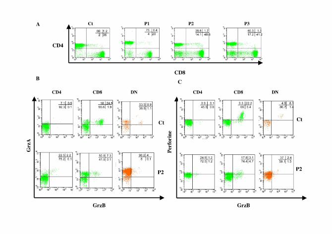
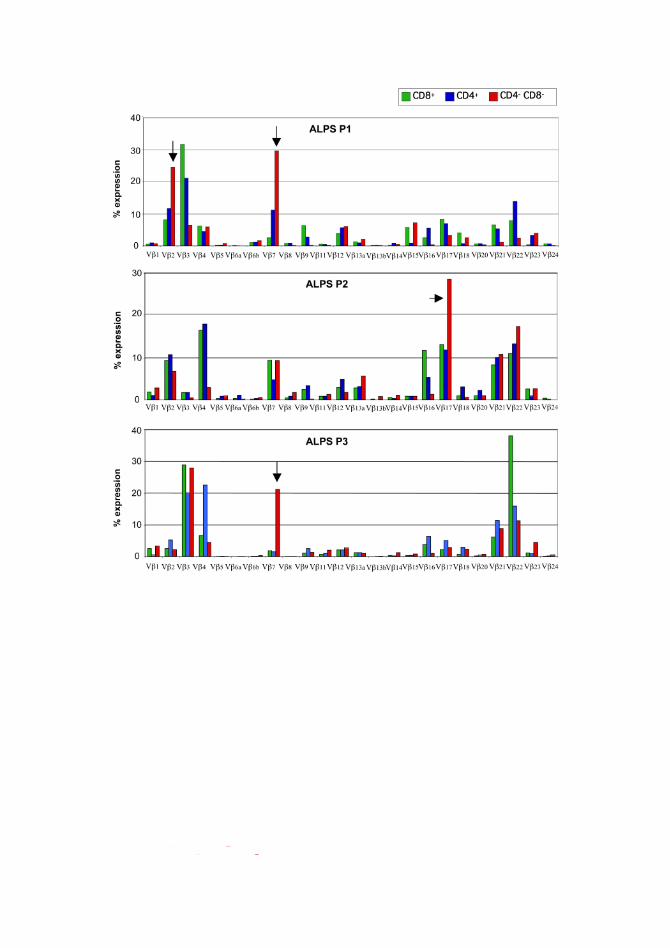
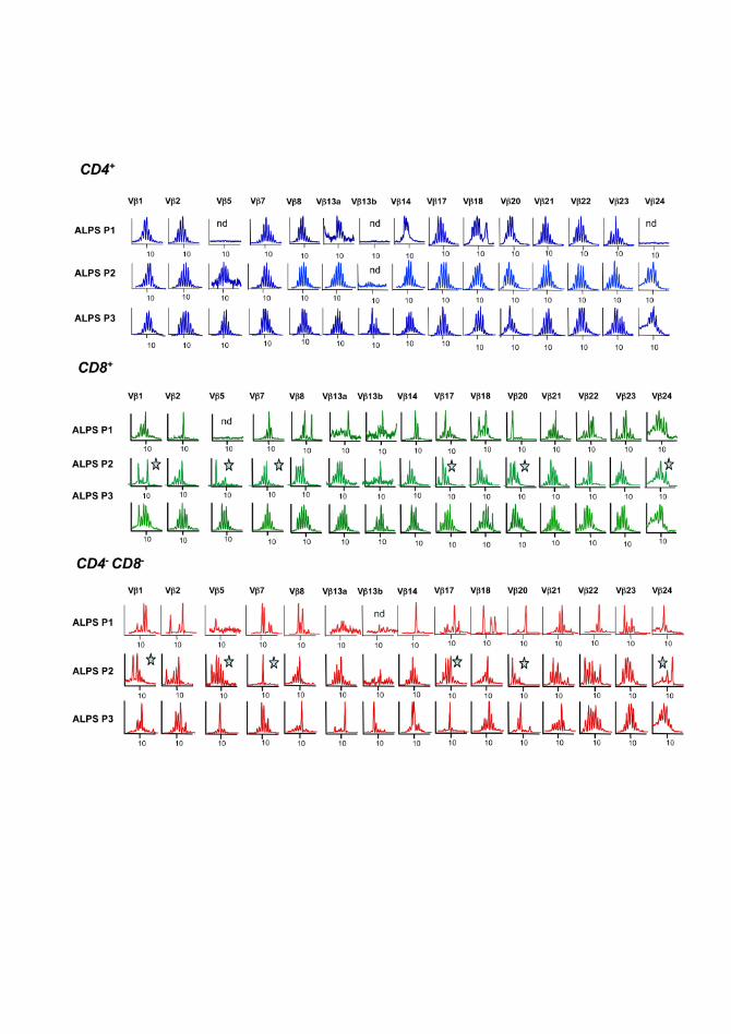
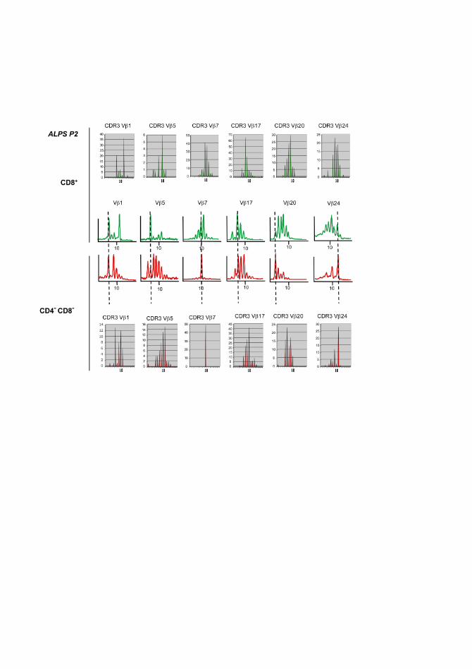
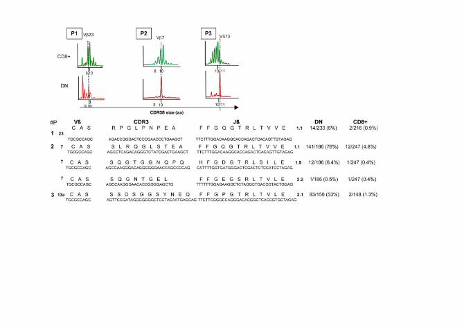
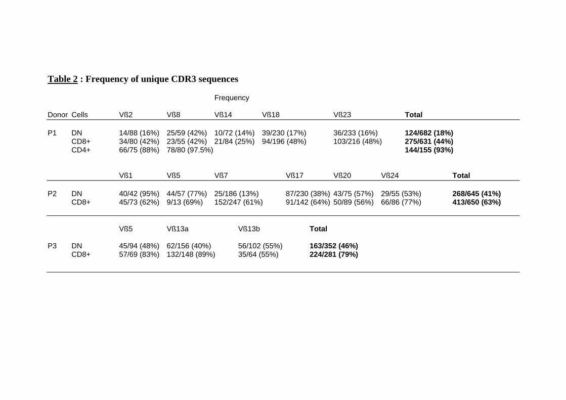
Table 2 : Frequency of unique CDR3 sequences Frequency Donor Cells Vß2 Vß8 Vß14 Vß18 Vß23 Total P1 DN 14/88 (16%) 25/59 (42%) 10/72 (14%) 39/230 (17%) 36/233 (16%) 124/682 (18%) CD8+ 34/80 (42%) 23/55 (42%) 21/84 (25%) 94/196 (48%) 103/216 (48%) 275/631 (44%) CD4+ 66/75 (88%) 78/80 (97.5%) 144/155 (93%) Vß1 Vß5 Vß7 Vß17 Vß20 Vß24 Total P2 DN 40/42 (95%) 44/57 (77%) 25/186 (13%) 87/230 (38%) 43/75 (57%) 29/55 (53%) 268/645 (41%) CD8+ 45/73 (62%) 9/13 (69%) 152/247 (61%) 91/142 (64%) 50/89 (56%) 66/86 (77%) 413/650 (63%) Vß5 Vß13a Vß13b Total P3 DN 45/94 (48%) 62/156 (40%) 56/102 (55%) 163/352 (46%) CD8+ 57/69 (83%) 132/148 (89%) 35/64 (55%) 224/281 (79%)


![IMMUNOGLOBULINE E T CELL RECEPTOR T. Strachan e A.P. … · B cell antigen receptor tetramero [ IgH 2 + IgL 2 (Ig oppure Ig )] T cell receptor (TCR) eterodimero TCR /TCR TCR /TCR](https://img.pdfslide.us/doc/110x75/5c017b5c09d3f26f1e8cc6a0/immunoglobuline-e-t-cell-receptor-t-strachan-e-ap-b-cell-antigen-receptor.jpg)