Embed Size (px)
Citation preview

Human Respiratory Syncytial Virus: Infectionand PathologyKaren Bohmwald, MSc1 Janyra A. Espinoza, MSc1 Emma Rey-Jurado, PhD1 Roberto S. Gómez, MSc1
Pablo A. González, PhD1 Susan M. Bueno, PhD1 Claudia A. Riedel, PhD2 Alexis M. Kalergis, PhD1,2,3,4
1Departamento de Genética Molecular y Microbiología, MillenniumInstitute on Immunology and Immunotherapy, Pontificia UniversidadCatólica de Chile, Santiago, Chile
2Departamento de Ciencias Biológicas y Facultad de Medicina,Millennium Institute on Immunology and Immunotherapy,Universidad Andrés Bello, Santiago, Chile
3Departamento de Endocrinología, Escuela de Medicina, MillenniumInstitute on Immunology and Immunotherapy, Pontificia UniversidadCatólica de Chile, Santiago, Chile
4 INSERM U1064, Nantes, France
Semin Respir Crit Care Med 2016;37:522–537.
Address for correspondence Alexis M. Kalergis, PhD, Departamentode Genética Molecular y Microbiología, Millennium Institute onImmunology and Immunotherapy, Facultad de Ciencias Biológicas,Pontificia Universidad Católica de Chile, Avenida Libertador BernardoO’Higgins #340, Santiago E-8331010, Chile(e-mail: [email protected]; [email protected]).
Acute lower respiratory tract infections (ALRTIs) are themajor cause of morbidity and mortality in young children,the elderly, and immunocompromised individuals world-wide.1 Importantly, the human respiratory syncytial virus(hRSV) is the principal microbial agent known to causeALRTIs.2–5Most clinical manifestations caused by hRSV rangefrom mild symptoms, such as rhinorrhea, cough, congestion,low-grade fever, reduced appetite, and respiratory distress, to
severe alveolitis, bronchiolitis, and pneumonia.6 The hetero-geneity of the diseases caused by hRSV depends, amongothers, on host risk factors, including preterm birth,7 con-genital heart disease,7,8 chronic lung diseases,9 andimmunosuppression.10
hRSV infections are considered highly contagious, affect-ing nearly 70% of infants before thefirst year of life and nearly100% of children by the age of 2.11Worldwide, approximately
Keywords
► human respiratorysyncytial virus
► virulence factors► innate immune
response► adaptive immune
response► extrapulmonary
manifestations► central nervous
system
Abstract The human respiratory syncytial virus (hRSV) is by far the major cause of acute lowerrespiratory tract infections (ALRTIs) worldwide in infants and children younger than2 years. The overwhelming number of hospitalizations due to hRSV-induced ALRTI eachyear is due, at least in part, to the lack of licensed vaccines against this virus. Thus, hRSVinfection is considered a major public health problem and economic burden in mostcountries. The lung pathology developed in hRSV-infected individuals is characterizedby an exacerbated proinflammatory and unbalanced Th2-type immune response. Inaddition to the adverse effects in airway tissues, hRSV infection can also causeneurologic manifestations in the host, such as seizures and encephalopathy. Althoughthe origins of these extrapulmonary symptoms remain unclear, studies with patientssuffering from neurological alterations suggest an involvement of the inflammatoryresponse against hRSV. Furthermore, hRSV has evolved numerous mechanisms tomodulate and evade the immune response in the host. Several studies have focused onelucidating the interactions between hRSV virulence factors and the host immunesystem, to rationally design new vaccines and therapies against this virus. Here, wediscuss about the infection, pathology, and immune response triggered by hRSV in thehost.
Issue Theme Respiratory ViralInfections; Guest Editor:Sunit K. Singh, PhD
Copyright © 2016 by Thieme MedicalPublishers, Inc., 333 Seventh Avenue,New York, NY 10001, USA.Tel: +1(212) 584-4662.
DOI http://dx.doi.org/10.1055/s-0036-1584799.ISSN 1069-3424.
522
Thi
s do
cum
ent w
as d
ownl
oade
d fo
r pe
rson
al u
se o
nly.
Una
utho
rized
dis
trib
utio
n is
str
ictly
pro
hibi
ted.
34 million new cases of hRSV-associated ALRTI occur inchildren younger than 5 years and as much as 200,000 deathsare estimated annually.12 Indeed, hRSV causes a minimum of3.4 million hospitalizations per year in the United States.12
Due to the high hospitalization rates and increased healthcare system costs, hRSV infections are considered a majorpublic health burden globally. For instance, medical costsrelated to hRSV infection in hospitalized individuals is esti-mated at 394million USD annually, just in the United States.13
Thus, safe and effective vaccines against this virus are urgent-ly needed.
hRSV spreads rapidly and efficiently throughout the pop-ulation by inhalation of aerosolized droplets of infectiousviral particles, or directly through the contact of thesedroplets with the ocular mucosa.14,15 One of the most rele-vant characteristics of hRSV infection is the capacity toproduce high reinfection rates in individuals during epidemicoutbreaks. For instance, epidemiological studies indicate thatapproximately 36% of individuals can be reinfected at leastonce during a given season.13,16,17 Based on the currentevidence, these reinfection episodes may be due to theelicitation of a deficient or hampered cellular and humoralimmune memory after a first exposure to the virus.4,18–20
Bronchiolitis is one of the most severe illnesses caused byhRSV infection. The term bronchiolitis is referred to distalbronchiole inflammation and obstruction. Such an obstruc-tion reduces the airflow into small airways and causes analteration in the exhalation capacity. This phenomenon canlead to lung hyperexpansion, lung function alterations, in-creased mucus production, atelectasis, and wheezing.21,22
Furthermore, infection of alveolar epithelium by hRSV leadsto pneumonia, which prevents an efficient gas-exchangeprocess. Such infection triggers distal airway inflammation,leading to severe pulmonary disease.22 The initial responseagainst hRSV infection is given by the airway epithelial cells(AECs), which promotes the recruitment of effector immunecells at the site of infection. hRSV-associated immunopathol-ogy is characterized by the expression of proinflammatorycytokines and the subsequent perivascular/peribronchial in-filtration by mononuclear cells, mainly neutrophils and lym-phocytes.21 This exacerbated inflammation is thought totrigger an unbalanced and pathogenic T helper (Th)2response.
Formore than 50 years,most research on hRSV has focusedon elucidating the mechanisms involved in respiratory pa-thology, as well as on the design of vaccines and therapiesagainst hRSV.23–25 Nowadays, it is known that hRSV is able tomigrate from the airways tovarious tissues in thehost, such asheart, kidney, liver, and brain, thereby producing diverseclinical manifestations, including cardiopathy, hepatitis, andencephalitis.6,26–28 As a consequence, extensive researchaimed at understanding the extrapulmonary manifestationsof hRSV infection has gained attention in the past few years.For instance, neurological abnormalities associated withhRSV infection in patients with severe bronchiolitis havebeen detected. Such patients have shown symptoms includ-ing seizures, apnea and altered proinflammatory cytokinelevels in cerebrospinal fluid (CSF).26However, the cellular and
molecular bases for the encephalopathy caused by hRSVremain unknown.
hRSV Characteristics
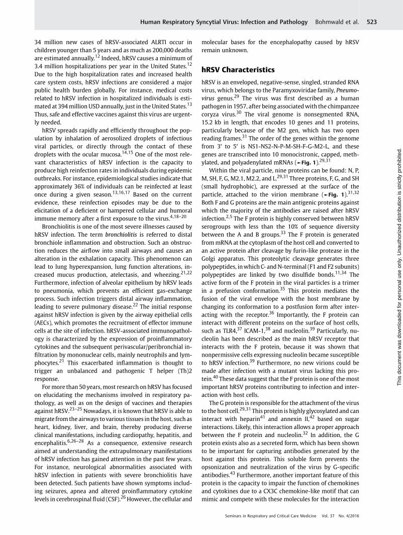
hRSV is an enveloped, negative-sense, singled, stranded RNAvirus, which belongs to the Paramyxoviridae family, Pneumo-virus genus.29 The virus was first described as a humanpathogen in 1957, after being associatedwith the chimpanzeecoryza virus.30 The viral genome is nonsegmented RNA,15.2 kb in length, that encodes 10 genes and 11 proteins,particularly because of the M2 gen, which has two openreading frames.31 The order of the genes within the genomefrom 3′ to 5′ is NS1-NS2-N-P-M-SH-F-G-M2-L, and thesegenes are transcribed into 10 monocistronic, capped, meth-ylated, and polyadenylated mRNAs (►Fig. 1).29,31
Within the viral particle, nine proteins can be found: N, P,M, SH, F, G, M2.1, M2.2, and L.29,31 Three proteins, F, G, and SH(small hydrophobic), are expressed at the surface of theparticle, attached to the virion membrane (►Fig. 1).31,32
Both F and G proteins are the main antigenic proteins againstwhich the majority of the antibodies are raised after hRSVinfection.2,5 The F protein is highly conserved between hRSVserogroups with less than the 10% of sequence diversitybetween the A and B groups.33 The F protein is generatedfrommRNA at the cytoplasm of the host cell and converted toan active protein after cleavage by furin-like protease in theGolgi apparatus. This proteolytic cleavage generates threepolypeptides, inwhich C- andN-terminal (F1 and F2 subunits)polypeptides are linked by two disulfide bonds.11,34 Theactive form of the F protein in the viral particles is a trimerin a prefusion conformation.35 This protein mediates thefusion of the viral envelope with the host membrane bychanging its conformation to a postfusion form after inter-acting with the receptor.36 Importantly, the F protein caninteract with different proteins on the surface of host cells,such as TLR4,37 ICAM-1,38 and nucleolin.39 Particularly, nu-cleolin has been described as the main hRSV receptor thatinteracts with the F protein, because it was shown thatnonpermissive cells expressing nucleolin became susceptibleto hRSV infection.39 Furthermore, no new virions could bemade after infection with a mutant virus lacking this pro-tein.40 These data suggest that the F protein is one of themostimportant hRSV proteins contributing to infection and inter-action with host cells.
The G protein is responsible for the attachment of the virusto thehost cell.29,31 This protein is highly glycosylated and caninteract with heparin41 and annexin II,42 based on sugarinteractions. Likely, this interaction allows a proper approachbetween the F protein and nucleolin.32 In addition, the Gprotein exists also as a secreted form, which has been shownto be important for capturing antibodies generated by thehost against this protein. This soluble form prevents theopsonization and neutralization of the virus by G-specificantibodies.43 Furthermore, another important feature of thisprotein is the capacity to impair the function of chemokinesand cytokines due to a CX3C chemokine-like motif that canmimic and compete with these molecules for the interaction
Seminars in Respiratory and Critical Care Medicine Vol. 37 No. 4/2016
Human Respiratory Syncytial Virus: Infection and Pathology Bohmwald et al. 523
Thi
s do
cum
ent w
as d
ownl
oade
d fo
r pe
rson
al u
se o
nly.
Una
utho
rized
dis
trib
utio
n is
str
ictly
pro
hibi
ted.
with their receptors and, thus, modulate CD8þ T-cell re-sponses.44,45 Moreover, the G protein was shown to displaystructural homology with tumor necrosis factor (TNF) recep-tors and is likely to interact with TNF family cytokine mem-bers, conducting to a misbalance in the inflammatoryresponse mediated by these molecules.46 Although G proteinis not totally necessary for hRSV infection, it plays an impor-tant role at modulating the immune response triggered byhRSV infection.
The SH protein locates at the surface membrane of thevirion has been shown to display two different forms thatvary in size depending on the hRSV serotype: one of 64amino acids (serotype A) or 65 amino acids (serotypeB).47,48 SH protein has been described as a viroporinbelonging to the family of small/highly hydrophobic viralproteins that are capable of forming ion channels in cellularmembranes.49 In fact, the hRSV-SH protein has been de-scribed to allow the entrance of low-molecular-weightcompounds and change the permeability of the cellular
membrane.49–51 In addition, the SH protein seems to beinvolved in the activation of the inflammasome, particu-larly through signal 2 by the activation of the NOD-likereceptor family, pyrin domain containing 3 (NLRP3), whichtriggers the cleavage of pro-IL1β and secretion of thiscytokine.50 Surprisingly, the SH protein has not been de-scribed to be involved in virus entry into host cells, as amutant virus lacking the SH protein can infect and replicateinside permissive cells (in vitro) and generate syncytiasimilar to wild-type virus.11,48 However, this mutant virus(hRSVΔSH) is attenuated in vivo, which suggests that thisprotein may work as a virulence factor during hRSV infec-tion.11,34,47 Other features described for the SH proteininclude an antiapoptotic effect that promotes viral replica-tion.48 Indeed, a hRSVΔSH mutant virus led to largersyncytia and more apoptosis rates, as compared with WThRSV.48 In addition, studies with the hRSVΔSH mutantvirus showed that the SH protein inhibit NFκB by theoverexpression of TNF-α48 Although the functions of the
Fig. 1 hRSV virion and genome structure. (A) Schematic representation of hRSV virion particle. In the rectangle, each protein are representedwith their principal associated function. (B) Schematic representation of hRSV genome. Transcription is mediated by L protein which generates 11viral mRNAs, with cap (vertical bars at the beginning) and polyA (horizontal bars at the ends). One mRNA for each proteins and width of each boxrepresent the quantity of transcription rate of each gene. Replication is mediated by L protein and is necessary for the generation of antigenomeproduct to generate new hRSV RNA. TrC segment in the 3′ is where replication promotor is located.
Seminars in Respiratory and Critical Care Medicine Vol. 37 No. 4/2016
Human Respiratory Syncytial Virus: Infection and Pathology Bohmwald et al.524
Thi
s do
cum
ent w
as d
ownl
oade
d fo
r pe
rson
al u
se o
nly.
Una
utho
rized
dis
trib
utio
n is
str
ictly
pro
hibi
ted.
SH protein have not been fully defined, it clearly promoteshRSV replication and dissemination.
Below the virus envelope lie the other viral proteins,namely, proteins N, P, L, M, and M2–1.29,31 The nucleoproteinN is in close contact with the viral genome and is thought toprotect the viral RNA from nucleases and together with P andL proteins constitute the hRSV ribonucleoprotein (RNP),which regulate the transcription and replication of the viralRNA.52,53 Importantly, the N protein prevents the genomicRNA from formingdouble-stranded RNA structures, aswell asRNA cleavage by host components.54 Noteworthy, its struc-ture with viral RNA has been recently determined.55,56 The Nprotein is generally located within cytoplasmic inclusionbodies, where it interacts with the M2–1, P, and L proteins.During the first hours after infection, the N protein has been
shown to associate within these structures with MDA5 andmitochondrial antiviral signaling (MAVS), which contributeto the innate immune response.57 The sequestering of thesemolecules by the N protein would cause a poor detection ofviral genome by these nucleic acid sensors, which coulddampen the antivirus interferon (IFN) response.57 Important-ly, it has been recently described that the N protein can beexpressed on the surface of infected epithelial and dendriticcells (DCs).58 Expression of this protein impairs the capacityof hRSV-infected DCs to activate T cells, probably due to ablockade of the interaction of peptide-MHC (pMHC; MHC,major histocompatibility complex) complexes with the T-cellreceptor (TCR).58 Such novel role for the N protein hasprovided new insights relative to the localization of thisprotein and how hRSV can interfere with the induction of
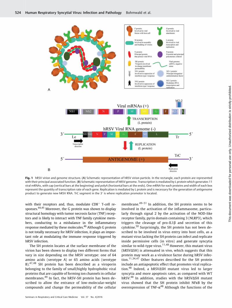
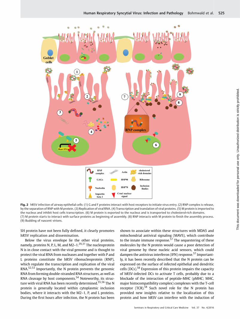
Fig. 2 hRSV infection of airway epithelial cells: (1) G and F proteins interact with host receptors to initiate virus entry. (2) RNP complex is release,by the separation of RNP with M protein. (3) Replication of viral RNA. (4) Transcription and translation of viral proteins. (5) M protein is imported tothe nucleus and inhibit host cells transcription. (6) M protein is exported to the nucleus and is transported to cholesterol-rich domains.(7) M protein starts to interact with surface proteins as beginning of assembly. (8) RNP interacts with M protein to finish the assembly process.(9) Budding of nascent virions.
Seminars in Respiratory and Critical Care Medicine Vol. 37 No. 4/2016
Human Respiratory Syncytial Virus: Infection and Pathology Bohmwald et al. 525
Thi
s do
cum
ent w
as d
ownl
oade
d fo
r pe
rson
al u
se o
nly.
Una
utho
rized
dis
trib
utio
n is
str
ictly
pro
hibi
ted.
protective T-cell responses, which is often impaired by hRSVinfection.3,19,58
The viral phosphoprotein P has been described as a cofac-tor of the RNP complex and the most important for the Lprotein. Indeed, the P protein can interact with N protein,allowing it to access the L protein.52,59 The P protein is highlystable as a tetramer and the C-terminal domain (PCTD from theresidue 161 to the residue 241) is critical for the interactionwith the L and N proteins.60–62 Consistent with this notion,the phosphorylation of the P protein has been shown to playan important role in the pathogenesis mediated by the virus,as a virus lacking thefive phosphorylation sites in this proteinshows reduced replication in vivo in mice and cotton rats, aswell as in vitro in HEp-2 sites.63 However, this recombinantvirus can replicate normally in Vero cells, thereby suggestingthat the phosphorylation of the P protein is necessary for anefficient viral replication.63
The RNA-dependent RNA polymerase (RdRp) L protein isthe lesser expressed of all viral proteins in the infected hostcells. The principal role of this protein is the replication andtranscription of the viral genome, regulated and supported bythe RNP complex.64 Because hRSV is a negative sense RNAvirus, the L protein transcripts the genome directly intomRNA for the expression of each hRSVgene.59 In this process,the L protein recognizes a promoter region in the 3′ extremeof the negative RNA strand and starts the transcription ofeach gene. Accumulating evidence suggests that transcriptionis modulated by the N protein.52,53,59 During the replicationprocess, the L protein copies the complete virus genome froma negative sense RNA into a positive sense RNA, which iscalled an antigenome. This RNA is then used as a template togenerate new negative sense RNA, which finally will beencapsided in the virions.59,65 A typical characteristic ofthe L protein is that in the process of transcription, it gen-erates a gradient of gene expression, from 3′ to 5′, producingmore mRNAs of the genes 3′ as compared with those 5′ in thegenome.66,67
The matrix protein M promotes viral assembly and isessential for hRSV replication.68 Early after infection, the Mprotein is located in the nucleus, where it is able to decreasethe transcriptional activity of the host cells genes.69–71 An-other role of matrix protein is to arrest the cell cycle in the G1phase, as shown in A549 cells. It also arrests the G1 and G2/Mphases in human bronchial epithelial cells.72 These actions,which are p53-dependent, increase hRSV replication.72 Inaddition, theMprotein is directly related to thematuration ofviral filaments.73 In this line, it has been described that hRSVstrains that are null for theMprotein showsignificantly lesserinfective progeny particles.73 Moreover, not only lesser newviral particles are generated but also protein trafficking isaffected, particularly with the N protein being concentratedin cytoplasmic inclusion bodies, before virus budding. Thisphenomenon suggests that M protein is important for trig-gering the trafficking of viral proteins to the budding site.73
Other studies show that the M protein is expressed ininclusion bodies and interact with the M2–1 protein, as ameans to interact with the RNP complex.74 The M protein isalso capable of inhibiting viral transcription and interact with
hRSV G and F proteins to signal the assembly of thevirions.75,76
The M2–1 protein is involved in the transcription processas part of the RNP complex, and acts as an antitermination/elongation factor promoting the transcription of all hRSVgenes, aiding the L protein to proceed with transcription ofviral genes.77 Interestingly, it was also showed that NS1 andNS2 genes can be transcribed by the L protein independentlyof the hRSV M2–1 protein, suggesting that several transcrip-tion mechanisms for viral genes exist.77 To exert its anti-termination functions, M2–1 needs to form as tetramers.78
Importantly, without this oligomerization, the protein cannotfunction correctly, which is supported by using a mutantvirus for M2–1 protein that cannot generate tetramers.78 AsM2–1 is part of the RNP complex, this protein can interactwith different protein of this complex.74,78–81 The interactionwith the N protein is particularly mediated through inter-actionswith the viral RNA, as treatmentwith RNAses disruptsthis binding.80 Another function of this protein is the activa-tion of nuclear factor-κB (NF-κB) and its association with theRelA protein.82 On the other hand, the M2–2 protein isinvolved in the regulation of transcription to the replicationby the virus polymerase.83,84 This effect was discovered bystudying an ΔM2–2 virus, in which the accumulation ofmRNA was higher in cells infected with this mutant virus,as compared with WT virus.84 In addition, viral titers werereduced over 1,000 times in the first 5 days and over 10 timesafter 7 to 8 days when the ΔM2–2 virus was used.84 Thus,these two proteins play a critical role in the regulation oftranscription and replication of hRSV RNA.
Besides the structural proteins mentioned earlier, thehRSV genome also encodes two nonstructural proteins,namely, NS1 and NS2, both with the capacity to interferewith host type 1 IFN innate response. This process negativelymodulates DCs maturation and T-cell responses.85,86 The NS1protein interferes with the activation of the IFN gene promo-tor by inhibiting the phosphorylation of interferon regulatoryfactor 3 (IRF-3).87NS2 also can interferewith the activation ofIRF-3 by its interaction with retinoic acid–inducible gene 1(RIG-I), inhibiting the activation of IFN response genes(IFNRs).88,89 The NS1 protein is able to interrupt the signalingof JAK/STAT pathways that are activated by IFN receptorpathways, particularly through the degradation of STAT-2.88,89 Both proteins, NS1 and NS2, are able to promotephosphoinositide 3-kinase (PI3K) pathways promoting thesurvival of infected cells, increasing viral yield.90 In this line,interference with the type 1 IFN response by NS1 and NS2proteins blocks DCs maturation.86 Concomitantly, ΔNS1/NS2and ΔNS1 viruses are able to increase the expression ofmaturation markers on DCs compared with WT hRSV.86
Furthermore, this effect in DCs could interfere with theircapacity to activate T cells.85,86 Indeed, human DCs infectedwith aΔNS1 virus show increased activation and proliferationof CD8þ T, increase the activation and proliferation of Th17protective cells, and decrease the activation of IL-4þ CD4þ Tcells, which are related to increased hRSV pathogenesis.85 Inaddition, a recent study showed that the expression of NS1and NS2 proteins by human bronchial epithelial cells
Seminars in Respiratory and Critical Care Medicine Vol. 37 No. 4/2016
Human Respiratory Syncytial Virus: Infection and Pathology Bohmwald et al.526
Thi
s do
cum
ent w
as d
ownl
oade
d fo
r pe
rson
al u
se o
nly.
Una
utho
rized
dis
trib
utio
n is
str
ictly
pro
hibi
ted.
decreases the polarization of T cells toward Th1, Th2, andTh17 phenotypes by the NS1 protein, and Th2 and Th17polarization by the NS2 protein.91 Thus, these two nonstruc-tural proteins are very important virulence factors thatdirectly affect the immune response of the host.
Viral Infection Cycle
The infection of target cells, such as airways epithelial cells(AECs), starts with the attachment of virions to the cellsurface aided by the G protein, which interacts with heparansulfates and chondroitin sulfate B glycosaminoglycans(GAGs).92 After this interaction, which helps the virusapproach the membrane of the cell to be infected, theF protein contacts its receptor, nucleolin (►Fig. 2).39 Theentry of hRSV has been described to occur particularly incholesterol-rich microdomains on the cell surface.93 Further,the fusion membranes require the participation of Pak-1 inthe rearrangement of actin filaments.93 Entry via endocyto-sis has been discarded because the use of Dynasoreshows that, despite dynamin-endocytic process is inhibited,viral fusion still occurs.93 Therefore, the fusion of the hRSVmembrane with the host cell membrane depends on theinteractions of hRSVG and F proteins with their receptor andthe rearrangement of actin filaments close to cholesterol-rich microdomains (►Fig. 2).32,93
The fusion of the viral and cell membranes triggers therelease of the viral nucleocapsid content into cytoplasm.Here, the nucleocapsid is dissociated from the RNP complexand repetitions of the M protein, which is mediated by thephosphorylation of the P protein (►Fig. 2).94 Importantly, thisprocess is mediated not only by viral proteins but also by hostcell enzymes, such as glycogen synthase kinase-3 (GSK-3) βand protein phosphatase 2A (PP2A).94 The transcriptionprocess mediated by the function of L protein mainly occursin cellular inclusion bodies together with N and P proteins.95
As mentioned earlier, the L proteins associated with the Pprotein are able to recognize the promotor region on the 3′ ofviral RNA, and initiate transcription of viral genes.59,67 Thepolymerase initiates transcription in gene start (GS) regionscarrying out mRNA capping and methylating the 5′ of themessenger.59,67 Then, the L polymerase recognizes a gene endsignal at the end of the mRNA and carries out polyadenyla-tion.59,67 This process goes on again after recognition of a newGS sequence downstream of a previously transcribed gene. Itis known that theminimal proteins required for transcriptionare N, P, and L. TheM2–1 protein also appears to be importantbecause of its ability to interact with all RNP proteins,78–81
includedwith theMprotein.74 Furthermore, host proteins arealso involved in the transcription process; for instance, pro-filin, an actin-modulatory protein, is required for an optimaltranscription.96 Additionally, host heat shock proteins (HSPs)are also involved in this process, particularly HSP90 andHSP70. Both proteins are expressed in lipid rafts and areassociated with the viral RNP complex. Recently, it wasdescribed that HSP90 is critical for the stability and function-ality of the L polymerase and that HSP70 is necessary forefficient RNA synthesis.97,98 When the viral genome is repli-
cated, the L polymerase recognizes the TrC promoter region atthe 3′ of the antigenome and generates genomic hRSV-RNA.99
This new RNA strand is immediately encapsulated by theN protein.59
Virus assembly, after viral RNA transcription and replica-tion, depends on the M protein localization and occurs atcholesterol-rich domains.73,100 As described earlier, at thebeginning of the infection cycle, the M protein is transportedto the nucleus by the interaction with importin-β1, where Mprotein can interferewith cellular transcription (►Fig. 2).69,70
The M protein is exported from the nucleus to the cytoplasmby a Crm1-dependant nuclear signal so that it localizes tolipids rafts.71 When the M protein is associated with thesedomains, the assembly and budding process begins andinvolves interactions with surface proteins F, G, and SH.101
Accordingly, a recent report showed that the F and G proteinsare expressed on the surface of ciliated cells.102 Thereafter,the interaction of the F protein with the M proteins promotesassembly of the new virions (►Fig. 2).103 On the other hand,the M2–1 protein has been shown to bind to the M protein,promoting its assembly with the RNP complex.74 The forma-tion of filaments that contain the virions is regulated by thehydroxymethylglutaryl coenzyme A reductase enzyme,which mediates changes in F-actin to generate viral filamen-tous projection that are involved in cell-to-cell transmis-sion.104 Finally, the budding process is not regulated by theendosomal sorting complex required for transport machin-ery, as occurs for other enveloped RNA viruses. In its place,hRSV budding is controlled by the RAB11 family interactingprotein 2 (FIP2),105 which has been described as a novelpathway for this type of process. Taken together, the hRSV-infective cycle depends of three main processes: (1) hRSVprotein localization, where inclusion bodies and rich choles-terol sites are principal placeswhere thehRSV proteins can befounded; (2) hRSV protein interaction, the particular interac-tion between RNP complex and accessory proteins for repli-cation and transcription and the interaction of M protein tosurface protein triggering the virion budding; and (3) inter-action of host cell proteinswith hRSV proteins and structures,principally how host cells help in the release of nucleocapsidat the beginning of the process and how a novel process ofbudding depends of host proteins (►Fig. 2).
Innate Immune System against RespiratorySyncytial Virus
Upon infection, AECs, DCs, andmacrophages play a key role inthe innate response against hRSV in the lungs.106 Patternrecognition receptors (PRRs) such as Toll-like receptors(TLRs), retinoic acid–inducible gene I (RIG-I)-like receptor(RLR) family members, and NOD-like receptors (NLR) areactivated following hRSV infection.107 TLRs have been shownto be fundamental for the recognition of hRSV.108–110 TLR4/TLR6 triggers a signaling cascade that activates innate im-mune responses by enhancing the production of TNF-α,interleukin (IL)-6, CCL2, and CCL5.110 HRSV is also sensedby endosomal TLR3 and TLR7, which triggers CCL5, IFN-α, andIFN-β production by TRIF-mediated- and MyD88 pathways,
Seminars in Respiratory and Critical Care Medicine Vol. 37 No. 4/2016
Human Respiratory Syncytial Virus: Infection and Pathology Bohmwald et al. 527
Thi
s do
cum
ent w
as d
ownl
oade
d fo
r pe
rson
al u
se o
nly.
Una
utho
rized
dis
trib
utio
n is
str
ictly
pro
hibi
ted.
respectively.108,109 The NLRP3 inflammasome, which belongsto theNOD-like receptor family, senses the SHprotein of hRSVand triggers pro-IL-β cleavage and secretion of IL-1βcytokine.50
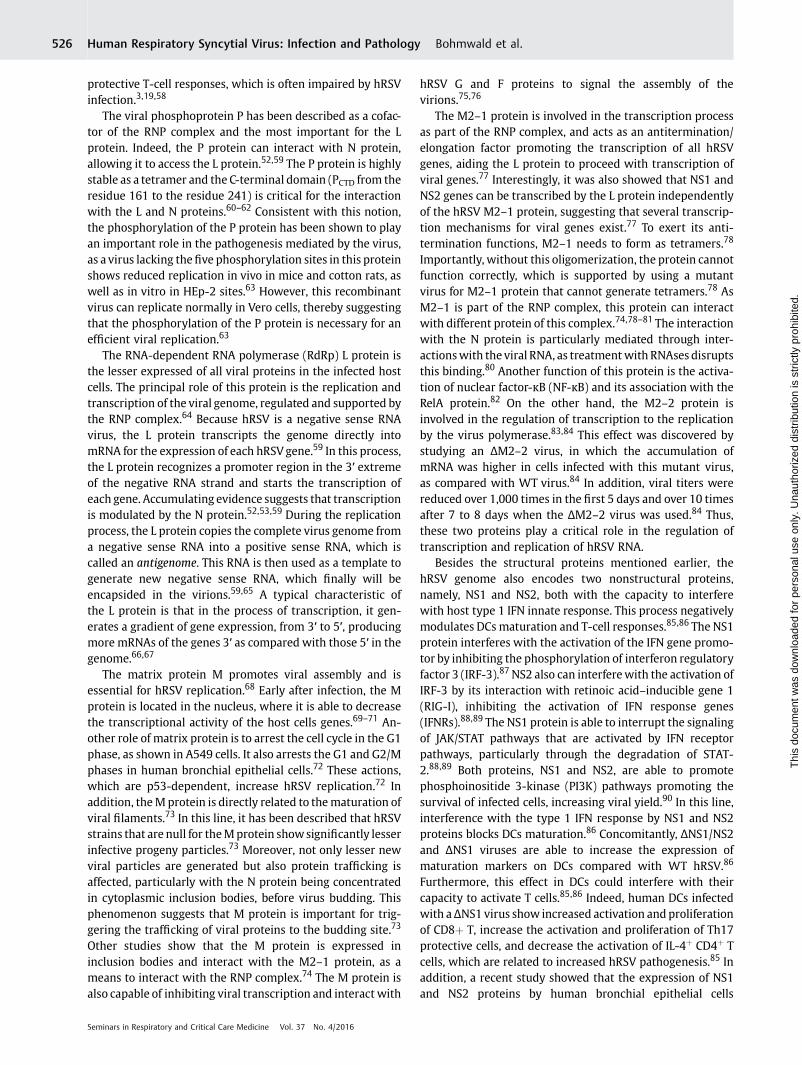
After PRRs are activated, NF-κB, IRFs, and ATF-2/cJun arepromoted.111 As a consequence, the expression of type 1 IFNsand the production of inflammatory cytokines, such as IL-8(IL-8/CXCL8), IL-4, IL-5, IL-6, and IL-10, as well as chemokinesand adhesion molecules are triggered.112,113 Such signalingcascades also prompt the recruitment of immune cells, suchas eosinophils,monocytes, and neutrophils to the lungs.114Asa result, exacerbated Th2-mediated airway inflammation istriggered, which contributes to lung damage (►Fig. 3).
hRSValso induces the secretion of both surfactant proteinsA andD (SP-A and SP-D) in the airways.115 These proteins playan important role in the regulation of the immune response inthe lung.116,117 Indeed, SP-A and SP-D can also stimulatemacrophage activation by increasing chemotaxis, phagocy-tosis, and increase cytokines secretion.118 Interestingly, SP-Dis able to bind hRSV G protein, thereby inhibiting hRSVinfection in vivo and in vitro.119 Recently, hRSV infection ofAECs has been shown to be involved in the production ofthymic stromal lymphopoietin (TSLP) and epithelial cell-derived IL-7, an IL-7-like cytokine.120,121 Interestingly, thesecytokines, together with IL-25 and IL-33, are related withacute exacerbations of asthma and Th2 inflammatory re-sponses triggered by viruses.122,123 Importantly, Qiao et alshowed the induction of functional maturation of myeloidDCs (mDCs) in hRSV-infected AECs through Th2-polarizing
molecules, such as thymus activation-regulated chemokine(TARC/CCL17) and OX40 ligand (OX40L) activation.121 Indeed,they suggested thatmDCs activationwasmediated by TSLP, asTSLP-targeted siRNA abrogated mDCs activation.121
Both hRSV NS1 and NS2 proteins inhibit the secretion oftype 1 IFNs in host cells by decreasing the levels of TNFreceptor-associated factor 3.124 Furthermore, survival of in-fected epithelial cells is achieved, thanks to NS1 and NS2,which activate the PI3K pathway as previously men-tioned.108,125 Consistent with this notion, suppression ofthese proteins resulted in accelerated apoptosis in hRSV-infected cells and consequently reduction in the virus yield.90
Importantly, the hRSV nucleoprotein could also attenuate theIFN response, as colocalization of this protein with RIG-1 andMAVS protein were found 6 hours postinfection.57
Adaptive Immune Response againstRespiratory Syncytial Virus
T cells play an important role in hRSV infection. CD4þ andCD8þ T cells have been shown to play pivotal roles in bothhRSV clearance and pathogenesis. Such a dichotomy in therole of virus-specific T cells has been observed for infection,and also lung damage after challenge with the virus.126 Forinstance, T cells expanded in mice experimentally infectedwith hRSV have been shown to be essential for the clearanceof the hRSV, although this immune response causes anexacerbated activation of the immune system within theairways.127 Similarly, mice immunized with a vaccine
Fig. 3 Immune response trigged by hRSV infection in respiratory airways. hRSV reaches the lower respiratory tract and is recognized forrespiratory epithelial cells by pattern recognition receptors (PRRs) expressed leading to the secretion of innate cytokines and chemokines such asTSLP, IL-13, and IL-25. Inflammatory cytokines and chemokines promote the recruitment of innate immune cells into the lungs, such as eosinophils,neutrophils, andmonocytes. The inflammatory environment induced by the innate immune cell recruitment and mucus production together withan excessive Th2 and Th17 response generate destruction of the respiratory epithelium and the obstruction of distal bronchiolar airways.
Seminars in Respiratory and Critical Care Medicine Vol. 37 No. 4/2016
Human Respiratory Syncytial Virus: Infection and Pathology Bohmwald et al.528
Thi
s do
cum
ent w
as d
ownl
oade
d fo
r pe
rson
al u
se o
nly.
Una
utho
rized
dis
trib
utio
n is
str
ictly
pro
hibi
ted.
consisting of formalin-inactivated virus suffered “vaccine-enhanced disease.”128 Pathology was observed as an exacer-bated increase in the immunological response of vaccinatedmice to the virus upon challenge, which was manifested byincreased eosinophil infiltration and Th2-like responses inthe lungs.128 Importantly, in this scenario, T cells weredescribed as a critical cell subset mediating the “vaccine-enhanced disease.”129 Furthermore, Th17 cells have also beenshown to contribute to hRSV airway pathology in humannewborns.130 On the contrary, mice immunized with BCGexpressing either the hRSV nucleoprotein (BCG-N) or M2protein (BCG-M2) showed a significant recruitment of IFN-γ-producing T cells in the lungs, promoting a Th1-response,which was protective and led to virus clearance withoutdetrimental inflammation.131
Cytotoxic CD8þ T cells (CTLs) are usually responsible forviral clearance by recognizing the F and N proteins of thehRSV.132 However, hRSV-specific CD8þT cells have also beenshown to play a role in a detrimental immune response.Consistent with this notion, depletion of CD8þ T cells reducedthe severity of hRSV-induced disease during primary andsecondary infection.133 Such detrimental responses havebeen suggested to occur because CD8þ T cells play a role inthe regulation and activation of the CD4þ T cells toward Th2polarized phenotypes.134 On the other hand, a reduction ofintracellular granzyme B content, diminished secretion ofIFN-γ, and impairment of perforin expression have beenobserved in CD8þ T cells in the lungs of hRSV-infectedindividuals.135,136
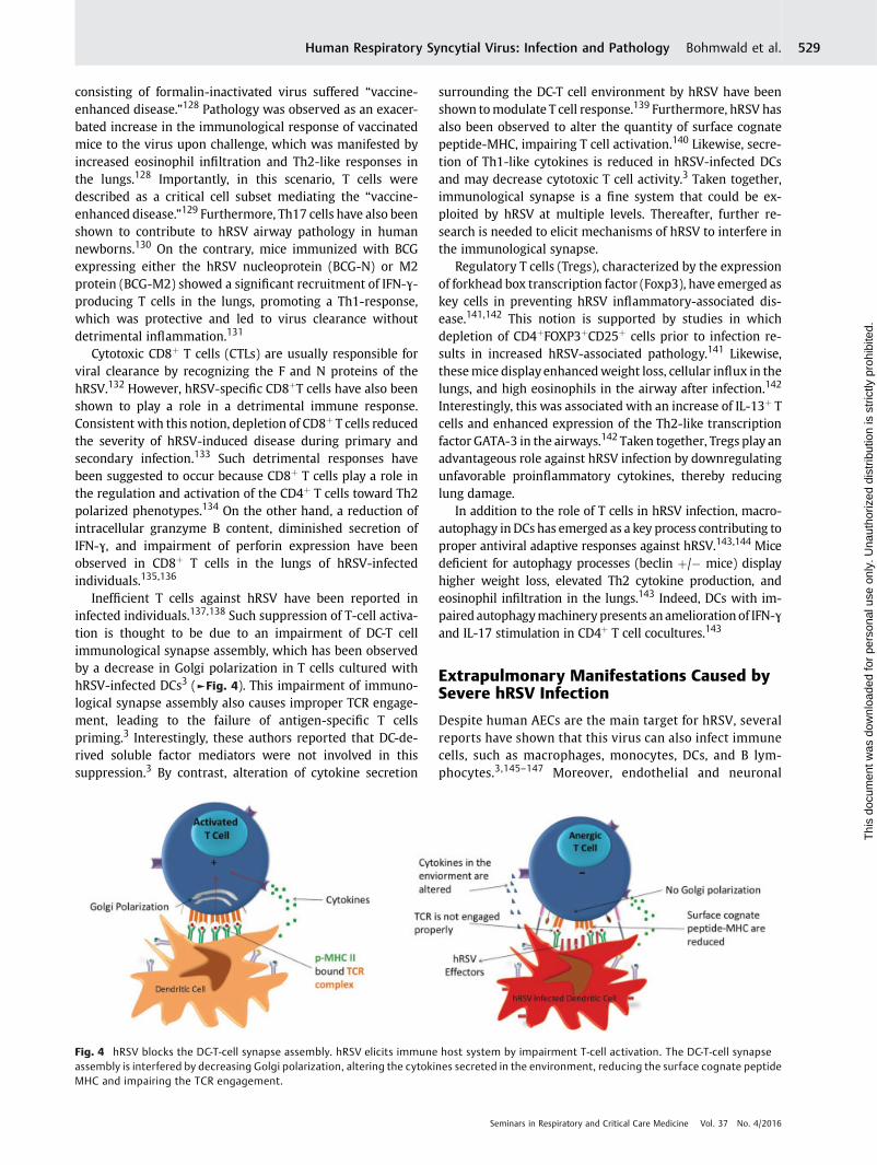
Inefficient T cells against hRSV have been reported ininfected individuals.137,138 Such suppression of T-cell activa-tion is thought to be due to an impairment of DC-T cellimmunological synapse assembly, which has been observedby a decrease in Golgi polarization in T cells cultured withhRSV-infected DCs3 (►Fig. 4). This impairment of immuno-logical synapse assembly also causes improper TCR engage-ment, leading to the failure of antigen-specific T cellspriming.3 Interestingly, these authors reported that DC-de-rived soluble factor mediators were not involved in thissuppression.3 By contrast, alteration of cytokine secretion
surrounding the DC-T cell environment by hRSV have beenshown tomodulate T cell response.139 Furthermore, hRSV hasalso been observed to alter the quantity of surface cognatepeptide-MHC, impairing T cell activation.140 Likewise, secre-tion of Th1-like cytokines is reduced in hRSV-infected DCsand may decrease cytotoxic T cell activity.3 Taken together,immunological synapse is a fine system that could be ex-ploited by hRSV at multiple levels. Thereafter, further re-search is needed to elicit mechanisms of hRSV to interfere inthe immunological synapse.
Regulatory T cells (Tregs), characterized by the expressionof forkhead box transcription factor (Foxp3), have emerged askey cells in preventing hRSV inflammatory-associated dis-ease.141,142 This notion is supported by studies in whichdepletion of CD4þFOXP3þCD25þ cells prior to infection re-sults in increased hRSV-associated pathology.141 Likewise,thesemice display enhancedweight loss, cellular influx in thelungs, and high eosinophils in the airway after infection.142
Interestingly, this was associated with an increase of IL-13þ Tcells and enhanced expression of the Th2-like transcriptionfactor GATA-3 in the airways.142 Taken together, Tregs play anadvantageous role against hRSV infection by downregulatingunfavorable proinflammatory cytokines, thereby reducinglung damage.
In addition to the role of T cells in hRSV infection, macro-autophagy in DCs has emerged as a key process contributing toproper antiviral adaptive responses against hRSV.143,144 Micedeficient for autophagy processes (beclin þ/� mice) displayhigher weight loss, elevated Th2 cytokine production, andeosinophil infiltration in the lungs.143 Indeed, DCs with im-paired autophagymachinery presents an amelioration of IFN-γand IL-17 stimulation in CD4þ T cell cocultures.143
Extrapulmonary Manifestations Caused bySevere hRSV Infection
Despite human AECs are the main target for hRSV, severalreports have shown that this virus can also infect immunecells, such as macrophages, monocytes, DCs, and B lym-phocytes.3,145–147 Moreover, endothelial and neuronal
Fig. 4 hRSV blocks the DC-T-cell synapse assembly. hRSV elicits immune host system by impairment T-cell activation. The DC-T-cell synapseassembly is interfered by decreasing Golgi polarization, altering the cytokines secreted in the environment, reducing the surface cognate peptideMHC and impairing the TCR engagement.
Seminars in Respiratory and Critical Care Medicine Vol. 37 No. 4/2016
Human Respiratory Syncytial Virus: Infection and Pathology Bohmwald et al. 529
Thi
s do
cum
ent w
as d
ownl
oade
d fo
r pe
rson
al u
se o
nly.
Una
utho
rized
dis
trib
utio
n is
str
ictly
pro
hibi
ted.
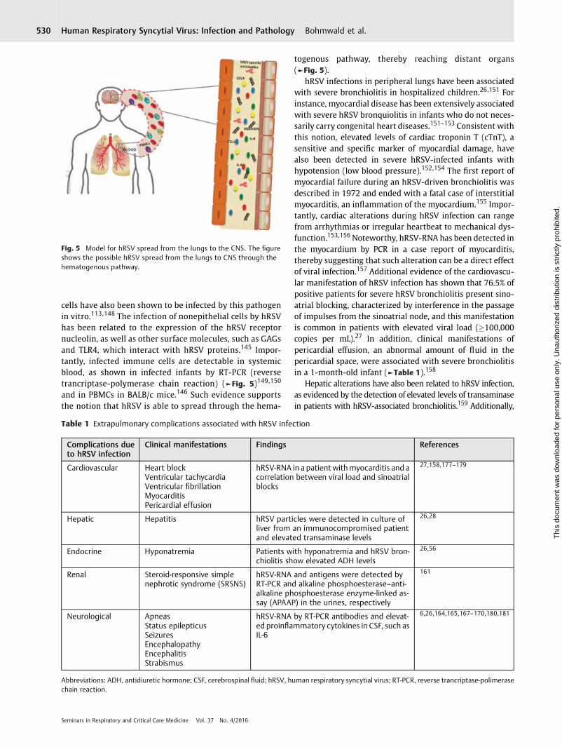
cells have also been shown to be infected by this pathogenin vitro.113,148 The infection of nonepithelial cells by hRSVhas been related to the expression of the hRSV receptornucleolin, as well as other surface molecules, such as GAGsand TLR4, which interact with hRSV proteins.145 Impor-tantly, infected immune cells are detectable in systemicblood, as shown in infected infants by RT-PCR (reversetrancriptase-polymerase chain reaction) (►Fig. 5)149,150
and in PBMCs in BALB/c mice.146 Such evidence supportsthe notion that hRSV is able to spread through the hema-
togenous pathway, thereby reaching distant organs(►Fig. 5).
hRSV infections in peripheral lungs have been associatedwith severe bronchiolitis in hospitalized children.26,151 Forinstance, myocardial disease has been extensively associatedwith severe hRSV bronquiolitis in infants who do not neces-sarily carry congenital heart diseases.151–153 Consistent withthis notion, elevated levels of cardiac troponin T (cTnT), asensitive and specific marker of myocardial damage, havealso been detected in severe hRSV-infected infants withhypotension (low blood pressure).152,154 The first report ofmyocardial failure during an hRSV-driven bronchiolitis wasdescribed in 1972 and ended with a fatal case of interstitialmyocarditis, an inflammation of the myocardium.155 Impor-tantly, cardiac alterations during hRSV infection can rangefrom arrhythmias or irregular heartbeat to mechanical dys-function.153,156Noteworthy, hRSV-RNA has been detected inthe myocardium by PCR in a case report of myocarditis,thereby suggesting that such alteration can be a direct effectof viral infection.157 Additional evidence of the cardiovascu-lar manifestation of hRSV infection has shown that 76.5% ofpositive patients for severe hRSV bronchiolitis present sino-atrial blocking, characterized by interference in the passageof impulses from the sinoatrial node, and this manifestationis common in patients with elevated viral load (�100,000copies per mL).27 In addition, clinical manifestations ofpericardial effusion, an abnormal amount of fluid in thepericardial space, were associated with severe bronchiolitisin a 1-month-old infant (►Table 1).158
Hepatic alterations have also been related to hRSV infection,as evidenced by the detection of elevated levels of transaminasein patients with hRSV-associated bronchiolitis.159 Additionally,
Fig. 5 Model for hRSV spread from the lungs to the CNS. The figureshows the possible hRSV spread from the lungs to CNS through thehematogenous pathway.
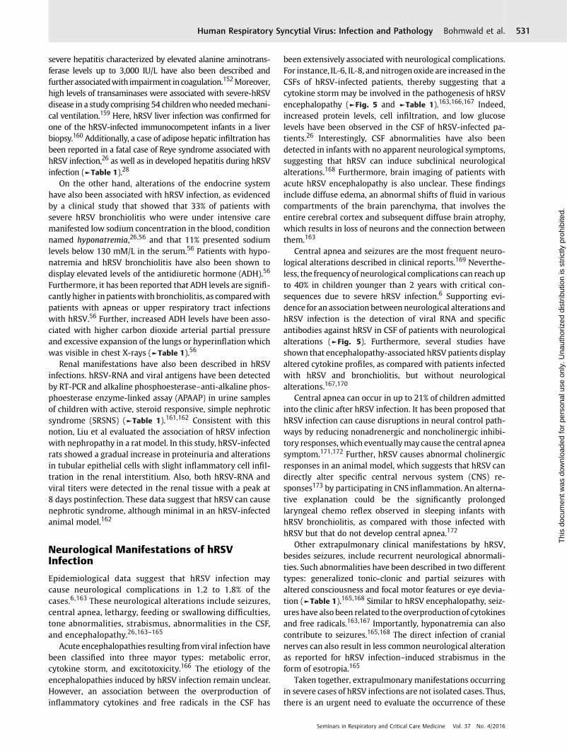
Table 1 Extrapulmonary complications associated with hRSV infection
Complications dueto hRSV infection
Clinical manifestations Findings References
Cardiovascular Heart blockVentricular tachycardiaVentricular fibrillationMyocarditisPericardial effusion
hRSV-RNA in a patient withmyocarditis and acorrelation between viral load and sinoatrialblocks
27,158,177–179
Hepatic Hepatitis hRSV particles were detected in culture ofliver from an immunocompromised patientand elevated transaminase levels
26,28
Endocrine Hyponatremia Patients with hyponatremia and hRSV bron-chiolitis show elevated ADH levels
26,56
Renal Steroid-responsive simplenephrotic syndrome (SRSNS)
hRSV-RNA and antigens were detected byRT-PCR and alkaline phosphoesterase–anti-alkaline phosphoesterase enzyme-linked as-say (APAAP) in the urines, respectively
161
Neurological ApneasStatus epilepticusSeizuresEncephalopathyEncephalitisStrabismus
hRSV-RNA by RT-PCR antibodies and elevat-ed proinflammatory cytokines in CSF, such asIL-6
6,26,164,165,167–170,180,181
Abbreviations: ADH, antidiuretic hormone; CSF, cerebrospinal fluid; hRSV, human respiratory syncytial virus; RT-PCR, reverse trancriptase-polimerasechain reaction.
Seminars in Respiratory and Critical Care Medicine Vol. 37 No. 4/2016
Human Respiratory Syncytial Virus: Infection and Pathology Bohmwald et al.530
Thi
s do
cum
ent w
as d
ownl
oade
d fo
r pe
rson
al u
se o
nly.
Una
utho
rized
dis
trib
utio
n is
str
ictly
pro
hibi
ted.
severe hepatitis characterized by elevated alanine aminotrans-ferase levels up to 3,000 IU/L have also been described andfurther associatedwith impairment in coagulation.152Moreover,high levels of transaminases were associated with severe-hRSVdisease in a study comprising 54 childrenwho neededmechani-cal ventilation.159 Here, hRSV liver infection was confirmed forone of the hRSV-infected immunocompetent infants in a liverbiopsy.160 Additionally, a case of adipose hepatic infiltration hasbeen reported in a fatal case of Reye syndrome associated withhRSV infection,26 as well as in developed hepatitis during hRSVinfection (►Table 1).28
On the other hand, alterations of the endocrine systemhave also been associated with hRSV infection, as evidencedby a clinical study that showed that 33% of patients withsevere hRSV bronchiolitis who were under intensive caremanifested low sodium concentration in the blood, conditionnamed hyponatremia,26,56 and that 11% presented sodiumlevels below 130 mM/L in the serum.56 Patients with hypo-natremia and hRSV bronchiolitis have also been shown todisplay elevated levels of the antidiuretic hormone (ADH).56
Furthermore, it has been reported that ADH levels are signifi-cantly higher in patientswith bronchiolitis, as comparedwithpatients with apneas or upper respiratory tract infectionswith hRSV.56 Further, increased ADH levels have been asso-ciated with higher carbon dioxide arterial partial pressureand excessive expansion of the lungs or hyperinflation whichwas visible in chest X-rays (►Table 1).56
Renal manifestations have also been described in hRSVinfections. hRSV-RNA and viral antigens have been detectedby RT-PCR and alkaline phosphoesterase–anti-alkaline phos-phoesterase enzyme-linked assay (APAAP) in urine samplesof children with active, steroid responsive, simple nephroticsyndrome (SRSNS) (►Table 1).161,162 Consistent with thisnotion, Liu et al evaluated the association of hRSV infectionwith nephropathy in a rat model. In this study, hRSV-infectedrats showed a gradual increase in proteinuria and alterationsin tubular epithelial cells with slight inflammatory cell infil-tration in the renal interstitium. Also, both hRSV-RNA andviral titers were detected in the renal tissue with a peak at8 days postinfection. These data suggest that hRSV can causenephrotic syndrome, although minimal in an hRSV-infectedanimal model.162
Neurological Manifestations of hRSVInfection
Epidemiological data suggest that hRSV infection maycause neurological complications in 1.2 to 1.8% of thecases.6,163 These neurological alterations include seizures,central apnea, lethargy, feeding or swallowing difficulties,tone abnormalities, strabismus, abnormalities in the CSF,and encephalopathy.26,163–165
Acute encephalopathies resulting fromviral infection havebeen classified into three mayor types: metabolic error,cytokine storm, and excitotoxicity.166 The etiology of theencephalopathies induced by hRSV infection remain unclear.However, an association between the overproduction ofinflammatory cytokines and free radicals in the CSF has
been extensively associated with neurological complications.For instance, IL-6, IL-8, and nitrogen oxide are increased in theCSFs of hRSV-infected patients, thereby suggesting that acytokine storm may be involved in the pathogenesis of hRSVencephalopathy (►Fig. 5 and ►Table 1).163,166,167 Indeed,increased protein levels, cell infiltration, and low glucoselevels have been observed in the CSF of hRSV-infected pa-tients.26 Interestingly, CSF abnormalities have also beendetected in infants with no apparent neurological symptoms,suggesting that hRSV can induce subclinical neurologicalalterations.168 Furthermore, brain imaging of patients withacute hRSV encephalopathy is also unclear. These findingsinclude diffuse edema, an abnormal shifts of fluid in variouscompartments of the brain parenchyma, that involves theentire cerebral cortex and subsequent diffuse brain atrophy,which results in loss of neurons and the connection betweenthem.163
Central apnea and seizures are the most frequent neuro-logical alterations described in clinical reports.169 Neverthe-less, the frequency of neurological complications can reach upto 40% in children younger than 2 years with critical con-sequences due to severe hRSV infection.6 Supporting evi-dence for an association between neurological alterations andhRSV infection is the detection of viral RNA and specificantibodies against hRSV in CSF of patients with neurologicalalterations (►Fig. 5). Furthermore, several studies haveshown that encephalopathy-associated hRSV patients displayaltered cytokine profiles, as compared with patients infectedwith hRSV and bronchiolitis, but without neurologicalalterations.167,170
Central apnea can occur in up to 21% of children admittedinto the clinic after hRSV infection. It has been proposed thathRSV infection can cause disruptions in neural control path-ways by reducing nonadrenergic and noncholinergic inhibi-tory responses, which eventuallymay cause the central apneasymptom.171,172 Further, hRSV causes abnormal cholinergicresponses in an animal model, which suggests that hRSV candirectly alter specific central nervous system (CNS) re-sponses173 by participating in CNS inflammation. An alterna-tive explanation could be the significantly prolongedlaryngeal chemo reflex observed in sleeping infants withhRSV bronchiolitis, as compared with those infected withhRSV but that do not develop central apnea.172
Other extrapulmonary clinical manifestations by hRSV,besides seizures, include recurrent neurological abnormali-ties. Such abnormalities have been described in two differenttypes: generalized tonic–clonic and partial seizures withaltered consciousness and focal motor features or eye devia-tion (►Table 1).165,168 Similar to hRSV encephalopathy, seiz-ures have also been related to the overproduction of cytokinesand free radicals.163,167 Importantly, hyponatremia can alsocontribute to seizures.165,168 The direct infection of cranialnerves can also result in less common neurological alterationas reported for hRSV infection–induced strabismus in theform of esotropia.165
Taken together, extrapulmonary manifestations occurringin severe cases of hRSV infections are not isolated cases. Thus,there is an urgent need to evaluate the occurrence of these
Seminars in Respiratory and Critical Care Medicine Vol. 37 No. 4/2016
Human Respiratory Syncytial Virus: Infection and Pathology Bohmwald et al. 531
Thi
s do
cum
ent w
as d
ownl
oade
d fo
r pe
rson
al u
se o
nly.
Una
utho
rized
dis
trib
utio
n is
str
ictly
pro
hibi
ted.
events in hospitalized childrenwith hRSV bronchiolitis and tostudy the possible consequences of acute neurological abnor-malities due to the infectionwith this virus. Furthermore, it isimperative to determine whether these extrapulmonaryeffects are due to direct effects of tissues with hRSV or byinflammatory mediators dispersed from the airways or re-sponding immune cells.
Possible Mechanisms behind theNeurological Alterations Caused by hRSV
The neurological complications observed upon hRSV infec-tion167 have encouraged researchers to understand themechanisms involved in CNS dysfunction. Studies performedin BALB/c mice and Sprague Dawley rats have detected hRSV-RNA and viral proteins in the brain of animals previouslyinfected intranasally with this pathogen.174 Studies havefound that immune cells are associated with hRSV in periph-eral blood from hRSV-infected patients. Consistent with thisnotion, hRSV-infected immune cellswouldmigrate to the CNSby the hematogenous pathway and trespass the blood–brainbarrier.174 An unexpected and important finding regardingthe access of hRSV to the CNS was the description thatimpairment in cognitive function is observed after pulmo-nary disease was resolved in mice and rats.174 Indeed, ourgroup recently described that mice and rats infected withhRSV have a deficient performance in tests that evaluate theseabilities. hRSV-infected mice performed significantly worsethan noninfected mice, both in the Marble Burying (MB) andMorris Water Maze (MWM) tests, several weeks after viralchallenge.174 The MB test consists in measuring the ability ofrodents to dig and hide marbles, which is controlled byhippocampal function.175 In addition, the MWM evaluatesthe animal’s ability for spatial learning through spatial locali-zation of relevant visual cues that are subsequently proc-essed, consolidated, retained, and then retrieved in the brainto successfully navigate and thereby locate a hidden platformto escape from the water.176 In both tasks, hRSV-infectedanimals showed significant alterations in behavioral andlearning processes, as compared with control animals. More-over, electrophysiological assays suggested that impairedcognitive function was due to a failure to efficiently inducelong-term potentiation responses in the stratum radiatum inthe hippocampus area. Our study supports the previouslyproposed idea that hRSV can alter CNS function. Accordingly,hRSV has been shown to infect primary neuronal cells invitro,as well as neural processes innervating the lungs.148
The association of an exacerbated immune responseagainst hRSV together with hRSV-induced cognitive im-pairment is supported by the observation that a vaccinethat induces protective T cell immunity prevents virus spreadinto the CNS, as well as neurological alterations caused byinfection.174 A possible explanation is that hRSV may enterthe CNS associated with leucocytes or freely, triggering anelevated secretion of proinflammatory cytokines that affectnormal neuronal function.
In summary, hRSV infection can cause important extrap-ulmonary symptoms, which can lead to important and long-
lasting health sequelae in children affected by this virus.Therefore, significant research efforts are required for thegeneration of vaccines and therapies to prevent or treat theinfection caused by this virus in the most susceptiblepopulation.
References1 Calvo C, García-García ML, Blanco C, et al. Multiple simultaneous
viral infections in infants with acute respiratory tract infectionsin Spain. J Clin Virol 2008;42(3):268–272
2 Ogra PL. Respiratory syncytial virus: the virus, the disease and theimmune response. Paediatr Respir Rev 2004;5(Suppl A ):S119–S126
3 González PA, Prado CE, Leiva ED, et al. Respiratory syncytial virusimpairs T cell activation by preventing synapse assembly withdendritic cells. Proc Natl Acad Sci U S A 2008;105(39):14999–15004
4 Bueno SM, González PA, Pacheco R, et al. Host immunity duringRSV pathogenesis. Int Immunopharmacol 2008;8(10):1320–1329
5 Collins PL, Melero JA. Progress in understanding and controllingrespiratory syncytial virus: still crazy after all these years. VirusRes 2011;162(1–2):80–99
6 Sweetman LL, Ng YT, Butler IJ, Bodensteiner JB. Neurologiccomplications associatedwith respiratory syncytial virus. PediatrNeurol 2005;32(5):307–310
7 Boyce TG, Mellen BG,Mitchel EF Jr, Wright PF, GriffinMR. Rates ofhospitalization for respiratory syncytial virus infection amongchildren in Medicaid. J Pediatr 2000;137(6):865–870
8 Wang EE, Law BJ, Boucher FD, et al. Pediatric InvestigatorsCollaborative Network on Infections in Canada (PICNIC) studyof admission and management variation in patients hospitalizedwith respiratory syncytial viral lower respiratory tract infection.J Pediatr 1996;129(3):390–395
9 Aujard Y, Fauroux B. Risk factors for severe respiratory syncytialvirus infection in infants. Respir Med 2002;96(Suppl B ):S9–S14
10 Navas L, Wang E, de Carvalho V, Robinson J; Pediatric Investi-gators Collaborative Network on Infections in Canada. Improvedoutcome of respiratory syncytial virus infection in a high-riskhospitalized population of Canadian children. J Pediatr 1992;121(3):348–354
11 Collins PL, GrahamBS. Viral and host factors in human respiratorysyncytial virus pathogenesis. J Virol 2008;82(5):2040–2055
12 Nair H, Nokes DJ, Gessner BD, et al. Global burden of acute lowerrespiratory infections due to respiratory syncytial virus in youngchildren: a systematic review and meta-analysis. Lancet 2010;375(9725):1545–1555
13 Paramore LC, Ciuryla V, Ciesla G, Liu L. Economic impact ofrespiratory syncytial virus-related illness in the US: an analysisof national databases. Pharmacoeconomics 2004;22(5):275–284
14 Halfhide CP, Flanagan BF, Brearey SP, et al. Respiratory syncytialvirus binds and undergoes transcription in neutrophils from theblood and airways of infants with severe bronchiolitis. J Infect Dis2011;204(3):451–458
15 Hall CB, Douglas RG Jr, Schnabel KC, Geiman JM. Infectivity ofrespiratory syncytial virus by various routes of inoculation. InfectImmun 1981;33(3):779–783
16 Storey S. Respiratory syncytial virus market. Nat Rev Drug Discov2010;9(1):15–16
17 Dudas RA, Karron RA. Respiratory syncytial virus vaccines. ClinMicrobiol Rev 1998;11(3):430–439
18 van Drunen Littel-van den Hurk S, Watkiss ER. Pathogenesis ofrespiratory syncytial virus. Curr Opin Virol 2012;2(3):300–305
19 González PA, Bueno SM, Riedel CA, Kalergis AM. Impairment of Tcell immunity by the respiratory syncytial virus: targeting
Seminars in Respiratory and Critical Care Medicine Vol. 37 No. 4/2016
Human Respiratory Syncytial Virus: Infection and Pathology Bohmwald et al.532
Thi
s do
cum
ent w
as d
ownl
oade
d fo
r pe
rson
al u
se o
nly.
Una
utho
rized
dis
trib
utio
n is
str
ictly
pro
hibi
ted.
virulence mechanisms for therapy and prophylaxis. Curr MedChem 2009;16(34):4609–4625
20 Bont L, Versteegh J, Swelsen WT, et al. Natural reinfection withrespiratory syncytial virus does not boost virus-specific T-cellimmunity. Pediatr Res 2002;52(3):363–367
21 Guo-Parke H, Canning P, Douglas I, et al. Relative respiratorysyncytial virus cytopathogenesis in upper and lower respiratorytract epithelium. Am J Respir Crit Care Med 2013;188(7):842–851
22 Pickles RJ, DeVincenzo JP. Respiratory syncytial virus (RSV) and itspropensity for causing bronchiolitis. J Pathol 2015;235(2):266–276
23 Rudraraju R, Jones BG, Sealy R, Surman SL, Hurwitz JL. Respiratorysyncytial virus: current progress in vaccine development. Viruses2013;5(2):577–594
24 Lanari M, Vandini S, Arcuri S, Galletti S, Faldella G. The use ofhumanized monoclonal antibodies for the prevention of respira-tory syncytial virus infection. Clin Dev Immunol 2013;2013:359683
25 Lanari M, Silvestri M, Rossi GA. Clinical and pharmacologicalaspects of immunoprophylaxis for respiratory syncytial virusinfection in high-risk infants. Curr Drug Metab 2013;14(2):216–225
26 Eisenhut M. Extrapulmonary manifestations of severe respirato-ry syncytial virus infection—a systematic review. Crit Care 2006;10(4):R107
27 Esposito S, Salice P, Bosis S, et al. Altered cardiac rhythm in infantswith bronchiolitis and respiratory syncytial virus infection. BMCInfect Dis 2010;10:305
28 Kirin BK, Topić RZ, Dodig S. Hepatitis during respiratory syncytialvirus infection—a case report. BiochemMed (Zagreb) 2013;23(1):112–116
29 Hacking D, Hull J. Respiratory syncytial virus—viral biology andthe host response. J Infect 2002;45(1):18–24
30 Chanock R, Roizman B, Myers R. Recovery from infants withrespiratory illness of a virus related to chimpanzee coryza agent(CCA). I. Isolation, properties and characterization. Am J Hyg1957;66(3):281–290
31 Collins P, Chanock R, Murphy B. Respiratory Syncytial Virus.Philadelphia, PA: Lippincott Williams & Wilkins; 2001
32 Lay MK, González PA, León MA, et al. Advances in understandingrespiratory syncytial virus infection in airway epithelial cells andconsequential effects on the immune response. Microbes Infect2013;15(3):230–242
33 McLellan JS, Chen M, Joyce MG, et al. Structure-based design of afusion glycoprotein vaccine for respiratory syncytial virus.Science 2013;342(6158):592–598
34 Becker Y. Respiratory syncytial virus (RSV) evades the humanadaptive immune system by skewing the Th1/Th2 cytokinebalance toward increased levels of Th2 cytokines and IgE,markers of allergy—a review. Virus Genes 2006;33(2):235–252
35 Ternette N, Tippler B, Uberla K, Grunwald T. Immunogenicityand efficacy of codon optimized DNA vaccines encoding theF-protein of respiratory syncytial virus. Vaccine 2007;25(41):7271–7279
36 Mastrangelo P, Hegele RG. RSV fusion: time for a new model.Viruses 2013;5(3):873–885
37 Marr N, Turvey SE. Role of human TLR4 in respiratory syncytialvirus-induced NF-κB activation, viral entry and replication. In-nate Immun 2012;18(6):856–865
38 Behera AK, Matsuse H, Kumar M, Kong X, Lockey RF, MohapatraSS. Blocking intercellular adhesion molecule-1 on human epithe-lial cells decreases respiratory syncytial virus infection. BiochemBiophys Res Commun 2001;280(1):188–195
39 Tayyari F, Marchant D, Moraes TJ, Duan W, Mastrangelo P, HegeleRG. Identification of nucleolin as a cellular receptor for humanrespiratory syncytial virus. Nat Med 2011;17(9):1132–1135
40 Openshaw PJ, Tregoning JS. Immune responses and diseaseenhancement during respiratory syncytial virus infection. ClinMicrobiol Rev 2005;18(3):541–555
41 Krusat T, Streckert HJ. Heparin-dependent attachment of respi-ratory syncytial virus (RSV) to host cells. Arch Virol 1997;142(6):1247–1254
42 Malhotra R,WardM, Bright H, et al. Isolation and characterisationof potential respiratory syncytial virus receptor(s) on epithelialcells. Microbes Infect 2003;5(2):123–133
43 Bukreyev A, Yang L, Fricke J, et al. The secreted form of respiratorysyncytial virus G glycoprotein helps the virus evade antibody-mediated restriction of replication by acting as an antigen decoyand through effects on Fc receptor-bearing leukocytes. J Virol2008;82(24):12191–12204
44 Harcourt J, Alvarez R, Jones LP, Henderson C, Anderson LJ, TrippRA. Respiratory syncytial virus G protein and G protein CX3Cmotif adversely affect CX3CR1þ T cell responses. J Immunol 2006;176(3):1600–1608
45 Tripp RA, Jones LP, Haynes LM, ZhengH,Murphy PM, Anderson LJ.CX3C chemokine mimicry by respiratory syncytial virus G glyco-protein. Nat Immunol 2001;2(8):732–738
46 Langedijk JP, de Groot BL, Berendsen HJ, van Oirschot JT. Struc-tural homology of the central conserved region of the attachmentprotein G of respiratory syncytial virus with the fourth subdo-main of 55-kDa tumor necrosis factor receptor. Virology 1998;243(2):293–302
47 Rixon HW, Brown G, Aitken J, McDonald T, Graham S, Sugrue RJ.The small hydrophobic (SH) protein accumulates within lipid-raft structures of the Golgi complex during respiratory syncytialvirus infection. J Gen Virol 2004;85(Pt 5):1153–1165
48 Fuentes S, Tran KC, Luthra P, Teng MN, He B. Function of therespiratory syncytial virus small hydrophobic protein. J Virol2007;81(15):8361–8366
49 Gan SW, Ng L, Lin X, Gong X, Torres J. Structure and ion channelactivity of the human respiratory syncytial virus (hRSV) smallhydrophobic protein transmembrane domain. Protein Sci 2008;17(5):813–820
50 Triantafilou K, Kar S, Vakakis E, Kotecha S, Triantafilou M. Humanrespiratory syncytial virus viroporin SH: a viral recognitionpathway used by the host to signal inflammasome activation.Thorax 2013;68(1):66–75
51 Gan SW, Tan E, Lin X, et al. The small hydrophobic protein of thehuman respiratory syncytial virus forms pentameric ion chan-nels. J Biol Chem 2012;287(29):24671–24689
52 Ruigrok RW, Crépin T. Nucleoproteins of negative strand RNAviruses; RNA binding, oligomerisation and binding to polymeraseco-factor. Viruses 2010;2(1):27–32
53 Tawar RG, Duquerroy S, Vonrhein C, et al. Crystal structure of anucleocapsid-like nucleoprotein-RNA complex of respiratorysyncytial virus. Science 2009;326(5957):1279–1283
54 Le Mercier P, Garcin D, Hausmann S, Kolakofsky D. Ambisensesendai viruses are inherently unstable but are useful to studyviral RNA synthesis. J Virol 2002;76(11):5492–5502
55 El Omari K, Dhaliwal B, Ren J, et al. Structures of respiratorysyncytial virus nucleocapsid protein from two crystal forms:details of potential packing interactions in the native helicalform. Acta Crystallogr Sect F Struct Biol Cryst Commun 2011;67(Pt 10):1179–1183
56 Hanna S, Tibby SM, Durward A, Murdoch IA. Incidence of hypo-natraemia and hyponatraemic seizures in severe respiratorysyncytial virus bronchiolitis. Acta Paediatr 2003;92(4):430–434
57 Lifland AW, Jung J, Alonas E, Zurla C, Crowe JE Jr, Santangelo PJ.Human respiratory syncytial virus nucleoprotein and inclusionbodies antagonize the innate immune response mediated byMDA5 and MAVS. J Virol 2012;86(15):8245–8258
58 Céspedes PF, Bueno SM, Ramírez BA, et al. Surface expression ofthe hRSV nucleoprotein impairs immunological synapse
Seminars in Respiratory and Critical Care Medicine Vol. 37 No. 4/2016
Human Respiratory Syncytial Virus: Infection and Pathology Bohmwald et al. 533
Thi
s do
cum
ent w
as d
ownl
oade
d fo
r pe
rson
al u
se o
nly.
Una
utho
rized
dis
trib
utio
n is
str
ictly
pro
hibi
ted.
formation with T cells. Proc Natl Acad Sci USA 2014;111(31):E3214–E3223
59 Cowton VM,McGivern DR, Fearns R. Unravelling the complexitiesof respiratory syncytial virus RNA synthesis. J Gen Virol 2006;87(Pt 7):1805–1821
60 Slack MS, Easton AJ. Characterization of the interaction of thehuman respiratory syncytial virus phosphoprotein and nucleo-capsid protein using the two-hybrid system. Virus Res 1998;55(2):167–176
61 Tran TL, Castagné N, Bhella D, et al. The nine C-terminal aminoacids of the respiratory syncytial virus protein P are necessaryand sufficient for binding to ribonucleoprotein complexes inwhich six ribonucleotides are contacted per N protein protomer.J Gen Virol 2007;88(Pt 1):196–206
62 Sourimant J, Rameix-Welti MA, Gaillard AL, et al. Fine mappingand characterization of the L-polymerase-binding domain of therespiratory syncytial virus phosphoprotein. J Virol 2015;89(8):4421–4433
63 Lu B, Ma CH, Brazas R, Jin H. The major phosphorylation sites ofthe respiratory syncytial virus phosphoprotein are dispensablefor virus replication in vitro. J Virol 2002;76(21):10776–10784
64 Grosfeld H, Hill MG, Collins PL. RNA replication by respiratorysyncytial virus (RSV) is directed by the N, P, and L proteins;transcription also occurs under these conditions but requires RSVsuperinfection for efficient synthesis of full-length mRNA. J Virol1995;69(9):5677–5686
65 Fearns R, Collins PL, Peeples ME. Functional analysis of thegenomic and antigenomic promoters of human respiratory syn-cytial virus. J Virol 2000;74(13):6006–6014
66 Hardy RW, Wertz GW. The product of the respiratory syncytialvirus M2 gene ORF1 enhances readthrough of intergenic junc-tions during viral transcription. J Virol 1998;72(1):520–526
67 Kuo L, Fearns R, Collins PL. The structurally diverse intergenicregions of respiratory syncytial virus do not modulate sequentialtranscription by a dicistronic minigenome. J Virol 1996;70(9):6143–6150
68 Teng MN, Collins PL. Identification of the respiratory syncytialvirus proteins required for formation and passage of helper-dependent infectious particles. J Virol 1998;72(7):5707–5716
69 Ghildyal R, Baulch-Brown C,Mills J, Meanger J. Thematrix proteinof Human respiratory syncytial virus localises to the nucleus ofinfected cells and inhibits transcription. Arch Virol 2003;148(7):1419–1429
70 Ghildyal R, Ho A, Wagstaff KM, et al. Nuclear import of therespiratory syncytial virus matrix protein is mediated by impor-tin beta1 independent of importin alpha. Biochemistry 2005;44(38):12887–12895
71 Ghildyal R, Ho A, Dias M, et al. The respiratory syncytial virusmatrix protein possesses a Crm1-mediated nuclear export mech-anism. J Virol 2009;83(11):5353–5362
72 Bian T, Gibbs JD, Örvell C, Imani F. Respiratory syncytial virusmatrix protein induces lung epithelial cell cycle arrest through ap53 dependent pathway. PLoS ONE 2012;7(5):e38052
73 Mitra R, Baviskar P, Duncan-Decocq RR, Patel D, Oomens AG. Thehuman respiratory syncytial virus matrix protein is required formaturation of viral filaments. J Virol 2012;86(8):4432–4443
74 Li D, Jans DA, Bardin PG,Meanger J, Mills J, Ghildyal R. Associationof respiratory syncytial virus M protein with viral nucleocapsidsis mediated by the M2-1 protein. J Virol 2008;82(17):8863–8870
75 Ghildyal R, Mills J, Murray M, Vardaxis N, Meanger J. Respiratorysyncytial virus matrix protein associates with nucleocapsids ininfected cells. J Gen Virol 2002;83(Pt 4):753–757
76 Ghildyal R, Li D, Peroulis I, et al. Interaction between therespiratory syncytial virus G glycoprotein cytoplasmic domainand the matrix protein. J Gen Virol 2005;86(Pt 7):1879–1884
77 Fearns R, Collins PL. Role of the M2-1 transcription antitermina-tion protein of respiratory syncytial virus in sequential transcrip-tion. J Virol 1999;73(7):5852–5864
78 Tran TL, Castagné N, Dubosclard V, et al. The respiratory syncytialvirusM2-1 protein forms tetramers and interacts with RNA and Pin a competitive manner. J Virol 2009;83(13):6363–6374
79 Kiss G, Holl JM, Williams GM, et al. Structural analysis ofrespiratory syncytial virus reveals the position of M2-1 betweenthe matrix protein and the ribonucleoprotein complex. J Virol2014;88(13):7602–7617
80 Cartee TL, Wertz GW. Respiratory syncytial virus M2-1 proteinrequires phosphorylation for efficient function and binds viralRNA during infection. J Virol 2001;75(24):12188–12197
81 Mason SW, Aberg E, Lawetz C, DeLong R, Whitehead P, Liuzzi M.Interaction between human respiratory syncytial virus (RSV)M2-1 and P proteins is required for reconstitution of M2-1-dependent RSV minigenome activity. J Virol 2003;77(19):10670–10676
82 Reimers K, Buchholz K, Werchau H. Respiratory syncytial virusM2-1 protein induces the activation of nuclear factor kappa B.Virology 2005;331(2):260–268
83 Cheng X, Park H, Zhou H, Jin H. Overexpression of the M2-2protein of respiratory syncytial virus inhibits viral replication.J Virol 2005;79(22):13943–13952
84 Bermingham A, Collins PL. The M2-2 protein of human respira-tory syncytial virus is a regulatory factor involved in the balancebetween RNA replication and transcription. Proc Natl Acad Sci U SA 1999;96(20):11259–11264
85 Munir S, Hillyer P, Le Nouën C, et al. Respiratory syncytial virusinterferon antagonist NS1 protein suppresses and skews thehumanT lymphocyte response. PLoS Pathog 2011;7(4):e1001336
86 Munir S, Le Nouen C, Luongo C, Buchholz UJ, Collins PL, BukreyevA. Nonstructural proteins 1 and 2 of respiratory syncytial virussuppress maturation of human dendritic cells. J Virol 2008;82(17):8780–8796
87 Spann KM, Tran KC, Collins PL. Effects of nonstructural proteinsNS1 and NS2 of human respiratory syncytial virus on interferonregulatory factor 3, NF-kappaB, and proinflammatory cytokines.J Virol 2005;79(9):5353–5362
88 Wright PF, Karron RA, Madhi SA, et al. The interferon antagonistNS2 protein of respiratory syncytial virus is an important viru-lence determinant for humans. J Infect Dis 2006;193(4):573–581
89 Elliott J, Lynch OT, Suessmuth Y, et al. Respiratory syncytial virusNS1 protein degrades STAT2 by using the Elongin-Cullin E3ligase. J Virol 2007;81(7):3428–3436
90 Wu W, Tran KC, Teng MN, et al. The interactome of the humanrespiratory syncytial virus NS1 protein highlightsmultiple effectson host cell biology. J Virol 2012;86(15):7777–7789
91 Qin L, PengD, Hu C, et al. Differentiation of Th subsets inhibited bynonstructural proteins of respiratory syncytial virus is mediatedby ubiquitination. PLoS ONE 2014;9(7):e101469
92 Lambert DM. Role of oligosaccharides in the structure andfunction of respiratory syncytial virus glycoproteins. Virology1988;164(2):458–466
93 San-Juan-Vergara H, Sampayo-Escobar V, Reyes N, et al. Choles-terol-rich microdomains as docking platforms for respiratorysyncytial virus in normal human bronchial epithelial cells. J Virol2012;86(3):1832–1843
94 Asenjo A, Rodríguez L, Villanueva N. Determination of phosphor-ylated residues from human respiratory syncytial virus P proteinthat are dynamically dephosphorylated by cellular phospha-tases: a possible role for serine 54. J Gen Virol 2005;86(Pt 4):1109–1120
95 Carromeu C, Simabuco FM, Tamura RE, Farinha Arcieri LE, Ven-tura AM. Intracellular localization of human respiratory syncytialvirus L protein. Arch Virol 2007;152(12):2259–2263
96 Burke E, Mahoney NM, Almo SC, Barik S. Profilin is required foroptimal actin-dependent transcription of respiratory syncytialvirus genome RNA. J Virol 2000;74(2):669–675
97 Brown G, Rixon HW, Steel J, et al. Evidence for an associationbetweenheat shock protein 70 and the respiratory syncytial virus
Seminars in Respiratory and Critical Care Medicine Vol. 37 No. 4/2016
Human Respiratory Syncytial Virus: Infection and Pathology Bohmwald et al.534
Thi
s do
cum
ent w
as d
ownl
oade
d fo
r pe
rson
al u
se o
nly.
Una
utho
rized
dis
trib
utio
n is
str
ictly
pro
hibi
ted.
polymerase complex within lipid-raft membranes during virusinfection. Virology 2005;338(1):69–80
98 Munday DC, Wu W, Smith N, et al. Interactome analysis of thehuman respiratory syncytial virus RNA polymerase complexidentifies protein chaperones as important cofactors that pro-mote L-protein stability and RNA synthesis. J Virol 2015;89(2):917–930
99 Mink MA, Stec DS, Collins PL. Nucleotide sequences of the 3′leader and 5′ trailer regions of human respiratory syncytial virusgenomic RNA. Virology 1991;185(2):615–624
100 Rodriguez R, Ramilo O. Respiratory syncytial virus: how, why andwhat to do. J Infect 2014;68(Suppl 1):S115–S118
101 Low KW, Tan T, Ng K, Tan BH, Sugrue RJ. The RSV F and Gglycoproteins interact to form a complex on the surface ofinfected cells. Biochem Biophys Res Commun 2008;366(2):308–313
102 Jumat MR, Yan Y, Ravi LI, et al. Morphogenesis of respiratorysyncytial virus in human primary nasal ciliated epithelial cellsoccurs at surfacemembranemicrodomains that are distinct fromcilia. Virology 2015;484:395–411
103 Shaikh FY, Cox RG, Lifland AW, et al. A critical phenylalanineresidue in the respiratory syncytial virus fusion protein cyto-plasmic tail mediates assembly of internal viral proteins into viralfilaments and particles. MBio 2012;3(1):270–271
104 Ravi LI, Liang L, Wong PS, Brown G, Tan BH, Sugrue RJ. Increasedhydroxymethylglutaryl coenzyme A reductase activity duringrespiratory syncytial virus infection mediates actin dependentinter-cellular virus transmission. Antiviral Res 2013;100(1):259–268
105 Utley TJ, Ducharme NA, Varthakavi V, et al. Respiratory syncytialvirus uses a Vps4-independent budding mechanism controlledby Rab11-FIP2. Proc Natl Acad Sci U S A 2008;105(29):10209–10214
106 AherneW, Bird T, Court SD, Gardner PS, McQuillin J. Pathologicalchanges in virus infections of the lower respiratory tract inchildren. J Clin Pathol 1970;23(1):7–18
107 Zeng R, Cui Y, Hai Y, Liu Y. Pattern recognition receptors forrespiratory syncytial virus infection and design of vaccines. VirusRes 2012;167(2):138–145
108 Groskreutz DJ, Monick MM, Powers LS, Yarovinsky TO, Look DC,Hunninghake GW. Respiratory syncytial virus induces TLR3protein and protein kinase R, leading to increased double-stranded RNA responsiveness in airway epithelial cells. J Immu-nol 2006;176(3):1733–1740
109 Lukacs NW, Smit JJ, Mukherjee S, Morris SB, Nunez G, Lindell DM.Respiratory virus-induced TLR7 activation controls IL-17-associ-ated increased mucus via IL-23 regulation. J Immunol 2010;185(4):2231–2239
110 Murawski MR, Bowen GN, Cerny AM, et al. Respiratory syncytialvirus activates innate immunity through Toll-like receptor 2.J Virol 2009;83(3):1492–1500
111 Baum A, García-Sastre A. Induction of type I interferon by RNAviruses: cellular receptors and their substrates. Amino Acids2010;38(5):1283–1299
112 Rudd BD, Burstein E, Duckett CS, Li X, Lukacs NW. Differential rolefor TLR3 in respiratory syncytial virus-induced chemokine ex-pression. J Virol 2005;79(6):3350–3357
113 Arnold R, KönigW. Respiratory syncytial virus infection of humanlung endothelial cells enhances selectively intercellular adhesionmolecule-1 expression. J Immunol 2005;174(11):7359–7367
114 Jaovisidha P, Peeples ME, Brees AA, Carpenter LR, Moy JN.Respiratory syncytial virus stimulates neutrophil degranulationand chemokine release. J Immunol 1999;163(5):2816–2820
115 Bals R, Hiemstra PS. Innate immunity in the lung: how epithelialcells fight against respiratory pathogens. Eur Respir J 2004;23(2):327–333
116 LeVine AM, Whitsett JA, Hartshorn KL, Crouch EC, Korfhagen TR.Surfactant protein D enhances clearance of influenza Avirus fromthe lung in vivo. J Immunol 2001;167(10):5868–5873
117 Crouch E, Wright JR. Surfactant proteins a and d and pulmonaryhost defense. Annu Rev Physiol 2001;63:521–554
118 Wright JR. Immunomodulatory functions of surfactant. PhysiolRev 1997;77(4):931–962
119 Hickling TP, Bright H, Wing K, et al. A recombinant trimericsurfactant protein D carbohydrate recognition domain inhibitsrespiratory syncytial virus infection in vitro and in vivo. Eur JImmunol 1999;29(11):3478–3484
120 LeeHC,HeadleyMB, Loo YM, et al. Thymic stromal lymphopoietinis induced by respiratory syncytial virus-infected airway epithe-lial cells and promotes a type 2 response to infection. J AllergyClin Immunol 2012;130(5):1187–1196.e5
121 Qiao J, Li A, Jin X. TSLP from RSV-stimulated rat airway epithelialcells activates myeloid dendritic cells. Immunol Cell Biol 2011;89(2):231–238
122 Zeng S, Wu J, Liu J, Qi F, Liu B. IL-33 receptor (ST2) signalling isimportant for regulation of Th2-mediated airway inflammationin a murine model of acute respiratory syncytial virus infection.Scand J Immunol 2015;81(6):494–501
123 Miazgowicz MM, Elliott MS, Debley JS, Ziegler SF. Respiratorysyncytial virus induces functional thymic stromal lymphopoietinreceptor in airway epithelial cells. J Inflamm Res 2013;6:53–61
124 Swedan S, Musiyenko A, Barik S. Respiratory syncytial virusnonstructural proteins decrease levels of multiple members ofthe cellular interferon pathways. J Virol 2009;83(19):9682–9693
125 Lindemans CA, Coffer PJ, Schellens IM, de Graaff PM, Kimpen JL,Koenderman L. Respiratory syncytial virus inhibits granulocyteapoptosis through a phosphatidylinositol 3-kinase and NF-kap-paB-dependent mechanism. J Immunol 2006;176(9):5529–5537
126 McDermott DS, Knudson CJ, Varga SM. Determining the breadthof the respiratory syncytial virus-specific T cell response. J Virol2014;88(6):3135–3143
127 Connors M, Kulkarni AB, Firestone CY, et al. Pulmonary histopa-thology induced by respiratory syncytial virus (RSV) challenge offormalin-inactivated RSV-immunized BALB/c mice is abrogatedby depletion of CD4þ T cells. J Virol 1992;66(12):7444–7451
128 Kruijsen D, Schijf MA, Lukens MV, et al. Local innate and adaptiveimmune responses regulate inflammatory cell influx into thelungs after vaccination with formalin inactivated RSV. Vaccine2011;29(15):2730–2741
129 Varga SM, Wang X, Welsh RM, Braciale TJ. Immunopathology inRSV infection is mediated by a discrete oligoclonal subset ofantigen-specific CD4(þ) T cells. Immunity 2001;15(4):637–646
130 Stoppelenburg AJ, de Roock S, Hennus MP, Bont L, Boes M.Elevated Th17 response in infants undergoing respiratory viralinfection. Am J Pathol 2014;184(5):1274–1279
131 Cautivo KM, Bueno SM, Cortes CM,Wozniak A, Riedel CA, KalergisAM. Efficient lung recruitment of respiratory syncytial virus-specific Th1 cells induced by recombinant bacillus Calmette-Guérin promotes virus clearance and protects from infection.J Immunol 2010;185(12):7633–7645
132 Brandenburg AH, de Waal L, Timmerman HH, Hoogerhout P, deSwart RL, Osterhaus AD. HLA class I-restricted cytotoxic T-cellepitopes of the respiratory syncytial virus fusion protein. J Virol2000;74(21):10240–10244
133 Tregoning JS, Yamaguchi Y, Harker J, Wang B, Openshaw PJ. Therole of T cells in the enhancement of respiratory syncytial virusinfection severity during adult reinfection of neonatally sensi-tized mice. J Virol 2008;82(8):4115–4124
134 Srikiatkhachorn A, Braciale TJ. Virus-specific CD8þ T lympho-cytes downregulate T helper cell type 2 cytokine secretion andpulmonary eosinophilia during experimental murine respiratorysyncytial virus infection. J Exp Med 1997;186(3):421–432
Seminars in Respiratory and Critical Care Medicine Vol. 37 No. 4/2016
Human Respiratory Syncytial Virus: Infection and Pathology Bohmwald et al. 535
Thi
s do
cum
ent w
as d
ownl
oade
d fo
r pe
rson
al u
se o
nly.
Una
utho
rized
dis
trib
utio
n is
str
ictly
pro
hibi
ted.

135 Chang J, Braciale TJ. Respiratory syncytial virus infection sup-presses lung CD8þ T-cell effector activity and peripheral CD8þ T-cell memory in the respiratory tract. Nat Med 2002;8(1):54–60
136 DiNapoli JM, Murphy BR, Collins PL, Bukreyev A. Impairment ofthe CD8þ T cell response in lungs following infectionwith humanrespiratory syncytial virus is specific to the anatomical site ratherthan the virus, antigen, or route of infection. Virol J 2008;5:105
137 de Graaff PM, de Jong EC, van Capel TM, et al. Respiratorysyncytial virus infection of monocyte-derived dendritic cellsdecreases their capacity to activate CD4 T cells. J Immunol2005;175(9):5904–5911
138 Bartz H, Türkel O, Hoffjan S, Rothoeft T, Gonschorek A, Schauer U.Respiratory syncytial virus decreases the capacity of myeloiddendritic cells to induce interferon-gamma in naïve T cells.Immunology 2003;109(1):49–57
139 Bromley SK, Peterson DA, Gunn MD, Dustin ML. Cutting edge:hierarchy of chemokine receptor and TCR signals regulating T cellmigration and proliferation. J Immunol 2000;165(1):15–19
140 Carreño LJ, Riquelme EM, González PA, et al. T-cell antagonism byshort half-life pMHC ligands can be mediated by an efficienttrapping of T-cell polarization toward the APC. Proc Natl AcadSci U S A 2010;107(1):210–215
141 Lee DC, Harker JA, Tregoning JS, et al. CD25þ natural regulatory Tcells are critical in limiting innate and adaptive immunity andresolving disease following respiratory syncytial virus infection.J Virol 2010;84(17):8790–8798
142 Durant LR, Makris S, Voorburg CM, Loebbermann J, Johansson C,Openshaw PJ. Regulatory T cells prevent Th2 immune responsesand pulmonary eosinophilia during respiratory syncytial virusinfection in mice. J Virol 2013;87(20):10946–10954
143 Reed M, Morris SH, Jang S, Mukherjee S, Yue Z, Lukacs NW.Autophagy-inducing protein beclin-1 in dendritic cells regulatesCD4 T cell responses and disease severity during respiratorysyncytial virus infection. J Immunol 2013;191(5):2526–2537
144 Reed M, Morris SH, Owczarczyk AB, Lukacs NW. Deficiency ofautophagy protein Map1-LC3b mediates IL-17-dependent lungpathology during respiratory viral infection via ER stress-associ-ated IL-1. Mucosal Immunol 2015;8(5):1118–1130
145 Angel Rico M, Trento A, Ramos M, et al. Human respiratorysyncytial virus infects and induces activation markers in mouseB lymphocytes. Immunol Cell Biol 2009;87(4):344–350
146 Torres JP, Gomez AM, Khokhar S, et al. Respiratory syncytial virus(RSV) RNA loads in peripheral blood correlates with diseaseseverity in mice. Respir Res 2010;11:125
147 Rivera-Toledo E, Gómez B. Respiratory syncytial virus persistencein macrophages alters the profile of cellular gene expression.Viruses 2012;4(12):3270–3280
148 Li XQ, Fu ZF, Alvarez R, Henderson C, Tripp RA. Respiratorysyncytial virus (RSV) infects neuronal cells and processes thatinnervate the lung by a process involving RSV G protein. J Virol2006;80(1):537–540
149 Tripp RA, Moore D, Barskey A IV, et al. Peripheral blood mononu-clear cells from infants hospitalized because of respiratory syn-cytial virus infection express T helper-1 and T helper-2 cytokinesand CC chemokine messenger RNA. J Infect Dis 2002;185(10):1388–1394
150 O’Donnell DR, McGarvey MJ, Tully JM, Balfour-Lynn IM, Open-shaw PJ. Respiratory syncytial virus RNA in cells from theperipheral blood during acute infection. J Pediatr 1998;133(2):272–274
151 Thorburn K, Hart CA. Think outside the box: extrapulmonarymanifestations of severe respiratory syncytial virus infection. CritCare 2006;10(4):159
152 EisenhutM, Sidaras D, Johnson R, Newland P, Thorburn K. CardiacTroponin T levels and myocardial involvement in children withsevere respiratory syncytial virus lung disease. Acta Paediatr2004;93(7):887–890
153 Thorburn K, Eisenhut M, Shauq A, Narayanswamy S, Burgess M.Right ventricular function in children with severe respiratorysyncytial virus (RSV) bronchiolitis. Minerva Anestesiol 2011;77(1):46–53
154 Checchia PA, Appel HJ, Kahn S, et al. Myocardial injury in childrenwith respiratory syncytial virus infection. Pediatr Crit Care Med2000;1(2):146–150
155 Giles TD, Gohd RS. Respiratory syncytial virus and heart disease.A report of two cases. JAMA 1976;236(10):1128–1130
156 Huang M, Bigos D, Levine M. Ventricular arrhythmia associatedwith respiratory syncytial viral infection. Pediatr Cardiol 1998;19(6):498–500
157 Fishaut M, Tubergen D, McIntosh K. Cellular response to respira-tory viruses with particular reference to children with disordersof cell-mediated immunity. J Pediatr 1980;96(2):179–186
158 Dabbah H, Glikman D, Zonis Z. Pericardial effusion in an infantwith severe respiratory syncytial virus bronchiolitis. CardiolYoung 2013;23(2):299–300
159 Eisenhut M, Thorburn K, Ahmed T. Transaminase levels inventilated childrenwith respiratory syncytial virus bronchiolitis.Intensive Care Med 2004;30(5):931–934
160 Nadal D, Wunderli W, Meurmann O, Briner J, Hirsig J. Isolation ofrespiratory syncytial virus from liver tissue and extrahepaticbiliary atresia material. Scand J Infect Dis 1990;22(1):91–93
161 Wu J, Wang Z, Wang D. Respiratory virus expression in renaltissues and urine of children with steroid responsive simplenephrotic syndrome. [in Chinese]. Sichuan Da Xue Xue Bao YiXue Ban 2007;38:969–972
162 Liu XM, Wang Z, Guo Y. Respiratory syncytial virus nephropathyin rats. Kidney Int 2007;71(5):388–396
163 Morichi S, Kawashima H, Ioi H, et al. Classification of acuteencephalopathy in respiratory syncytial virus infection. J InfectChemother 2011;17(6):776–781
164 Eisenhut M. Cerebral involvement in respiratory syncytial virusdisease. Brain Dev 2007;29(7):454
165 Millichap JJ, Wainwright MS. Neurological complications ofrespiratory syncytial virus infection: case series and review ofliterature. J Child Neurol 2009;24(12):1499–1503
166 Miyamoto K, FujisawaM, Hozumi H, et al. Systemic inflammatoryresponse syndrome and prolonged hypoperfusion lesions in aninfant with respiratory syncytial virus encephalopathy. J InfectChemother 2013;19(5):978–982
167 Kawashima H, Kashiwagi Y, Ioi H, et al. Production of chemo-kines in respiratory syncytial virus infection with centralnervous system manifestations. J Infect Chemother 2012;18(6):827–831
168 Kho N, Kerrigan JF, Tong T, Browne R, Knilans J. Respiratorysyncytial virus infection and neurologic abnormalities: retro-spective cohort study. J Child Neurol 2004;19(11):859–864
169 Antonucci R, Fanos V. Acute encephalopathy associated withrespiratory syncytial virus infections in childhood. A literaturereview. Minerva Pediatr 2005;57(3):137–142
170 Otake Y, Yamagata T, Morimoto Y, et al. Elevated CSF IL-6 in apatient with respiratory syncytial virus encephalopathy. BrainDev 2007;29(2):117–120
171 RayyanM, Naulaers G, Daniels H, Allegaert K, Debeer A, DevliegerH. Characteristics of respiratory syncytial virus-related apnoea inthree infants. Acta Paediatr 2004;93(6):847–849
172 Lindgren C, Grögaard J. Reflex apnoea response and inflammato-ry mediators in infants with respiratory tract infection. ActaPaediatr 1996;85(7):798–803
173 Piedimonte G. Pathophysiological mechanisms for the respirato-ry syncytial virus-reactive airway disease link. Respir Res 2002;3(Suppl 1):S21–S25
174 Espinoza JA, Bohmwald K, Céspedes PF, et al. Impaired learningresulting from respiratory syncytial virus infection. Proc NatlAcad Sci U S A 2013;110(22):9112–9117
Seminars in Respiratory and Critical Care Medicine Vol. 37 No. 4/2016
Human Respiratory Syncytial Virus: Infection and Pathology Bohmwald et al.536
Thi
s do
cum
ent w
as d
ownl
oade
d fo
r pe
rson
al u
se o
nly.
Una
utho
rized
dis
trib
utio
n is
str
ictly
pro
hibi
ted.
175 Deacon RM. Digging andmarble burying inmice: simplemethodsfor in vivo identification of biological impacts. Nat Protoc 2006;1(1):122–124
176 Vorhees CV, Williams MT. Morris water maze: procedures forassessing spatial and related forms of learning and memory. NatProtoc 2006;1(2):848–858
177 Donnerstein RL, Berg RA, Shehab Z, Ovadia M. Complex atrialtachycardias and respiratory syncytial virus infections in infants.J Pediatr 1994;125(1):23–28
178 Menchise A. Myocarditis in the setting of RSV bronchiolitis. FetalPediatr Pathol 2011;30(1):64–68
179 Puchkov GF, Min’kovich BM. Respiratory syncytial infection in achild complicated by interstitial myocarditis with fatal outcome[in Russian]. Arkh Patol 1972;34(1):70–73
180 Akins PT, Belko J, Uyeki TM, Axelrod Y, Lee KK, Silverthorn J. H1N1encephalitis with malignant edema and review of neurologiccomplications from influenza. Neurocrit Care 2010;13(3):396–406
181 Zlateva KT, Van Ranst M. Detection of subgroup B respiratorysyncytial virus in the cerebrospinal fluid of a patient withrespiratory syncytial virus pneumonia. Pediatr Infect Dis J2004;23(11):1065–1066
Seminars in Respiratory and Critical Care Medicine Vol. 37 No. 4/2016
Human Respiratory Syncytial Virus: Infection and Pathology Bohmwald et al. 537
Thi
s do
cum
ent w
as d
ownl
oade
d fo
r pe
rson
al u
se o
nly.
Una
utho
rized
dis
trib
utio
n is
str
ictly
pro
hibi
ted.