Embed Size (px)
Citation preview
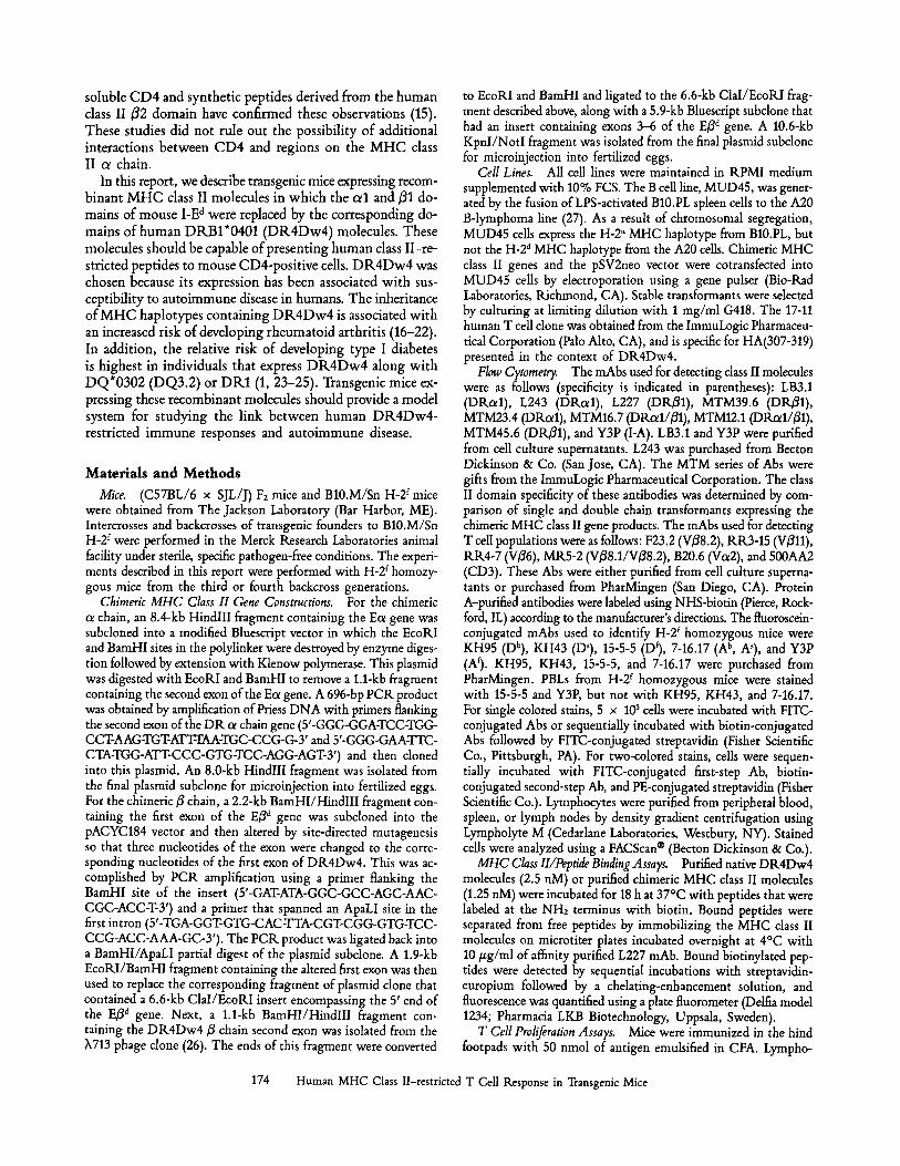
Human Major Histocompatibility Complex Class II-restricted T Cell Responses in Transgenic Mice By Andrea Woods,* Howard Y. Chen,~ Myrna E. Trumbauer,~ Anna Sirotina,S Rich Cummings, ll and Dennis M. Zaller*
From the Departments of *Molecular Immunology, ~Genetics & Molecular Biology, Slmmunology, and lIBiophysical Chemistry, Merck Research Laboratories, Rahway, New Jersey 07065
Stllnllllary
Transgenic mice expressing human major histocompatibility complex (MHC) class II molecules would provide a valuable model system for studying human immunology. However, attempts to obtain human class II-restricted T cell responses in such transgenic mice have had only limited success, possibly due to an inability of mouse CD4 to interact efficiently with human MHC class II molecules. To circumvent this problem, we constructed recombinant MHC class II genes in which the peptide-binding domain was derived from human DR sequences whereas the CD4- binding domain was derived from mouse I-E sequences. Purified chimeric human/mouse MHC class II molecules were capable of specifically binding DR-restricted peptides. Human B cell transformants that expressed these chimeric MHC class II molecules could present peptide antigens to human T cell clones. Expression of these chimeric class II molecules in transgenic mice led to the intrathymic deletion of T cells expressing superantigen-reactive V/3 gene segments, indicating that the chimeric class II molecules could influence the selection of the mouse T cell repertoire. These transgenic mice were fully capable of mounting human DR-restricted immune responses after challenge with peptide or whole protein antigens. Thus, the chimeric class II molecules can serve as functional antigen presentation molecules in vivo. In addition, transgenic mice expressing chimeric class II molecules could be used to generate antigen-specific mouse T cell hyhridomas that were capable of interacting with human antigen-presenting cells.
M HC class II proteins are cell surface heterodimers com- posed of a nearly invariant o~ chain and a highly poly-
morphic/5 chain. The primary function of these molecules is to present peptide fragments derived from processed an- tigens to CD4-bearing T cells. Interactions between MHC class II-peptide complexes and TCRs play a central role in the selection of the T cell repertoire and in the elicitation of an immune response. The inheritance of particular MHC class II alleles is genetically associated with susceptibility to certain autoimmune diseases such as type I diabetes and rheu- matoid arthritis (1, 2). The basis of the association between MHC class II genes and autoimmune disease is not known, but could be due to several immunological mechanisms. Poly- morphic amino acid residues in the class II peptide-binding groove might differentially bind potential autoantigens. Al- ternatively, the expression of particular MHC class II alleles might dictate whether autoreactive T cells reach functional maturity during development in the thymus. Another ex- planation that has been proposed for HLA disease associa- tions is molecular mimicry between environmental pathogens and MHC class II proteins (3, 4). Sequence homologies be- tween CMV proteins (5), EBV proteins (6, 7), and disease-
associated MHC class II molecules have been described, al- though the mechanisms by which these homologies could lead to autoimmune disease are not clear.
A model system in which human MHC class II molecules were expressed in transgenic mice would be of great benefit for studying the role of these molecules in the induction of autoimmune disease. However, previous attempts to generate human class II-restricted T cells responses in such transgenic mice have had only limited success (8, 9). One possible reason for this is an inability of mouse CD4 to interact efficiently with human MHC class II molecules (10-13). This xenogeneic barrier would be expected to result in inefficient interactions between mouse CD4-positive T cells and human class II-ex- pressing APCs in the transgenic mice. It seemed reasonable that this barrier could be overcome by employing chimeric human/mouse MHC class II molecules in which the CD4- binding domain of the human protein is replaced by the cor- responding domain of the mouse protein. Experiments using human class II molecules which had been altered by exon shuffling (12, 13) or point mutation (14) have identified a region on the/32 domain of human class II molecules that is involved in binding to CD4. Direct binding studies using
173 J. Exp. Med. �9 The Rockefeller University Press ~ 0022-1007/94/07/0173/09 $2.00 Volume 180 July 1994 173-181
soluble CD4 and synthetic peptides derived from the human class II B2 domain have confirmed these observations (15). These studies did not rule out the possibility of additional interactions between CD4 and regions on the M H C class II ot chain.
In this report, we describe transgenic mice expressing recom- binant M H C class II molecules in which the or1 and/31 do- mains of mouse I-E d were replaced by the corresponding do- mains of human DRB1*0401 (DR4Dw4) molecules. These molecules should be capable of presenting human class II-re- stricted peptides to mouse CD4-positive cells. D R 4 D w 4 was chosen because its expression has been associated with sus- ceptibility to autoimmune disease in humans. The inheritance of M H C haplotypes containing D R 4 D w 4 is associated with an increased risk of developing rheumatoid arthritis (16-22). In addition, the relative risk of developing type I diabetes is highest in individuals that express D R 4 D w 4 along with DQ*0302 (DQ3.2) or DR1 (1, 23-25). Transgenic mice ex- pressing these recombinant molecules should provide a model system for studying the link between human DR4Dw4- restricted immune responses and autoimmune disease.
Materials and Methods Mice. (C57BL/6 x SJL/J) Fz mice and B10.M/Sn H-2 f mice
were obtained from The Jackson Laboratory (Bar Harbor, ME). Intercrosses and backcrosses of transgenic founders to B10.M/Sn H-2 f were performed in the Merck Research Laboratories animal facility under sterile, specific pathogen-free conditions. The experi- ments described in this report were performed with H-2 f homozy- gous mice from the third or fourth backcross generations.
Chimeric MHC Class II Gene Constructions. For the chimeric a chain, an 8.4-kb HindlII fragment containing the Ea gene was subcloned into a modified Bluescript vector in which the EcoRI and BamHI sites in the polylinker were destroyed by enzyme diges- tion followed by extension with Klenow polymerase. This plasmid was digested with EcoRI and BamHI to remove a 1.1-kb fragment containing the second exon of the Eot gene. A 696-bp PCR product was obtained by amplification of Priess DNA with primers flanking the second exon of the DR a chain gene (5'-GGG-GGA-TCC-TGG- CCT-AAG-TGT-AT'IZFAA-TGC-CCG-G-3' and 5'-GGG-GAA-TTC- CTA-TGG-ATT-CCC-GTG-TCC-AGG-AGT-3') and then cloned into this plasmid. An 8.0-kb HindlII fragment was isolated from the final plasmid subclone for microinjection into fertilized eggs. For the chimeric/~ chain, a 2.2-kb BamHI/HindlII fragment con- taining the first exon of the E3 d gene was subcloned into the pACYC184 vector and then altered by site-directed mutagenesis so that three nucleotides of the exon were changed to the corre- sponding nucleotides of the first exon of DR4Dw4. This was ac- complished by PCR amplification using a primer flanking the BamHI site of the insert (5'-GAT-ATA-GGC-GCC-AGC-AAC- CGC-ACC-T-Y) and a primer that spanned an ApaLI site in the first intron (5'-TGA-GGT-GTG-CAC-TTA-CGT-CGG-GTG-TCC- CCG-ACC-AAA-GC-Y). The PCR product was ligated back into a BamHI/ApaLI partial digest of the plasmid subclone. A 1.9-kb EcoRI/BamHI fragment containing the altered first exon was then used to replace the corresponding fragment of plasmid clone that contained a 6.6-kb ClaI/EcoRI insert encompassing the 5' end of the E3 a gene. Next, a 1.1-kb BamHI/HindlII fragment con- taining the DR4Dw4 ~ chain second exon was isolated from the X713 phage clone (26). The ends of this fragment were converted
to EcoRI and BamHI and ligated to the 6.6-kb ClaI/EcoRI frag- ment described above, along with a 5.9-kb Bluescript subclone that had an insert containing exons 3-6 of the E3 d gene. A 10.6-kb KpnI/NotI fragment was isolated from the final plasmid subclone for microinjection into fertilized eggs.
Cell Lines. All cell lines were maintained in RPMI medium supplemented with 10% FCS. The B cell line, MUD45, was gener- ated by the fusion of LPS-activated B10.PL spleen cells to the A20 B-lymphoma line (27). As a result of chromosomal segregation, MUD45 cells express the H-2 u MHC haplotype from B10.PL, but not the H-2 a MHC haplotype from the A20 cells. Chimeric MHC class II genes and the pSV2neo vector were cotransfected into MUD45 cells by electroporation using a gene pulser (Bio-Rad Laboratories, Richmond, CA). Stable transformants were selected by culturing at limiting dilution with 1 mg/ml G418. The 17-11 human T cell clone was obtained from the ImmuLogic Pharmaceu- tical Corporation (Palo Alto, CA), and is specific for HA(307-319) presented in the context of DR4Dw4.
Flow Cytometry. The mAbs used for detecting class II molecules were as follows (specificity is indicated in parentheses): LB3.1 (DRotl), L243 (DRotl), L227 (DRB1), MTM39.6 (DRB1), MTM23.4 (DRotl), MTM16.7 (DRot1/31), MTM12.1 (DRot1/31), MTM45.6 (DRY1), and Y3P (I-A). LB3.1 and Y3P were purified from cell culture supernatants. L243 was purchased from Becton Dickinson & Co. (San Jose, CA). The MTM series of Abs were gifts from the ImmuLogic Pharmaceutical Corporation. The class II domain specificity of these antibodies was determined by com- parison of single and double chain transformants expressing the chimeric MHC class II gene products. The mAbs used for detecting T cell populations were as follows: F23.2 (V~8.2), RR3-15 (V~11), RR4-7 (V36), MR5-2 (V38.1/V38.2), B20.6 (Va2), and 500AA2 (CD3). These Abs were either purified from cell culture superna- tants or purchased from PharMingen (San Diego, CA). Protein A-purified antibodies were labeled using NHS-biotin (Pierce, Rock- ford, IL) according to the manufacturer's directions. The fluoroscein- conjugated mAbs used to identify H-2 r homozygous mice were KH95 (Db), KH43 (Ds), 15-5-5 (Dr), 7-16.17 (A b, As), and Y3P (Af). KH95, KH43, 15-5-5, and 7-16.17 were purchased from PharMingen. PBLs from H-2 f homozygous mice were stained with 15-5-5 and Y3P, but not with KH95, KH43, and 7-16.17. For single colored stains, 5 x 10 s cells were incubated with FITC- conjugated Abs or sequentially incubated with biotin-conjugated Abs followed by FITC-conjugated streptavidin (Fisher Scientific Co., Pittsburgh, PA). For two-colored stains, cells were sequen- tially incubated with FITC-conjugated first-step Ab, biotin- conjugated second-step Ab, and PE-conjugated streptavidin (Fisher Scientific Co.). Lymphocytes were purified from peripheral blood, spleen, or lymph nodes by density gradient centrifugation using Lympholyte M (Cedarlane Laboratories, Westbury, NY). Stained cells were analyzed using a FACScan | (Becton Dickinson & Co.).
MHC Class II/Peptide Binding Assays. Purified native DR4Dw4 molecules (2.5 riM) or purified chimeric MHC class II molecules (1.25 nM) were incubated for 18 h at 37~ with peptides that were labeled at the NH2 terminus with biotin. Bound peptides were separated from free peptides by immobilizing the MHC class II molecules on microtiter plates incubated overnight at 4~ with 10/~g/ml of affinity purified L227 mAb. Bound biotinylated pep- tides were detected by sequential incubations with streptavidin- europium followed by a chelating-enhancement solution, and fluorescence was quantified using a plate fluorometer (Delfia model 1234; Pharmacia LKB Biotechnology, Uppsala, Sweden).
T Cell Proliferation Assays. Mice were immunized in the hind footpads with 50 nmol of antigen emulsified in CFA. Lympho-
174 Human MHC Class II-restricted T Cell Response in Transgenic Mice
cytes were isolated from popliteal, inguinal, and paraaortic lymph nodes 10 d after immunization. 4 x 10³ cells were then cultured in microtiter plates for 4 d in RPMI/10% FCS with serial dilu- tions of antigen. For human T cell clones, 3 x 10⁴ T cells and 10 s glutaraldehyde-fixed APCs were incubated for 2 d with serial dilutions of antigen. 1 #Ci of [3H]thymidine was added to the wells during the last 16 h of culture. Plates were then harvested and counts per minute was determined by liquid scintillation counting.
T Cell Hybridomas. Lymph node cells from immunized mice were cultured for 3 d in the presence of 100/zM of antigen. Live cells were purified over lympholyte M gradients and fused to a BW5147 variant cell line (28). Hybridomas were selected in cul- ture medium supplemented with HAT. The specificity of the hy- bridomas were assessed in IL-2 release assays. Single cell clones were obtained by culture a limiting dilution.
1L-2 Release Assays. Hybridoma cells and APCs were cocul- tured overnight with serial dilutions of antigen in flat-bottom mi- crotiter plates. Supernatants were then transferred to culture wells containing 5 x 10³ HT-2 indicator cells. The HT-2 cells were then incubated for 24 h with 1 #Ci of [3H]thymidine added during the last 5 h of culture. Plates were harvested and counts per minute was determined by liquid scintillation counting.
Results
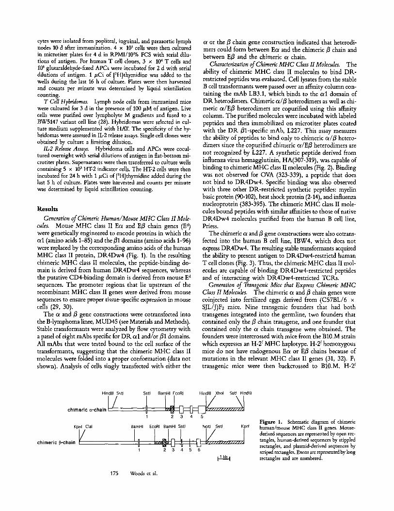
Generation of Chimeric Human~Mouse MHC Class II Mole- cules. Mouse MHC class II Ecx and E/3 chain genes (E a) were genetically engineered to encode proteins in which the oel (amino acids 1-85) and the/31 domains (amino acids 1-96) were replaced by the corresponding amino acids of the human MHC class II protein, DR4Dw4 (Fig. 1). In the resulting chimeric MHC class II molecules, the peptide-binding do- main is derived from human DR4Dw4 sequences, whereas the putative CD4-binding domain is derived from mouse E a sequences. The promoter regions that lie upstream of the recombinant MHC class II genes were derived from mouse sequences to ensure proper tissue-specific expression in mouse cells (29, 30).
The cx and/3 gene constructions were cotransfected into the B-lymphoma linee, MUD45 (see Materials and Methods). Stable transformants were analyzed by flow cytometry with a panel of eight mAbs specific for DR oll and/or/31 domains. All mAbs that were tested bound to the cell surface of the transformants, suggesting that the chimeric MHC class II molecules were folded into a proper conformation (data not shown). Analysis of cells singly transfected with either the
ot or the/3 chain gene construction indicated that heterodi- mers could form between Ecx and the chimeric fl chain and between E3 and the chimeric c~ chain.
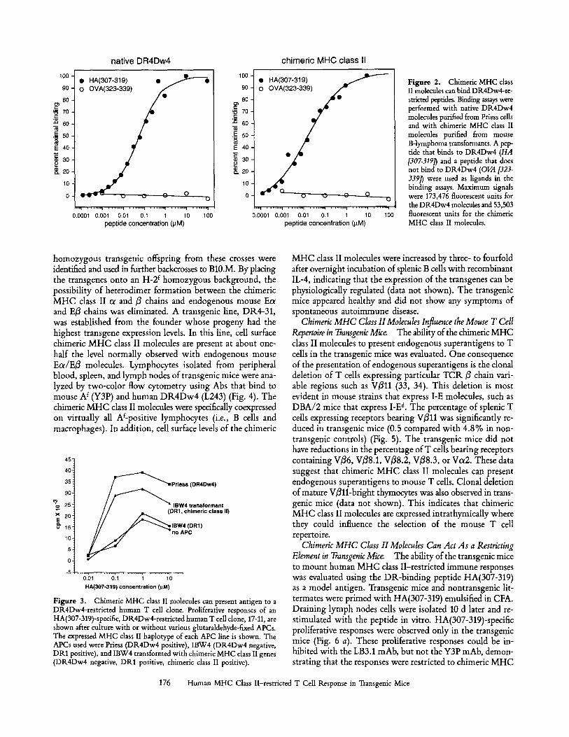
Characterization of Chimeric MHC Class II Molecules. The ability of chimeric MHC class II molecules to bind DP,- restricted peptides was evaluated. Cell lysates from the stable B cell transformants were passed over an affinity column con- taining the mAb LB3.1, which binds to the cxl domain of DR heterodimers. Chimeric cx//3 heterodimers as well as chi- meric cx/E3 heterodimers are copurified using this affinity column. The purified molecules were incubated with labeled peptides and then immobilized on microtiter plates coated with the DR/31-specific mAb, L227. This assay measures the ability of peptides to bind only to chimeric cx/3 hetero- dimers since the copurified chimeric ol/E/3 heterodimers are not recognized by L227. A synthetic peptide derived from influenza virus hemagglutinin, HA(307-319), was capable of binding to chimeric MHC class II molecules (Fig. 2). Binding was not observed for OVA (323-339), a peptide that does not bind to DR4Dw4. Specific binding was also observed with three other DR-restricted synthetic peptides: myelin basic protein (90-102), heat shock protein (2-14), and influenza nucleoprotein (383-395). The chimeric MHC class II mole- cules bound peptides with similar affinities to those of native DR4Dw4 molecules purified from the human B cell line, Priess.
The chimeric ot and/3 gene constructions were also cotrans- fected into the human B cell line, IBW4, which does not express DR4Dw4. The resulting stable transformants acquired the ability to present antigen to DR4Dw4-restrictd human T cell clones (Fig. 3). Thus, the chimeric MHC class II mol- ecules are capable of binding DR4Dw4-restricted peptides and of interacting with DR4Dw4-restricted TCRs.
Generation of Transgenic Mice that Express Chimeric MHC Class II Molecules. The chimeric cx and/3 chain genes were coinjected into fertilized eggs derived from (C57BL/6 x SJL/J)F2 mice. Nine transgenic founders that had both transgenes integrated into the germline, two founders that contained only the/3 chain transgene, and one founder that contained only the cx chain transgene were obtained. The founders were intercrossed with mice from the B10.M strain which expresses an H-2 f MHC haplotype. H-2 f homozygous mice do not have endogenous Eol or E/3 chains because of mutations in the relevant MHC class II genes (31, 32). F1 transgenic mice were then backcrossed to B10.M. H-2 f
Hindlll Sstl Sstl BamHI EcoRI
1
Kpnl Clal BamHI
chimeric~-chain V I , II 1
Hindlll Xhol Sstl Hindlll
/ 2 3 4 5
EcoRI BarnHl Sstl Notl Sstl Kpnl
2 3 4 5 6
Ilkbl
Figure 1. Schematic diagram of chimeric human/mouse MHC class II genes. Mouse- derived sequences are represented by open rec- tangles, human-derived sequences by stippled rectangles, and plasmid-derived sequences by striped rectangles. Exons are represented by long rectangles and are numbered.
175 Woods et al.
100
90
80 == ~ zo 5~ 60
.~ 50
E 40
m 30
g. eO
10
native DR4Dw4
�9 HA(307-319) �9 ~ , . , . ' ~ - ' O
o o o o o
0 . 0 0 0 1 0 . 0 0 1 0 . 0 1 0 . 1 1 1 0 1 0 0
peptide concentration (gM)
100
90
8O == ~ 70 "~ 60
. ~ 5 0
E 40
g 30 p ~. 20
chimeric MHC class II
�9 HA(307-319)
u ~, 0 Q
�9 ,,~ . . . . . . . . i . . . . . . . . i . . . . . . ,~ . . . . . . . . i . . . . . . . . i . . . . . . . i 0.0001 0.001 0.01 0.1 1 10 100
peptide concentration (gM)
F igure 2. Chimeric MHC class II molecules can bind DK4Dw4-re- stricted peptides. Binding assays were performed with native DR.4Dw4 molecules purified from Priess cells and with chimeric MHC class II molecules purified from mouse B-lymphoma transformants. A pep- tide that binds to DR4Dw4 (HA [307-3191) and a peptide that does not bind to DR4Dw4 (OVA [323- 3391" ) were used as ligands in the binding assays. Maximum signals were 173,476 fluorescent units for the DR4Dw4 molecules and 53,503 fluorescent units for the chimeric MHC class II molecules.
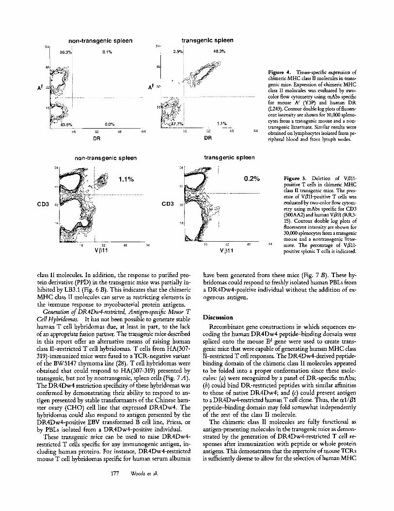
homozygous transgenic offspring from these crosses were identified and used in further backcrosses to B10.M. By placing the transgenes onto an H-2 f homozygous background, the possibility of heterodimer formation between the chimeric MHC class II ot and 3 chains and endogenous mouse Ec~ and Eft chains was eliminated. A transgenic line, DR4-31, was established from the founder whose progeny had the highest transgene expression levels. In this line, cell surface chimeric MHC class II molecules are present at about one- half the level normally observed with endogenous mouse Ec~/E3 molecules. Lymphocytes isolated from peripheral blood, spleen, and lymph nodes of transgenic mice were ana- lyzed by two-color flow cytometry using Abs that bind to mouse A f (Y3P) and human DR4Dw4 (L243) (Fig. 4). The chimeric MHC class II molecules were specifically coexpressed on virtually all Af-positive lymphocytes (i.e., B cells and macrophages). In addition, cell surface levels of the chimeric
45"
40i 35 i
30-
2 5 "
x 2 0 -
E u 15"
10- / F ~ ' ~ ~ P r i ess (DR4Dw4)
~ I B W 4 transformant
BW4 (DR1) no APC
. . . . . . i . . . . . . . . , . . . . . . . . , . . . . . . . . i . �9 0.01 0.1 1 10
HA(307-319) concentration (pM)
Figure 3. Chimeric MHC class II molecules can present antigen to a DR4Dw4-restricted human T cell clone. Proliferative responses of an HA(307-319)-specific, DR4Dw4-restricted human T cell clone, 17-11, are shown after culture with or without various glutaraldehyde-fixed APCs. The expressed M H C class II haplotype of each APC line is shown. The APCs used were Priess (DR4Dw4 positive), IBW4 (DR4Dw4 negative, DR1 positive), and IBW4 transformed with chimeric MHC class II genes (DR4Dw4 negative, DR1 positive, chimeric class II positive).
MHC class II molecules were increased by three- to fourfold after overnight incubation of splenic B cells with recombinant IL-4, indicating that the expression of the transgenes can be physiologically regulated (data not shown). The transgenic mice appeared healthy and did not show any symptoms of spontaneous autoimmune disease.
Chimeric MHC Class II Molecules Influence the Mouse T Cell Repertoire in Transgenic Mice The ability of the chimeric MHC class II molecules to present endogenous superantigens to T cells in the transgenic mice was evaluated. One consequence of the presentation of endogenous superantigens is the clonal deletion of T cells expressing particular TCR fl chain vari- able regions such as Vflll (33, 34). This deletion is most evident in mouse strains that express I-E molecules, such as DBA/2 mice that express I-E d. The percentage of splenic T cells expressing receptors bearing V311 was significantly re- duced in transgenic mice (0.5 compared with 4.8% in non- transgenic controls) (Fig. 5). The transgenic mice did not have reductions in the percentage of T cells bearing receptors containing Vfl6, Vfl8.1, Vf18.2, Vf18.3, or Vot2. These data suggest that chimeric MHC class II molecules can present endogenous superantigens to mouse T cells. Clonal deletion of mature Villi-bright thymocytes was also observed in trans- genic mice (data not shown). This indicates that chimeric MHC class II molecules are expressed intrathymically where they could influence the selection of the mouse T cell repertoire.
Chimeric MHC Class II Molecules Can Act As a Restricting Element in Transgenic Mice. The ability of the transgenic mice to mount human MHC class II-restricted immune responses was evaluated using the DR-binding peptide HA(307-319) as a model antigen. Transgenic mice and nontransgenic lit- termates were primed with HA(307-319) emulsified in CFA. Draining lymph nodes cells were isolated 10 d later and re- stimulated with the peptide in vitro. HA(307-319)-specific proliferative responses were observed only in the transgenic mice (Fig. 6 a). These proliferative responses could be in- hibited with the LB3.1 mAb, but not the Y3P mAb, demon- strating that the responses were restricted to chimeric MHC
176 Human MHC Class II-restricted T Cell Response in Transgenic Mice
++I 48
A f 32
non-transgenic spleen
56.3%: 0.1%
~ 4 3 . 5 % 0.0% 16 32 48
DR 64
A f
transgenic spleen 64[ ! 48.3%
, 2-9% I
32~
1.1% 16 32 48 64
DR
Figure 4. Tissue-specific expression of chimeric MHC class II molecules in trans- genic mice. Expression of chimeric MHC class II molecules was evaluated by two- color flow cytometry using mAbs specific for mouse A f (Y3P) and human DR. (I.243). Contour double log plots of fluores- cent intensity are shown for 10,000 spleno- cytes from a transgenic mouse and a non- transgenic littermate. Similar results were obtained on lymphocytes isolated from pe- ripheral blood and from lymph nodes.
CD3
non-transgenic spleen
~ , ~ 1.1% %
16 32 48 Vl311
CD3
transgenic spleen
0.2%
16 32 48 VL311
Figure 5. Deletion of V~ll- positive T cells in chimeric MHC class II transgenic mice. The pres- ence of VB11-positive T cells was evaluated by two-color flow cytom- etry using mAbs specific for CD3 (500AA2) and human V/311 (RR3- 15). Contour double log plots of fluorescent intensity are shown for 30,000 splenocytes from a transgenic mouse and a nontransgenic litter- mate. The percentage of V/~11- positive splenic T cells is indicated.
class II molecules. In addition, the response to purified pro- tein derivative (PPD) in the transgenic mice was partially in- hibited by LB3.1 (Fig. 6 B). This indicates that the chimeric MHC class II molecules can serve as restricting elements in the immune response to mycobacterial protein antigens.
Generation of DR 4Dw4-restricted, Antigen-specific Mouse T CellHybridomas. It has not been possible to generate stable human T cell hybridomas due, at least in part, to the lack of an appropriate fusion partner. The transgenic mice described in this report offer an alternative means of raising human class II-restricted T cell hybridomas. T cells from HA(307- 319)-immunized mice were fused to a TCR-negative variant of the BW5147 thymoma line (28). T cell hybridomas were obtained that could respond to HA(307-319) presented by transgenic, but not by nontransgenic, spleen cells (Fig. 7 A). The DR4Dw4 restriction specificity of these hybridomas was confirmed by demonstrating their ability to respond to an- tigen presented by stable transformants of the Chinese ham- ster ovary (CHO) cell line that expressed DR4Dw4. The hybridomas could also respond to antigen presented by the DR4Dw4-positive EBV transformed B cell line, Priess, or by PBLs isolated from a DR4Dw4-positive individual.
These transgenic mice can be used to raise DR4Dw4- restricted T cells specific for any immunogenic antigen, in- cluding human proteins. For instance, DR4Dw4-restricted mouse T cell hybridomas specific for human serum albumin
have been generated from these mice (Fig. 7 B). These hy- bridomas could respond to freshly isolated human PBLs from a DR4Dw4-positive individual without the addition of ex- ogenous antigen.
D i s c u s s i o n
Recombinant gene constructions in which sequences en- coding the human DR4Dw4 peptide-binding domain were spliced onto the mouse E a gene were used to create trans- genic mice that were capable of generating human MHC class II-restricted T cell responses. The DR4Dw4-derived peptide- binding domain of the chimeric class II molecules appeared to be folded into a proper conformation since these mole- cules: (a) were recognized by a panel of DR-specific mAbs; (b) could bind DR-restricted peptides with similar affinities to those of native DR4Dw4; and (c) could present antigen to a DR4Dw4-restricted human T cell done. Thus, the od/B1 peptide-binding domain may fold somewhat independently of the rest of the class II molecule.
The chimeric class II molecules are fully functional as antigen-presenting molecules in the transgenic mice as demon- strated by the generation of DR4Dw4-restricted T cell re- sponses after immunization with peptide or whole protein antigens. This demonstrates that the repertoire of mouse TCRs is sufficiently diverse to allow for the selection of human MHC
177 Woods et al.
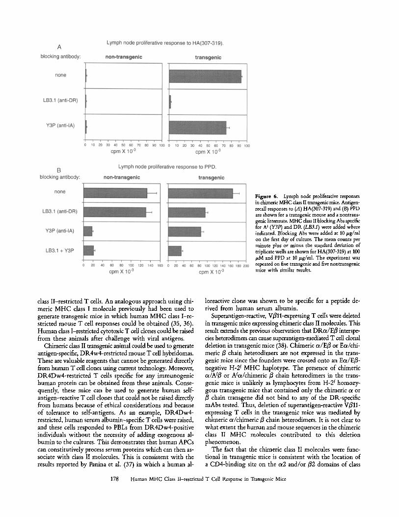
Figure 6. Lymph node proliferative responses in chimeric MHC dass II transgenic mice. Antigen- recall responses to (A) HA(307-319) and (B) PPD are shown for a transgenic mouse and a nontrans- genic littermat~ MHC class II blocking Abs specific for A t (Y3P) and DR (LB3.1) were added where indicated. Blocking Abs were added at 10/zg/ml on the first day of culture. The mean counts per minute plus or minus the standard deviation of triplicate wells are shown for HA(307-319) at 100 /zM and PPD at 10/~g/ml. The experiment was repeated on five transgenic and five nontransgenic mice with similar results.
class II-restricted T cells. An analogous approach using chi- meric MHC class I molecule previously had been used to generate transgenic mice in which human MHC class I-re- stricted mouse T cell responses could be obtained (35, 36). Human class I-restricted cytotoxic T cell clones could be raised from these animals after challenge with viral antigens.
Chimeric dass II transgenic animal could be used to generate antigen-specific, DR4w4-restricted mouse T cell hybridomas. These are valuable reagents that cannot be generated directly from human T cell dones using current technology. Moreover, DR4Dw4-restricted T cells specific for any immunogenic human protein can be obtained from these animals. Conse- quently, these mice can be used to generate human self- antigen-reactive T cell clones that could not be raised directly from humans because of ethical considerations and because of tolerance to self-antigens. As an example, DR4Dw4- restricted, human serum albumin-specific T ceils were raised, and these cells responded to PBLs from DR4Dw4-positive individuals without the necessity of adding exogenous al- bumin to the cultures. This demonstrates that human APCs can constitutively process serum proteins which can then as- sociate with class II molecules. This is consistent with the results reported by Panina et al. (37) in which a human al-
loreactive clone was shown to be specific for a peptide de- rived from human serum albumin.
Superantigen-reactive, Villi-expressing T cells were deleted in transgenic mice expressing chimeric class II molecules. This result extends the previous observation that DRoe/Efl interspe- des heterodimers can cause superantigen-mediated T cell donal deletion in transgenic mice (38). Chimeric ol/Efl or E~/chi- meric fl chain heterodimers are not expressed in the trans- genic mice since the founders were crossed onto an Eoe/Efl- negative H-2 f MHC haplotype. The presence of chimeric c~/Af3 or Aloe/chimeric fl chain heterodimers in the trans- genic mice is unlikely as lymphocytes from H-2 t homozy- gous transgenic mice that contained only the chimeric o~ or fl chain transgene did not bind to any of the DR-specific mAbs tested. Thus, deletion of superantigen-reactive Villi- expressing T cells in the transgenic mice was mediated by chimeric c~/chimeric fl chain heterodimers. It is not clear to what extent the human and mouse sequences in the chimeric class II MHC molecules contributed to this deletion phenomenon.
The fact that the chimeric class II molecules were func- tional in transgenic mice is consistent with the location of a CD4-binding site on the c~2 and/or f12 domains of class
178 Human MHC Class II-restricted T Cell Response in Transgenic Mice
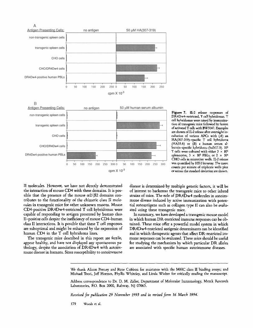
Figure 7. Ib2 release responses of DR4Dw4-restricted, T call hybridomas. T cell hybridomas were raised by immuniza- tion of transgenic mice followed by fusion of activated T cells with BW5147. Examples are shown of Ib2 release after overnight in- cubation of various APCs with (.4) an HA(307-319)-specific T cell hybridoma (HA33.6) or (B) a human serum al- bumin-speeific hybridoma (halb17.9). 105 T cells were cultured with either 3 x 10 s splenocytes, 3 x 10⁵ PBLs, or 2 x 10⁴ CHO cells in microtiter wells. Ib2 release was quantified by HT-2 bioassay. The mean counts per minute of triplicate wells plus or minus the standard deviation are shown.
II molecules. However, we have not directly demonstrated the interaction of mouse CD4 with these domains. It is pos- sible that the presence of the mouse ot2//~2 domains con- tributes to the functionality of the chimeric class II mole- cules in transgenic mice for other unknown reasons. Mouse CD4-positive DR4Dw4-restricted T cell hybridomas were capable of responding to antigen presented by human class II-positive cells despite the inefficiency of mouse CD4-human class II interactions. It is possible that these T cell responses are suboptimal and might be enhanced by the expression of human CD4 in the T cell hybridoma lines.
The transgenic mice described in this report are fertile, appear healthy, and have not displayed any spontaneous pa- thology, despite the association of DR4Dw4 with autoim- mune disease in humans. Since susceptibility to autoimmune
disease is determined by multiple genetic factors, it will be of interest to backcross the transgenic mice to other inbred strains of mice. The role of DK4Dw4 molecules in autoim- mune disease induced by active immunization with poten- tial autoantigens such as collagen type II can also be evalu- ated using these transgenic mice.
In summary, we have developed a transgenic mouse model in which human DR-restricted immune responses can be ob- tained. These mice offer a powerful model system in which DR4Dw4-restricted antigenic determinants can be identified and in which therapeutic agents that affect DR-restricted im- mune responses can be evaluated. These mice should be useful for studying the mechanism by which particular DR alleles are associated with specific human autoimmune diseases.
We thank Alison Pressey and Rose Cubbon for assistance with the MHC class II binding assays; and Michael Tocci, Jeff Hermes, Phyllis Whiteley, and Linda Wicker for critically reading the manuscript.
Address correspondence to Dr. D. M. Zaller, Department of Molecular Immunology, Merck Research Laboratories, P.O. Box 2000, Rahway, NJ 07065.
Received for publication 29 November 1993 and in revised form 16 March 1994.
179 Woods et al.
References 1. Nepom, G.T., and H. Erlich. 1991. MHC class-II molecules
and autoimmunity. Annu. Rev. Immunol. 9:493. 2. Tirawi, J., and P. Terasaki. 1985. HLA and Disease Associa-
tions. Springer-Verlag New York Inc., New York. 472 pp. 3. Oldstone, M.B. 1987. Molecular mimicry and autoimmune
disease. Cell. 50:819. 4. Oldstone, M.B. 1989. Molecular mimicry as a mechanism for
the cause and a probe uncovering etiologic agent(s) of autoim- mune disease. Curt. Top. Microbiol. Immunol. 145:127.
5. Fujinami, R.S., J.A. Nelson, L. Walker, and M.B. Oldstone. 1988. Sequence homology and immunologic cross-reactivity of human cytomegalovirus with HLA-DR beta chain: a means for graft rejection and immunosuppression. J. Virol. 62:100.
6. Horn, G.T., T.L. Bugawan, C.M. Long, and H.A. Erlich. 1988. Allelic sequence variation of the HLA-DQ loci: relationship to serology and to insulin-dependent diabetes susceptibility. Proc. Natl. Acad. Sci. USA. 85:6012.
7. Roudier, J., G. Rhodes, J. Petersen, J.H. Vaughan, and D.A. Carson. 1988. The Epstein-Barr virus glycoprotein gp110, a molecular link between HLA DR4, HLA DR1, and rheuma- toid arthritis. Stand. J. Immunol, 27:367.
8. Sasazuki, T., T. Iwanaga, T. Inamitsu, Y. Yanagawa, M. Yasunami, A. Kimura, K. Hirokawa, and Y. Nishimura. 1989. Expression and function of human major histocompatibility complex HLA-DQw6 genes in the C57BL/6 mouse. Cold Spring Harbor Symtx Quant. Biol. 1:513.
9. Nishimura, Y., T. Iwanaga, T. Inanitsu, Y. Yanagawa, M. Yasunami, A. Kimura, K. Hirokawa, and T. Sasazuki. 1990. Expression of the human MHC, HLA-DQw6 genes alters the immune response in C57B1/6 mice. J. Immunol. 145:353.
10. Sleckman, B.P., A. Peterson, W.K. Jones, J.A. Foran, J.L. Greenstein, B. Seed, and S.J. Burakoff. 1987. Expression and function of CD4 in a routine T cell hybridoma. Nature (Lond.), 328:351.
11. Lamarre, D., A. Ashkenazi, S. Fleury, D.H. Smith, R.P. Sekaly, and D.J. Capon. 1989. The MHC-binding and gp120-binding functions of CD4 are separable. Science (Wash. DC). 245:743.
12. Lombardi, G., L. Barber, G. Aichinger, T. Heaton, S. Sidhu, J.R. Batchelor, and R.I. Lechler. 1991. Structural analysis of anti-DR1 allorecognition by using DR1/H-2Ek hybrid mole- cules. Influence of the beta 2-domain correlates with CD4 de- pendence. J. Immunol. 147:2034.
13. Vignali, D.A.A., J. Moreno, D. Schiller, and G.J. Hiimmer- ling. 1992. Species-specific binding of CD4 to the ~ 2 domain of major histocompatibility complex class II molecules.J. Exp. Med. 175:925.
14. Konig, R., L.Y. Huang, and R.N. Germain. 1992. MHC class II interaction with CD4 mediated by a region analogous to the MHC class I binding site for CD8. Nature (Lond.). 356:796.
15. Cammarota, G., A. Scheirle, R Takacs, D.M. Doran, R. Knott, W. Bannwarth, J. Guardiola, and F. Sinigaglia. 1992. Iden- tification of a CD4 binding site on the beta 2 domain of HLA-DR molecules. Nature (Lond.). 356:799.
16. Nepom, G.T., J. Hansen, and B. Nepom. 1986. The molecular basis for HLA class II associations with rheumatoid arthritis. J. Clin. Immunol. 7:1.
17. Stasny, P. 1978. Associations of the B-cell alloantigen DRw4 with rheumatoid arthritis. IV. Engl. J. Med. 289:869.
18. Nepom, B.S., G.T. Nepom, J. Schaller, E. Mickelson, J. SchaUer, P. Antonelli, and A. Hansen. 1984. Specific HLA-DR4- associated histocompatibility molecules in patients with
seropositivejuvenile rhematoid arthritis.J. Clin. Invest. 74:287. 19. Zoschke, D., and M. Segall. 1986. Dw subtypes of DR4 in
rheumtoid arthritis: evidence for a preferential association with Dw4. Hum. Immunol. 15:118.
20. Oilier, W., D. Carthy, S. Cutbush, R. Okoye, J. Awad, A. Fielder, A. Silman, and H. Festenstein. 1988. DR4-associated Dw types in rheumatoid arthritis. Tissue Antigens. 33:33.
21. Nepom, G.T., P. Byers, C. Seyfried, L.A. Healey, K.R. W'dske, D. Stage, and B.S. Nepom. 1989. HLA genes associated with rheumatoid arthritis. Arthritis Rheum. 32:15.
22. Wordsworth, B.P., J.S. Lanchbury, L.I. Sakkas, K.I. Welsh, G.S. Panayi, and J.I. Bell. 1989. HLA-DR4 subtype frequen- cies in rheumatoid arthritis indicate that DRB1 is the major susceptibility locus within the HLA class II region. Proc. Natl. Acad. Sci. USA. 86:10049.
23. Tait, B.D., G. Mraz, and L.C. Harrison. 1988. Association of HLA-DQw3 (TA10-) with type I diabetes occurs with DR3/4 but not DR1/4 patients. Diabetes. 37:926.
24. Sheehy, M.J., S.J. Scharf, J.R. Rowe, M.H. Neme de Gimenez, L.M. Meske, H.A. Erlich, and B.S. Nepom. 1989. A diabetes- susceptible HLA haplotype is best defined by a combination of HLA-DR and -DQ alleles. J. Clin. Invest. 83:830.
25. Erlich, H.A., T.L. Bugawan, S. Scharf, G.T. Nepom, B. Tait, and R.L. Griffith, 1990. HLA-DQ beta sequence polymor- phism and genetic susceptibility to IDDM. Diabetes. 39:96.
26. Andersson, G., D. Larhammar, E. Widmark, B. Servenius, P.A. Peterson, and L. Rash. 1987. Class II genes of the human major histocompatibility complex. Organization and evolutionary relationship of the DR beta genes (published erratum appears inJ. Biol. Chem. 1988. Jun 15;263117]:8551). j . Biol. Chem. 262:8748.
27. Kim, K.J., C. Kannellopoulos-Langevin, R.M. Merwin, D.H. Sachs, and R. Asofsky. 1979. Establishment and characteriza- tion of BALB/c lymphoma lines with B cell properties. J. Ira. munol. 122:549.
28. White, J., M. Blackman, J. Bill, J. Kappler, P. Marrack, D.P. Gold, and W. Born. 1989. Two better cell lines for making hybridomas expressing specific T cell receptors. J. Immunol. 143:1822.
29. Benoist, C., and D. Mathis. 1990. Regulation of major histocompatibility complex class-II genes: X, Y and other letters of the alphabet. Annu. Rev. Immunol. 8:681.
30. Dorn, A., B. Durand, C. Marring, M.M. Le, C. Benoist, and D. Mathis. 1987. Conserved major histocompatibility com- plex class II boxes-X and Y-are transcriptional control ele- ments and specifically bind nuclear proteins. Proc. Natl. Acad. Sci. USA. 84:6249.
31. Vu, T.H., A.B. Begovich, C.F. Tacchini, and P.P. Jones. 1989. Molecular defects in the non-expressed H-2 E alpha genes of the f and q haplotypes. J. Immunol. 142:2936.
32. Begovich, A.B., T.H. Vu, and P.P. Jones. 1990. Characteriza- tion of the molecular defects in the mouse E beta f and E beta q genes. Implications for the origin of MHC polymorphism.
J. Immunol. 144:1957. 33. Bill, J., O. Kanagawa, D.L. Woodland, and E. Palmer. 1989.
The MHC molecule I-E is necessary but not sufficient for the clonal deletion of V~811-bearing T cells.J. Exp. Med. 169:1405.
34. Gao, E.-K., O. Kanagawa, and J. Sprent. 1989. Capacity of unprimed CD4 § and CD8 § T cells expressing Vj811 receptors to respond to I-E alloantigens in vivo. J. Exp. Med. 170:1947.
35. Salter, R.D., R.J. Benjamin, P.K. Wesley, S.E. Buxton, T.P.
180 Human MHC Class II-restricted T Cell Response in Transgenic Mice
Garrett, C. Clayberger, A.M. Krensky, A.M. Norment, D.K. Littman, and P. Parham. 1990. A binding site for the T-cell co-receptor CD8 on the alpha 3 domain of HLA-A2. Nature (Lond.). 345:41.
36. Vitiello, A., D. Marchesini, J. Furze, L.A. Sherman, and R.W. Chesnut. 1991. Analysis of the HLA-restricted influenza-specific cytotoxic T lymphocyte response in transgenic mice carrying a chimeric human-mouse class I major histocompatibility com- plex. J. Exp. Med. 173:1007.
37. Panina, B.P., G. Corradin, E. Roosnek, A. Sette, and A. Lan- zavecchia. 1991. Recognition by class II alloreactive T cells of processed determinants from human serum proteins. Science (Wash. DC). 252:1548.
38. Lawrance, S.K., L. Karlsson, J. Price, V. Quaranta, Y. Ron, J. Sprent, and P.A. Peterson. 1989. Transgenic HLA-DK alpha faithfully reconstitutes IE-controlled immune functions and induces cross-tolerance to E alpha and E alpha 0 mutant mice. Cell. 58:583.
181 Woods et al.