Embed Size (px)
Citation preview
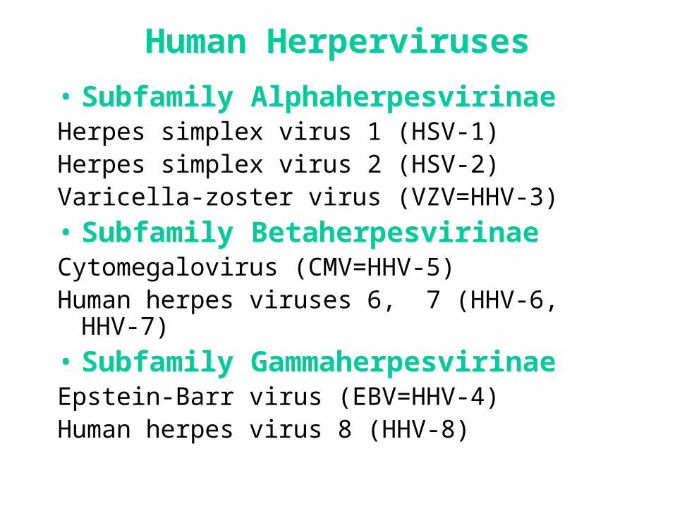
Human Herperviruses
• Subfamily AlphaherpesvirinaeHerpes simplex virus 1 (HSV-1)Herpes simplex virus 2 (HSV-2)Varicella-zoster virus (VZV=HHV-3)
• Subfamily BetaherpesvirinaeCytomegalovirus (CMV=HHV-5)Human herpes viruses 6, 7 (HHV-6, HHV-7)
• Subfamily GammaherpesvirinaeEpstein-Barr virus (EBV=HHV-4)Human herpes virus 8 (HHV-8)
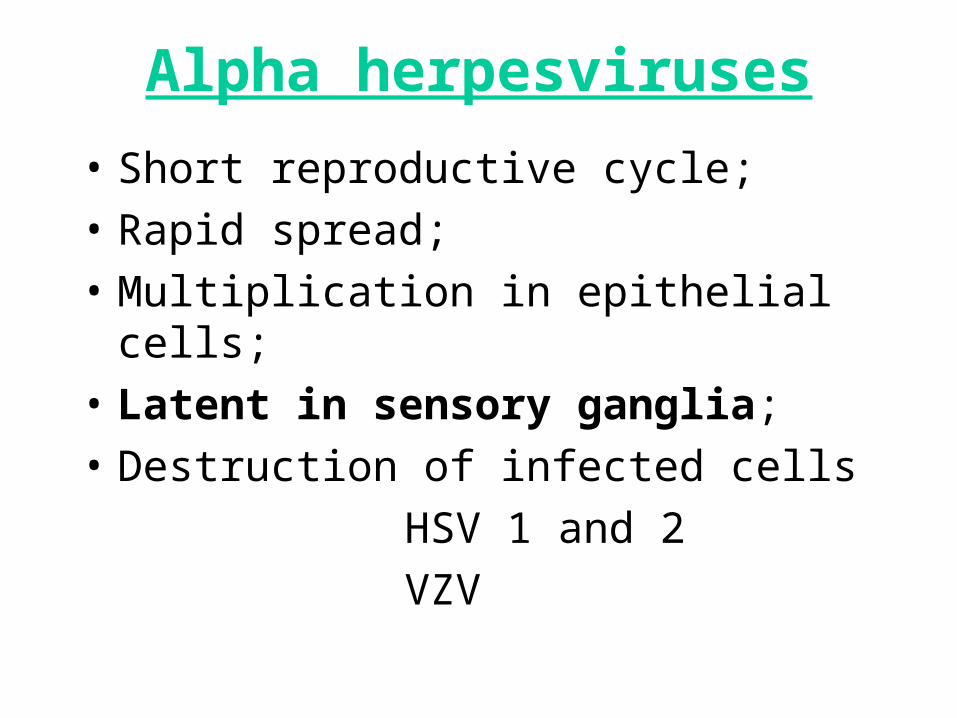
Alpha herpesviruses
• Short reproductive cycle;
• Rapid spread;
• Multiplication in epithelial cells;
• Latent in sensory ganglia;
• Destruction of infected cells
HSV 1 and 2
VZV
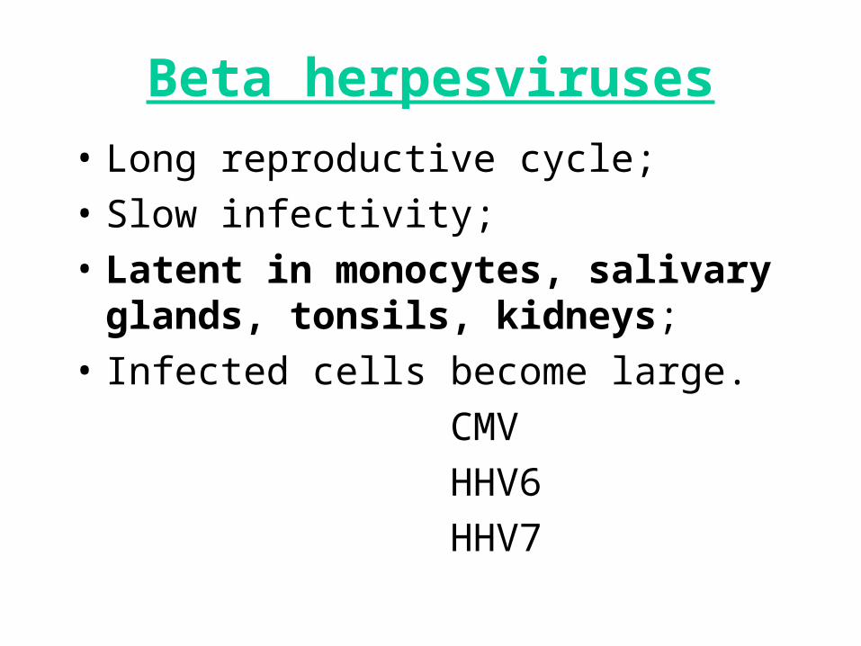
Beta herpesviruses
• Long reproductive cycle;
• Slow infectivity;
• Latent in monocytes, salivary glands, tonsils, kidneys;
• Infected cells become large.
CMV
HHV6
HHV7
Gamma herpesviruses
• Infection specific to T or B lymphocytes;
• Latent in lymphoid tissue, lymphocytes, salivary glands, epithelial cells of mouth and pharynx.
• Proliferation of B-lymphocytes.
EBV
HHV 8
Herpes Simplex Viruses
- Herpetic gingivostomatitis;
- Herpes labialis (cold sore);
- Herpetic keratoconjunctivitis;
- Skin manifestations;
- HS meningitis and encephalitis;
- Genital herpes;
- Neonatal herpes.
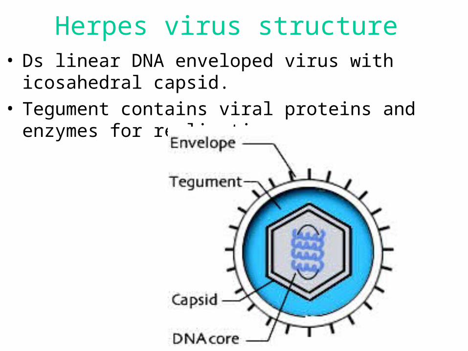
Herpes virus structure • Ds linear DNA enveloped virus with icosahedral
capsid.• Tegument contains viral proteins and enzymes for
replication.
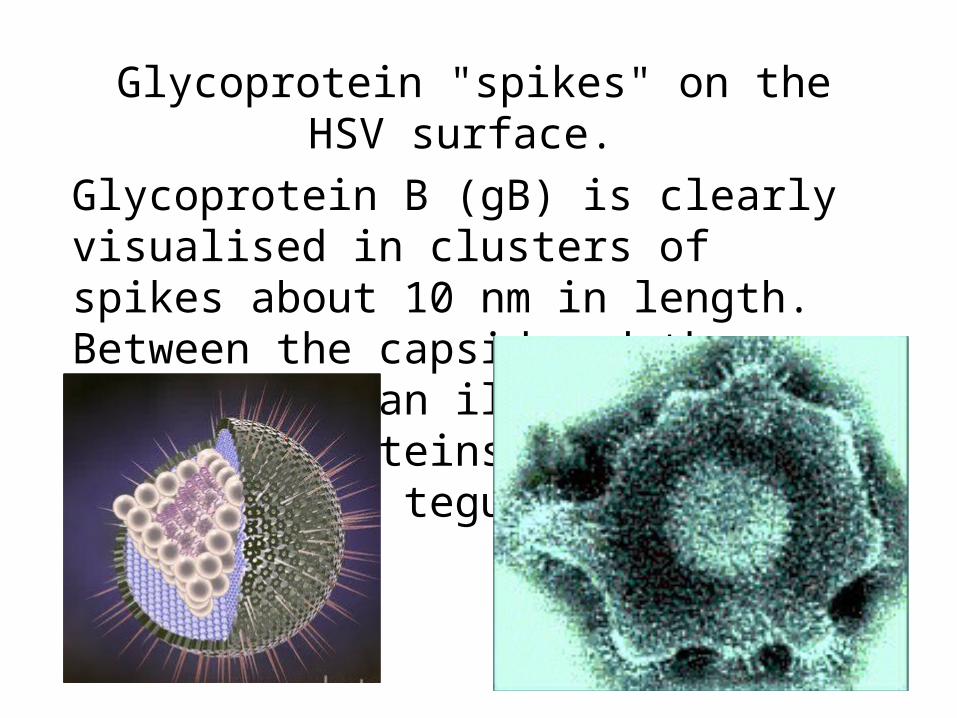
Glycoprotein "spikes" on the HSV surface.
Glycoprotein B (gB) is clearly visualised in clusters of spikes about 10 nm in length. Between the capsid and the envelope is an ill-defined layer of proteins, collectively known as the tegument.
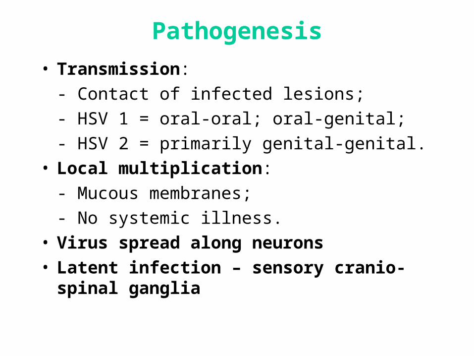
Pathogenesis
• Transmission:
- Contact of infected lesions;
- HSV 1 = oral-oral; oral-genital;
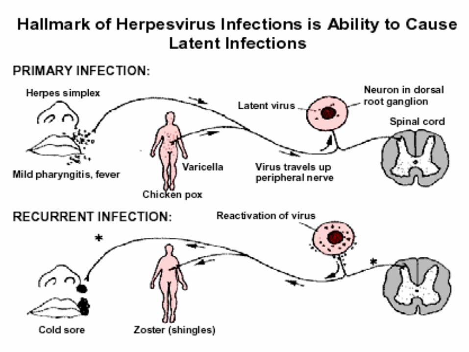
- HSV 2 = primarily genital-genital.• Local multiplication:
- Mucous membranes;
- No systemic illness.• Virus spread along neurons• Latent infection – sensory cranio-spinal ganglia
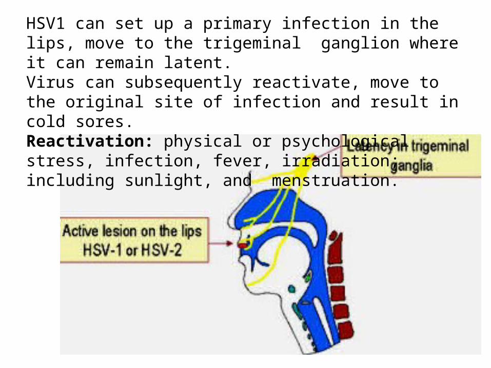
HSV1 can set up a primary infection in the lips, move to the trigeminal ganglion where it can remain latent. Virus can subsequently reactivate, move to the original site of infection and result in cold sores.Reactivation: physical or psychological stress, infection, fever, irradiation; including sunlight, and menstruation.
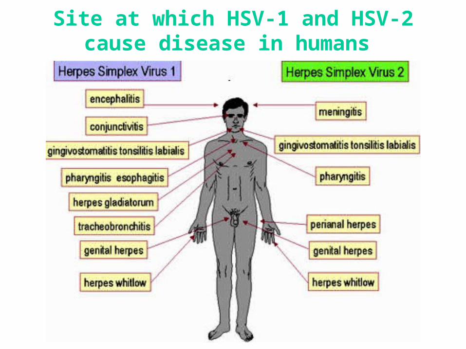
Site at which HSV-1 and HSV-2 cause disease in humans
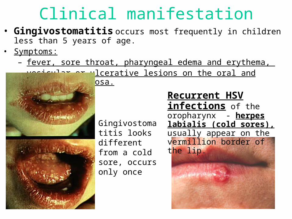
Clinical manifestation• Gingivostomatitis occurs most frequently in children less than 5 years of age. • Symptoms:
– fever, sore throat, pharyngeal edema and erythema, – vesicular or ulcerative lesions on the oral and pharyngeal mucosa.
Gingivostomatitis looks different from a cold sore, occurs only once
Recurrent HSV infections of the oropharynx - herpes labialis (cold sores), usually appear on the vermillion border of the lip.
Genital Herpes• is caused by HSV 2 and HSV 1. • Primary infection usually involves:
– In women: the vulva, vagina, and cervix.– In men: the glans penis, prepuce or penile shaft.
• Clinical manifestation: fever, malaise, anorexia, bilateral inguinal lymphadenopathy, dysuria and urinary retention due to urethral involvement.
• As many as 10 % of individuals develop an aseptic meningitis.
• Recurrence – 60%.
• The complete healing takes several weeks.

Clinical manifestation• Herpetic Keratitis
– It is usually caused by HSV 1 and is accompanied by conjunctivitis.
– The characteristic lesions are dendritic ulcers. – Deep stromal involvement may result in
visual impairment.
• Skin Manifestations – At any skin site. – Lesions on abraded skin of the fingers, known
as herpetic whitlows. – Wrestlers because of physical contact may
develop herpes gladiatorum.
• Herpes Simplex Encephalitis – hemorrhagic necrosis of the inferiomedial
portion of the temporal lobe. – clinical manifestations: headache, fever,
altered consciousness, and abnormalities of speech and behavior.
– mortality rate 70-100%.
Neonatal Herpes Simplex Virus Infection
• It usually results from contact of the fetus with infected maternal genital secretions at the time of delivery.
• Manifestations: • 1) Skin, eye and mouth disease consists of cutaneous
lesions and does not involve other organ systems. • 2) Encephalitis.• 3) Disseminated infection involves multiple organ
systems and can produce disseminated intravascular coagulation, hemorrhagic pneumonitis, encephalitis, and cutaneous lesions.
• The mortality rate:- zero for skin, eye and mouth disease; - 15 % for encephalitis; - 60 % for neonates with disseminated infection.
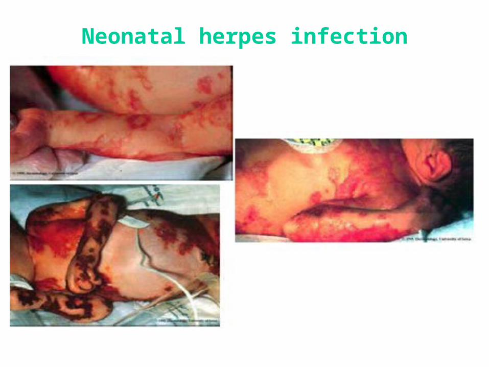
Neonatal herpes infection
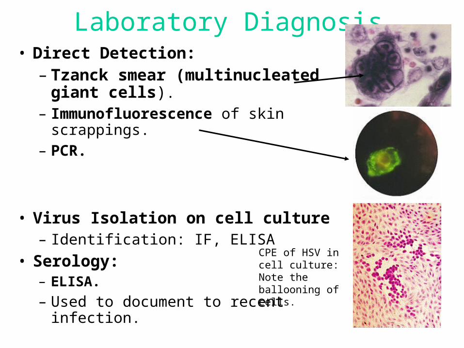
Laboratory Diagnosis• Direct Detection:
– Tzanck smear (multinucleated giant cells).
– Immunofluorescence of skin scrappings.
– PCR.
• Virus Isolation on cell culture – Identification: IF, ELISA
• Serology:– ELISA.
– Used to document to recent infection.
CPE of HSV in cell culture: Note the ballooning of cells.
Varicella-zoster virus
• Differs from HSV:
- Respiratory secretions transmission;
- Systemic disease;
- Rash itches;
- Latent in multiple sensory ganglia.
• Live attenuated vaccine
Major Diseases Associated withHZV
• Varicella or chicken pox
• Zoster or shingles
• Post-infectious encephalitis
• Neonatal infection
Varicella
• Varicella or chickenpox is the manifestation of primary varicella-zoster virus infection.
• The incubation period - from 11 to 23 days.
• The rash begins on the face and trunk and spreads to the extremities. – The average duration of lesion formation is 3 to 5 days
in the normal child; however, it is usually longer in adolescents, adults and in the immunocompromised
• Latency - in the cerebral or posterior root ganglia.
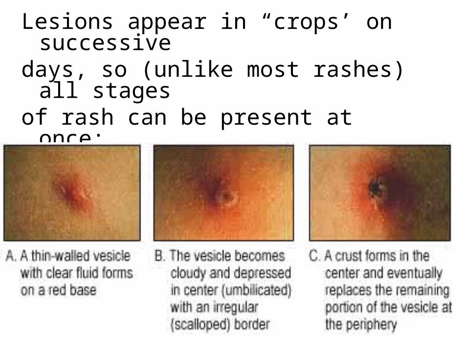
Lesions appear in “crops’ on successivedays, so (unlike most rashes) all stagesof rash can be present at once:
papules, vesicles, scabs
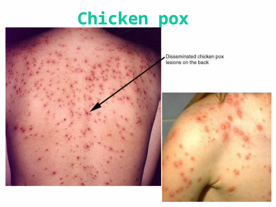
Chicken pox
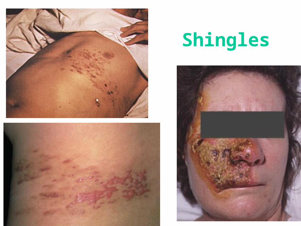
Herpes Zoster or shingles• is the recurrent form of varicella-zoster virus.
• It is a reactivation of latent virus, manifests as a localized vesicular rash with a dermatomal distribution.
• The virus reactivates in the ganglion and tracks down the sensory nerve to the area of the skin innervated by the nerve.
• It is accompanied by intensive pain which may last for months (postherpetic neuralgia)
Shingles
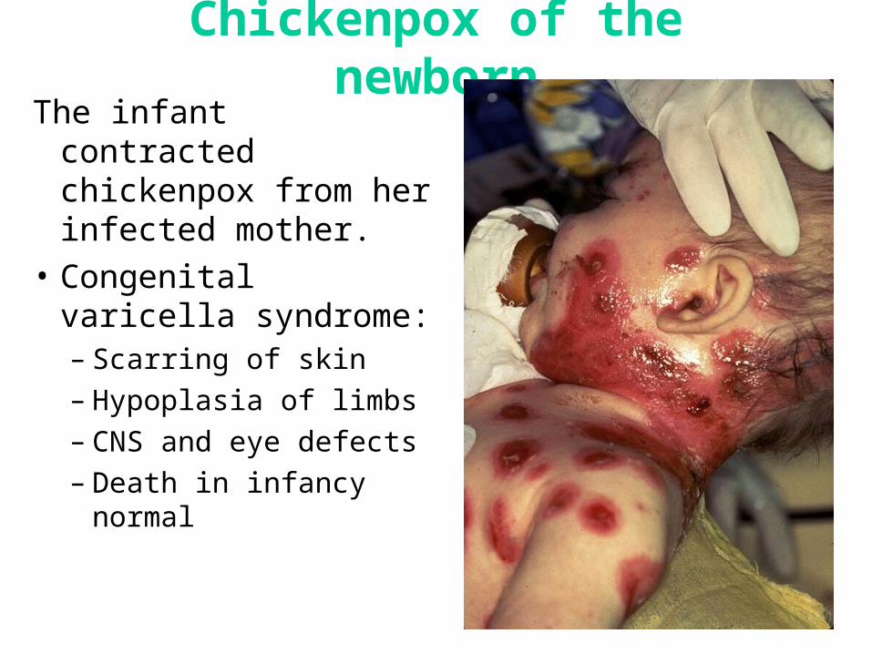
Chickenpox of the newbornThe infant contracted
chickenpox from her infected mother.
• Congenital varicella syndrome:– Scarring of skin– Hypoplasia of limbs– CNS and eye defects– Death in infancy normal
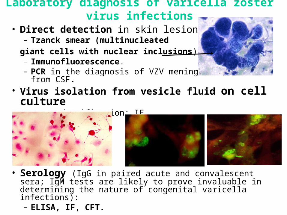
Laboratory diagnosis of varicella zoster virus infections
• Direct detection in skin lesions:– Tzanck smear (multinucleated
giant cells with nuclear inclusions). – Immunofluorescence. – PCR in the diagnosis of VZV meningoencephalitis from CSF.
• Virus isolation from vesicle fluid on cell culture – CPE Identification: IF
• Serology (IgG in paired acute and convalescent sera; IgM tests are likely to prove invaluable in determining the nature of congenital varicella infections):– ELISA, IF, CFT.
Cytomegalovirus
• Asymptomatic shedding:
• Urine
• Oral secretions
• Cervical secretions
• Semen
• Breast milk
• No destruction of infected cells.
• Cytomegaly.
Pathogenesis of cytomegalovirus• Routs of transmission:
- direct contact with infected body fluids: saliva, breast milk, urine;- sexual intercourse;- blood transfusions;
- respiratory;- fecal-oral;- trancplacentally;- organ transplantation.
• Infects epithelial cells and leukocytes.• Spread to all organs. Virus shed in body fluids many years.• Latent in neutrophils, monocytes, salivary glands, tonsils,
kidneys.
Clinical manifestation• Congenital cytomegalovirus infection - 1 % of all live
births. • Children acquire infection through:
– contact with infected maternal genital secretions,
– transplacentally,
– breast milk.
• Severe symptomatic disease:– hepatosplenomegaly,
– myocarditis,
– optic atrophy,
– deafness,
– pneumonitis,
– involvement of the CNS.
Clinical manifestation• Mononucleosis-like syndrome.
– in approximately 10 % of primary infections in older children and adults;
• Manifestations:– fever, – malaise, – atypical lymphocytosis, – pharyngitis, – rarely, cervical adenopathy or hepatitis.
Clinical manifestation
• Cytomegalovirus infection in severely immunocompromised individuals - life-threatening disease from either primary or reactivated infection.
• Infection can involve:– lungs, – gastrointestinal tract, – liver,– retina, – CNS.
• Individuals at high risk for severe disease (CMV pneumonia):– organ transplant recipients, particularly bone marrow transplant
recipients, – individuals with human immunodeficiency virus infection.
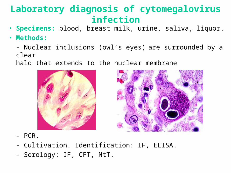
Laboratory diagnosis of cytomegalovirus infection
• Specimens: blood, breast milk, urine, saliva, liquor.
• Methods:
- Nuclear inclusions (owl’s eyes) are surrounded by a clearhalo that extends to the nuclear membrane
- PCR.
- Cultivation. Identification: IF, ELISA.
- Serology: IF, CFT, NtT.
Epstein-Barr virus
• Infectious mononucleosis.
• Associated with human tumors:
– Burkitt’s lymphoma;
– Nasopharyngeal carcinoma;
– B-lymphoma;
– Hodgkin's lymphoma;
– Hair-like oral leukoplakia
Pathogenesis Epstein-Barr virus infection
• Transmission:
– by contact with saliva, in particularly through kissing,– transfusions and organ transplantation can spread
infected leukocytes.
• Virus multiplies in:– epithelial cells of mouth, – local lymph tissue,– T- and B-lymphocytes.
• Carriers shed virus for lifetime.
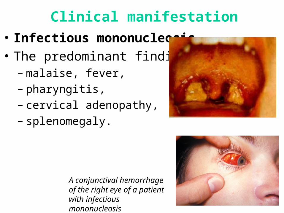
Clinical manifestation
• Infectious mononucleosis.
• The predominant findings: – malaise, fever,– pharyngitis, – cervical adenopathy, – splenomegaly.
A conjunctival hemorrhage of the right eye of a patient with infectious mononucleosis
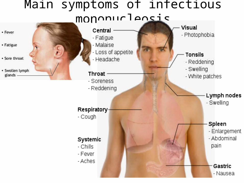
Main symptoms of infectious mononucleosis
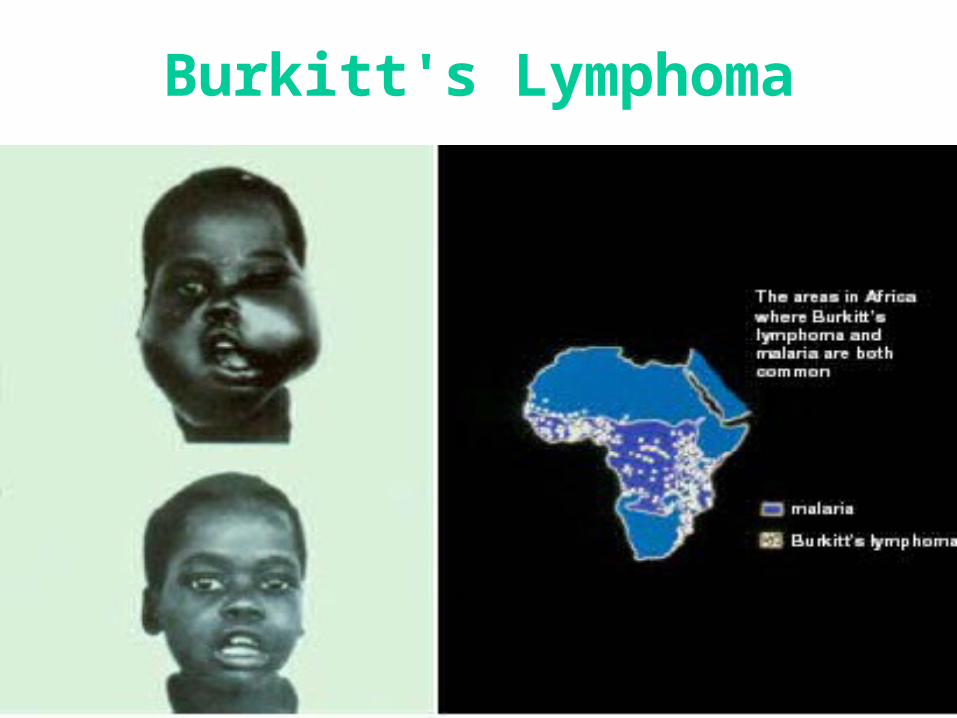
Burkitt's Lymphoma
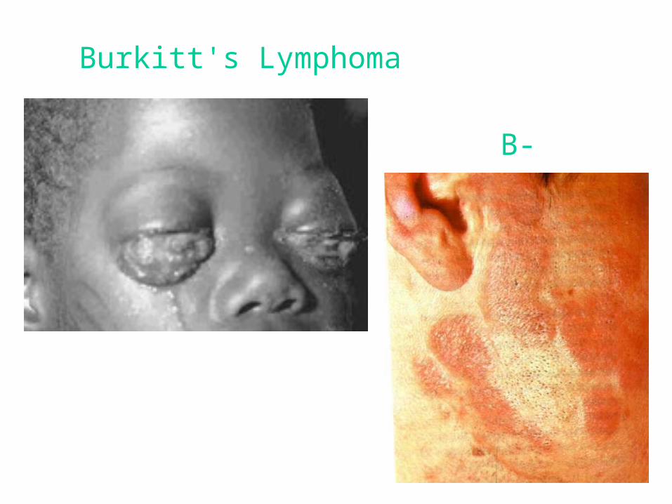
Burkitt's Lymphoma
B-lymphoma
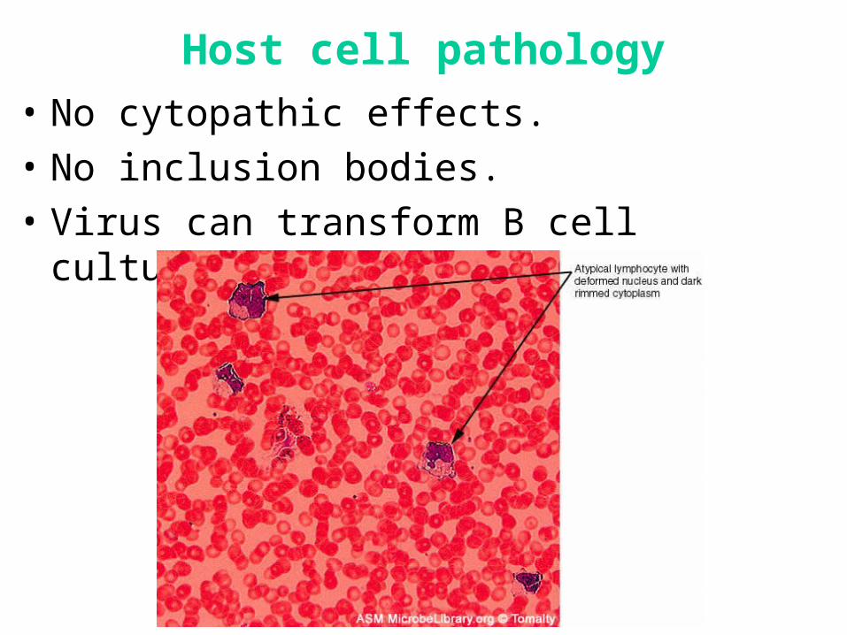
Host cell pathology
• No cytopathic effects.
• No inclusion bodies.
• Virus can transform B cell cultures (proliferation)
Host immune response
• Stimulated (infected) B cells produce:– Polyclone immunoglobulins (heterphile Abs);– Viral membrane antigens.
• Increase in activated T cells:– “Atypical lymphocytes”;– Abnormal lymphocytes in blood (Downy cells).
• Cytotoxic T cells reduce number infected B cells.
• Immunosuppression = lymphomas.
Diagnosis EBV
• Histology: Downy cells (atypical lymphocytes).
• Detection of heterophile antibodies:– Agglutinate sheep RBCs;
• Serology:– Ig M against the Capsid Antigen = early;– Raising of the titer of the anti-EBV Nuclear Antigen.
3-6 weeks post infection
HHV-6 and 7• Transmission: through contact with saliva and breast feeding.• The main target cells are the T-lymphocyte and B-lymphocytes.• HHV-6 and HHV-7 become latent following primary infection and
are reactivated from time to time, especially during periods of immunosuppression.
• HHV-6 infection is firmly associated with:– roseala infantum, – meningitis, encephalitis, – symptoms in transplant recipients such as fever, liver and CNS
manifestations. • HHV-6 and 7 is associated with chronic fatigue syndrome:
– fatigue, – arthralgia, – sweating,– lymphadenopathy.
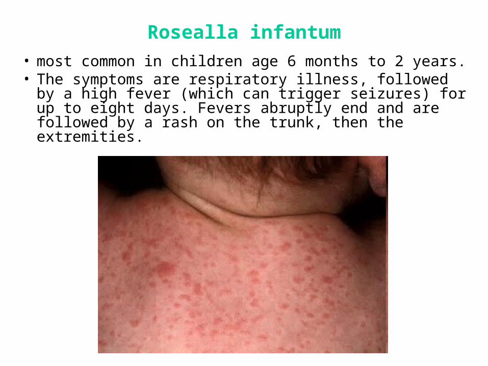
Rosealla infantum
• most common in children age 6 months to 2 years. • The symptoms are respiratory illness, followed by a
high fever (which can trigger seizures) for up to eight days. Fevers abruptly end and are followed by a rash on the trunk, then the extremities.
Human Herpes Virus 8
• Is associated with Kaposi’s sarcoma, malignancies such as Castleman’s disease and primary effusion lymphomas.
• HHV-8 DNA is found in almost 100% of cases of Kaposi’s sarcoma.
• Most patients with KS have antibodies against HHV-8.• The seroprevalence of HHV-8 is low among the general
population but is high in groups of individuals susceptible to KS, such as homosexuals.
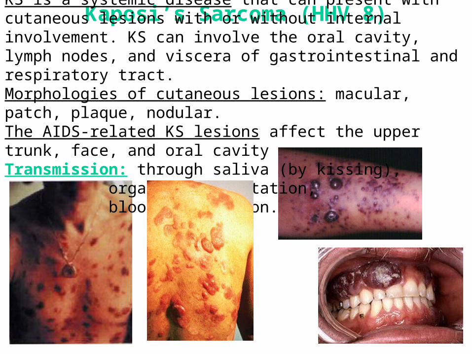
Kaposi’s Sarcoma (HHV 8)KS is a systemic disease that can present with cutaneous lesions with or without internal involvement. KS can involve the oral cavity, lymph nodes, and viscera of gastrointestinal and respiratory tract. Morphologies of cutaneous lesions: macular, patch, plaque, nodular. The AIDS-related KS lesions affect the upper trunk, face, and oral cavityTransmission: through saliva (by kissing),
organ transplantation, blood transfusion.
Treatment of herpesviruses infections
• Acyclovir is the treatment of choice for:– mucocutaneous HSV infections, – herpes simplex encephalitis, – neonatal HSV infections, – varicella-zoster virus infections in the
immunocompromised individuals.
• Valaciclovir, and famciclovir is the treatment for shingles.
• Ganciclovir and foscarnet are used for the treatment of cytomegalovirus infection in immunocompromised individuals.
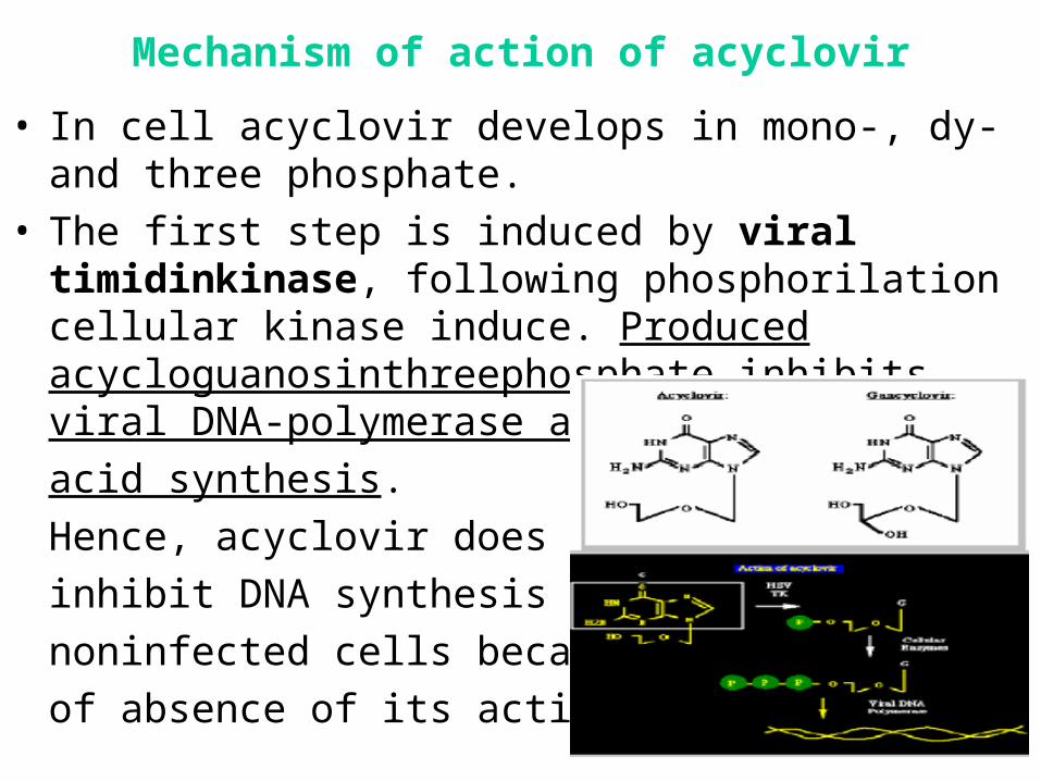
Mechanism of action of acyclovir
• In cell acyclovir develops in mono-, dy- and three phosphate.
• The first step is induced by viral timidinkinase, following phosphorilation cellular kinase induce. Produced acycloguanosinthreephosphate inhibits viral DNA-polymerase and so nucleic
acid synthesis.
Hence, acyclovir does not
inhibit DNA synthesis in
noninfected cells because
of absence of its active form.


![HSV-UL18 DNA vaccine construction and …...Herpes simplex virus (HSV) is a globally spreading DNA virus that can cause oral and genital ulcers [1]. Few cases have shown that HSV infections](https://img.pdfslide.us/doc/110x75/5fc53ff7524eae6ffe5a41d8/hsv-ul18-dna-vaccine-construction-and-herpes-simplex-virus-hsv-is-a-globally.jpg)