Embed Size (px)
Citation preview
Pakistan J. Zool., vol. 48(1), pp. 235-240, 2016. Human Glial Fibrillary Acidic Protein Gene Promoter Targets Hepatic and Pancreatic Stellate Cells in Transgenic Bama Mini-Pigs Xiangxing Zhu,1,2,3 Shouneng Quan,1,2,3 Junyu Nie,1,2,3 Kehuan Lu1,2,3,* and Shengsheng Lu1,2,3,*
1State Key Laboratory for Conservation and Utilization of Subtropical Agro-Bioresources, Nanning 530004, China 2Guangxi High Education Key Laboratory for Animal Reproduction and Biotechnology, Nanning 530004, China 3College of Animal Science and Technology, Guangxi University, Nanning 530004, China A B S T R A C T Hepatic and pancreatic stellate cells (HSCs and PSCs) have been considered to be closely associated with the pathogenesis of fibrosis, offering a hopeful therapeutic target for treating hepatic and pancreatic fibrosis. Recently, we have generated transgenic mini-pigs carrying astrocyte-specific expression of DsRed regulated by the 2.2-kb human glial fibrillary acidic protein (hGFAP) gene promoter. This study was intended to examine whether the targeted transgene expression of DsRed will be present in the porcine HSCs and PSCs. Histological fluorescent imaging showed several DsRed-expressing cells were interspersed between the parenchyma cells. Moreover, these DsRed-expressing cells were checked as indeed HSCs and PSCs by co-labeling with GFAP immunoreactivity. Thus, our results demonstrate the 2.2-kb hGFAP gene promoter holds matched regulatory elements therefore is capable of introducing specific expression of transgene in porcine HSCs and PSCs, providing a novel strategy to selectively manipulate the HSCs and PSCs in a large animal model.
INTRODUCTION
Hepatic and pancreatic stellate cells (HSCs and PSCs) are two types of nonparenchymal cells with long cytoplasmic processes giving them a typical stellate-shaped appearance. HSCs are resident liver cells located in the perisinusoidal space of Disse, between the hepatocytes and sinusoidal endothelial cells of the hepatic lobule (Friedman, 2008; Yin et al., 2013; Bhatia et al., 2014). PSCs are resident exocrine pancreas cells located in the periacinar, periductal and perivascular spaces of the pancreas (Omary et al., 2007; Erkan et al., 2011). In normal physiological conditions, both HSCs and PSCs are quiescent and play critical roles in normal tissue architecture by regulating extracellular matrix production (Omary et al., 2007; Friedman, 2008; Erkan et al., 2011; Yin et al., 2013). Whereas in response to injuries, quiescent HSCs and PSCs will be activated, following synthesize and secrete a large amount of extracellular matrix components to generate a temporary ________________________ * Corresponding authors: [email protected] (S.S. Lu)
or [email protected] (K.H. Lu) 0030-9923/2016/0001-0235 $ 8.00/0 Copyright 2016 Zoological Society of Pakistan
scar at the site of injury to protect the tissue from further damage (Omary et al., 2007; Friedman, 2008; Erkan et al., 2011; Hernandez-Gea and Friedman, 2011; Yin et al., 2013). This process is termed as ‘fibrosis’, which bears a strong resemblance to the ‘reactive astrogliosis’ mainly contributed by the astrocytes located in the central nervous system (Schachtrup et al., 2011; Burda and Sofroniew, 2014). Furthermore, both HSCs and PSCs, quiescent or activated, consistently express glial fibrillary acidic protein (GFAP) (Omary et al., 2007; Friedman, 2008; Erkan et al., 2011; Yin et al., 2013), a reliable marker for the astrocytes (Burda and Sofroniew, 2014). Chronic sustained injuries may result in accumulation of activated stellate cells, leading to a progressive substitution of parenchyma by scar tissue and often generate cirrhosis, which may has a poor outcome and high mortality (Friedman, 2008, 2010; Hernandez-Gea and Friedman, 2011; Bhatia et al., 2014). Thus, due to their pivotal roles in pathogenesis, stellate cells, HSCs and PSCs, may sever as a hopeful therapeutic target for treating fibrosis (Omary et al., 2007; Friedman, 2008, 2010; Erkan et al., 2011; Hernandez-Gea and Friedman, 2011; Yin et al., 2013). Numerous biological functions of HSCs have been explored by experiments using cultured HSCs. However, cultured HSCs always undergo spontaneous activation
Article Information Received 3 April 2015 Revised 1 September 2015 Accepted 19 September 2015 Available online 1 January 2016 Authors’ Contributions: KL and SL conceived and designed the study. XZ, SQ and JN executed the experiments. XZ, SQ, JN, KL and SL analyzed the data. XZ and SL wrote the article. Key words Hepatic stellate cell (HSC), pancreatic stellate cell (PSC), human glial fibrillary acidic protein (GFAP) gene promoter; transgenic pig, disease model.
X. ZHU ET AL. 236
and do not faithfully reproduce the underlying changes in gene expression observed in vivo where they are accompanied by other cell types (De Minicis et al., 2007; Yin et al., 2013). Whereas the field of PSCs biology is very young, their functions usually be analogized from their hepatic counterparts (Omary et al., 2007; Erkan et al., 2011). Fortunately, transgenic animal models generated for manipulating the number and activity of HSCs may offer an appealing strategy to address this issue. A transgenic mouse model expressing Cre/loxP system regulated by a 2.2-kb human GFAP (hGFAP) gene promoter has already been employed for tracing the cell fate of HSCs in vivo (Yang et al., 2008). More recently, by using transgenic mouse models bearing herpes simplex virus thymidine kinase (HSV-TK) under the control of a mouse GFAP (mGFAP) gene promoter element, two independent groups achieved depletion of proliferating HSCs, providing an ingenious strategy to explore the potential functions of HSCs in vivo (Puche et al., 2013; Stewart et al., 2014). For modeling human disease, pigs, especially mini-pigs enjoy numerous advantages (Prather et al., 2013; Liu et al., 2014). Recently, we have generated transgenic Bama mini-pigs (hGFAP-DsRed) carrying astrocyte-specific expression of DsRed regulated by the 2.2-kb hGFAP gene promoter (Zhu et al., 2015), which had already been demonstrated to be capable of specifically driving a transgene expression in cultured rodent HSCs (Maubach et al., 2006) and PSCs (Ding et al., 2009). Applicative evaluation of these transgenic mini-pigs in neuroscience research has not yet been completed, while, in the present study, we first checked whether the targeted transgene expression will be present in the transgenic porcine HSCs and PSCs.
MATERIALS AND METHODS All animal procedures used in this study were carried out in accordance with the Guide for Care and Use of Laboratory Animals (8th edition, prepared by the National Research Council, USA) and were approved by the Institutional Animal Care and Use Committee (IACUC) of Guangxi University. Unless otherwise stated, all organic and inorganic reagents were purchased from Sigma-Aldrich Co. (St. Louis, MO, USA).
Tissue processing One 30-day-old transgenic mini-pig generated in our another study (Zhu et al., 2015) was deeply anesthetized by intramuscular injection of sodium pentobarbital solution (50 mg/kg). After then a midventral sternal thoracotomy was performed and a
cannula was inserted in the aorta through the left ventricle. The right atrium was opened and one liter of normal saline was injected through the cannula by gravity flow, followed by perfusion with two liters of ice-cold 4% paraformaldehyde in 0.1 M PBS solution. After then, the liver and pancreas were quickly picked out, followed by cut into 3-mm-thick coronal blocks and immediately fixed overnight at 4°C with paraformaldehyde-PBS solution. Fixed tissues were used for histological sectioning. One age-matched wild-type pig was used as a negative control.
Histological sectioning and fluorescence detection Histological sectioning and staining were performed as described by Al-Doaiss et al. (2013), fixed tissues were routinely dehydrated, paraffin-embedded, sectioned (5 µm) and stained with hematoxylin and eosin (HE). Unstained sections were employed to detect the presence of DsRed using a Nikon 50i fluorescence microscope (Nikon, Tokyo, Japan). Fluorescent micrographs were acquired using a NIS Elements image system (Nikon) and processed with Adobe Photoshop CS5 software (Adobe, San Jose, CA, USA).
Double-labeling immunofluorescence assay The astrocyte-specific expression of DsRed was examined using a double-labeling immunofluorescence assay, which was performed as described by Mederacke et al. (2013). Briefly, fixed tissues were embedded in Tissue Freezing Medium (Leica, Nussolch, Germany), followed by cryo-sectioned into 5-µm-thick sections using a Leica CM1950 freezing microtome. Sections were pasted onto poly-lysine-coated glasses and washed three times in ice-cold PBS for 10 minutes each to remove sucrose, followed by submerged in blocking/permeablizing buffer solution (5% normal donkey serum and 0.5% Triton-X100 in PBS) at 4°C for 1 h to prevent non-specific binding of IgG. After this step, the sections were incubated overnight at 4°C with primary antibodies of goat anti-GFAP polyclonal IgG (diluted at 1:100 in 20% blocking/permeablizing buffer solution; Santa Cruz sc-6171) and mouse anti-DsRed monoclonal IgG (1:100; Santa Cruz sc-390909). After washed three times in ice-cold PBS for 10 minutes each, sections were incubated overnight at 4°C with secondary antibodies of donkey anti-mouse IgG-R (1:200; Santa Cruz sc-2300) and donkey anti-goat IgG-FITC (1:200; Santa Cruz sc-2024). After extensively washed and air-dried at room temperature, the sections were mounted with UltraCruz™ Mounting Medium (Santa Cruz) followed by analyzed using a Nikon A1 confocal laser scanning microscope system (Nikon). Fluorescent images
HGFAP GENE PROMOTER TARGETS STELLATE CELLS IN TRANSGENIC 237
were captured and processed with Adobe Photoshop CS5 software.
RESULTS AND DISCUSSION
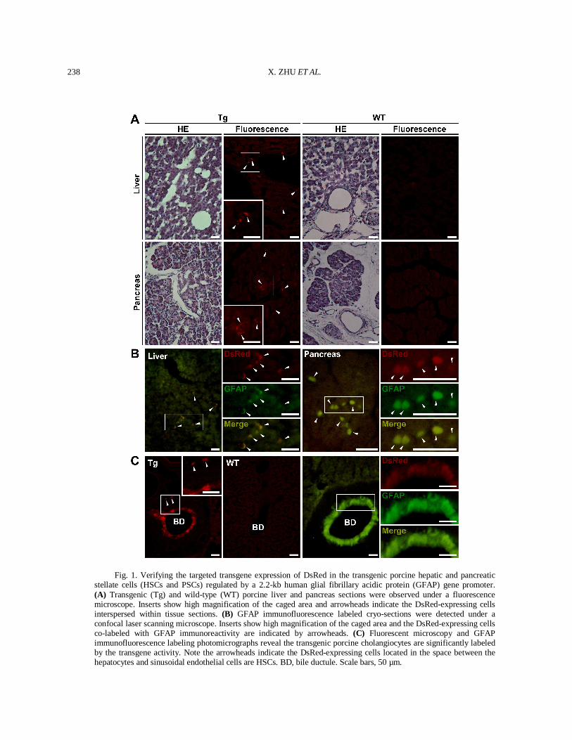
Histological fluorescent microscopy and GFAP immunofluorescence labeling assay were employed for verifying the targeted transgene expression of DsRed in the hGFAP-DsRed transgenic porcine liver and pancreas. As is shown in Figure 1A, a number of interspersed DsRed-expressing cells were detected in the space between the hepatocytes and sinusoidal endothelial cells. Similarly, several DsRed-expressing cells were also present in the interstitium between pancreatic acini. On the contrary, DsRed-expressing cells were absent in the tissues from a wild-type mini-pig as a negative control. For verifying these DsRed-expressing cells were indeed stellate cells, a dual immuno-fluorescence labeling assay was performed. As is shown in Figure 1B, all of the DsRed-expressing cells located in the transgenic porcine liver and pancreas were labeled with GFAP immunoreactivity, indicating they were exact HSCs and PSCs. Besides, as expected, we also noticed the transgenic porcine cholangiocytes were significantly labeled with transgene activity (Fig. 1C). In rodents and human, HSCs and PSCs are defined as main vitamin A-storing cells and constitute several percents of all cells in the normal liver and pancreas, respectively (Omary et al., 2007; Erkan et al., 2011; Yin et al., 2013). The present study has been checked their porcine counterparts. In addition, the HSCs and PSCs-specific expression of DsRed could permit the strategy that utilizes the hGFAP gene promoter for specifically introducing targeted transgene expression in porcine HSCs and PSCs. Such strategy has been exploited by two independent studies (Puche et al., 2013; Stewart et al., 2014), in which mouse HSCs-depletion was generated by using a mGFAP gene promoter, providing an in vivo tool for investigating the potential roles of HSCs contributed to liver fibrosis. Fate-tracing could be used for identifying the all progeny of interest cells marked with certain specific expression of a transgene. Thus utilize hGFAP-Cre transgenic mice plus a reporter strain with conditional expression of fluorescent proteins, the fate of HSCs during development, differentiation and pathogenesis could be traced. By using this strategy, Yang et al. (2008) confirmed the hGFAP gene promoter was capable of targeting mouse HSCs and further verified the HSCs were indeed one type of epithelial progenitors in adult mouse livers. However, in a recent study, Mederacke et al. (2013) pointed out that the cholangiocytes, but not
HSCs, could be efficiently marked by both of the hGFAP and mGFAP gene promoter. The transgene activity introduced into cholangiocytes has been reported by Yang et al. (2008) and also be reproduced in our transgenic mini-pig situation, while the finding that neither hGFAP nor mGFAP gene promoter could be efficiently mark the rodent HSCs was diverged far from our present study and those previous studies in rodents performed in vitro (Maubach et al., 2006; Ding et al., 2009) and in vivo (Yang et al., 2008; Puche et al., 2013; Stewart et al., 2014), and also be contradicted with the evidence that the GFAP was really present in rhesus monkey HSCs (Jin et al., 2011). Coincidentally, controversies also exist in PSCs. Some researchers even alleged the GFAP expression is absent in quiescent PSCs, whereas in the view of experienced investigators in this field, this is more likely to be resulted from the differences in the quality of the antibody used for immunostaining of rodent GFAP (Erkan et al., 2011). Possibly, this arbitration could also be appropriate for the condition in HSCs. Pigs, especially mini-pigs enjoy numerous advantages for modeling human diseases. Advances in porcine genetic modification confer efficient creation of transgenic large animal models for investigating human diseases (Garrels et al., 2012; Prather et al., 2013; Liu et al., 2014). To date, a vast amount of genetically modified pig models have already provided deeper insights into the pathogenesis and therapies of human hepatic, pancreatic and gastrointestinal diseases, for instance the hereditary tyrosinemia type I disease (Hickey et al., 2011), cystic fibrosis (Rogers et al., 2008a,b; Stoltz et al., 2013), diabetes (Renner et al., 2010; 2013) as well as familial adenomatous polyposis (Flisikowska et al., 2012). In addition, genetically modified pigs also hold enormous application potentials in the studies on pancreas regeneration (Matsunari et al., 2013, 2014). Therefore, it’s expectable that genetically modified mini-pigs may serve as a novel preclinical large animal model for investigating the biology and pathology of HSCs and PSCs. Regardless of the debates in rodent models, in the present study, the fact that the 2.2-kb hGFAP gene promoter holds matched regulatory elements therefore be capable of targeting porcine HSCs and PSCs has already been testified evidently. Thus, our findings first demonstrate that it’s feasible to specifically manipulate HSCs and PSCs in pigs by using the hGFAP gene promoter, providing a practical means to create preclinical large animal models for answering pathogenic mechanisms and developing new therapeutic approaches to the hepatic and pancreatic fibrosis.
X. ZHU ET AL. 238
Fig. 1. Verifying the targeted transgene expression of DsRed in the transgenic porcine hepatic and pancreatic stellate cells (HSCs and PSCs) regulated by a 2.2-kb human glial fibrillary acidic protein (GFAP) gene promoter. (A) Transgenic (Tg) and wild-type (WT) porcine liver and pancreas sections were observed under a fluorescence microscope. Inserts show high magnification of the caged area and arrowheads indicate the DsRed-expressing cells interspersed within tissue sections. (B) GFAP immunofluorescence labeled cryo-sections were detected under a confocal laser scanning microscope. Inserts show high magnification of the caged area and the DsRed-expressing cells co-labeled with GFAP immunoreactivity are indicated by arrowheads. (C) Fluorescent microscopy and GFAP immunofluorescence labeling photomicrographs reveal the transgenic porcine cholangiocytes are significantly labeled by the transgene activity. Note the arrowheads indicate the DsRed-expressing cells located in the space between the hepatocytes and sinusoidal endothelial cells are HSCs. BD, bile ductule. Scale bars, 50 µm.
HGFAP GENE PROMOTER TARGETS STELLATE CELLS IN TRANSGENIC 239
ACKNOWLEDGEMENTS This study was jointly supported by the National Natural Science Foundation of China (No. 31260553) and the Graduate Programs for Innovational Research founded by the Guangxi Provincial Department of Education (No. YCSZ2013003). The authors declare no conflict of interest.
REFERENCES
Al-Doaiss, A.A., Alarif, S.A. and Jarrar, B.M., I.J., 2013. Hepatic histological and histochemical alterations induced by rosuvastatin therapeutic doses. Pakistan J. Zool., 45: 149-157.
Bhatia, S.N., Underhill, G.H., Zaret, K.S. and Fox, I.J., 2014. Cell and tissue engineering for liver disease. Sci. Transl. Med., 6:245sr2.
Burda, J.E. and Sofroniew, M.V., 2014. Reactive gliosis and the multicellular response to CNS damage and disease. Neuron, 81:229-248.
De Minicis, S., Seki, E., Uchinami, H., Kluwe, J., Zhang, Y.H., Brenner, D.A. and Schwabe, R.F., 2007. Gene expression profiles during hepatic stellate cell activation in culture and in vivo. Gastroenterology, 132:1937-1946.
Ding, Z., Maubach, G., Masamune, A. and Zhuo, L., 2009. Glial fibrillary acidic protein promoter targets pancreatic stellate cells. Digest. Liver Dis., 41:229-236.
Erkan, M., Adler, G., Apte, M.V., Bachem, M.G., Buchholz, M., Detlefsen, S., Esposito, I., Friess, H., Gress, T.M., Habisch, H.J., Hwang, R.F., Jaster, R., Kleeff, J., Klöppel, G., Kordes, C., Logsdon, C.D., Masamune, A., Michalski, C.W., OH, J., Phillips, P.A., Pinzani, M., Reiser-Erkan, C., Tsukamoto, H. and Wilson, J., 2011. StellaTUM: current consensus and discussion on pancreatic stellate cell research. Gut, 61:172-178.
Flisikowska, T., Merkl, C., Landmann, M., Eser, S., Rezaei, N., Cui, X.X., Kurome, M., Zakhartchenko, V., Kessler, B., Wieland, H., Rottmann, O., Schmid, R.M., Schneider, G., Kind, A., Wolf, E., Saur, D. and Schnieke, A., 2012. A porcine model of familial adenomatous polyposis. Gastroenterology, 143:1173-1175.
Friedman, S.L., 2008. Hepatic stellate cells: protean, multifunctional, and enigmatic cells of the liver. Physiol. Rev., 88:125-172.
Friedman, S.L., 2010. Evolving challenges in hepatic fibrosis. Nat. Rev. Gastro. Hepat., 7:425-436.
Garrels, W., Ivics, Z. and Kues, W.A., 2012. Precision genetic engineering in large mammals. Trends Biotechnol., 30:386-393.
Hernandez-Gea, V. and Friedman, S.L., 2011. Pathogenesis of liver fibrosis. Annu. Rev. Pathol. Mech., 6:425-456.
Hickey, R.D., Lillegard, J.B., Fisher, J.E., McKenzie, T.J., Hofherr, S.E., Finegold, M.J., Nyberg, S.L. and Grompe,
M., 2011. Efficient production of Fah-Null heterozygote pigs by chimeric adeno-associated virus-mediated gene knockout and somatic cell nuclear transfer. Hepatology, 54:1351-1359.
Jin, L.F., Guo, X.Y., Ji, S.H., Ji, W.Z. and Sun, A.J., 2011. Demonstration of expression of hepatic stellate cell markers in cells from liver epithelial progenitor cells of rhesus monkey. Liver Int., 31:744-746.
Liu, H.B., Lv, P.R., Zhu, X.X., Wang, X.W., Yang, X.G., Zuo, E.W., Lu, Y.Q., Lu, S.S. and Lu, K.H., 2014. In vitro development of porcine transgenic nuclear-transferred embryos derived from newborn Guangxi Bama mini-pig kidney fibroblasts. In vitro Cell, Dev. Anim., 50:811-821.
Matsunari, H., Kobayashi, T., Watanabe, M., Umeyama, K., Nakano, K., Kanai, T., Matsuda, T., Nagaya, M., Hara, M., Nakauchi, H. and Nagashima, H., 2014. J. Transgenic pigs with pancreas-specific expression of green fluorescent protein. Reprod. Develop., 60:230-237.
Matsunari, H., Nagashima, H., Watanabe, M., Umeyama, K., Nakano, K., Nagaya, M., Kobayashi, T., Yamaguchi, T., Sumazaki, R., Herzenberg, L.A. and Nakauchi, H., 2013. Blastocyst complementation generates exogenic pancreas in vivo in apancreatic cloned pigs. Proc. natl. Acad. Sci. USA, 110:4557-4562.
Maubach, G., Lim, M.C.C., Zhang, C.Y. and Zhuo, L., 2006. GFAP promoter directs lacZ expression specifically in a rat hepatic stellate cell line. World J. Gastroenterol., 12:723-730.
Mederacke, I., Hsu, C.C., Troeger, J.S., Huebener, P., Mu, X.R., Dapito, D.H., Pradere, J.P. and Schwabe, R.F., 2013. Fate-tracing reveals hepatic stellate cells as dominant contributors to liver fibrosis independent of its etiology. Nat. Commun., 4:2823.
Omary, M.B., Lugea, A., Lowe, A.W. and Pandol, S.J., 2007. The pancreatic stellate cell: a star on the rise in pancreatic diseases. J. clin. Invest., 117:50-59.
Puche, J.E., Lee, Y.M.A., Jiao, J.J., Aloman, C., Fiel, M.I., Munoz, U., Kraus, T., Lee, T.F., Yee, H.F. Jr. and Friedman, S.L., 2013. A novel murine model to deplete hepatic stellate cells uncovers their role in amplifying liver damage in mice. Hepatology, 57:340-350.
Prather, R.S., Lorson, M., Ross, J.W., Whyte, J.J. and Walters, E., 2013. Genetically engineered pig models for human diseases. Annu. Rev. Anim. Biosci., 1:203-219.
Renner, S., Fehlings, C., Herbach, N., Hofmann, A., von Waldthausen, D.C., Kessler, B., Ulrichs, K., Chodnevskaja, I., Moskalenko, V., Amselgruber, W., Göke, B., Pfeifer, A., Wanke, R. and Wolf, E., 2010. Glucose intolerance and reduced proliferation of pancreatic β-cells in transgenic pigs with impaired glucose-dependent insulinotropic polypeptide function. Diabetes, 59:1228-1238.
Renner, S., Braun-Reichhart, C., Blutke, A., Herbach, N., Emrich, D., Streckel, E., Wünsch, A., Kessler, B., Kurome, M., Bähr, A., Klymiuk, N., Krebs, S., Puk, O.,
X. ZHU ET AL. 240
Nagashima, H., Graw, J., Blum, H., Wanke, R. and Wolf, E., 2013. Permanent neonatal diabetes in INSC94Y transgenic pigs. Diabetes, 62:1505-1511.
Rogers, C.S., Hao, Y.H., Rokhlina, T., Samuel, M., Stoltz, D.A., Li, Y.H., Petroff, E., Vermeer, D.W., Kabel, A.C., Yan, Z.Y., Spate, L., Wax, D., Murphy, C.N., Rieke, A., Whitworth, K., Linville, M.L., Korte, S.W., Engelhardt, J.F., Welsh, M.J. and Prather, R.S., 2008a. Production of CFTR-null and CFTR-∆F508 heterozygous pigs by adeno-associated virus-mediated gene targeting and somatic cell nuclear transfer. J. clin. Invest., 118:1571-1577.
Rogers, C.S., Stoltz, D.A., Meyerholz, D.K., Ostedgaard, L.S., Rokhlina, T., Taft, P.J., Rogan, M.P., Pezzulo, A.A., Karp, P.H., Itani, M.A., Kabel, A.C., Wohlford-Lenane, C.L., Davis, G.J., Hanfland, R.A., Smith, T.L., Samuel, M., Wax, D., Murphy, C.N., Rieke, A., Whitworth, K., Uc, A., Starner, T.D., Brogden, K.A., Shilyansky, J., McCcray, P.B. Jr., Zabner, J., Prather, R.S. and Welsh, M.J., 2008b. Disruption of the CFTR gene produces a model of cystic fibrosis in newborn pigs. Science, 321:1837-1841.
Schachtrup, C., Le Moan, N., Passino, M.A. and Akassoglou, K., 2011. Hepatic stellate cells and astrocytes, stars of scar formation and tissue repair. Cell Cycle, 10:1764-1771.
Stewart, R.K., Dangi, A., Huang, C., Murase, N., Kimura, S., Stolz, D.B., Wilson, G.C., Lentsch, A.B. and Gandhi, C.R., 2014. A novel mouse model of depletion of stellate cells
clarifies their role in ischemia/reperfusion- and endotoxin-induced acute liver injury. J. Hepatol., 60:298-305.
Stoltz, D.A., Rokhlina, T., Ernst, S.E., Pezzulo, A.A., Ostedgaard, L.S., Karp, P.H., Samuel, M.S., Reznikov, L.R., Rector, M.V., Gansemer, N.D., Bouzek, D.C., Abou Alaiwa, M.H., Hoegger, M.J., Ludwig, P.S., Taft, P.J., Wallen, T.J., Wohlford-Lenane, C., McMenimen, J.D., Chen, J.H., Bogan, K.L., Adam, R.J., Hornick, E.E., Nelson, G.A. IV., Hoffman, E.A., Chang, E.H., Zabner, J., McCray, P.B. Jr., Prather, R.S., Meyerholz, D.K. and Welsh, M.J., 2013. Intestinal CFTR expression alleviates meconium ileus in cystic fibrosis pigs. J. clin. Invest., 123:2685-2693.
Yang, L., Jung, Y.M., Omenetti, A., Witek, R.P., Choi, S., Vandongen, H.M., Huang, J.W., Alpini, G.D. and Diehl, A.M., 2008. Fate-mapping evidence that hepatic stellate cells are epithelial progenitors in adult mouse livers. Stem Cells, 26:2104-2113.
Yin, C.Y., Evason, K.J., Asahina, K. and Stainier, D.Y.R., 2013. Hepatic stellate cells in liver development, regeneration, and cancer. J. clin. Invest., 123:1902-1910.
Zhu, X.X., Quan, S.N., Zeng, Y.L., Nie, J.Y., Xu, H.Y., Yang, X.G., Lu, Y.Q., Lu, K.H. and Lu, S.S., 2015. Increasing the number of transferred embryos results in delivery of viable transgenic cloned Bama mini-pigs Rom. Biotechnol. Lett., (In press)