Embed Size (px)
Citation preview
Clin. exp. Immunol. (1975) 22, 210-222.
HUMAN FAECAL IMMUNOGLOBULINS INHEALTHY INFANTS AND CHILDREN, AND IN SOME
WITH DISEASES AFFECTING THE INTESTINALTRACT OR THE IMMUNE SYSTEM
B. HANEBERG AND D. AARSKOGDepartment of Pediatrics and Broegelmann Research Laboratory for Microbiology,
University of Bergen, Norway
(Received 14 April 1975)
SUMMARY
IgA, IgG and 1gM in faeces were quantified by single radial immunodiffusion usingextracts of freeze-dried faeces. IgA in small specimens of faeces seemed to mirror thetotal amount ofIgA secreted into the gut at the time of sampling. Presumptive normalvalues for faecal IgA concentrations in infants and children were established.Agglutinins to rabbit erythrocytes served as markers for the antibody activity.Infants and children just recovered from enteritis of probably infectious origin hadhigher concentrations of both IgA and agglutinins in faeces. Faeces from three outof five patients with ulcerative colitis in remission, contained IgG in markedly in-creased concentrations. Two patients with IgA deficiency had no detectable IgA infaeces, but had increased levels of faecal IgM which also agglutinated rabbiterythrocytes. Immunoglobulins were not demonstrated in faeces from three patientswith agammaglobulinaemia. The findings indicate that faeces can be used for assayof immunoglobulins of the intestinal tract.
INTRODUCTION
Immunoglobulins are normally found in extracts of freeze-dried faeces from healthy infantsand children (Haneberg, 1974b; Haneberg & T0nder, 1973). IgA is the dominant class,and agglutinins to rabbit erythrocytes can be used as markers for immunological activity(Haneberg, 1974a). The methods developed using faecal material seemed practical for theinvestigation of intestinal immunoglobulins which have been the subject of much interest,as can be seen from the review by Shearman, Parkin & McClelland (1972).'
In this paper we describe the results of experiments designed to clarify the limitationsand the usefulness of faeces as a material for the investigation of intestinal immunoglobulins.Normal levels of immunoglobulins in faeces from infants and children of various age groupshave been established. They were compared with the findings in faeces from patients justrecovered from enteritis of probably infectious origin, or with verified septicemia and/or
Correspondence: Dr Bj0rn Haneberg, Barneklinikken, Haukeland sykehus, N-5016 Bergen, Norway.
210
Faecal Ig in health and disease 211clinical signs of osteomyelitis. Children with ulcerative colitis in remission, as well as infantsand children with some immunoglobulin deficiency states, were also investigated.
MATERIALS AND METHODS
Individuals. The group of healthy infants and children comprised 161 individuals, aged from 1 day to 12years, randomly selected at well-baby clinics and among children of hospital employees. Some were alsopatients at the Children's Hospital, Bergen, admitted for undescended testicles, nocturnal enuresis, orcongenital heart disease without signs of failure. None of them had signs of infection or gastrointestinaldisease. The infants did not receive human milk for 2 weeks or more prior to the sampling of faeces, andwere fed solids from the age of 2-3 months.
Seventeen individuals (5 months-10 years of age) were studied 3-7 days after a bout of diarrhoea lastingfor 2-3 days, sometimes accompanied by vomiting. Since the enteritis affected previously healthy individualsin minor epidemics, an infectious aetiology was assumed. At the time faeces were collected, they had allrecovered and had regular, formed stools.
Fifteen infants and children (6 months to 9 years of age) with septic infections, were studied. In seven ofthese, repeated blood cultures were positive for either Staphylococcus aureus, fJ-haemolytic Streptococcus,Hemophilus influenzae or Pneumococcus. In the remaining cases the diagnosis of osteomyelitis was based onradiological and clinical signs. None of the patients had signs of gastrointestinal dysfunction. Samples offaeces were collected 1-3 weeks after initiation of fever.
Five patients with ulcerative colitis were studied when in clinical remission, i.e. when their stools wereregular and formed, and without blood. The diagnosis was based on history of bloody diarrhoea and recto-scopic as well as X-ray findings.One patient with low (> 2 s.d. for age) but easily measurable serum IgA concentration and two others with
IgA deficiency (serum levels <4 mg/100 ml) were also included in the study. One of them was detectedamong the patients with osteomyelitis and another was found among the children with ulcerative colitis.The third was mentally retarded and had neurological and clinical signs of ataxia telangiectasia. In additionthree other individuals with agammaglobulinaemia, probably sex-linked, were included. One of these(H.0.) had frequent loose stools during infancy, all had seemingly normal bowel function at the time ofstudy.
Extracts offaeces. Small batches (1-2 g) of faeces were collected in plastic containers and frozen within2 hr after defaecation. The samples obtained at 1-3 days of age consisted of meconium. As outlined before(Haneberg & Tonder, 1973), extracts were prepared from the faeces after freeze-drying, using 10 ml ofphosphate-buffered saline (PBS), pH 7-2, per gram of dry matter. From fourteen individuals the totalamount of faeces was also collected over 3 days. All the faecal samples studied were negative for blood by thebenzidine test (Haneberg & Tonder, 1973).
Sera. From fifty-two of the healthy individuals and from all patients serum was obtained at the time offaecal sampling. Pooled serum from ten healthy blood donors served as normal human serum (NHS), theIgA concentration being 198 mg/100 ml. Standard human serum (Batch number 173, Behringwerke AG,Marburg-Lahn, Germany) which, according to the manufacturer's manual, contained 230 mg of IgA/100 mland were related to aWHO reference sample, were used as control for immunoglobulin measurements.Normal rabbit serum and rabbit antisera to whole human serum, human faecal extracts, human salivary
IgA, serum IgG and IgM were obtained as outlined before (Haneberg & T0nder, 1973). Commercial rabbitantisera to human serum IgA, IgG (Fab), albumin, a1-antitrypsin, a2-macroglobulin and to free secretorycomponent (Behringwerke AG) were also used.
All sera were stored at -20'C and those used for agglutination techniques were heated to 560C for 30min to destroy complement activity.
Agglutination of rabbit erythrocytes. Titres of agglutinins to rabbit erythrocytes were determined asdescribed previously (Haneberg, 1974a) with faecal extracts, fractions of these and sera. Inhibition ofagglutination and antiglobulin test were carried out with antisera to IgA, IgM and IgG, also as described.
Gel filtration. Extracts of faeces, either dialysed or concentrated by negative pressure dialysis againstTris-NaCl buffer (0 05 M Tris-NaCI+0*14 M NaCl), pH8, were separated through a Sephadex G-200column measuring 1-5 x 45 cm, as in a former study (Haneberg & Tonder, 1973).
Purification offaecal IgA. Faecal IgA was isolated as outlined for milk IgA (Brandtzaeg, Fjellanger &
212 B. Haneberg and D. AarskogGjeruldsen, 1970). Extracts of faeces were filtered through Sephadex G-200 column, and the fractionscontaining IgA were pooled and dialysed against 0 01 M phosphate buffer, pH 7 5, before being applied to aDEAE-cellulose column measuring 1 2 x 27 cm. Elution was stepwise, the same buffer being used with0-04, 01,, -2, 0 3, 04 and 1 M NaCl. The flow rate was kept relatively constant at 11 ml/cm2/hr, and fractionsabout 1 ml each were collected during continuous registration of u.v.-light transmittance at 280 nm (UvicordAbsorptiometer, LKB-Produkter AB, Stockholm, Sweden).
Other methods. Single radial immunodiffusion, double diffusion and immunoelectrophoresis in agar werecarried out as described previously (Haneberg & T0nder, 1973).
Protein concentrations in fractions of faecal extracts were determined according to the method of Folin-Ciocalteu (Kabat & Mayer, 1964), using Beckman DU-2 spectrophotometer (Beckman Instruments In-corporated, Palo Alto, U.S.A.). As standards were used dilutions in PBS of Gammaglobulin 16-5% (KabiAB. Stockholm, Sweden).The pH of faeces was measured as described earlier (Haneberg, 1974b) with Lyphan indicator paper
(Dr Gerhard Kloz, West Berlin, Germany).Conventional statistical methods were applied, including Student's t-test.
RESULTSQuantitative determination of IgA
Since diffusion of IgA in agar is dependent on its molecular size, it was necessary topurify IgA from faecal extracts to obtain a correction factor for faecal IgA values in singleradial immunodiffusion based on serum IgA as standard.By gel filtration and ion exchange chromatography, IgA was isolated from: pooled
extracts of faeces from three healthy children; an extract of faeces from one child justrecovered from 'infectious' enteritis; and an extract of faeces from one child with ulcerativecolitis. Reasonably pure preparations were obtained with eluting buffer containing 0-2 MNaCl; only IgA could be demonstrated in the unconcentrated fractions. However, after100-fold concentration, traces of a1-antitrypsin were demonstrated while IgG and IgMamounted to less than 0-03 and 1 3% of total protein concentrations, respectively. Noother serum or faecal components were demonstrated using antisera to whole humanserum, faecal extract and to IgG (Fab) and a2-macroglobulin.
Results of gel filtration of the IgA preparation from the healthy individuals gave evidencethat IgA in the purified preparation had roughly the same distribution of molecular sizes asthe IgA of the initial material. In this experiment IgA was demonstrated in all the fractionsranging from those representing the molecular size of IgM to those of IgG while the highestconcentrations were found in the fractions between these.A linear relationship was evident between total protein concentrations in dilutions of
these IgA-preparations and the corresponding concentrations of IgA, measured as thepercentage of NHS in single radial immunodiffusion. Therefore, correction factors wereestablished for concentrations of IgA in extracts of faeces from different individuals (Table1). The results were dependent on the antiserum used; with our antiserum to secretory IgAnot reacting with free secretory component the values were 63-52,% higher than with anantiserum to serum IgA (Behringwerke). When compared to concentrations (w/v) of IgAin NHS, the correction factors were 5 3 and 3 3, respectively. In the following, the cor-rections of IgA concentrations were thus based on values obtained with the same type andthe same batch of antiserum throughout the study, i.e. our antiserum to secretory IgA.
Small-molecular impurities in the IgA preparations were not responsible for a substantialpart of protein concentrations; less than 5% difference was found for correction factorsafter prolonged dialysis.
Faecal Ig in health and disease
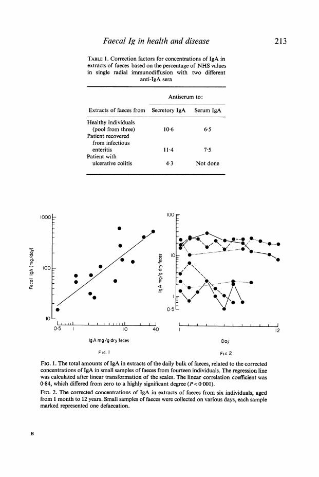
TABLE 1. Correction factors for concentrations of IgA inextracts of faeces based on the percentage of NHS valuesin single radial immunodiffusion with two different
anti-IgA sera
Antiserum to:
Extracts of faeces from Secretory IgA Serum IgA
Healthy individuals(pool from three) 10-6 6-5
Patient recoveredfrom infectiousenteritis 11-4 7-5
Patient withulcerative colitis 4-3 Not done
100
0
V)
'0'
E0'
l
10 40
Ig A mg /g dry feces
FIG.
12
Day
FIG.2
FIG. 1. The total amounts of IgA in extracts of the daily bulk of faeces, related to the correctedconcentrations of IgA in small samples of faeces from fourteen individuals. The regression linewas calculated after linear transformation of the scales. The linear correlation coefficient was0-84, which differed from zero to a highly significant degree (P< 0001).FIG. 2. The corrected concentrations of IgA in extracts of faeces from six individuals, agedfrom 1 month to 12 years. Small samples of faeces were collected on various days, each samplemarked represented one defaecation.
B
213
1000
'o
E
-
a)1u
100 0
00
00
0
10 Lwl l
05I ..
214 B. Haneberg and D. Aarskog
The concentrations of IgA related to the dry weight of faeces were not dependent on thewater content of fresh faeces. This seemed valid even though the wet-weight/dry-weightratio in faeces from the healthy individuals varied between 18 and 7 0, with mean value3-8 and standard deviation + 1 9. For meconium obtained at 1-3 days of age, this meanratio was 5 3, otherwise there was no difference in ratio with age.The IgA concentrations in extracts of small samples of faeces correlated well with the
total daily amounts of IgA in dry faeces excreted over a 3-day period (Fig. 1). Thus, theconcentration of IgA in a single small sample of faeces can for practical purposes be usedas a measure of the excretion of IgA with faeces at that time. The finding that the faecalIgA concentrations, in the same individuals, varied little from day to day (Fig. 2), supportsthis.
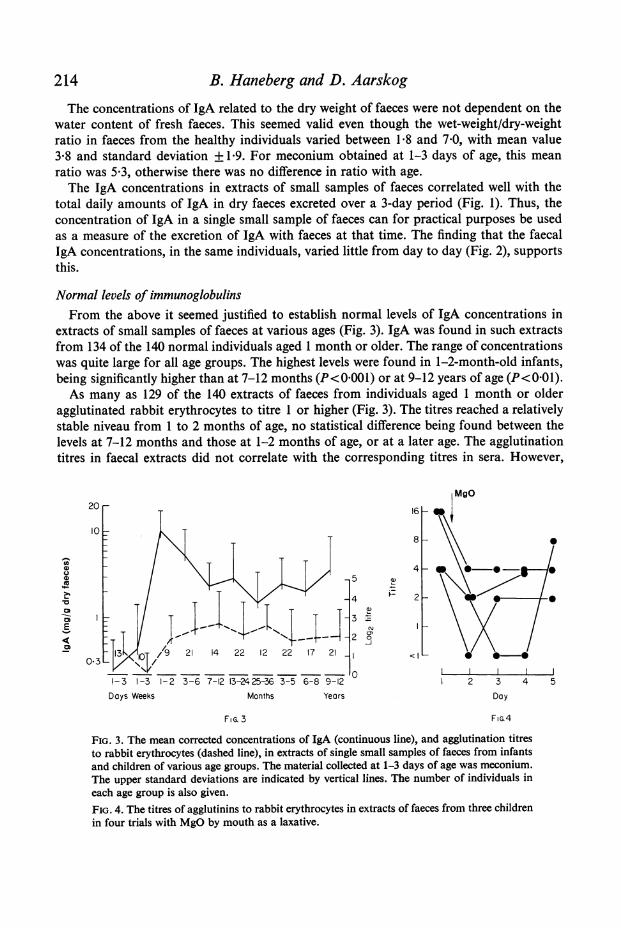
Normal levels of immunoglobulinsFrom the above it seemed justified to establish normal levels of IgA concentrations in
extracts of small samples of faeces at various ages (Fig. 3). IgA was found in such extractsfrom 134 of the 140 normal individuals aged 1 month or older. The range of concentrationswas quite large for all age groups. The highest levels were found in 1-2-month-old infants,being significantly higher than at 7-12 months (P<0 001) or at 9-12 years of age (P< 0-01).As many as 129 of the 140 extracts of faeces from individuals aged 1 month or older
agglutinated rabbit erythrocytes to titre 1 or higher (Fig. 3). The titres reached a relativelystable niveau from 1 to 2 months of age, no statistical difference being found between thelevels at 7-12 months and those at 1-2 months of age, or at a later age. The agglutinationtitres in faecal extracts did not correlate with the corresponding titres in sera. However,
MgO20 -1 2
I08
0 ~~~~~~~~~~~~~~~~~4
Cu~~~~~~~~~~~~~~~~~~~~~~~C
2~~~~~~~~~~~~~~~
O~313I~1/9 213- 712 22 12 22 17 21
I-3 13I23671213-24253 3-5 6-8 9-12 2 3 4 5
Days Weeks Months Years Day
FiG 3 FIG.4
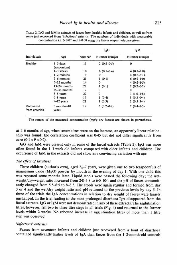
FIG. 3. The mean corrected concentrations of IgA (continuous line), and agglutination titresto rabbit erythrocytes (dashed line), in extracts of single small samples of faeces from infantsand children of various age groups. The material collected at 1-3 days of age was meconium.The upper standard deviations are indicated by vertical lines. The number of individuals ineach age group is also given.FIG. 4. The titres of agglutinins to rabbit erythrocytes in extracts of faeces from three childrenin four trials with MgO by mouth as a laxative.
Faecal Ig in health and disease 215
TABLE 2. IgG and IgM in extracts of faeces from healthy infants and children, as well as fromsome just recovered from 'infectious' enteritis. The numbers of individuals with measurable
concentration i.e. >007 and >0-08 mg/g dry faeces respectively, are given
IgG IgM
Individuals Age Number Number (range) Number (range)
Healthy 1-3 days 13 2 (02-0 5) 0(meconium)1-3 weeks 10 6 (0 1-0 4) 4 (0-2-2 8)1-2 months 9 0 4 (0-8-3-1)3-6 months 21 1 (0 1) 6 (02-10)7-12 months 14 0 4 (02-1V5)13-24 months 22 1 (0 1) 2 (0-2-02)25-36 months 12 0 03-5 years 22 0 3 (1PO1-8)6-8 years 17 1 (0 4) 3 (0 3-04)9-12 years 21 1 (03) 2 (05-3-4)
Recovered 5 months-10 17 5 (02-08) 7 (0-41-5)from enteritis years
The ranges of the measured concentration (mg/g dry faeces) are shown in parentheses.
at 1-6 months of age, when serum titres were on the increase, an apparently linear relation-ship was found; the correlation coefficient was 0 45 but did not differ significantly fromzero (0f1 <P<0-2).IgG and IgM were present only in some of the faecal extracts (Table 2). IgG was more
often found in the 1-3-week-old infants compared with older infants and children. Theoccurrence of IgM in the extracts did not show any convincing variation with age.
The effect of laxativesThree children (author's own), aged 24-7 years, were given one to two teaspoonfuls of
magnesium oxide (MgO) powder by mouth in the evening of day 1. With one child thiswas repeated some months later. Liquid stools were passed the following day; the wet-weight/dry-weight ratio increased from 2-8-3-8 to 6O-101 and the pH of faeces concomit-antly changed from 5 5-6*5 to 8-8*5. The stools were again regular and formed from day3 or 4 and the wet/dry weight ratio and pH returned to the previous levels by day 5. Inthree of the trials the IgA concentrations in relation to dry weight of faeces were largelyunchanged. In the trial leading to the most prolonged diarrhoea IgA disappeared from thefaecal extracts. IgG or IgM were not demonstrated in any of these extracts. The agglutinationtitres, however, fell two to three titre steps in all trials (Fig. 4) and returned to the formerlevels within 2 weeks. No rebound increase in agglutination titres of more than 1 titrestep was observed.
'Infectious' enteritisFaeces from seventeen infants and children just recovered from a bout of diarrhoea
contained significantly higher levels of IgA than faeces from the 1-2-month-old controls
B. Haneberg and D. Aarskog
w COO ~~~~~4)
> ~~~~~~~~~C-
to0
bo
cis
cd) cb
C)~~ ~~~~~C
4)~~~~~~~~Z
Cd
4-~~~~~~~~~~-
0~~~~~~~~0-~ ~~~I
to 0 5
1-d
ccd
~ ~ bo 0 4
~ ~ ~ ~ W
4)0 ~ ~
216
Faecal Ig in health and disease
having the highest levels of faecal IgA among the healthy individuals (Table 3). The IgAconcentrations were corrected with the same factor as that established for normal controls.The correction factor made out for one patient with enteritis was not lower than thiscontrol factor (Table 1), indicating that the higher levels of IgA in faeces from these patientswere not based on increased amounts of low molecular weight IgA. Also, the agglutinationtitres were higher than in another control group, 7-12 months of age, having the greatestnormal agglutinin activity.IgG was found in faeces from nearly one-third of the patients, whereas in the controls
aged 1 month or older IgG was measurable in only four out of 138 (Table 2). The occurrenceof IgM in faeces did not differ greatly between these groups of individuals.The serum levels of IgA and the agglutination titres in the patient group did not differ
from the controls at the time of collection of faeces.
Septic infectionsIn fifteen patients with septicaemia or obvious osteomyelitis, but no signs of gastro-
intestinal dysfunction, the faecal or serum IgA concentrations, and agglutination titreswere found to be substantially the same as in twenty-one controls of the same age. Onepatient (T.L.), however, had low serum IgA concentration and was subsequently includedin the group with immunoglobulin deficiencies.
Ulcerative colitisIn four out of the five patients normal levels of faecal IgA were measured (Table 4). The
correction factor established for IgA in faeces from one of these was low (Table 1), suggestingan abundance of low molecular weight IgA compared to the controls. One patient (V.H.)had no detectable IgA in faeces and very low serum IgA concentration, and is thereforeincluded in the immunodeficiency group.
Unusually high levels of IgG were found in faeces from three of these patients (O.E.,A.I. and V.H.) (Table 4). Their faecal agglutination titres were in the normal range. Anti-IgGdid not increase the titres in the antiglobulin test, indicating that IgG in faeces was notactive against rabbit erythrocytes. Albumin was found in faecal extracts from two of them(O.O. and A.I.), in faeces from the others no albumin was detected. The high levels ofIgG in faeces from two patients (O.E. and V.H.) with no detectable faecal albumin, cannottherefore be explained by a mere leakage of serum through an inflamed colonic mucosa.
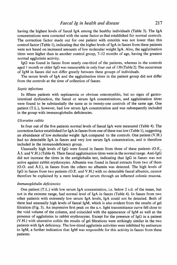
Immunoglobulin deficienciesOne patient (T.L.) with low serum IgA concentration, i.e. below 2 s.d. of the mean, but
not in the extreme range, had normal level of IgA in faeces (Table 4). In faeces from twoother patients with extremely low serum IgA levels, IgA could not be detected. Both ofthese had unusually high levels of faecal IgM, which is also evident from the results of gelfiltration (Fig. 5). An impressive first peak on the u.v. light transmittance curve fell close tothe void volume of the column, and coincided with the appearance of IgM as well as thepresence of agglutinins to rabbit erythrocytes. Except for the presence of IgG in a patient(V.H.) with ulcerative colitis the results of gel filtrations were strikingly similar in the twopatients with IgA deficiency. The low-titred agglutinin activities were inhibited by antiserumto IgM, a further indication that IgM was responsible for this activity in faeces from thesepatients.
217
B. Haneberg and D. Aarskog
mt 00 o 00 Nt
1r4 N
e'
,t l -4 -M
v v v
r6 -6 -6 -6 -
t C
e -r'Oin C Co o4 0 =% 0 0
r-IC,400 o 10 00- -4 ,4 0I~c W)itn W) 4 N N-4
-O
00 Nv
00000 0 00 00 004C4v v v
000 0 0en 0 0o 0 CC'1en iN- -m e -
00 0O \soI 'rs 00 .
(q - en - 0
00~O 0
0 0C It
000
't I'D It Itv- v
,so -" en N r- r- r-0-0-c4 I-
O-; ~_ - j "LO O -- - I, :> ; E-4
CO
0
*
C,~~ C,~ (-CC
CIS Ud
Q .=~~~~~~~~.
V) CO
o, o oooCsoY 0 *
~~ 00 o000o00aQ V Ce C C-t
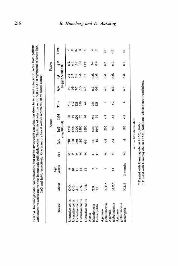
218
&n
L.4
a)
00 -,1*S.a)4-vCd
0
04ao
t0
Eto
2to
E0 otw
E_
Cd
oO 0
C. _
C O-
cdo0 004-
Cd u
C.
a)0.-t 0.=CO a)
cn d
c.) .C
o Y-'w eE
sCa)
04 EC
.-. a
°) s 00C)* 04
O
o
00>
O0*
o-C8 aSCO34-C.)
a)
0z1O
0*-
4:2
Cd
00
0
-0o
to to._ ._
Cd Cd
o o
a)a)5. s-
* 4
Cd Cd
Ct Cd
xa)(Ui
a--00c
COa)._
Faecal Ig in health and disease 219
40_
40 +++ Agglutinins60 1gM
E
a) 80 & 0 << \cm 8 | antitrypsin
100 _____,,,,,,,,,,,,,,,,,,,,,,,,,,,,,,
60
Albumin
80_/a \a,antitrypsin
100II -IIII 11
10 20 30 40 50
Fraction number
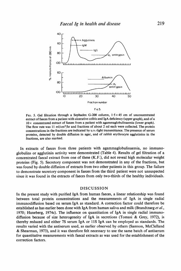
FIG.5.
FIG. 5. Gel filtration through a Sephadex G-200 column, 15 x 45 cm of unconcentratedextract of faeces from a patient with ulcerative colitis and IgA deficiency (upper graph), and of a10 x concentrated extract of faeces from a patient with agammaglobulinaemia (lower graph).The flow rate was 11 ml/cm2/hr and fractions of about 2 ml each were collected. The proteinconcentrations in the fractions are indicated by u.v.-light transmittance. The presence of serumproteins, detected by double diffusion in agar, and of rabbit erythrocyte agglutinins in thefractions, are also marked.
In extracts of faeces from three patients with agammaglobulinaemia, no immuno-globulins or agglutinin activity were demonstrated (Table 4). Results of gel filtration of aconcentrated faecal extract from one of these (K.F.), did not reveal high molecular weightproteins (Fig. 5). Secretory component was not demonstrated in any of the fractions, butwas found by double diffusion of extracts from two other patients in this group. The failureto demonstrate secretory component in faeces from the third patient were not unsuspectedsince it was found in the extracts of faeces from only two-thirds of the healthy individuals.
DISCUSSIONIn the present study with purified IgA from human faeces, a linear relationship was foundbetween total protein concentrations and the measurements of IgA in single radialimmunodiffusion based on serum IgA as standard. A correction factor could therefore beestablished as has earlier been done with IgA from human saliva and milk (Brandtzaeg et al.,1970; Haneberg, 1974c). The influence on quantitation of IgA in single radial immuno-diffusion because of size heterogeneity of IgA in secretions (Tomasi & Grey, 1972), isthereby reduced and either 7S serum IgA or IIS IgA can be employed as standards. Theresults varied with the antiserum used, as earlier observed by others (Samson, McClelland& Shearman, 1973), and it was therefore felt necessary to use the same batch of antiserumfor quantitative measurements with faecal extracts as was used for the establishment of thecorrection factors.
220 B. Haneberg and D. Aarskog
Differences in quantitative relationship between high and low molecular weight IgA willinfluence the correction factor; with more low molecular weight IgA, the factor will besmaller. Since the factor for faecal IgA from one individual just recovered from 'infectious'enteritis was nearly the same as for the controls, the higher IgA values in the enteritis groupmost likely reflect increased intestinal IgA production. On the other hand, the low correctionfactor established for one patient with ulcerative colitis might suggest a passive leakage oflow molecular IgA through the mucosal epithelium. Evidence for this is given by histologicalstudies using immunofluorescence technique (Gelzayd et al., 1968).The molecular weight distribution of IgA in faeces may vary even between healthy
individuals. For accurate estimation of IgA it would therefore be essential to establish acorrection factor with a purified preparation from each individual sample. The excretion ofdry faecal matter may also differ from time to time and between individuals. Therefore, thedetermination of IgA, related to dry weight of faeces, can be semi-quantitative at best.However, it is likely that the IgA in small samples of faeces reflects the secretion of IgA intothe gut at the time when faeces were collected.The IgA concentrations in small samples of faeces from healthy infants and children indi-
cate that the intestinal mucosa is immunologically mature at least from the age of 1 month.Moreover, the higher level at this early age, compared to that found at a later age, mayreflect a vivid response to the introduction into the gut of dietary as well as microbialantigens (Shearman et al., 1972).
Testing for agglutinins to rabbit erythrocytes has proved useful for the assay of IgAactivity in faeces (Haneberg, 1974a); rabbits immunized with their own erythrocytes,agglutinated by human faecal extracts, produced precipitating antibodies to human IgA.In the healthy individuals the agglutination titres, like IgA concentrations in faeces, reacheda relatively stable level from 1 month of age. The relatively high titres with extracts ofmeconium may be explained by swallowed amniotic fluid (Rule et al., 1971), or by transu-dation of antibodies from the blood (Roulet & von Muralt, 1961). It was difficult to excludecontamination of blood in meconium by the benzidine test. The agglutinins in meconium,therefore, were not thought of as being of solely intestinal origin.
Diarrhoea induced by laxatives seemed to 'wash-out' the agglutinins recovered in faeces,and when diarrhoea was prolonged IgA also disappeared. In cases with ongoing severediarrhoea, as in cholera and shigellosis, the specific faecal antibody activity (Freter et al.,1965; Reed & Williams, 1971) would therefore be highly significant.The high levels of faecal IgA in the patients with enteritis indicate a local stimulus by a
supposedly infectious agent. The increased jejunal IgA synthesis demonstrated by othersfollowing experimental infection with the Norwalk agent (Agus et al., 1974) is in line withthis. Increased serum IgA levels in premature infants with gastroenteritis (Panayotou et al.,1971) is further in support of increased intestinal production of IgA since tissues along theintestinal tract are main sources of IgA (Tomasi & Grey, 1972). The concomitant increasedagglutinin activity against rabbit erythrocytes as demonstrated in our study is suggestive ofan antigenic relationship between the surface of the rabbit erythrocytes and the presumptiveinfectious agent. Samples of faeces were not taken until after recovery from the disease, inorder to obtain samples comparable to the controls. This also allowed time for the pro-duction of local antibody response.The presence and the high levels of IgG in faeces from patients with ulcerative colitis,
are analogous to the findings in saliva from individuals with inflamed gingival mucosa
Faecal Ig in health and disease 221
(Brandtzaeg et al., 1970). Signs of increased local IgG synthesis seem to be a general findingin chronic inflammatory conditions (Brandtzaeg, 1972; Brandtzaeg et al., 1974), althoughsome results of immunofluorescence studies obtained with rectal mucosa have been con-flicting (Gelzayd et al., 1968). The high levels of IgG in faeces, even in formed stools, freefrom blood, may therefore indicate a continuous inflammation of the colon.The apparent lack of IgA in faeces from two patients with isolated serum IgA deficiency
parallels the findings in saliva, parotid secretions and intestinal juice (Brandtzaeg, Fjellanger& Gjeruldsen, 1968; Savilahti, 1973; South et al., 1968). The increased faecal IgM con-centrations in our patients clearly indicate that IgM in intestinal secretions, as in salivarygland secretions (Brandtzaeg et al., 1968; Savilahti, 1973) can compensate for IgA. This issubstantiated by the demonstrations of low numbers of IgA- and increased numbers ofIgM-containing plasma cells in the intestinal mucosa of patients with IgA deficiency(Crabbe & Heremans, 1966; Savilahti, 1973). Also faecal IgM in our patients seemed immuno-logically active against rabbit erythrocytes. We have not earlier been able to demonstratesuch activity of faecal immunoglobulins other than for IgA. Possibly part of the largeamounts of IgM may escape inactivation by proteolytic enzymes.The absence of detectable immunoglobulins in faeces from three patients with agamma-
globulinaemia was interesting. At least as far as IgA is concerned, this parallels the findingsin saliva (South et al., 1968). The importance of local immunoglobulins for the normalgross functioning of the intestines might therefore be questioned since none of the patientswith agammaglobulinaemia had signs of gastrointestinal dysfunction.
In any event, the results so far give evidence that faecal material us useful for the evaluationof inmmunoglobulins of at least the low intestinal tract.
This study was supported in part by L. Meltzers h0yskolefond.
REFERENCE S
AGUS, S.G., FALCHUK, Z.M., SESSOMS, C.S.WYATT, R.G. & DOLIN, R. (1974) Increasedjejunal IgA synthesis in vitro during acute in-fectious nonbacterial gastroenteritis. Amer. J.dig. Dis. 19, 127.
BRANDTZAEG, P. (1972) Local formation and trans-port of immunoglobulins related to the oralcavity. Host Resistance to Commensal Bacteria(ed. by T. MacPhee), p. 116. Churchill Living-stone, Edinburgh.
BRANDTZAEG, P., BAKLIEN, K., FAUSA, 0. &HOEL, P.S. (1974) Immunohistochemical char-acterization of local immunoglobulin formationin ulcerative colitis. Gastroenterology, 66, 1123.
BRANDTZAEG, P., FJELLANGER, I., & GJERULDSEN,S.T. (1968) Immunoglobulin M: local synthesisand selective secretion in patients with immuno-globulin A deficiency. Science, 160, 789.
BRANDTZAEG, P., FJELLANGER, I. & GJERULDSEN,S.T. (1970) Human secretory immunoglobulins.I. Salivary secretions from individuals with normalor low levels of serum immunoglobulins. Scand.J. Haemat. supplement 12.
CRABBt, P.A. & HEREMANS, J.F. (1966) Lack of
B*
gamma A-immunoglobulin in serum of patientswith steatorrhoea. Gut, 7, 119.
FRETER, R., DE, S.P., MONDAL, A., SHRIVASTAVA,D.L. & SUNDERMAN, F.W. JR. (1965) Copro-antibody and serum antibody in cholera patients.J. infect. Dis. 115, 83.
GELZAYD, E.A., KRAFT, S.C., FITCH, F.W. &KIRSNER, J.B. (1968) Distribution of immuno-globulins in human rectal mucosa. II. Ulcerativecolitis and abnormal mucosal control subjects.Gastroenterology, 54, 341.
HANEBERG, B. (1974a) Human fecal agglutinins torabbit erythrocytes. Scand. J. Immunol. 3, 71.
HANEBERG, B. (1974b) Immunoglobulins in fecesfrom infants fed human or bovine milk. Scand. J.Immunol. 3, 191.
HANEBERG, B. (1974c) Human milk immunoglobulinsand agglutinins to rabbit erythrocytes. Int. Arch.Allergy, 47, 716.
HANEBERG, B. & T0NDER, 0. (1973) Immuno-globulins and other serum proteins in feces frominfants and children. Scand. J. Immunol. 2, 375.
KABAT, E.A. & MAYER, M.F. (1964) Estimation ofprotein with the Folin-Ciocalteu phenol reagent.
222 B. Haneberg and D. AarskogExperimental Immunochemistry, 2nd edn, p. 556.Charles C. Thomas, Springfield, Illinois.
PANAYOTOU, P., PAPADATOS, C., PAPAEVANGELOU,G., ALEXIOU, D., SKARDOUTSOU, A. & KOUREA, E.(1971) Immunoglobulin A and M levels in pre-mature infants with gastroenteritis. Arch. Dis.Childh. 46, 671.
REED, W.P. & WILLIAMS, R.C. (1971) Intestinalimmunoglobulins in shigellosis. Gastroenterology,61, 35.
ROULET, D.L.A. & VON MURALT, G. (1961) Anti-genanalytische Untersuchungen an Fruchtwasser-und Meconiumproteinen. Schweiz. med. Wschr.91, 74.
RULE, A.H., LAWRENCE, D., HAGER, H.J., HYSLOP,N. JR. & SHWACHMAN, H. (1971) IgA: presencein meconium obtained from patients with cysticfibrosis. Pediatrics, 48, 601.
SAMSON, R.R., MCCLELLAND, D.B.L. & SHEARMAN,
D.J.C. (1973) Studies on the quantitation ofimmunoglobulin in human intestinal secretions.Gut, 14, 616.
SAVILAHTI, E. (1973) IgA deficiency in children.Immunoglobulin-containing cells in the intestinalmucosa, immunoglobulins in secretions andserum IgA levels. Clin. exp. Immunol. 13, 395.
SHEARMAN, D.J.C., PARKIN, D.M. & MCCLELLAND,D.B.L. (1972) The demonstration and function ofantibodies in the gastrointestinal tract. Gut, 13,483.
SOUTH, M.A., COOPER, M.D., WOLLHEIM, F.A. &GOOD, R.A. (1968) The IgA system. II. Theclinical significance of IgA deficiency: studies inpatients with agammaglobulinemia and ataxiatelangiectasia. Amer. J. Med. 44, 168.
ToMASI, T.B. & GREY, H.M. (1972) Structure andfunction of immunoglobulin A. Progr. Allergy,16, 81.